HAL Id: inserm-02409310
https://www.hal.inserm.fr/inserm-02409310
Submitted on 13 Dec 2019
HAL is a multi-disciplinary open access archive for the deposit and dissemination of sci-entific research documents, whether they are pub-lished or not. The documents may come from teaching and research institutions in France or abroad, or from public or private research centers.
L’archive ouverte pluridisciplinaire HAL, est destinée au dépôt et à la diffusion de documents scientifiques de niveau recherche, publiés ou non, émanant des établissements d’enseignement et de recherche français ou étrangers, des laboratoires publics ou privés.
mitochondrial respiratory chain to SDHI pesticides and
its consequence on the impact of SDHIs on human
cultured cells
Paule Bénit, Agathe Kahn, Dominique Chretien, Sylvie Bortoli, Laurence
Huc, Manuel Schiff, Anne-Paule Gimenez-Roqueplo, Judith Favier, Pierre
Gressens, Malgorzata Rak, et al.
To cite this version:
Paule Bénit, Agathe Kahn, Dominique Chretien, Sylvie Bortoli, Laurence Huc, et al.. Evolutionarily conserved susceptibility of the mitochondrial respiratory chain to SDHI pesticides and its consequence on the impact of SDHIs on human cultured cells. PLoS ONE, Public Library of Science, 2019, 14 (11), pp.e0224132. �10.1371/journal.pone.0224132�. �inserm-02409310�
Evolutionarily conserved susceptibility of the
mitochondrial respiratory chain to SDHI
pesticides and its consequence on the impact
of SDHIs on human cultured cells
Paule Be´nit1, Agathe Kahn1, Dominique Chretien1, Sylvie Bortoli2, Laurence Huc3, Manuel SchiffID1,4, Anne-Paule Gimenez-Roqueplo5,6, Judith Favier6, Pierre Gressens1,4,
Malgorzata Rak1, Pierre RustinID1*
1 Universite´ de Paris, NeuroDiderot, INSERM, Paris, France, 2 Universite´ de Paris, INSERM, UMR-S 1124, Paris, France, 3 INRA UMR 1331 ToxAlim (Research Center in Food Toxicology), Universite´ de Toulouse ENVT, INP, UPS, 180 Chemin de Tournefeuille, France, 4 Assistance Publique-Hoˆpitaux de Paris (AP-HP), Hoˆpital Robert Debre´, Service de neurologie et maladies me´taboliques, Paris, France, 5 Universite´ de Paris, PARCC, INSERM, Equipe Labellise´ e par la Ligue contre le Cancer, Paris, France, 6 Assistance Publique-Hoˆpitaux de Paris (AP-HP), Hoˆ pital Europe´en Georges Pompidou, Service de Ge´ne´tique, Paris, France *pierre.rustin@inserm.fr
Abstract
Succinate dehydrogenase (SDH) inhibitors (SDHIs) are used worldwide to limit the prolifera-tion of molds on plants and plant products. However, as SDH, also known as respiratory chain (RC) complex II, is a universal component of mitochondria from living organisms, highly conserved through evolution, the specificity of these inhibitors toward fungi warrants investigation. We first establish that the human, honeybee, earthworm and fungal SDHs are all sensitive to the eight SDHIs tested, albeit with varying IC50values, generally in the
micro-molar range. In addition to SDH, we observed that five of the SDHIs, mostly from the latest generation, inhibit the activity of RC complex III. Finally, we show that the provision of glu-cose ad libitum in the cell culture medium, while simultaneously providing sufficient ATP and reducing power for antioxidant enzymes through glycolysis, allows the growth of RC-defi-cient cells, fully masking the deleterious effect of SDHIs. As a result, when glutamine is the major carbon source, the presence of SDHIs leads to time-dependent cell death. This pro-cess is significantly accelerated in fibroblasts derived from patients with neurological or neurodegenerative diseases due to RC impairment (encephalopathy originating from a par-tial SDH defect) and/or hypersensitivity to oxidative insults (Friedreich ataxia, familial Alzhei-mer’s disease).
Introduction
Succinate dehydrogenase (SDH; EC 1.3.5.1), also known as electron transport chain (ETC) complex II (CII), is a universal and key component of the mitochondrial respiratory chain (RC) of all living organisms [1]. This protein transfers electrons derived from the oxidation of
a1111111111 a1111111111 a1111111111 a1111111111 a1111111111 OPEN ACCESS
Citation: Be´nit P, Kahn A, Chretien D, Bortoli S,
Huc L, Schiff M, et al. (2019) Evolutionarily conserved susceptibility of the mitochondrial respiratory chain to SDHI pesticides and its consequence on the impact of SDHIs on human cultured cells. PLoS ONE 14(11): e0224132.
https://doi.org/10.1371/journal.pone.0224132 Editor: Annalisa Pastore, National Institute for
Medical Research, Medical Research Council, London, UNITED KINGDOM
Received: August 8, 2019 Accepted: October 4, 2019 Published: November 7, 2019
Peer Review History: PLOS recognizes the
benefits of transparency in the peer review process; therefore, we enable the publication of all of the content of peer review and author responses alongside final, published articles. The editorial history of this article is available here:
https://doi.org/10.1371/journal.pone.0224132 Copyright:© 2019 Be´nit et al. This is an open access article distributed under the terms of the
Creative Commons Attribution License, which permits unrestricted use, distribution, and reproduction in any medium, provided the original author and source are credited.
succinate to fumarate to a dedicated pool of ubiquinone without concomitant proton extru-sion. The SDH complex is composed of four proteins (SDHA-D) that were largely conserved during evolution [2]. As part of the Krebs cycle, SDH is the only enzyme of the cycle that has no counterpart in any other cellular compartment and no soluble redox cofactors, such as NAD+/NADH, which is possibly balanced out by the activity of other dehydrogenases; thus, SDH is irreplaceable in mitochondrial and cellular metabolism (S1 Fig). As a result, any block-ade, even partial, of SDH activity can lead to changes in the cellular metabolome and have del-eterious consequences for the cell [3]. Associated with its role in electron transfer, the enzyme is also favored in the RC for one-electron reactions through its iron-sulfur clusters, resulting in the generation of deleterious superoxides, especially in the presence of downstream SDH inhibitors (SDHIs) [4–6].
In humans, mutations of SDH-coding genes lead to pronounced defects of varying degrees in SDH activity that are associated with a wide spectrum of diseases, with frequent neurologi-cal expression [3]. Complete abolition of SDH activity results in profound modification of the metabolome, transcriptome and epigenome and to the development of various tumors and cancers [3,7]. Generally, mitochondrial dysfunction is now recognized as a contributing factor to the early pathology of multiple human conditions, including major neurodegenerative dis-eases [8]. This finding is consistent with the role of mitochondria as signaling organelles with a multitude of functions, ranging from energy production as heat or ATP [9] to regulation of cellular metabolism [10,11], energy homeostasis [12], stress response [13], and cell fate [14].
Given the central role of SDH, SDHIs are now widely used in agriculture worldwide to fight fungal proliferation [15]. These compounds are used on cereal crops; for preservation of fruits, vegetables, and seeds; and for care of public lawns and golf courses. However, because of the vir-tually universal function of SDH in cell respiration and mitochondrial metabolism, it can be assumed that any living organisms exposed to these substances could also be affected, and the consequences of exposure to SDHIs for non-target organisms appear a major issue. These effects depend on SDHI sensitivity of SDH, which may vary slightly based on the SDHI and the species used. The dose, time, frequency and duration of exposure; SDHI absorption and meta-bolization; and the eventual use of additional toxic substances are obvious factors to consider.
The fungicide qualifier associated with SDHIs infers some degree of specificity with respect to fungi, which cannot be confirmed based on basic scientific information comparing the action of the SDHI predecessor (carboxin) on SDHs from other species, including mammals [16]. This gap served as an incentive to investigate the effects of eight SDHIs, including the lat-est generation of molecules, on SDH activity and, more generally, on mitochondrial ETC qui-none-dependent activities in four different species:Botrytis cinerea, as the target organism,
andHomo sapiens, Lumbricus terrestris, and Apis mellifera, as non-target species. In addition
to the predictable, general effect of the tested SDHIs against SDHs from different species, albeit with variable sensitivity, we observed that several of the latest-generation SDHIs also inhibited RC complex III (CIII) in addition to SDH (CII), indicating the potential binding of these SDHIs to other quinone-binding sites in the cell. We finally showed that in nonpermissive cul-ture conditions for RC-deficient cells, these inhibitors readily impaired the growth and, ulti-mately, the survival of SDHI-treated human cultured cells presumably because of SDHI engendered oxidative stress.
Material and methods
Biological material
Four biological materials,i.e., B. cinerea fungi, A. mellifera, L. terrestris, and human cultured
cells were used to test the effect of SDHIs on the mitochondrial RC. To avoid interference with
Data Availability Statement: All relevant data are
within the manuscript and its Supporting Information files.
Funding: No specific funding was initially provided
for this work. After the work realization, PB and PR received a specific support grant from the French Association POLLINIS (https://www.pollinis.org/). General expenses were covered by grants from AAJI (Association pour l’Aide aux Jeunes Infirmes & aux Personnes Handicape´es); AFAF (Association Franc¸aise de l’Ataxie de Friedreich); Association OLY (Ouvrir Les Yeux); ANR MITOXDRUGS (ANR-16-CE18-0010). Sponsors or funders had no role in the study design, data collection and analysis, decision to publish, or preparation of the manuscript.
Competing interests: The authors have declared
contaminating factors and to maintain similar experimental conditions for the measurement of RC activities, different preparation methods had to be used for cultured cells, whole organ-isms and tissues [17].
Botrytis cinerea
The mixed population ofB. cinerea fungi used in this study was kindly provided by Dr. Joelle
Dupont (Museum National d’Histoire Naturelle, Paris, France). Conidia fromBotrytis cinerea
were obtained from 15–30 d cultures grown on malt medium agar plates. Mycelial colonies were prepared by seeding conidia from 10 colonies in liquid medium (10 g of glucose, 2 g of KH2PO4, 0.5 g of MgSO47H2O, 1 g of (NH4)2SO4, 2 g of yeast extract and deionized water up
to 1 l, pH 6.2). Incubation was performed at 23˚C until hyphae totally covered the surface of the bottle. The hyphae (9-cm-diameter disks) were harvested, washed twice with refrigerated distilled water and finally with mitochondria extraction medium containing 20 mM Tris (pH 7.2), 0.25 M sucrose, 40 mM KCl, 2 mM EGTA, 1 mg/ml BSA, and no cysteine. The hyphae were suspended in 150 ml of extraction medium, cut in small pieces (1 cm2) and then dis-rupted at high speed for 1×2 sec and at low speed for 2×3 sec using a MultiMoulinex mixer (Moulinex, France). The suspension of disrupted hyphae was then filtered through two layers of nylon cloth (clearance 150μm). The cloth was washed with 50 ml of extraction medium. After low-speed centrifugation (700g for 15 min), the mitochondria were collected at 10,000 g
for 20 min. The pellet was suspended in 250μl of extraction medium, aliquoted (± 30 μl) and kept frozen at -80˚C.
Apis mellifera
A. mellifera individuals (n = 5) were a kind gift from Jacques Kemp (beekeeper;
Saint-Re´my-lès-Chevreuse, Yvelines, France). Whole bodies of individuals frozen in dry ice, except heads, were homogenized using a 1 ml glass-glass Potter-Elvehjem homogenizer in an ice-cold medium consisting of 20 mM Tris (pH 7.2), 0.25 M sucrose, 40 mM KCl, 2 mM EGTA, and 1 mg/ml BSA. The homogenate was centrifuged at 1,500g for 5 min. The mitochondria
con-tained in the supernatant were spun down at 10,000g for 10 min. The pellet of crude
mito-chondria was distributed as 20-μl aliquots and kept frozen at -80˚C.
Lumbricus terrestris
L. terrestris individuals (n = 5) were collected manually in a household compost in Epernon
(France 28230) and identified by the person (Paule Be´nit) in charge of this compost. Before use, the worms were washed 5 times with 1× phosphate-buffered saline. A fresh homogenate was prepared from the segments located between the prostomium and clitellum of the 5 indi-viduals. Tissues were placed in 500μl of an ice-cold medium consisting of 20 mM Tris (pH 7.2), 0.25 M sucrose, 40 mM KCl, 2 mM EGTA, and 1 mg/ml BSA and homogenized using a 1-ml glass-glass Potter-Elvehjem homogenizer. Low-speed centrifugation at 1,500g for 5 min
allowed heavy debris to be discarded. The supernatant was distributed as 50-μl aliquots and kept frozen at -80˚C.
Human cultured fibroblasts and HEK cells
Fibroblasts were derived from skin biopsies obtained from SDH-deficient patient, Friedreich ataxia (FRDA) patients, familial Alzheimer disease (FAD) and three anonymous healthy indi-viduals, all of European ancestry. Written informed consent for research was obtained from patients and/or family members according to protocols in accordance to the Necker Hospital
(SDH-deficient patient; 1993) or Robert Debre´ (FRDA patient; 2011) Hospital ethical commit-tees (Paris, France). None of these patients were recruited specifically for this study which used already available skin fibroblast cultures from the above mentioned Hospitals or from com-mercial source (FAD patient; Coriell Institute cell repository; AG08064). The SDH-deficient patient presented Leigh syndrome resulting from a homozygous R554W mutation in the SDH flavoprotein subunit (SDHA) [18]. The FRDA fibroblasts were from a female patient harbor-ing a biallelic long GAA expansion (>2.6 kb) in the frataxin (FXN) gene (patient FRDA4; [19]. This expansion caused silencing of both the FXN and PIP5K1B genes [20]. Loss of pip5k1β
function results in a decrease in PI(4,5)P2 levels, causing destabilization of the actin network and impaired Keap1-Nrf2 superoxide dismutase (SOD) signaling [19,21]. As a result, these cells were highly sensitive to superoxides [21,22]. The FAD fibroblasts were from a clinically affected male patient with FAD. Such Alzheimer’s disease fibroblasts were previously shown to display abnormalities in the SOD-signaling-related Nrf2 pathway [23] and to be highly sensi-tive to oxidasensi-tive insults [24,25].
The cells were grown in T75 flasks using 10 ml Dulbecco’s modified Eagle’s minimal essen-tial medium (DMEM) either containing 1 or 4.5 g/l glucose, 4 mM glutamine (glutamax; Gibco Thermo Fisher Scientific, MA), 2 mM pyruvate and 200μM uridine (permissive medium, thereafter referred as GlucoMax) or lacking glucose, pyruvate and uridine but con-taining 4 mM glutamine (nonpermissive medium, thereafter referred as MitoMax;S1 Table). All media were supplemented with 10% fetal calf serum and 100 U/mL each penicillin and streptomycin. HEK293 human embryonic kidney cells were cultured in DMEM containing 4.5 g/L glucose, 4 mM glutamine, 10% fetal calf serum, 200μM uridine, 2 mM pyruvate, and 100 U/mL each penicillin and streptomycin [9]. Cell pellets (1,500g for 5 min) were kept
fro-zen (-80˚C), and for biochemical studies, the pellets were permeabilized by two freeze-thaw cycles before enzyme assays [26].
To test SDHIs on growing skin fibroblasts, cells trypsinized from a confluent 75-cm2flask culture were seeded in 4×75-cm2flasks (20–25% confluency) containing GlucoMax medium (S1 Table). After allowing the culture to stand for one night, the medium was changed to the desired conditions,i.e., GlucoMax medium or MitoMax medium (S1 Table), supplemented with the chosen amount of SDHI (control conditions compensated for DMSO used to solubi-lize SDHI; 14 mM max). The number of cells was estimated from random images using the ImageJ processing program for 15–20 days without changing the medium (one unique expo-sure to SDHI).
Enzyme measurements and the potential effects of SDHIs
RC enzyme activities were spectrophotometrically measured using a pseudo-double-wave-length spectrophotometer (Cary 60, Agilent). Malonate-sensitive succinate quinone dichloro-phenolindophenol (DCPIP) reductase (SQDR), as an indicator of the activity of the isolated SDH enzyme (isolated CII), and glycerol-3-phosphate quinone DCPIP reductase (GQDR), as a measure of the activity of the isolated glycerol-3-phosphate dehydrogenase (G3PDH), were assayed as previously described [17]. To study the effect of SDHIs on the RC, we had to take into consideration the fact that these inhibitors act through binding to quinone-binding sites in the RC. Accordingly, we observed that the extent of the inhibitory effects of these substances varies with the addition of exogenous quinones (Fig 1). When possible, the potential effects of SDHIs on RC enzymes were therefore studied under experimental conditions that did not require the addition of exogenous quinones. With this aim, we measured the malonate-sensi-tive succinate cytochrome c reductase (SDH+CIII), glycerol-3-phosphate cytochromec
Fig 1. Effect of SDHI (bixafen) on the respiratory chain as a function of quinone (decylubiquinone) concentration. A, SDH activity from
honeybee spectrophotometrically measured as reduction in DCPIP levels in the presence of succinate and exogenous quinone (SQDR). SDH activity was measured before and after the addition of exogenous decylubiquinone (80μM) in the presence of bixafen (100 μM; trace b) or DMSO (trace a: control condition). Percent inhibition refers to the effect of bixafen in the absence or presence of decylubiquinone. B, Succinate cytochromec reductase (SCCR) from freeze-thawed HEK cells measured in the presence of DMSO (trace a; control condition), bixafen (trace b),
or malonate (trace c). Percent inhibition refers to the effect of bixafen (0.5μM) measured before or after the addition of decylubiquinone (80 μM) on the malonate-sensitive cytochromec reduction. C, Modulation by decylubiquinol of complex III inhibition by SDHI. A selected sequence of
addition allows successful measurement of malonate-sensitive SCCR, glycerol-3-phosphate cytochromec reductase (GCCR), and the
antimycin-sensitive decylubiquinol cytochromec reductase (QCCR) of HEK cells. The effect of bixafen (20 μM; traces b,d,f) on each of the three activities
was observed by comparing the rates to the rates measured under control conditions (DMSO) and in the presence of various amounts of decylubiquinol: 25μM (a,b), 50 μM (c,d), and 100 μM (e,f). Percent inhibition refers to the effect of bixafen on the antimycin-sensitive decylubiquinol cytochromec reductase (CIII). Aa, antimycin; DCPIP (dichlorophenolindophenol); DcQ and DcQH2, the oxidized and reduced
forms of decylubiquinone, respectively; G3P, glycerol-3-phosphate.
previously described [26]. The effects of eight different SDHIs were studied, namely, flutolanil, fluopyram, boscalid, fluxapyroxad, penflufen, penthiopyrad, isopyrazam and bixafen (S2 Fig), all of which are known to bind the quinone-binding site of SDH (S3 Fig). As DMSO, the sol-vent used for SDHIs and quinones, tends to reduce the activity of most RC enzymes, it was added in control conditions to compensate for DMSO added with SDHI (always below 25μl/ ml, corresponding to 352 mM).
Total SOD activity was measured in freeze-thawed cell lysates resuspended in 50 mM KH2PO4(pH 7.8). Activity was determined by monitoring the autoxidation of pyrogallol at
420 nm and expressed as IU/mg protein [27].
Protein concentration was estimated using the Bradford assay.
SDHI IC
50determination
The effects of eight different SDHIs (up to 500μM;S2 Fig) were determined on four different biological materials. Because IC50are known to be affected by the actual absolute activity of
the tested enzyme, these IC50were quantified using similar levels of SDH enzyme activity
dur-ing the assay. IC50values were determined using the AAT Bioquest IC50calculator tool
(https://www.aatbio.com/tools/ic50-calculator).
Statistics
Data are presented as mean± SD for all experiments. Statistical significance was calculated by standard unpaired t-test or one-way ANOVA with Bonferroni post-test correction for more than two conditions. A p < 0.05 was considered statistically significant (GraphPad Prism).
Results
Effects of SDHIs on the mitochondrial respiratory chain
We determined the effects of eight different SDHIs (S2 Fig) on the mitochondrial RC activity in freeze-thawed HEK cells, earthworms, honeybees, andB. cinerea fungi (Fig 2andTable 1). The IC50values for the different SDHIs with SDH were first estimated from the succinate
cytochromec reductase (SCCR) activity (Fig 2). All the SDHIs tested were found to exert an inhibitory effect on SDH regardless of the biological origin of the enzyme, albeit to varying degrees. As a reference, the ability of SDHIs to block SDH activity was studied on the enzyme ofB. cinerea, the target organism, and was seen to vary depending on the SDHI, with flutolanil
exhibiting relative ineffectiveness (Fig 2,Table 1). SDH showed highly pronounced sensitivity for the last-generation SDHIs, with an IC50below 0.1μM for the fungal enzyme. Human SDH
appears to be relatively less sensitive to fluopyram (IC50>150μM) than to flutolanil, boscalid,
penthiopyrad and fluxapyroxad (IC50values ranging from 18.6 to 2.1μM). The earthworm
enzyme appeared to be particularly sensitive to boscalid, flutolanil, and fluxapyroxad, whereas the honeybee enzyme was more sensitive to flutolanil and fluopyram than the enzymes of other non-target organisms. Last-generation SDHIs (S2 Fig) were particularly effective in blocking the human enzyme. The IC50values calculated for penflufen, isopyrazam and bixafen
were all in the order of 1μM or less. A similar variability among the effects of the various SDHIs was observed for the earthworm and the honeybee SDHs, which exhibited similar and higher sensitivity, respectively, to the last-generation SDHIs than to the SDHI predecessors. For most SDHIs, the IC50values were lower for the enzyme from theB. cinerea fungi, with the
notable exception of flutolanil, which was particularly active against the earthworm and hon-eybee enzymes. Disturbingly, investigation of the SDHI sensitivity of SDH enzymes from only
4 species was enough to reveal that at least one of these species was quite sensitive to at least one of these SDHIs (Fig 2).
We next studied the effect of SDHIs on additional coenzyme Q-dependent RC activities, such as the activities of glycerol-3-phosphate cytochromec reductase (GCCR) and ubiquinol
cytochromec reductase (QCCR), also known as RC CIII. Interestingly, isopyrazam and, even
more efficiently, bixafen were found to also inhibit human GCCR activity (IC5021.1μM)
(Table 1). Under our experimental conditions, no inhibitory effect of bixafen (up to 500μM) could be detected on the glycerol 3-phosphate quinone-DCPIP reductase (not shown), while the molecule strongly inhibited CIII activity (IC5015.5μM), albeit less efficiently than it
inhib-ited SCCR activity (IC500.34μM) (Table 1). The effect of SDHIs was not restricted to human
CIII and was exhibited by fluxapyroxad, penflufen, penthiopyrad, isopyrazam and bixafen on the fungal CIII and by penthiopyram, isopyrazam and bixafen on CIII from all 4 species. This unreported inhibitory effect of several SDHIs on RC CIII was modulated by the amount of exogenous quinones (DcQH2) used to measure CIII activity, with 60 to 40% inhibition
observed upon increasing DcQH2from 25 to 100μM (Fig 1C). Notably, as adding reduced
quinone, the substrate of the reaction, is necessary to measure CIII directly, the IC50values
measured for CIII are only indicative. As expected, in case of CIII inhibition and similarly to the GCCR (Table 1), the NADH oxidase activity was also reduced (not shown), as would be observed following a treatment with rotenone or paraquat.
Fig 2. IC50values of SDHIs on the succinate cytochromec reductase of Homo sapiens, Lombricus terrestris, Apis mellifera, and Botrytis cinerea. A-H, IC50
(n � 3) of boscalid (A), penflufen (B), flutolanil (C), fluopyram (D), isopyrazam (E), penthiopyrad (F), fluxapyroxad (G), and bixafen (H) on the succinate cytochromec reductase of Homo sapiens (green), Lumbricus terrestris (purple), Apis mellifera (red), and Botrytis cinerea (black). Note the variable Y scales.
SCCR, succinate cytochromec reductase.
https://doi.org/10.1371/journal.pone.0224132.g002
Table 1. Comparative inhibitory effects (IC50values) of eight SDHIs on the RC activities of 4 different species.
Homo sapiens Lumbricus terrestrix. Apis mellifera Botrytis cinerea
Flutolanil SCCR: 18.7±3.5 μM GCCR > 500μM QCCR > 500μM SCCR: 0.36±0.02 μM GCCR: 450±70 μM QCCR > 500μM SCCR: 0.8±0.5 μM GCCR > 500μM GCCR > 500μM SCCR: 8.6±4.5 μM GCCR > 500μM QCCR > 500μM Fluopyram SCCR 160±40 μM GCCR > 500μM QCCR > 500μM SCCR 30.6±7.6 μM GCCR > 500μM QCCR > 500μM SCCR 3.8±0.3 μM GCCR > 500μM QCCR > 500μM SCCR 0.2±0.1 μM GCCR > 500μM QCCR > 500μM Boscalid SCCR 4.8±0.2 μM GCCR > 500μM QCCR > 500μM SCCR 0.5±0.3 μM GCCR > 500μM QCCR > 500μM SCCR 76.7±6.0 μM GCCR > 500μM QCCR > 500μM SCCR 0.8±1.1 μM GCCR > 500μM QCCR > 500μM Fluxapyroxad SCCR 2.1±0.7 μM GCCR > 500μM QCCR > 500μM SCCR 0.71±0.07 μM GCCR > 500μM QCCR > 500μM SCCR 11.7±3.4 μM GCCR 205±34 μM QCCR 300±5 μM SCCR 0.095±0.008 μM GCCR 60±56 μM QCCR 67±53 μM Penflufen SCCR 1.3±0.3 μM GCCR > 500μM QCCR > 500μM SCCR 1.13±0.16 μM GCCR > 500μM QCCR > 500μM SCCR 7.5±0.7 μM GCCR > 500μM QCCR > 500μM SCCR 0.13±0.06 μM GCCR 80±57 μM QCCR 433±58 μM Penthiopyrad SCCR 3.7±1.1 μM GCCR 211±15 μM QCCR 349±72 μM SCCR 0.70±0.01 μM GCCR 254±65 μM QCCR 297±4 μM SCCR 10±2 μM GCCR 251±75 μM QCCR 268±44 μM SCCR 0.045±0.023 μM GCCR 93.5±47 μM QCCR 105±7.1 μM Isopyrazam SCCR 0.63±0.18 μM GCCR 51.3±5.8 μM QCCR 125±35 μM SCCR 0.46±0.17 μM GCCR 83±5 μM QCCR 76±21 μM SCCR 5.1±2.3 μM GCCR 97±9 μM QCCR 87±18 μM SCCR 0.023±0.004 μM GCCR 27.9±10.6 μM QCCR 14.2±5.6 μM Bixafen SCCR 0.34±0.12 μM GCCR 21.1±9.3 μM QCCR 15.5±13.4 μM SCCR 6.0±3.6 μM GCCR 450±71 μM QCCR � 500μM SCCR 3.3±0.33 μM GCCR 24.1±11.4 μM QCCR 5.7±5.3 μM SCCR 0.07±0.06 μM GCCR 22.5±3.5 μM QCCR 22.2±14.4 μM SCCR, succinate cytochromec reductase; GCCR, glycerol-3-phosphate cytochrome c reductase; QCCR, quinol cytochrome c reductase
The SDHI-binding site is highly conserved during evolution
We next examined the molecular basis of susceptibility to account for the nonspecific effect of SDHIs on SDH from the different species studied. With this aim, we aligned the amino acid sequences of the three SDH subunits (B, C, D) involved in the SDHI- or quinone-binding site of the SDH (S3 Fig) by using the sequences available in NCBI from 22 organisms. Through examination of the SDH sequences of these 22 organisms, selected for the coverage that they provide in terms of evolution and/or susceptibility to SDHI exposure in nature, a fairly precise idea can be obtained for the probability of interaction of the SDHIs with SDH. We used the Cobalt alignment tool to provide an estimate of the conservation status of each amino acid [28]. A general examination revealed a high degree of conservation of a large portion of the SDH sequences throughout the 22 organisms regardless of the subunit considered (S4 Fig). This result is even more evident when one limits the comparison to the amino acids known to play an important role in the interaction of quinones with SDH or that have been shown to be the source of resistance to SDHIs when mutated [29] (S3 Fig). According to this analysis (Table 2), 100% identity among the 22 organisms was observed for more than half of the 15 amino acids considered, while all of these residues are annotated as conserved residues by the Cobalt tool [28].
SDHIs and the viability of cultured human cells
Given the substantial effect of SDHIs on the activity of human SDH, we next assessed the effects of these compounds on the viability of human cells in culture (primary skin fibroblasts) (Figs3,4and5). It should be noted that the half-life of these extremely stable molecules
Table 2. Amino acid residues involved in the ubiquinone-binding site and in SDHI binding in the SDH of 22 spe-cies (Glomeromycota Rhizophagus irregularis, Botrytis cinerea, Rhizobium leguminosarum, Zymoseptoria tritici, Acropora digitifera, Medicago trunculata, Drosophila melanogaster, Nephila clavipes, Pieris rapae, Caenorhabditis elegans, Apis mellifera, Xenopus laevis, Danio rerio, Esox lucius, Sorex araneus, Oryctolagus cuniculus, Gallus gal-lus, Felis catus, Canis lupus, Ovis aries, Sus scrofa, Homo sapiens).
Involved in Q binding (% of the 22 cases)
Involved in SDHI binding (% of the 22 cases)
SDHB H267: identity 100% P220: identity 100% I269: identity 100% H267: identity 100% W224: identity 100% W224: identity 100% W223: identity 100%
P220: identity 100%
SDHC S83: identity 100% L71: identity 48%; conserved 100%
R87: identity 100% W80: identity 19%; conserved 100% A84: identity 0.05%; conserved 100% S83: identity 100%
W80: identity 4; conserved 100% A84�: identity 0.05%; conserved 100% P76: identity 9; conserved 100% L85�: identity 1%; conserved 100% L71: identity 10; conserved 100% R87: identity 100%
V88�: identity 0.05%; conserved 100% SDHD D126: identity 100% A123�: identity 0.05%; conserved 100%
Y127: identity 90%; conserved 100% Y127: identity 90%; conserved 100%
Residues are noted as being either identical across species or conserved according to the prediction from theCobalt
tool (set to 2 bits) included in the NCBI toolkit [28]. The SDH fromBotrytis cinerea was used as the reference. Bold
characters indicate residues known to be located in the SDH polar cavity �amino acids found in the surface groove and not involved in carboxin binding.
exceeds months, thus reducing the number and frequency needed for their application on crops (1 or 2per year according to distributors). We previously demonstrated using
RC-defec-tive fibroblasts from patients that the amount of glucose used under standard growing condi-tions (1 to 4.5 g/l) renders mitochondrial funccondi-tions nonessential for long duracondi-tions [30]. Accordingly, we observed that, despite the presence of 1μM or even 20 μM bixafen, the cells grew actively for at least 10 days in a culture medium containing 1 g/l (5.5 mM) glucose (Fig 3A, 3B and 3C). Accelerated yellowing of the GlucoMax medium was nevertheless observed after 18 days in the presence of 20μM bixafen, indicating increased acidification presumably due to abnormal accumulation of lactate [31]. Such lactic accumulation is one of the frequent
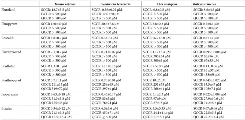
Fig 3. Masked by glucose, the consequences of SDH inhibition by SDHIs in human cells in culture. A, D, Control fibroblasts
cultured for ten days in the presence (A) or absence (D) of glucose, glutamine being the sole source of carbon in the latter condition. The culture medium was not changed during the experiment. B, E, Cells were grown for a similar duration (ten days) without changing the medium in the presence of 1μM bixafen. Note the presence of numerous white spots in the absence of glucose (E), indicative of the presence of dead cells in the low-density culture. C, F, Despite the presence of 20μM bixafen, after 10 days of cultivation, no sign of cellular suffering was observed in the presence of glucose (C). A mirror experiment carried out in the absence of glucose (F) resulted in massive cell death at as low as 5μM bixafen. In each figure, the black bar represents 200 μm.
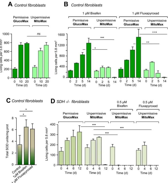
Fig 4. Death of control and SDH-deficient patient cells induced by SDHIs in permissive GlucoMax and nonpermissive MitoMax media. A,
Control cells from three different individuals were allowed to grow in culture media either permissive or nonpermissive for defective mitochondria (seeμ Table). In addition to glucose (4.5 g/l; hyperglycemic), the permissive medium contains 4 mM glutamine, 2 mM pyruvate (both a potential additional carbon source and a potent H2O2scavenger), and 200μM uridine, an amino acid, possibly in insufficient amount when a profound defect
affects the terminal part of the respiratory chain (CIII or CIV;S1 Fig) [35]. B, SDHIs (1μM) were tested (n = 3) in permissive and nonpermissive media. Both bixafen and fluxapyroxad resulted in massive cell death after 14 days of cultivation under nonpermissive conditions. Noticeably, similar attempts to test for the effect of boscalid at effective concentration (computed from IC50determination toward human SDH;Fig 2) resulted in cell
death as well, but data were obscured due to the formation of a precipitate in the culture medium. C, Superoxide dismutase activity of control fibroblasts (n = 4) grown under permissive conditions (glucose 1 g/l) in the absence (control DMSO) or presence of 1μM bixafen or 1 μM
fluxapyroxad. D, Effect of bixafen and fluxapyroxad on patient fibroblasts with 60% residual SDH activity [18]. In nonpermissive medium, massive cell death was observed at 4 days of cultivation in the presence of 0.5μM bixafen and 6 days for the same concentration of fluxapyroxad. Noticeably, the
deleterious consequences observed in patients with mitochondrial RC defects and could lead to major brain dysfunction [32]. Notably, regular changing of the buffered cell culture medium artificially reduces the incidence of lactate accumulation, resulting from the effect of SDHIs on the mitochondria. In cells grown under this condition in the presence of 20μM bixafen, the malonate-sensitive SCCR activity was found to be 80% inhibited, despite washing twice with 10 ml of PBS, trypsinization (2 ml), subsequent addition of 10 ml of trypsin stop buffer fol-lowed by cell centrifugation and a final wash in 50 ml of PBS. Simultaneously, we observed a significant doubling of total SOD activity after 13 days of cultivation in the presence of either 1μM bixafen or 1 μM fluxapyroxad in a permissive medium (1 g/l glucose) (Fig 4C). Such induction of SOD activity is known to specifically reflect increased superoxide production by the mitochondrial RC [33], an increase that may account for the previously reported genotoxic effect of bixafen [34], and was previously observed in the presence of downstream inhibitors of the SDH complex [6].
Control fibroblasts were next cultured in a medium supplemented with glutamine (4 mM) as the only carbon source (Fig 3D–3F). In this challenging medium (nonpermissive MitoMax medium), energy metabolism became fully dependent on mitochondrial activity, allowing detection of the effect of SDH inhibition by bixafen. Under these conditions, using 1μM bixa-fen for 10 days, we observed a significant reduction in the number of cells accompanied by the appearance of numerous dead cells (refractive white spots under the light microscope). A simi-lar duration (10 days) of exposure to 5μM bixafen resulted in the death of almost all cells (Fig 3F).
We next enriched the glucose-containing medium, making it even more favorable for the growth of RC-deficient cells, by increasing the glucose concentration to 4.5 g/l (25 mM) and adding 2 mM pyruvate and 200μM uridine (permissive GlucoMax medium) (S1 Table). Upon cultivation in such a permissive medium for the growth of mitochondria-defective cells, fibro-blasts can cope with most type of RC deficiency or inhibition, including the nonspecific effects of the SDHIs targeting CIII. Notably, the growth of the control fibroblasts was similar in both GlucoMax medium, which was permissive for defective mitochondria, and MitoMax medium, which lacked pyruvate and uridine and contained glutamine as the only carbon source (chal-lenging medium for mitochondria) (Fig 4A). As expected, the presence of bixafen did not affect the growth of cells in the permissive GlucoMax medium. However, bixafen caused the death of almost all cells after 14 days of cultivation in the challenging MitoMax medium (Figs
4Band5A). Notably, the observed effect was not restricted to bixafen, which was shown to inhibit both SDH (CII) and CIII (Table 1), and a similar effect was observed with fluxapyr-oxad, another new-generation SDHI (Fig 4B) that did not inhibit CIII in human cells (Table 1).
SDHIs potently affect human cells with defective mitochondria
We next investigated the effect of SDHIs on cultured skin fibroblasts from patients with mito-chondrial defects linked to either a partial SDH deficiency [18] or a hypersensitivity to oxida-tive insults due to impaired SOD signaling resulting from defecoxida-tive nuclear Nrf2 translocation [21,23,36]. The patient with an SDH deficiency presenting as Leigh syndrome [18] had 60% residual SCCR activity (1.8± 0.3 nmol/min/mg prot; control 3.0 ± 0.3 nmol/min/mg prot, n = 4) in fibroblasts. We first compared the growth of these SDH-deficient cells in the permis-sive GlucoMax medium to that in the challenging MitoMax medium (Fig 4D). After an initial
used SDHI concentration (0.5μM) is below the ADI for each of these molecules (0.58 μM) (S2 Fig). Culture media were not changed for the duration of the experiment (n = 3).
slow growth rate observed in the MitoMax medium, cell death took over, resulting in near-total cell loss at 12 days. In the presence of 0.5μM bixafen, total cell loss was observed after 4 days of cultivation (Fig 4D). In the presence of 0.5μM fluxapyroxad, total cell loss was observed at day 6.
We next studied the effect of bixafen on fibroblasts selected for their hypersensitivity to oxi-dative insults from patients suffering from either FRDA [33] or FDA [24] [37] (Fig 5). Com-pared to control fibroblasts (Fig 5A), FRDA fibroblasts showed acceleration of cell death that was particularly marked at the highest concentration of bixafen (1μM) for cells grown in MitoMax medium (Fig 5B). An identical, although less marked, phenomenon was observed for FAD cells (Fig 5C). After 4 days of growth in MitoMax medium, these cells exhibited sus-ceptibility to 1μM bixafen (Fig 5C), while control cells (Fig 5A) remained perfectly viable. Thus, abnormality in the functioning of mitochondria that directly affects the RC (defective SDH activity) or the ability to respond to oxidative stress (defective SOD induction) leads to hypersensitivity to the inhibitory effect of SDHIs.
Discussion
The above data establish the inhibitory effects of eight SDHIs on SDHs from four biological species, namely, the fungusB. cinerea, the earthworm L. terrestris, the honeybee A. mellifera,
and humans (H. sapiens), with varying inhibition strengths. This finding reflects the high
con-servation of the SDH B-D subunits that constitute the ubiquinone-reducing site of SDH (S2–
S4Figs). The lack of species specificity of the inhibitory effect of carboxin, the predecessor of most SDHIs, was recognized as early as 1976 [16], when the authors noted the extremely high affinity of carboxin for mammalian SDH [38]. Once widely sprayed as a fungicide on Cana-dian wheat, carboxin, used in doses similar to those used for the SDHIs derived from it, is indi-cated to be a major environmental hazard due to its nonspecificity [38]. In this work, we extended this observation to a series of SDHIs derived from carboxin, with which they share a common mode of action for inhibition of SDH.
The lack of species specificity of the inhibitory effect of SDHIs due to this mechanism of action is indeed known to agrochemical manufacturers, part of the SDHIs were reported to also be efficient against nematodes [39]. Similarly, the hazard represented by SDHIs for aquatic organisms has long been recognized, as indicated in the specification sheets, and confirmed by recent data obtained forDanio rerio with boscalid or penthiopyrad [40,41] and forXenopus tropicalis embryos with isopyrazam or bixafen [42]. Simultaneously, the toxic effects of bosca-lid on honeybees have been demonstrated to occur as long as the duration of exposure is suffi-cient [43]. The inhibitory effect of SDHIs presumably extends to most living organisms, modulated by the propensity of each organism to absorb, metabolize and/or excrete each of the SDHIs. Thus, the leaves of plants that exhibit poor absorption of SDHIs will be spared as long as the pesticide does not come into contact with the roots, which are likely to absorb the SDHI. Finally, the toxicological interactions resulting from the frequent use of a mixture of pesticides have been largely ignored, especially those associated with compounds such as CIII inhibitors (e.g., strobilurin), which are often added to commercial preparations of SDHIs [42]. The toxic effects on the fauna of additional permeabilizing agents which are found in several SDHI mixtures have been similarly largely ignored.
Fig 5. Death of control, FRDA and FAD patient cells induced by bixafen. A, Control cells were allowed to grow
(n = 3) in either GlucoMax or MitoMax culture medium, which are permissive or nonpermissive for defective mitochondria (seeTable 2), respectively, in the absence or presence of 0.5μM or 1 μM bixafen. A similar experiment was carried out on fibroblasts from a Friedreich ataxia patient (FRDA fibroblasts) (B) and from a patient suffering from familial Alzheimer’s Disease (C). Culture media were not changed for the duration of the experiment (n = 3).
It is extremely hazardous to compare the IC50values obtainedin vitro under laboratory
conditions with the concentrations of SDHI that might result from the application of these pesticides on crops. One can however reliably determine the concentration of SDHI at the out-let of the diffusion nozzles. In the case of bixafen, it is about 0.6 to 1.8 mM according to the rec-ommendations distributed to farmers, resulting in a cloud of nebulization, corresponding to 75–125 g/hectare of bixafen. The final exposition depends on numerous factors hard to con-trol, including the spreading conditions, the nature of the ground, its vegetation cover, etc. From a regulatory point of view, the ADI for example for boscalid and bixafen amounts to 0.04 and 0.02 mg / kg / day, respectively (see legendS2 Fig). For an average adult (60 kg / blood volume 5 l), this correspond to a blood concentration of approximately 1.40 and 0.58μM, respectively.
To date, SDHIs have been reported to potently inhibit SDH activity, but the effect of SDHIs on additional sites, especially in the RC, has not been described. Here, we show that several of the latest-generation SDHIs inhibit CIII in addition to SDH, possibly increasing both their fungicidal activity and toxicity toward other organisms. To date, this additional effect has not been considered in the marketing of any of these molecules. Interestingly, however, SDHI-spe-cific non-target site resistance has been recently described as being indicative of at least a sec-ond site of SDHI action [44].
The effects of RC inhibition on the growth of human cells in culture are greatly dependent on the balance between glycolysis and mitochondrial activity. Thus, so-called rho˚ cells, which lack mitochondrial DNA and therefore lack RC activity, are auxotrophic for uridine but can generate ATP from available glucose through glycolysis [45], producing a large amount of lac-tate (S1 Fig). Notably, complete disruption of the terminal part of the RC (CIII, CIV) results in the blockade of dihydroorotate dehydrogenase [46]. This latter enzyme normally produces the orotate required for pyrimidine synthesis, which is mandatory for cellular proliferation (S1 Fig), making uridine supplementation necessary to for strong growth of cells defective in CIII or CIV activity [46] or completely lacking mitochondrial DNA and therefore lacking a func-tional RC [45]. This is demonstrated by comparison of the permissive GlucoMax medium and the nonpermissive MitoMax medium, where the latter alone allows the detection of cells har-boring mitochondria with deficient RCs. Notably, the use of this nonpermissive MitoMax medium is not intended to represent a given physiological situation but to allow detecting a possible deleterious effect of a given substance toward mitochondria. This MitoMax medium should be used a standard for testing the toxicity of any substance targeting mitochondria in cell culture in the context of hazard assessment in non-target organisms. In this study, we showed effective application of this strategy for SDHI-treated cells. Indeed, using standard glu-cose-containing media makes it impossible to detect even severe blockade of SDH in human cells. These experiments suggest the need for reevaluation of SDHI toxicity in human cultured cells using mitochondria-challenging MitoMax medium containing glutamine as the sole source of carbon. In any case, given the complexity of the phenomena possibly resulting from a mitochondrial stress (overproduction of superoxides, lactate accumulation, shortage of ATP, metabolic imbalance, etc.) it is central to respect a sufficient duration (>15 days) for these eval-uations, based on the concentration of the inhibitor used.
Another aspect shown by this study is the increased sensitivity of cells with defective mito-chondria to SDHIs. This finding holds true regardless of the nature of the mitomito-chondrial defect, directly resulting from a partial deficiency of the RC or from hypersensitivity to oxida-tive stress due to poor induction of SOD. Isolated and partial SDH defects are rare human con-ditions that are generally associated with neurological or cardiac phenotypes [3]. However, in addition to the profound SDH defects evidenced in phaeochromocytomas and paragangliomas or gastrointestinal stromal tumor tissues of patients with germline mutations in the SDH
tumor suppressor genes [47–50], a partial SDH deficiency has been observed in a number of human diseases that affect one or several organs,i.e., FRDA [51], Barth syndrome [52], various leukoencephalopathies [53] [54] [55], asthenozoospermia male infertility [56], myopathy [57], rare hemolytic uremic syndromes and rhabdomyolysis [58]. This feature defines a subpopula-tion that might be particularly at risk when in contact with SDHIs. Our results suggest that in this subpopulation and but also in populations, such as FRDA and Alzheimer’s disease, known to present increased susceptibility to oxidative stress, a very common feature among human diseases, SDHIs could contribute to acceleration of disease progression.
Conclusions
This work first establishes that, similar to their predecessor molecule carboxin [16], all tested SDHIs inhibit SDH in all the species considered, albeit with varying efficiency. Moreover, the tested next-generation SDHIs containing a methyl-pyrazol moiety (S2 Fig) also inhibit RC CIII. This lack of specificity represents a major problem considering the current widespread use of these SDHIs. While this lack of selectivity might be the source of the efficiency of last-generation SDHIs, it might also constitute an additional hazard for the exposed organisms. Our study next established that the standard conditions used to test the eventual toxicity of SDHIs (as well as of any other mitochondria-targeted pesticides) can mask potential toxic effect. Thus, we recommend regulatory testing for determination of molecular toxicity and the use of MitoMax growth medium in which glutamine is the sole source of carbon.
Finally, we show that a pre-existing mitochondrial defect, such as partial SDH dysfunction or hypersensitivity to oxidative insults (FRDA, FAD), increases the susceptibility to SDHIs, suggesting a particular risk for individuals with such a dysfunction.
Supporting information
S1 Table. Permissiveversus nonpermissive culture media for mitochondrial respiratory chain-deficient human cells.
(PDF)
S1 Fig. Succinate dehydrogenase: A crossroad between the respiratory chain and interme-diary metabolism. Fum, fumarate; KG,α-ketoglutarate; Succ, succinate; TAC, tricarboxylic acid cycle; I-V, the various complexes of the respiratory chain (II, complex II or SDH). (PDF)
S2 Fig. SDHI molecular structure. The yellow and blue parts for each molecule are
reminis-cent of the predecessor carboxin structure (top). The red dotted lines underline the methyl-pyrazol moiety present in the different SDHIs of the recent generation that also inhibit respira-tory chain complex III. Boscalid (ADI: 0.04 mg/kg/d) was introduced in 2003 in the USA by BASF, fluopyram (ADI: 0.012 mg/kg/d) in 2010 in the USA by Bayer, flutolanil (0.09 mg/kg/d) in 1981 in the USA by Nichino America, penflufen (ADI: 0.04 mg/kg/d) in 2012 in the USA by Bayer, isopyrazam (ADI: 0.03 mg/kg/d) in 2010 in England by Syngenta, penthiopyrad (ADI: 0.1 mg/kg/d) in 2011 in the USA by Dupont-Fontelis, fluxapyroxad (ADI: 0.02 mg/kg/d)in 2011 in France by BASF, and bixafen (ADI: 0.02 mg/kg/d) in 2011 in England by Bayer. ADI; Acceptable Daily Intake according to European regulations.
(PDF)
S3 Fig. Amino acids that are involved in the SDH quinone-binding site or mutation of which confers SDHI resistance. A, Crystal structure of the quinone-binding site of the
por-cine (Sus scrofa) heart mitochondrial SDH (EC 1.3.5.1) drawn based on information from the
using Roman characters, while the italic characters outside the figure correspond to the num-bering of theBotrytis cinerea sequence. B, Schematic depiction of the ubiquinone-binding site
of SDH featuring some of the amino acids that have been said to favor fungal resistance to SDHIs (Sierotzki and Scalliet 2013). Encircled by the dotted line, a simplified representation of SDHIs shows in yellow the part of the molecule accommodated by the polar cavity of the ubi-quinone-binding site of SDH; the part accommodated by the hydrophobic pocket is shown in blue.
(PDF)
S4 Fig. Sequence analysis of the amino acid compositions of succinate dehydrogenase sub-units B, C, and D. A, List of the 22 organisms selected for alignment with the references of
each protein sequence as deposited in the NCBI database. B, C, D, For each subunit, the graphic representation illustrates the conservation (indicated in red) of the subunit among the 22 species selected, followed by the alignment showing the amino acids present at the positions (gray background/dark arrows) supposedly involved in the resistance of fungi to SDHIs or/ and in the binding of coenzyme Q (S3 Fig) [29]. Alignment was performed using the COBALT multiple sequence alignment tool (https://www.ncbi.nlm.nih.gov/tools/cobalt/re_cobalt.cgi). (PDF)
Author Contributions
Conceptualization: Paule Be´nit, Laurence Huc, Manuel Schiff, Anne-Paule
Gimenez-Roque-plo, Judith Favier, Pierre Gressens, Pierre Rustin.
Funding acquisition: Paule Be´nit.
Investigation: Paule Be´nit, Agathe Kahn, Dominique Chretien, Malgorzata Rak, Pierre
Rustin.
Methodology: Pierre Rustin. Supervision: Pierre Gressens.
Validation: Anne-Paule Gimenez-Roqueplo.
Writing – original draft: Paule Be´nit, Sylvie Bortoli, Laurence Huc, Manuel Schiff,
Anne-Paule Gimenez-Roqueplo, Judith Favier, Malgorzata Rak, Pierre Rustin.
Writing – review & editing: Sylvie Bortoli, Laurence Huc, Manuel Schiff, Anne-Paule
Gime-nez-Roqueplo, Judith Favier, Pierre Gressens, Pierre Rustin.
References
1. Ackrell B, Johnson M, Gunsalus R, Cecchini G. Structure and function of succinate dehydrogenase and fumarate reductase. Muller F, editor. Boca Raton, FL CRC Press; 1990.
2. Her YF, Maher LJ 3rd. Succinate Dehydrogenase Loss in Familial Paraganglioma: Biochemistry, Genetics, and Epigenetics. Int J Endocrinol. 2015; 2015:296167. Epub 2015/08/22.https://doi.org/10. 1155/2015/296167PMID:26294907.
3. Be´nit P, Letouze E, Rak M, Aubry L, Burnichon N, Favier J, et al. Unsuspected task for an old team: suc-cinate, fumarate and other Krebs cycle acids in metabolic remodeling. Biochim Biophys Acta. 2014; 1837(8):1330–7. Epub 2014/04/05. S0005-2728(14)00100-5 [pii]https://doi.org/10.1016/j.bbabio.2014. 03.013PMID:24699309.
4. Francis K, Smitherman C, Nishino SF, Spain JC, Gadda G. The biochemistry of the metabolic poison propionate 3-nitronate and its conjugate acid, 3-nitropropionate. IUBMB Life. 2013; 65(9):759–68. Epub 2013/07/31.https://doi.org/10.1002/iub.1195PMID:23893873.
5. Teplova VV, Belosludtsev KN, Kruglov AG. Mechanism of triclosan toxicity: Mitochondrial dysfunction including complex II inhibition, superoxide release and uncoupling of oxidative phosphorylation. Toxicol
Lett. 2017; 275:108–17. Epub 2017/05/10. S0378-4274(17)30172-8 [pii]https://doi.org/10.1016/j.toxlet. 2017.05.004PMID:28478158.
6. Kruspig B, Valter K, Skender B, Zhivotovsky B, Gogvadze V. Targeting succinate:ubiquinone reductase potentiates the efficacy of anticancer therapy. Biochim Biophys Acta. 2016; 1863(8):2065–71. Epub 2016/05/04. S0167-4889(16)30126-4 [pii]https://doi.org/10.1016/j.bbamcr.2016.04.026PMID:
27140478.
7. Morin A, Letouze E, Gimenez-Roqueplo AP, Favier J. Oncometabolites-driven tumorigenesis: From genetics to targeted therapy. Int J Cancer. 2014; 135(10):2237–48. Epub 2014/08/16.https://doi.org/ 10.1002/ijc.29080PMID:25124653.
8. Flannery PJ, Trushina E. Mitochondrial dynamics and transport in Alzheimer’s disease. Mol Cell Neu-rosci. 2019; 98:109–20. Epub 2019/06/20. S1044-7431(19)30094-6 [pii]https://doi.org/10.1016/j.mcn. 2019.06.009PMID:31216425.
9. Chre´tien D, Be´nit P, Ha HH, Keipert S, El-Khoury R, Chang YT, et al. Mitochondria are physiologically maintained at close to 50 degrees C. PLoS Biol. 2018; 16(1):e2003992. Epub 2018/01/26.https://doi. org/10.1371/journal.pbio.2003992pbio.2003992 [pii]. PMID:29370167.
10. Neagu M, Constantin C, Popescu ID, Zipeto D, Tzanakakis G, Nikitovic D, et al. Inflammation and Metabolism in Cancer Cell-Mitochondria Key Player. Front Oncol. 2019; 9:348. Epub 2019/05/30.
https://doi.org/10.3389/fonc.2019.00348PMID:31139559.
11. Spinelli JB, Haigis MC. The multifaceted contributions of mitochondria to cellular metabolism. Nat Cell Biol. 2018; 20(7):745–54. Epub 2018/06/29.https://doi.org/10.1038/s41556-018-0124-1[pii]. PMID:
29950572.
12. Canto C, Menzies KJ, Auwerx J. NAD(+) Metabolism and the Control of Energy Homeostasis: A Balanc-ing Act between Mitochondria and the Nucleus. Cell Metab. 2015; 22(1):31–53. Epub 2015/06/30. S1550-4131(15)00266-1 [pii]https://doi.org/10.1016/j.cmet.2015.05.023PMID:26118927.
13. Manoli I, Alesci S, Blackman MR, Su YA, Rennert OM, Chrousos GP. Mitochondria as key components of the stress response. Trends Endocrinol Metab. 2007; 18(5):190–8. Epub 2007/05/15. S1043-2760 (07)00069-0 [pii]https://doi.org/10.1016/j.tem.2007.04.004PMID:17500006.
14. Zhang H, Menzies KJ, Auwerx J. The role of mitochondria in stem cell fate and aging. Development. 2018; 145(8). Epub 2018/04/15. 145/8/dev143420 [pii]https://doi.org/10.1242/dev.143420PMID:
29654217.
15. Be´nit P, Bortoli S, Chre´tien D, Rak M, Rustin P. Pathologies lie´s au cycle de Krebs. Rev Francophone Laboratoires. 2018; 501:49–57.
16. Mowery PC, Ackrell BA, Singer TP. Carboxins: powerful selective inhibitors of succinate oxidation in animal tissues. Biochem Biophys Res Commun. 1976; 71(1):354–61. Epub 1976/07/12. 0006-291X (76)90290-4 [pii].https://doi.org/10.1016/0006-291x(76)90290-4PMID:962926.
17. Rustin P, Chre´tien D, Bourgeron T, Ge´rard B, Rotig A, Saudubray JM, et al. Biochemical and molecular investigations in respiratory chain deficiencies. Clin Chim Acta. 1994; 228(1):35–51.https://doi.org/10. 1016/0009-8981(94)90055-8PMID:7955428.
18. Bourgeron T, Rustin P, Chre´tien D, Birch-Machin M, Bourgeois M, Viegas-Pequignot E, et al. Mutation of a nuclear succinate dehydrogenase gene results in mitochondrial respiratory chain deficiency. Nat Genet. 1995; 11(2):144–9.https://doi.org/10.1038/ng1095-144PMID:7550341.
19. Bayot A, Reichman S, Lebon S, Csaba Z, Aubry L, Sterkers G, et al. Cis-silencing of PIP5K1B evi-denced in Friedreich’s ataxia patient cells results in cytoskeleton anomalies. Hum Mol Genet. 2013. Epub 2013/04/05. ddt144 [pii]https://doi.org/10.1093/hmg/ddt144PMID:23552101.
20. Bayot A, Reichman S, Lebon S, Csaba Z, Aubry L, Sterkers G, et al. Cis-silencing of PIP5K1B evi-denced in Friedreich’s ataxia patient cells results in cytoskeleton anomalies. Hum Mol Genet. 2013; 22 (14):2894–904. Epub 2013/04/05. ddt144 [pii]https://doi.org/10.1093/hmg/ddt144PMID:23552101.
21. Paupe V, Dassa EP, Goncalves S, Auchere F, Lonn M, Holmgren A, et al. Impaired nuclear Nrf2 trans-location undermines the oxidative stress response in friedreich ataxia. PLoS ONE. 2009; 4(1):e4253.
https://doi.org/10.1371/journal.pone.0004253PMID:19158945.
22. Takeda M, Tatebayashi Y, Nishimura T. Change in the cytoskeletal system in fibroblasts from patients with familial Alzheimer’s disease. Prog Neuropsychopharmacol Biol Psychiatry. 1992; 16(3):317–28. Epub 1992/05/01.https://doi.org/10.1016/0278-5846(92)90083-qPMID:1589589.
23. Strachan GD, Morgan KL, Otis LL, Caltagarone J, Gittis A, Bowser R, et al. Fetal Alz-50 clone 1 inter-acts with the human orthologue of the Kelch-like Ech-associated protein. Biochemistry. 2004; 43 (38):12113–22. Epub 2004/09/24.https://doi.org/10.1021/bi0494166PMID:15379550.
24. Tesco G, Latorraca S, Piersanti P, Piacentini S, Amaducci L, Sorbi S. Alzheimer skin fibroblasts show increased susceptibility to free radicals. Mech Ageing Dev. 1992; 66(2):117–20. Epub 1992/11/01. 0047-6374(92)90129-2 [pii].https://doi.org/10.1016/0047-6374(92)90129-2PMID:1365838.
25. Tesco G, Latorraca S, Piersanti P, Sorbi S, Piacentini S, Amaducci L. Free radical injury in skin cultured fibroblasts from Alzheimer’s disease patients. Ann N Y Acad Sci. 1992; 673:149–53. Epub 1992/12/26.
https://doi.org/10.1111/j.1749-6632.1992.tb27446.xPMID:1485712.
26. Be´nit P, Goncalves S, Philippe Dassa E, Brière JJ, Martin G, Rustin P. Three spectrophotometric assays for the measurement of the five respiratory chain complexes in minuscule biological samples. Clin Chim Acta. 2006; 374:81–6.https://doi.org/10.1016/j.cca.2006.05.034PMID:16828729.
27. Marklund S, Marklund G. Involvement of the superoxide anion radical in the autoxidation of pyrogallol and a convenient assay for superoxide dismutase. Eur J Biochem. 1974; 47(3):469–74.https://doi.org/ 10.1111/j.1432-1033.1974.tb03714.xPMID:4215654.
28. Papadopoulos JS, Agarwala R. COBALT: constraint-based alignment tool for multiple protein
sequences. Bioinformatics. 2007; 23(9):1073–9. Epub 2007/03/03. btm076 [pii]https://doi.org/10.1093/ bioinformatics/btm076PMID:17332019.
29. Sierotzki H, Scalliet G. A review of current knowledge of resistance aspects for the next-generation suc-cinate dehydrogenase inhibitor fungicides. Phytopathology. 2013; 103(9):880–7. Epub 2013/04/19.
https://doi.org/10.1094/PHYTO-01-13-0009-RVWPMID:23593940.
30. Be´nit P, Pelhaitre A, Saunier E, Bortoli S, Coulibaly A, Rak M, et al. Paradoxical Inhibition of Glycolysis by Pioglitazone Opposes the Mitochondriopathy Caused by AIF Deficiency. EBioMedicine. 2017; 17:75–87. Epub 2017/02/24. S2352-3964(17)30072-5 [pii]https://doi.org/10.1016/j.ebiom.2017.02.013
PMID:28229909.
31. Manel N, Kinet S, Kim FJ, Taylor N, Sitbon M, Battini JL. [GLUT-1 is the receptor of retrovirus HTLV]. Med Sci (Paris). 2004; 20(3):277–9. Epub 2004/04/07. 007846ar [pii]https://doi.org/10.1051/medsci/ 2004203277PMID:15067572.
32. El-Hattab AW, Adesina AM, Jones J, Scaglia F. MELAS syndrome: Clinical manifestations, pathogene-sis, and treatment options. Mol Genet Metab. 2015. Epub 2015/06/23. S1096-7192(15)30024-X [pii]
https://doi.org/10.1016/j.ymgme.2015.06.004PMID:26095523.
33. Chantrel-Groussard K, Geromel V, Puccio H, Koenig M, Munnich A, Rotig A, et al. Disabled early recruitment of antioxidant defenses in Friedreich’s ataxia. Hum Mol Genet. 2001; 10(19):2061–7.
https://doi.org/10.1093/hmg/10.19.2061PMID:11590123.
34. Graillot V, Tomasetig F, Cravedi JP, Audebert M. Evidence of the in vitro genotoxicity of methyl-pyra-zole pesticides in human cells. Mutat Res. 2012; 748(1–2):8–16. Epub 2012/06/30. S1383-5718(12) 00224-0 [pii]https://doi.org/10.1016/j.mrgentox.2012.05.014PMID:22743356.
35. Bourgeron T, Chre´tien D, Rotig A, Munnich A, Rustin P. Fate and expression of the deleted mitochon-drial DNA differ between human heteroplasmic skin fibroblast and Epstein-Barr virus-transformed lym-phocyte cultures. J Biol Chem. 1993; 268(26):19369–76. PMID:8396136.
36. Mota SI, Costa RO, Ferreira IL, Santana I, Caldeira GL, Padovano C, et al. Oxidative stress involving changes in Nrf2 and ER stress in early stages of Alzheimer’s disease. Biochim Biophys Acta. 2015; 1852(7):1428–41. Epub 2015/04/11. S0925-4439(15)00106-4 [pii]https://doi.org/10.1016/j.bbadis. 2015.03.015PMID:25857617.
37. Majd S, Power JHT. Oxidative Stress and Decreased Mitochondrial Superoxide Dismutase 2 and Per-oxiredoxins 1 and 4 Based Mechanism of Concurrent Activation of AMPK and mTOR in Alzheimer’s Disease. Curr Alzheimer Res. 2018; 15(8):764–76. Epub 2018/02/24. CAR-EPUB-88760 [pii]https:// doi.org/10.2174/1567205015666180223093020PMID:29473507.
38. Singer TP, Ramsay RR, Ackrell BA. Deficiencies of NADH and succinate dehydrogenases in degenera-tive diseases and myopathies. Biochim Biophys Acta. 1995; 1271(1):211–9. Epub 1995/05/24. 0925-4439(95)00030-8 [pii].https://doi.org/10.1016/0925-4439(95)00030-8PMID:7599211.
39. Faske TR, Hurd K. Sensitivity of Meloidogyne incognita and Rotylenchulus reniformis to Fluopyram. J Nematol. 2015; 47(4):316–21. Epub 2016/03/05. PMID:26941460.
40. Qian L, Zhang J, Chen X, Qi S, Wu P, Wang C. Toxic effects of boscalid in adult zebrafish (Danio rerio) on carbohydrate and lipid metabolism. Environ Pollut. 2019; 247:775–82. Epub 2019/02/06. S0269-7491(18)34233-7 [pii]https://doi.org/10.1016/j.envpol.2019.01.054PMID:30721868.
41. Qian L, Qi S, Cao F, Zhang J, Li C, Song M, et al. Effects of penthiopyrad on the development and behaviour of zebrafish in early-life stages. Chemosphere. 2019; 214:184–94. Epub 2018/09/29. S0045-6535(18)31773-9 [pii]https://doi.org/10.1016/j.chemosphere.2018.09.117PMID:30265925.
42. Wu S, Lei L, Liu M, Song Y, Lu S, Li D, et al. Single and mixture toxicity of strobilurin and SDHI fungi-cides to Xenopus tropicalis embryos. Ecotoxicol Environ Saf. 2018; 153:8–15. Epub 2018/02/07. S0147-6513(18)30055-1 [pii]https://doi.org/10.1016/j.ecoenv.2018.01.045PMID:29407742.
43. Simon-Delso N, San Martin G, Bruneau E, Hautier L. Time-to-death approach to reveal chronic and cumulative toxicity of a fungicide for honeybees not revealed with the standard ten-day test. Sci Rep. 2018; 8(1):7241. Epub 2018/05/10.https://doi.org/10.1038/s41598-018-24746-9[pii]. PMID:
44. Yamashita M, Fraaije B. Non-target site SDHI resistance is present as standing genetic variation in field populations of Zymoseptoria tritici. Pest Manag Sci. 2018; 74(3):672–81. Epub 2017/10/13.https://doi. org/10.1002/ps.4761PMID:29024365.
45. Morais R, Gregoire M, Jeannotte L, Gravel D. Chick embryo cells rendered respiration-deficient by chloramphenicol and ethidium bromide are auxotrophic for pyrimidines. Biochem Biophys Res Com-mun. 1980; 94(1):71–7. Epub 1980/05/14. S0006-291X(80)80189-6 [pii].https://doi.org/10.1016/ s0006-291x(80)80189-6PMID:6248067.
46. Ge´rard B, Bourgeron T, Chre´tien D, Rotig A, Munnich A, Rustin P. Uridine preserves the expression of respiratory enzyme deficiencies in cultured fibroblasts. Eur J Pediatr. 1993; 152(3):270.https://doi.org/ 10.1007/bf01956163PMID:8383055.
47. Favier J, Brière JJ, Strompf L, Amar L, Filali M, Jeunemaitre X, et al. Hereditary Paraganglioma/Pheo-chromocytoma and Inherited Succinate Dehydrogenase Deficiency. Horm Res. 2005; 63(4):171–9.
https://doi.org/10.1159/000084685PMID:15795514.
48. Boikos SA, Xekouki P, Fumagalli E, Faucz FR, Raygada M, Szarek E, et al. Carney triad can be (rarely) associated with germline succinate dehydrogenase defects. Eur J Hum Genet. 2016; 24(4):569–73. Epub 2015/07/16. ejhg2015142 [pii]https://doi.org/10.1038/ejhg.2015.142PMID:26173966.
49. Miettinen M, Lasota J. Succinate dehydrogenase deficient gastrointestinal stromal tumors (GISTs)—a review. Int J Biochem Cell Biol. 2014; 53:514–9. Epub 2014/06/03. S1357-2725(14)00191-5 [pii]https:// doi.org/10.1016/j.biocel.2014.05.033PMID:24886695.
50. Dubard Gault M, Mandelker D, DeLair D, Stewart CR, Kemel Y, Sheehan MR, et al. Germline SDHA mutations in children and adults with cancer. Cold Spring Harb Mol Case Stud. 2018; 4(4). Epub 2018/ 08/03. mcs.a002584 [pii]https://doi.org/10.1101/mcs.a002584PMID:30068732.
51. Rotig A, de Lonlay P, Chre´tien D, Foury F, Koenig M, Sidi D, et al. Aconitase and mitochondrial iron-sul-phur protein deficiency in Friedreich ataxia. Nat Genet. 1997; 17(2):215–7.https://doi.org/10.1038/ ng1097-215PMID:9326946.
52. Dudek J, Cheng IF, Chowdhury A, Wozny K, Balleininger M, Reinhold R, et al. Cardiac-specific succi-nate dehydrogenase deficiency in Barth syndrome. EMBO Mol Med. 2016; 8(2):139–54. Epub 2015/12/ 25. emmm.201505644 [pii]https://doi.org/10.15252/emmm.201505644PMID:26697888.
53. Ardissone A, Invernizzi F, Nasca A, Moroni I, Farina L, Ghezzi D. Mitochondrial leukoencephalopathy and complex II deficiency associated with a recessive SDHB mutation with reduced penetrance. Mol Genet Metab Rep. 2015; 5:51–4. Epub 2016/03/01.https://doi.org/10.1016/j.ymgmr.2015.10.006
S2214-4269(15)30041-0 [pii]. PMID:26925370.
54. Helman G, Caldovic L, Whitehead MT, Simons C, Brockmann K, Edvardson S, et al. Magnetic reso-nance imaging spectrum of succinate dehydrogenase-related infantile leukoencephalopathy. Ann Neu-rol. 2016; 79(3):379–86. Epub 2015/12/09.https://doi.org/10.1002/ana.24572PMID:26642834.
55. Ishiyama A, Sakai C, Matsushima Y, Noguchi S, Mitsuhashi S, Endo Y, et al. IBA57 mutations abrogate iron-sulfur cluster assembly leading to cavitating leukoencephalopathy. Neurol Genet. 2017; 3(5):e184. Epub 2017/09/16.https://doi.org/10.1212/NXG.0000000000000184NG2017005421 [pii]. PMID:
28913435.
56. Tomar R, Mishra AK, Mohanty NK, Jain AK. Altered expression of succinic dehydrogenase in astheno-zoospermia infertile male. Am J Reprod Immunol. 2012; 68(6):486–90. Epub 2012/09/19.https://doi. org/10.1111/aji.12023PMID:22985091.
57. Rodrigues Ade S, Kiyomoto BH, Oliveira AS, Gabbai AA, Schmidt B, Tengan CH. Progressive myopa-thy with a combined respiratory chain defect including Complex II. J Neurol Sci. 2008; 264(1–2):182–6. Epub 2007/09/14. S0022-510X(07)00526-6 [pii]https://doi.org/10.1016/j.jns.2007.08.002PMID:
17850823.
58. Micheletti MV, Lavoratti G, Gasperini S, Donati MA, Pela I. Hemolytic uremic syndrome and rhabdomy-olysis in a patient with succinate coenzyme Q reductase (complex II) deficiency. Clin Nephrol. 2011; 76 (1):68–73. Epub 2011/07/05. 8753 [pii].https://doi.org/10.5414/cn106681PMID:21722608.