HAL Id: hal-02174911
https://hal.archives-ouvertes.fr/hal-02174911
Submitted on 10 Nov 2020
HAL is a multi-disciplinary open access archive for the deposit and dissemination of sci-entific research documents, whether they are pub-lished or not. The documents may come from teaching and research institutions in France or abroad, or from public or private research centers.
L’archive ouverte pluridisciplinaire HAL, est destinée au dépôt et à la diffusion de documents scientifiques de niveau recherche, publiés ou non, émanant des établissements d’enseignement et de recherche français ou étrangers, des laboratoires publics ou privés.
cyclase DacA by direct interaction with the
phosphoglucosamine mutase GlmM
Tommaso Tosi, Fumiya Hoshiga, Charlotte Millership, Rahul Singh, Charles
Eldrid, Delphine Patin, Dominique Mengin-Lecreulx, Konstantinos
Thalassinos, Paul Freemont, Angelika Gründling
To cite this version:
Tommaso Tosi, Fumiya Hoshiga, Charlotte Millership, Rahul Singh, Charles Eldrid, et al.. Inhibi-tion of the Staphylococcus aureus c-di-AMP cyclase DacA by direct interacInhibi-tion with the phospho-glucosamine mutase GlmM. PLoS Pathogens, Public Library of Science, 2019, 15 (1), pp.e1007537. �10.1371/journal.ppat.1007537�. �hal-02174911�
Inhibition of the Staphylococcus aureus
c-di-AMP cyclase DacA by direct interaction with
the phosphoglucosamine mutase GlmM
Tommaso TosiID1, Fumiya Hoshiga1, Charlotte Millership1, Rahul SinghID1,Charles EldridID2,3, Delphine PatinID4, Dominique Mengin-LecreulxID4,
Konstantinos ThalassinosID2,3, Paul Freemont5*, Angelika Gru¨ ndlingID1*
1 Section of Microbiology and MRC Centre for Molecular Bacteriology and Infection, Imperial College London, London, United Kingdom, 2 Institute of Structural and Molecular Biology, Birkbeck College, University of London, Malet Street, London, United Kingdom, 3 Institute of Structural and Molecular Biology, Division of Biosciences, University College London, London, United Kingdom, 4 Institute for Integrative Biology of the Cell, CEA, CNRS, Univ Paris-Sud and Universite´ Paris-Saclay, Gif-sur-Yvette, France, 5 Section of Structural Biology, Department of Medicine, Imperial College London, London, United Kingdom
*p.freemont@imperial.ac.uk(PF);a.grundling@imperial.ac.uk(AG)
Abstract
c-di-AMP is an important second messenger molecule that plays a pivotal role in regulating fundamental cellular processes, including osmotic and cell wall homeostasis in many Gram-positive organisms. In the opportunistic human pathogen Staphylococcus aureus, c-di-AMP is produced by the membrane-anchored DacA enzyme. Inactivation of this enzyme leads to a growth arrest under standard laboratory growth conditions and a re-sensitization of methi-cillin-resistant S. aureus (MRSA) strains to ß-lactam antibiotics. The gene coding for DacA is part of the conserved three-gene dacA/ybbR/glmM operon that also encodes the pro-posed DacA regulator YbbR and the essential phosphoglucosamine mutase GlmM, which is required for the production of glucosamine-1-phosphate, an early intermediate of peptido-glycan synthesis. These three proteins are thought to form a complex in vivo and, in this manner, help to fine-tune the cellular c-di-AMP levels. To further characterize this important regulatory complex, we conducted a comprehensive structural and functional analysis of the S. aureus DacA and GlmM enzymes by determining the structures of the S. aureus GlmM enzyme and the catalytic domain of DacA. Both proteins were found to be dimers in solution as well as in the crystal structures. Further site-directed mutagenesis, structural and enzy-matic studies showed that multiple DacA dimers need to interact for enzyenzy-matic activity. We also show that DacA and GlmM form a stable complex in vitro and that S. aureus GlmM, but not Escherichia coli or Pseudomonas aeruginosa GlmM, acts as a strong inhibitor of DacA function without the requirement of any additional cellular factor. Based on Small Angle X-ray Scattering (SAXS) data, a model of the complex revealed that GlmM likely inhibits DacA by masking the active site of the cyclase and preventing higher oligomer formation.
Together these results provide an important mechanistic insight into how c-di-AMP produc-tion can be regulated in the cell.
a1111111111 a1111111111 a1111111111 a1111111111 a1111111111 OPEN ACCESS
Citation: Tosi T, Hoshiga F, Millership C, Singh R,
Eldrid C, Patin D, et al. (2019) Inhibition of the
Staphylococcus aureus c-di-AMP cyclase DacA by
direct interaction with the phosphoglucosamine mutase GlmM. PLoS Pathog 15(1): e1007537.
https://doi.org/10.1371/journal.ppat.1007537
Editor: Michael Otto, National Institutes of Health,
UNITED STATES
Received: September 26, 2018 Accepted: December 18, 2018 Published: January 22, 2019
Copyright:© 2019 Tosi et al. This is an open access article distributed under the terms of the
Creative Commons Attribution License, which permits unrestricted use, distribution, and reproduction in any medium, provided the original author and source are credited.
Data Availability Statement: Structure coordinates
were deposited in the Protein Database, under PDB codes 6GYW (nucleotide-free DacACD), 6GYX (ApCpp-bound DacACD), 6GYY (DacACD-C mutant), 6GYZ (GlmM). SAXS models were deposited in the SASBDB database, under accession codes SASDE78 (DacACD/GlmM complex), SASDE88 (GlmM) and SASDE98 (DacACD). These data have been released under the following links:https://www.rcsb.org/ structure/6GYW https://www.rcsb.org/structure/ 6GYX https://www.rcsb.org/structure/6GYZ
Author summary
c-di-AMP has recently emerged as an essential molecule in Gram-positive bacteria, con-trolling osmotic stress resistance and virulence. The molecule has also been linked to anti-biotic resistance/sensitivity, as alterations in its levels have been shown to modulate the effect of cell wall-targeting antibiotics. c-di-AMP is produced and degraded by dedicated enzymes and the activities of these enzymes are tightly regulated in the cell for optimal bacterial growth. In the human pathogenStaphylococcus aureus, c-di-AMP is produced
by the di-adenylate cyclase enzyme DacA, which is thought to form a complex and be reg-ulated by YbbR and GlmM, two other conserved proteins. There are ongoing efforts to devise strategies to inhibit di-adenylate cyclase enzymes, as inhibition of c-di-AMP pro-duction will negatively impact bacterial growth and also re-sensitize methicillin-resistant
S. aureus (MRSA) strains to ß-lactam antibiotics. Here, we investigated the activity and
regulation of theS. aureus c-di-AMP cyclase DacA. We determined the atomic structures
of theS. aureus DacA and GlmM enzymes and show that GlmM readily binds to DacA in vitro and blocks its activity by masking its active site. With this, our work provides
impor-tant insight into possible ways to ablate c-di-AMP production in bacterial cells.
Introduction
For pathogenic bacteria, the ability to rapidly adapt to the host cell environment or different host cell niches is essential for their infectivity. Nucleotide second-messenger molecules are critical components involved in such adaptive responses, allowing bacteria to simultaneously regulate multiple cellular processes [1]. c-di-AMP has emerged as an important second-mes-senger, in particular in Gram-positive bacteria [2,3]. Several recent studies have shown that c-di-AMP binds to and regulates cellular transport systems for potassium and osmolytes and in this manner likely controls the cellular turgor [2,4–9]. Therefore c-di-AMP has an important role in preserving the integrity and viability of the cell when osmotic conditions change. Fur-thermore, in a number of Gram-positive bacteria, including the human pathogens Staphylo-coccus aureus and Listeria monocytogenes, c-di-AMP impacts the susceptibility to ß-lactam
antibiotics [10–14]. More specifically, high cellular c-di-AMP levels lead to increased ß-lactam resistance and low cellular c-di-AMP levels to decreased resistance [12–15]. Although the molecular mechanism behind this is still unclear, it has been suggested that c-di-AMP might at least in part impact ß-lactam resistance through its regulatory function of potassium and osmolyte transporters and changes in osmotic balance and pressure [9,16]. These findings indicate that blocking c-di-AMP production could potentially be used to re-sensitize patho-gens such as methicillin-resistantS. aureus (MRSA) strains to ß-lactam antibiotics. In order to
exploit such strategies, a better knowledge of the structure and function of c-di-AMP synthesis enzymes and the modulation of their activity is required.
For optimal fitness, bacteria need to tightly regulate c-di-AMP production [8,17–19] [6,11,
20–22]. Its cellular levels are regulated by the balance between its synthesis by dedicated di-adenylate cyclase enzymes (DacA inS. aureus) and its degradation to pApA or AMP by
spe-cific phosphodiesterase enzymes [12,23–28]. These enzymes are part of three main classes, represented by DisA, DacA (CdaA) and CdaS [23,24,29]. Structural and biochemical charac-terization of members of the different c-di-AMP cyclase families have revealed important information on their regulation. The first di-adenylate cyclase to be characterized was the DNA integrity scanning protein DisA fromThermotoga maritima [23]. DisA is a modular
https://www.sasbdb.org/search/?q=SASDE78 https://www.sasbdb.org/search/?q=SASDE88 https://www.sasbdb.org/search/?q=SASDE98.
Funding: This work was supported by the MRC
grant MR/P011071/1 to AG and PF, as well as the Wellcome Trust grant 100289 to AG. We would also like to acknowledge funding from the Wellcome Trust grant 104913/Z/14/Z for the mass spectrometry instrumentation in the KT laboratory and CE is funded by the BBSRC BB/L015382/1 iCASE studentship with Waters. The funders had no role in study design, data collection and analysis, decision to publish, or preparation of the manuscript.
Competing interests: The authors have declared
cytoplasmic protein, in which the di-adenylate cyclase domain (from here on referred to as DAC domain) is linked by a helical linker region to a DNA-binding domain. The protein was present as an octamer (tetramer of dimers) in the crystal structure, with the DAC domains in a head-to-head conformation [23,30]. DisA dependent c-di-AMP production is regulated through multiple mechanisms. Both, the interaction of DisA with the DNA repair protein RadA and the binding of the DisA-associated DNA-binding domain to damaged DNA, nega-tively impact its activity [23,31,32].
Besides DisA, the structure of the CdaS cyclase fromBacillus cereus is available (PDB 2FB5)
and the homologous proteins fromBacillus subtilis and Bacillus thuringiensis have been
char-acterized biochemically [33,34]. The cyclase domain in CdaS proteins is preceded by an N-ter-minal regulatory YojJ domain that is composed of two long alpha-helices. The structural and biochemical analyses of CdaS proteins from differentBacillus species indicate that the protein
is a hexamer in the crystal structure as well as in solution [33,34]. However, in contrast to the oligomeric DisA protein, theB. subtilis CdaS enzyme seemed to be only weakly active in this
higher oligomeric form, as the DAC domains were not found in a head-to-head conformation [33]. Deletion of the N-terminal regulatory helices of theB. subtilis CdaS protein disrupted
hexamer formation, yielding protein dimers with high enzymatic activity [33]. In contrast, deletion of the N-terminal regulatory domain in theB. thuringiensis CdaS protein greatly
inhibited the enzymatic activity of this enzyme. Therefore, while it is clear that the c-di-AMP activity of CdaS-type proteins is regulated by the N-terminal YojJ domain, the exact regulatory mechanism is still unclear and might also differ between differentBacillus spp.
CdaA-type (DacA-type) enzymes are widely distributed among bacteria. As many impor-tant Gram-positive pathogens produce this type of cyclase, inhibiting the function of members of this class of enzyme will most likely have the biggest therapeutic potential. The soluble enzy-matic domain of the membrane-bound CdaA enzyme fromL. monocytogenes has been
crystal-lized [35]. While the CdaA enzyme fromL. monocytogenes was present as a crystallographic
tetramer in the structure, none of the subunits were engaged in a catalytically competent head-to-head conformation.
ThecdaA genes (including dacA in S. aureus) encoded by Firmicutes are located in a highly
conserved three- or four-gene operon that includes the conserved genesybbR and glmM [17,
18], revealing a potentially interesting connection to cell wall biosynthesis. GlmM codes for the essential phosphoglucosamine mutase enzyme, required for the conversion of glucos-amine-6-phosphate to the early peptidoglycan synthesis precursor glucosamine-1-phosphate [36,37]. Recent work has highlighted a direct interaction between CdaA, YbbR and GlmM [17,18]. Bacterial two-hybrid (B2H) assays performed with theB. subtilis or Lactococcus lactis
proteins indicated that YbbR interacts with the transmembrane region of CdaA, whilst GlmM interacts with the cytoplasmic catalytic domain of CdaA [17,38]. Further work inL. lactis
indi-cated that the interaction of GlmM with CdaA leads to a decrease in the cellular levels of c-di-AMP, suggesting an inhibitory role of GlmM on the CdaA c-di-AMP cyclase enzyme [17]. However, biochemical and atomic level details of the GlmM/CdaA (DacA) interaction are cur-rently lacking.
As part of this study, we investigated the regulatory mechanism of theS. aureus c-di-AMP
cyclase DacA, including the interaction of GlmM with DacA, structurally and biochemically. We determined the structures of the two essentialS. aureus enzymes, GlmM and the enzymatic
domain of DacA and also showed that these proteins form a stable complexin vitro without
the requirement of any additional factors. Upon binding, GlmM completely abolished the activity of the DacA cyclase enzyme, while GlmM activity was not significantly affected upon complex formation. SAXS envelope data of the purified DacA/GlmM complex allowed us to propose a molecular model for how GlmM inhibits the activity of the c-di-AMP cyclase by
blocking access to its catalytic site. Thus, our study provides important new insight into how c-di-AMP production can be blocked, paving the way to devising new strategies to interfere with c-di-AMP production in important human pathogens such asS. aureus.
Results
Apo-structure of the
S. aureus c-di-AMP cyclase DacA
CDTheS. aureus c-di-AMP cyclase enzyme DacA has three N-terminal transmembrane helices,
which are followed by the cytoplasmically located enzymatic DisA_N domain. To gain further insight into the function and catalytic mechanism of theS. aureus c-di-AMP cyclase DacA, the
soluble catalytic domain starting from amino acid F101 (hereafter referred to as DacACD) was
expressed and purified as N-terminal His-tag fusion protein. Following removal of the His-tag, the protein was crystallized and the structure of the nucleotide-free DacACDdomain was
obtained by molecular replacement using theL. monocytogenes CdaA protein (PDB 4RV7) as
a search model [35] (Fig 1andS1 Table). The two proteins have an identify of 51.7% and the structures overlapped with a root-mean-square deviation (RMSD) of 0.438Å. The S. aureus DacACDprotein displayed the expected globular fold, with a centralβ-sheet made up of 7
anti-parallel strands flanked by 5 helices (Fig 1A, left). While theL. monocytogenes CdaACDprotein
was found to be a tetramer in the asymmetric unit (although dimeric in solution) [35], theS. aureus DacACDwas present as a symmetric dimer, with two protomers interacting via the
backbone ofα3, β2 and the loop connecting α3 and β3 (Fig 1A and 1B). At the interface, resi-due N166 from one protomer formed a hydrogen bond interaction with resiresi-due T172 from the second protomer and a van der Waals interaction was formed between residue P173 and residues N166 and I165 of the neighbouring protomer (Fig 1B). Further analysis using the PdbePISA program [39] revealed a total buried surface area of 1450Å2at the dimer interface with aΔG (int) of -8.3 kcal/mol and a ΔG (diss) of 2.1 kcal/mol, indicative of a strong protein/ protein interaction.
Structure of
S. aureus DacA
CDbound to ApCpp
In order to condense two ATP molecules to c-di-AMP, two protomers need to be arranged in a head-to-head conformation [23,35]. However, in theS. aureus DacACDdimer found in the
crystallographic asymmetric unit, the active sites of the protomers are outward facing and hence are too far apart for c-di-AMP synthesis (Fig 1A). In addition, analysis of the crystal lat-tice did not reveal any symmetry-related molecule arranged with the catalytic sites in a head-to-head orientation. To determine whether nucleotide binding could trigger a conformational change of the protein leading to inter-protomer rearrangement, theS. aureus DacACDprotein
was crystallized in the presence of ApCpp, a non-hydrolyzable ATP substrate as well as 2 mM MnCl2. Crystals were readily obtained and diffracted to 2.7Å (Fig 1andS1 Table). The
struc-ture was solved by molecular replacement using the strucstruc-ture of the nucleotide-freeS. aureus
DacACDdomain as a search model. The DacACDprotein co-crystallized with ApCpp had the
same dimeric arrangement as observed for the nucleotide-free protein, but extra densities for the ApCpp nucleotide were observed in each protomer (Fig 1). As expected, the ApCpp mole-cule was found in the catalytic pocket with residues D176 and G177 interacting with the ribose moiety, residue T207 interacting with the phosphate backbone and residue Y192 engaged in a π-stacking interaction with the adenine base (Fig 1B, left). The overall protein structures of the nucleotide-free and the nucleotide-bound proteins were very similar and overlapped with an RMSD of 0.34Å (Fig 1A). Our data suggest that nucleotide binding does not trigger any major conformational changes leading to a rearrangement of the DacACDdimer. However, we did
within the crystal lattice (S1A Fig). While the individual protomers in neighbouring units are too far apart to be engaged in a catalytic reaction, the data do suggest that the protein can form a catalytically active dimeric conformation by forming higher-order oligomers. Furthermore, overlaying the DacACD-ApCpp structure with a dimer of theB. subtilis DisA structure
highlighted that there is sufficient space for twoS. aureus DacACDdimers to interact in a
head-to-head orientation as required for catalysis (S1B Fig).
Enzymatic activity and characterization of the
S. aureus DacA
CDdimer
interface
To determine whether the observed DacACDdimer could be physiologically relevant and
enzy-matically active, we next performed enzyme activity assays using different DacACDvariants to
test the relevance of the observed dimer interface. Two key interacting residues in the dimer interface are amino acids N166 and T172 (Fig 1B). To potentially disrupt this interaction, we mutated these two residues to lysines, yielding variant DacACD-K. To “lock” the dimer
confor-mation, we also mutated these residues to cysteines (DacACD-C variant) to facilitate the
forma-tion of disulfide bonds between the two protomers. WT DacACDand the DacACD-K and
Fig 1. Crystal structure of theS. aureus DacACDprotein in the nucleotide-free and nucleotide-bound states. (A) Protein structure of theS.
aureus DacACDprotein with and without ApCpp, shown in ribbon representation. The apoS. aureus DacACDstructure was solved by molecular replacement using theL. monocytogenes CdaA protein (PDB 4RV7) as template. The two monomers in the crystallographic unit are shown in
teal and purple (for apo-DacACD) and in cyan and brown (for ApCpp-DacACD). The dimer is shown in two views with the second view in a 90˚ angle with the predicted active site residues D176, G177, T207 shown in stick representation in the 90˚ view (right). While thedacA sequence
starting from codon F101 were included in the construct, only residues Y110 to G260 were visible in chain A and residues S111 to T261 in chain B. (B) Zoomed-in view of theS. aureus ApCpp-DacACDbinding site (left) and DacACDdimerization interface (right). Electron densities corresponding to ApCpp and the metal ion are displayed. ApCpp and the DacACDinteracting residues are shown in stick representation. Amino acid residues N166, T172 and P173 providing hydrogen bonding or van der Waals interactions are also shown in stick representation.
DacACD-C variants were purified and the oligomeric state of the proteins was estimated by
size exclusion chromatography. The WT DacACDand DacACD-C variant had similar elution
profiles and eluted at a retention volume as expected for a DacACDdimer (S2 Fig). The main
peak for the DacACD-K variant eluted with the same retention volume as the WT DacACDand
DacACD-C variants indicating that the protein was still able to form a dimer. However,
addi-tional faster and slower eluting peaks were observed for the DacACD-K variant indicative of
protein unfolding or aggregation (earlier eluting peak) and monomer formation (later eluting peak) (S2 Fig). These data indicate that introduction of lysine residues partially disrupts DacACDdimer formation. To further assess if the DacACD-C variant could form
disulfide-bridge locked dimers, the purified protein was crystallized and its structure determined. The protein crystallized in the same dimeric conformation as the DacACDprotein and electron
densities consistent with the formation of two disulfide bonds were observed for the DacACD
-C variant, which were absent in the DacACDprotein (Fig 2andS1 Table).
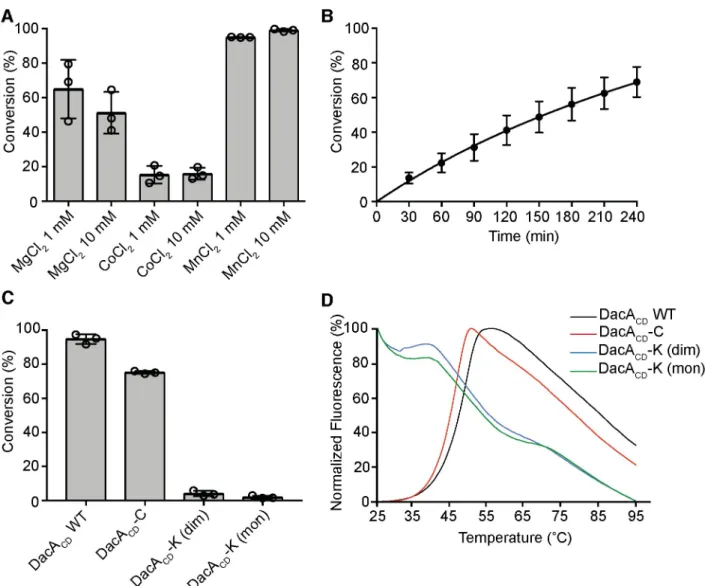
Next, the purified proteins were tested for enzyme activity by quantifying the conversion of α-P32
-labelled ATP into P32-labelled c-di-AMP. First, the metal dependency of the WT DacACDprotein was assessed. To this end, DacACDwas incubated with ATP spiked with a
small amount ofα-P32-labelled ATP in buffers containing 1 or 10 mM of one of the divalent cations Mg2+, Co2+or Mn2+. After 4 h the reactions were stopped and the input ATP and c-di-AMP reaction product quantified by phosphorimaging. This analysis revealed that theS. aureus DacACDprotein is most active in the presence of Mn2+and hence all subsequent
exper-iments were performed in the presence of 10 mM MnCl2(Fig 3A). Next, a time course
experi-ment was performed and the DacACDenzyme displayed anin vitro catalytic activity with an
average catalytic rate of 2.28x10⁻¹⁰M/min (Fig 3B). This activity is slow, but consistent with previous observations [23]. To assess the activity of the different DacACDvariants, enzyme
activity assays were performed with the DacACD, DacACD-C, or DacACD-K (either obtained
from the predicted monomer or dimer peak) proteins. The DacACD-C variant displayed very
similar activity to the WT DacACDprotein (Fig 3C), suggesting that locking the dimer through
two disulfide bridges does not influence the activity of the enzyme. In contrast, the DacACD-K
variant derived either from the late eluting (predicted monomer) or earlier eluting (predicted dimer) peaks displayed very low activity (Fig 3C). We reasoned that the activity loss could be due to an intrinsic instability of this protein variant. To test this, the stability of the DacACD-K
variant was assessed using a thermofluor assay and compared to that of the WT DacACDand
Fig 2. Structure of the DacACD-C variant. (A) X-ray structure of the DacACD-C variant. The structure of the tag-less DacACD-C variant was solved by molecular replacement using the WTS. aureus DacACDprotein shown inFig 1as template. The DacACD-C protein was found as a dimer in the crystallographic unit. (B) Electron density of the dimer interface in DacACD-C, showing electron densities consistent with the formation of two disulfide bonds between two DacACD-C monomers at sigma 1.3.
DacACD-C variant. The melting curves for the DacACDand DacACD-C proteins were
sigmoi-dal, which is typical for well-folded proteins (Fig 3D). On the other hand, the DacACD-K
vari-ant displayed high levels of fluorescence from the beginning of the melting curve (Fig 3D), which is indicative of an unfolded protein and likely explains the low enzymatic activity of the DacACD-K variant. Taken together, these data show that WT DacACDand the ‘locked-dimer’
DacACD-C variant show similar enzymatic activities, suggesting that the stable dimer we
observed in our crystal structures will likely transiently interact with other dimers to form higher oligomers for catalysis and c-di-AMP product formation.
Fig 3. Activity assays of DacACDand its variants. (A) Enzyme activity assay and metal-dependency of the WT DacACDprotein. 5μM DacACDwas incubated with 100μM M ATP spiked with a small amount of radiolabeled ATP in buffer containing 1 mM or 10 mM of Mg2+
, Co2+or Mn2+metal ions. The reactions were incubated for 4 h at 37˚C and % ATP to c-di-AMP conversion determined. The average values and standard deviations from three independent experiments were plotted. (B) DacACDcyclase activity time-course experiment. 5μM DacACDwas incubated with 100μM ATP spiked with radiolabeled ATP in buffer containing 10 mM Mn2+and the reactions were stopped at the indicated time points. The % ATP that was converted to c-di-AMP was determined and the average values and standard deviations from three independent experiments plotted. (C) Cyclase activity of WT DacACD, DacACD-C and DacACD-K variants. The indicated protein was incubated with ATP for 4 h as described in panel B and the average values of % ATP conversion and standard deviations from three independent experiments were plotted. (D) Thermofluor melting curves. 5μM of the indicated protein was incubated with SYPRO Orange dye, the solution was heated from 25 to 95˚C in 1˚C increments and the fluorescence readings determined. The melting curve for WT DacACDis shown in black, for the DacACD-C variant in red, for the DacACD-K variant derived from the presumed dimer peak in blue and the DacACD-K variant derived from the presumed monomer peak in green.
S. aureus DacA
CDand GlmM form a complex
in vitro
Results fromin vivo crosslinking and bacterial two-hybrid experiments suggested that the
phosphoglucosamine mutase GlmM can interact with the DacA homologs inB. subtilis and L. lactis [17,18]. To assess whether theS. aureus DacACDprotein can interact with theS. aureus
GlmM protein, a plasmid for the overexpression of a C-terminally His-taggedS. aureus GlmM
protein (GlmM-His) was generated. Next, expression of the His-DacACDand GlmM-His
pro-teins was induced inE. coli, the cells lysed and the proteins purified by Ni-affinity and size
exclusion chromatography, orE. coli lysates from His-DacACDand GlmM-His overexpressing
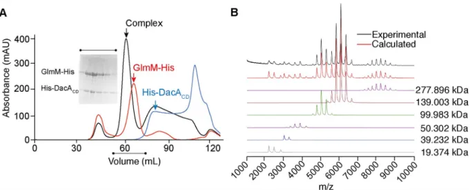
strains mixed before affinity and size exclusion chromatography. When the two proteins were purified together, a new faster eluting peak corresponding to a species of higher molecular weight and indicating the formation of a complex, was obtained (Fig 4A). SDS-PAGE analysis of aliquots from the different elution fractions confirmed the co-elution of the His-DacACD
and GlmM-His proteins in the high molecular weight peak (Fig 4A). To further confirm the interaction of DacACDwith GlmMin vitro, a pull-down assay was performed. For this
pur-pose, the His-tag was removed from DacACDand the purified tag-less protein subsequently
mixed in equimolar amount with purified GlmM-His. The protein mixture was then applied to a Ni-NTA column and after two wash steps, bound proteins were eluted. The untagged DacACDprotein quantitatively co-eluted with GlmM-His, confirming the protein/protein
interaction (S3 Fig). As control, the tag-less DacACDprotein was subjected to the same
proce-dure in the absence of GlmM-His. In this case, the protein was found in the load and wash fractions. Taken together, these results show that DacA and GlmM can form a stable complex
in vitro, and that the two proteins can be co-purified as a single species. To estimate the size
and stoichiometry of the complex, the purified DacACD-GlmM complex was further analyzed
by SEC-MALS as well as native mass spectrometry. Based on the SEC-MALS elution profile,
Fig 4. TheS. aureus DacACDand GlmM proteins form a stable complexin vitro. (A) Size exclusion profiles of the His-DacACD(blue line), GlmM-His (red line) proteins and the His-DacACD/GlmM-His complex (black line). The insert shows a Coomassie-stained gel with aliquots of fractions from the complex peak. The experiment was performed three times and a representative result is shown. (B) Native mass-spectrometry experiment of the DacA/GlmM complex. The spectrum was deconvoluted using the software Amphitrite [53]. Experimental and calculated spectra are shown in black and red, respectively. Charge state distributions corresponding to the different detected species are depicted in different colours. The molecular weights reported correspond to: DacA/GlmM dimer complex (purple line, 277.896 kDa, four copies of DacACD with four copies of GlmM); DacA/GlmM complex (brown line, 139.003 kDa, two copies of DacACDwith two copies of GlmM–main DacA/ GlmM complex); GlmM dimer (green line, 99.983 kDa); GlmM monomer (purple line—50.302 kDa); DacACDdimer (blue line—39.232 kDa); DacACDmonomer (grey line—19.374 kDa) (S2 Table).
the complex had an estimated molecular weight of 129 kDa± 0.4% (S4 Fig) and based on the native mass spectrometry analysis, the major species in the spectrum corresponded to a com-plex of 139.003 kDa± 10.41 Da (Fig 4B). These data are consistent with a DacACD-GlmM
complex comprising one GlmM dimer (theoretical mass 99.462 kDa) interacting with one DacACDdimer (theoretical mass 19.374 kDa), with a theoretical molecular weight of 138.21
kDa (S2 Table).
S. aureus GlmM is a negative regulator of DacA activity both in vitro and in
vivo
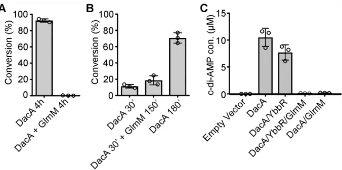
A study withL. lactis indicated that GlmM might inhibit the activity of DacA upon binding
[17]. To investigate this further, c-di-AMP production by theS. aureus DacACDenzyme was
assessed in the absence or presence of GlmM. DacACDwas incubated alone or mixed at a 1:2
molar ratio with GlmM, and conversion of ATP to c-di-AMP assessed after 4 h incubation at 37˚C. No ATP conversion was observed in the presence of GlmM (Fig 5A). In a second experi-ment, a 2-fold molar excess of GlmM was added to a DacACDreaction, 30 min after initiating
the reaction, and the sample subsequently incubated for a further 150 min (180 min total reac-tion time). As controls, 30 and 180 min DacACDenzyme reactions were also set up in the
absence of GlmM. When GlmM was added 30 min following the initiation of the DacACD
enzymatic cycle, the production of c-di-AMP was arrested (Fig 5B). Taken together, these data show that GlmM can effectively block the activity of the DacACDcyclase domainin vitro
with-out the need for any additional cofactors.
Fig 5.S. aureus GlmM negatively impacts the activity of DacACDin vitro and in vivo. (A) DacACDenzyme activity in the presence of GlmM. Enzyme assays were set up as described inFig 3in the absence or presence of 10μM GlmM added at the start of the reactions and reactions stopped after 4 h incubation. (B) Enzyme assays were set up with 5μM DacACDand 10μM GlmM added after 30 min incubation and the reactions incubated for further 150 min (180 min total). As a control, DacACDenzyme reactions were also set up in the absence of GlmM and incubated for 30 or 180 min. The average values of % ATP to c-di-AMP conversion and standard deviations from three independent experiments are plotted. (C) ELISA determination of c-di-AMP levels inE. coli. Cell extracts were prepared from E. coli strains containing the
empty pBAD33 vector or producing DacA, DacA/YbbR, DacA/YbbR/GlmM or DacA/GlmM. The cellular c-di-AMP levels were determined by ELISA and the average values (μM c-di-AMP per ml E. coli culture with an A600of 10) and standard deviations of three independent
experiments plotted.
To investigate this further and confirm that GlmM can also inhibit the activity of the full-length membrane-anchored DacA enzyme, full-full-length DacA was expressed inE. coli either
alone, in the presence of YbbR or GlmM or in the presence of both, GlmM and YbbR, and c-di-AMP production assessed by ELISA. To this end,dacA, ybbR or the complete dacA-ybbR-glmM operon was cloned into plasmid pBAD33, allowing for arabinose inducible DacA,
DacA/YbbR or DacA/YbbR/GlmM expression. For expression of DacA and GlmM only, the YbbR start codon was mutated and thedacA-no-ybbR-glmM operon inserted into plasmid
pBAD33. Next, DacA and YbbR production from the different constructs was confirmed by western blot using protein-specific antibodies and GlmM production was assessed by visualiz-ing proteins by Coomassie stainvisualiz-ing. With the exception of the empty vector control strain, similar DacA amounts were produced in all strains regardless whether or not YbbR, GlmM or YbbR and GlmM were co-expressed with the cyclase (S5 Fig). A clearly visible Coomassie stainable band of the expected size for GlmM was observed for the two strains overproducing GlmM (S5 Fig). YbbR production was also observed in theE. coli strain containing plasmids
pBAD33-dacA-ybbR or pBAD33-dacA-ybbR-glmM. Mutating the YbbR start codon drastically
reduced the production of YbbR but did not completely abolish its production, indicating that a second internal start codon might be utilized (S5 Fig). Next, the c-di-AMP production was assessed in the different strains. As expected, no c-di-AMP could be detected inE. coli
contain-ing the empty vector (Fig 5C). High amounts of c-di-AMP were detected upon DacA expres-sion or expresexpres-sion of DacA/YbbR. On the other hand, c-di-AMP levels were drastically reduced upon co-expression of GlmM, which is consistent with the data reported by Zhuet al.
[17] (Fig 5C). Taken together, these data show that GlmM is a negative regulator of theS. aureus c-di-AMP cyclase DacA both in vivo and in vitro. Of note, when GlmM activity was
assessedin vitro in the presence of DacACDusing a previously reported coupled enzyme assay
[40], no significant enzyme inhibition was observed. The specific activity of the pure GlmM enzyme was estimated at 15.9± 3.1 μmol/min/mg of protein in the assay conditions used. Addition of DacA in a 5- to 50-fold excess over GlmM did not at all inhibit the phosphogluco-samine mutase activity and its presence in 400-fold excess only led to a slight 15% reduction of GlmM activity. Taken together, these data indicate that only GlmM can impact the activity of the c-di-AMP cyclase enzyme but likely notvice versa.
GlmM of Gram-negative bacteria do not regulate or interact with the
S.
aureus DacA protein
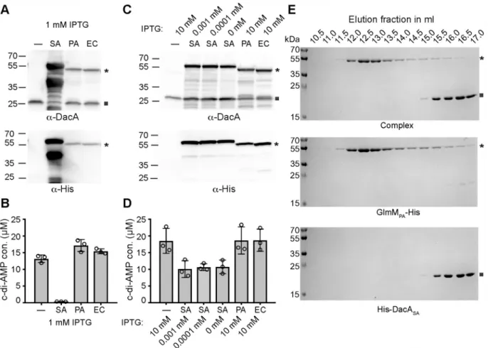
Next, we tested if GlmM proteins from unrelated Gram-negative bacteria can also interact and influence the activity of theS. aureus DacA protein. To this end, we overexpressed the S. aureus DacA protein from pBAD33-dacA along with C-terminally His-tagged versions of the E. coli (EC), Pseudomonas aeruginosa (PA) and as control S. aureus (SA) GlmM proteins in E. coli and measured c-di-AMP levels by ELISA (Fig 6). As expected, expression of theS. aureus
GlmM-His protein (SA) blocked c-di-AMP production, while c-di-AMP levels were not reduced upon expression of theE. coli or P. aeruginosa GlmM proteins (Fig 6B). However, when we analyzed the GlmM protein levels by western-blot following induction with 1 mM IPTG, we noted that theS. aureus proteins was expressed at significant higher levels as
com-pared to the other two GlmM proteins (Fig 6A). Therefore, we repeated the experiment using 10 mM IPTG for the induction of theE. coli and P. aeruginosa GlmM proteins but no (0 mM)
or very low levels (0.001 or 0.0001 mM) of IPTG for the induction of theS. aureus GlmM
pro-tein. In this case, similar GlmM amounts were observed for all strains (Fig 6C). Again, only expression of theS. aureus GlmM protein (be it to a lesser extent) but not expression of the
levels (Fig 6D). Next, we investigated the interaction of theS. aureus DacA and P. aeruginosa
GlmM proteinsin vitro. The expression of the S. aureus His-DacACDandP. aeruginosa
GlmM-His proteins were induced inE. coli, the cells lysed and the individual proteins purified
by Ni-affinity followed by size exclusion chromatography. In addition, the lysates from theS. aureus His-DacACDandP. aeruginosa GlmM-His overexpressing strains were mixed before
affinity and size exclusion chromatography. In contrast to the observation with theS. aureus
GlmM protein (Fig 4A), theP. aeruginosa GlmM-His protein did not coelute with the S. aureus His-DacACD(Fig 6E). Taken together, these data highlight a specificity in the
interac-tion between theS. aureus DacA and GlmM proteins, as no interaction was detected between
theS. aureus DacA protein and the GlmM proteins from the Gram-negative bacteria E. coli
andP. aeruginosa.
Fig 6. GlmM proteins fromE. coli or P. aeruginosa do not interact or impact the activity of the S. aureus DacA enzyme. (A and C) Detection of DacA and GlmM-His by western blot. Protein samples were prepared fromE. coli XL1-Blue pBAD33-dacA containing either an
empty pTrcHis60 vector (-) or a plasmid for expression of the S. aureus GlmM-His (SA), the P. aeruginosa GlmM-His (PA) or the E. coli
GlmM-His (EC). DacA or the GlmM-His proteins were detected by western-blot using an anti-DacA or anti-His antibody as indicated below the respective panel. DacA protein expression was induced with 0.2% arabinose and GlmM protein expression with 1 mM IPTG in (A) or with the amount of IPTG as indicated in (C). (B and D). ELISA determination of c-di-AMP levels inE. coli. Cell extracts were prepared from the E. coli strains used in panels A and C. The cellular c-di-AMP levels in μM per ml E. coli culture with an A600of 10 were determined by ELISA and the average values and standard deviations of three independent experiments plotted. (E) Coomassie stained gels. Aliquots from the size exclusion fractions of the complex (top), theP. aeruginosa GlmMPA-His (middle) or theS. aureus His-DacASA(bottom) were separated on 12% PAA gels and proteins visualized by Coomassie staining. The experiment was performed twice, and a representative result is shown. GlmM and DacA proteins are indicated with an asterisk and a square, respectively in panels A, C and E.
S. aureus GlmM protein structure and small-angle X-ray scattering analysis
of the DacA
CD/GlmM complex
In order to gain structural information on theS. aureus DacACD/GlmM complex, we first set
out to determine the structure of theS. aureus GlmM protein in isolation. To this end, the
C-terminal His-taggedS. aureus GlmM protein was purified, the His-tag was removed, and the
purified protein crystallized in the presence of Mg2+and glucosamine-6-P. The structure of GlmM was obtained by molecular replacement using the GlmM protein structure from Bacil-lus anthracis (PDB 3PDK) as a search model [41] (Fig 7andS1 Table). While the protein was crystallized in the presence of metal ion and substrate, no corresponding extra electron density was observed. TheS. aureus GlmM protein and the B. anthracis protein, which share 67%
identity on a protein level, displayed a similar fold and dimeric arrangement and the structures overlapped with an RMSD of 0.996Å. One GlmM monomer consisted of four α-β domains with the fourth most C-terminal domain linked by a flexible loop (Fig 7). Next, we attempted to crystallize and solve the structure of theS. aureus DacACD/GlmM complex. While crystals
were obtained under several conditions, poor diffraction prevented the structural determina-tion of the complex. In order to gain structural informadetermina-tion of the DacA/GlmM complex, the single proteins and complex were subjected to small-angle X-ray scattering (SAXS) analysis (Fig 8,S6andS7Figs andS3 Table). The reconstructed model for the GlmM sample showed an elongated particle, consistent with the dimeric arrangement found in the crystal structure (Fig 8B) and with previously published findings [42]. In the case of DacA, the model was most consistent with the particle being a DacACDdimer, however an extra density was observed to
one extremity (Fig 8A), which we speculate is due to the presence of extra N-terminal amino acids, not visible in the X-ray structure, and hence appear as a flexible and unstructured region of the protein. The reconstructed envelope of the complex displays a bigger particle size as compared to GlmM dimer alone, consistent with a DacACDdimer interacting with a GlmM
dimer (Fig 8C). A sequential fitting of the GlmM dimer structure followed by the fitting of the DacA dimer structure into the SAXS envelope of the complex with Chimera [43] yielded the best fit for a model with a correlation score of 0.9196 in which a DacA dimer is positioned on top of a GlmM dimer (Fig 8C). In this model, the active sites of DacA protomers are occluded through binding to the GlmM protein (Fig 8D), while the GlmM active sites are still accessible. To validate the SAXS analysis, the reconstructed envelopes were used to calculate theoretical Collision Cross Section (CCS) values using the program EM\IM [44]. These theoretical values were subsequently compared to those experimentally determined by Ion-Mobility Mass Spec-trometry. The theoretical values agree with the CCS determination (S4 Table), showing that
Fig 7.S. aureus GlmM structure. Structure of the S. aureus GlmM protein shown in ribbon representation. The protein was crystallized in the presence of GlcN-6P and MgCl2but no densities for the metal ion or substrate were present in the structure. The structure was solved by molecular replacement using theB. anthracis GlmM protein (PDB 3PDK) as template. The protein crystallized as dimer and the two different
monomers are shown in orange and purple. The dimer is shown in two views with the second view rotated by 90˚.
the structural models of DacA, GlmM and their complex obtained by SAXS correspond to the shapes of the proteins in solution. Taken together, our model provides an explanation as to how the interaction of DacA with GlmM inhibits c-di-AMP production without affecting the activity of the GlmM enzyme.
Discussion
In this study, we have determined the structures of theS. aureus enzymes DacA and GlmM
and provide structural and functional information on the complex formed between these two enzymes. Previously, these enzymes have been shown to interact, and using the information
Fig 8. Small Angle X-ray Scattering (SAXS) data forS. aureus proteins DacACD, GlmM and the DacACD/GlmM complex. Reconstructed
envelopes of the purified DacACD(A), GlmM (B) and DacACD/GlmM complex (C). DacACD(12 mg/ml), GlmM (8 mg/ml) and the DacACD/ GlmM complex (12 mg/ml) were injected onto a Superdex 200 5/150 column coupled to a Small-Angle X-Ray beam. All data were analyzed and envelopes reconstructed using ScÅtter. Conversion of envelopes to maps and subsequent structure fitting were performed using Chimera. (D) Zoomed in views of the DacACDactive sites in the DacACD/GlmM SAXS model. Catalytic residues on both DacACDprotomers are shown in stick representations. The interaction of GlmM with DacACDcovers the active sites, thus preventing DacACDdimers from interacting and forming catalytically active species.
obtained here, we can now propose a speculative molecular mechanism for the regulation of the cyclase activity of DacA by GlmM. Specifically, our data suggest that GlmM blocks access to the active site of DacA and in this manner prevents the formation of catalytically active head-to-head DacA oligomers.
c-di-AMP production is essential for the growth ofS. aureus under standard laboratory
conditions [20,45] and the only enzyme in this bacterium that produces c-di-AMP is DacA. TheS. aureus DacACDcatalytic domain investigated as part of this study has, as expected, a
very similar overall fold to the enzymatic domain of theL. monocytogenes CdaA cyclase [35]. However, there were also notable and interesting differences. As expected, the side chains of amino acids D176 and G177 in the HDG motif and T207 before the RHR motif in theS. aureus
DacACDenzyme made contacts with the ribose and the phosphate backbone of ApCpp,
respectively (Fig 1). However, in addition to these residues, we also observe an additional π-stacking interaction between residue Y192 and the adenine base (Fig 1) in the DacACD-ApCpp
structure. This residue is located in a loop region between ß4 andα4. A structure-based align-ment using STRAP [46] showed that this residue is not conserved inT. maritima and B. subti-lis DisA proteins, or in B. cereus CdaS, but present in L. monocytogenes CdaA (S8 Fig). This indicates that this residue could potentially play an important role in fine-tuning c-di-AMP production inS. aureus and other DacA/CdaA-containing bacteria by affecting the binding of
the nucleotide substrate.
Similar to other c-di-AMP cyclases, which require a metal ion as cofactor (usually Mg2+, Mn2+and/or Co2+) [23,35,47], we found thatS. aureus DacACDhas the highestin vitro
enzyme activity in the presence of Mn2+, followed by Mg2+and was least active in the presence of Co2+(Fig 3). This is very similar to what has been described for theMycobacterium tubercu-losis c-di-AMP cyclase Rv3586 (a DisA family enzyme), but differs from what has been
reported for theL. monocytogenes CdaA enzyme, which was most active in the presence of Co2 +
, whereas Mg2+did not support enzyme catalysis [35,47]. In addition to ATP, for some c-di-AMP cyclases it has been shown that ADP can also be used as substrate [47]. Interestingly, when theM. tuberculosis c-di-AMP cyclase Rv3586 was provided with ADP as substrate, the
highest activity was seen in the presence of Mg2+, rather than Mn2+[47]. While the actual metal preference and requirement for the activity of c-di-AMP cyclasesin vivo is not known, it
is conceivable that metal ion availability as well as substrate availability (ATP versus ADP) might be an important factor in adjusting c-di-AMP levels in the cell.
c-di-AMP cyclase enzymes form higher-order oligomeric complexes in solution. The CdaS-and DisA-type enzymes form hexamers CdaS-and octamers, respectively [23,33,34], whereas the catalytic domain of theL. monocytogenes cyclase CdaA was reported to be a dimer in solution
[35]. Indeed, for the c-di-AMP condensation reaction to take place, the enzyme requires two catalytic sites in a symmetric head-to-head conformation. Here, we show that theS. aureus
DacACDprotein is a dimer (S2 FigandFig 8). However, based on the data presented in this
study, we suggest that this dimer is not the active unit per se, as the protomers are not found in a head-to-head conformation (Fig 1). We propose that multiple dimers need to form higher oligomers (e.g. tetramers) in order to produce c-di-AMP (Figs3and9). Therefore, by regulat-ing the ability of DacACDto form higher oligomers its activity can be regulated and as
dis-cussed below, we proposed that this is the mechanism by which GlmM inhibits the activity of DacA.
The same inactive (non-head-to-head) dimer conformation as observed in our DacACD
structure has also been observed in the hexamer model of the CdaS di-adenylate cyclase from
Bacillus spp. (PDB 2FB5) [33] (S9 Fig). Mehneet al. proposed that the hexamerization of CdaS
is driven by two sets of interactions: the interactions between the two N-terminal helices (YojJ domain) and the interactions between residues found at the dimer interface between two DAC
domains. The latter interactions are similar the ones we observed in theS. aureus DacACD
dimer (Fig 1andS9 Fig) [33]. After deletion of the N-terminal helices, theB. subtilis CdaS
enzyme formed dimers in solution and this truncated protein had high catalytic activity [33]. However, it was not further investigated if the CdaS dimers were the active unit or if they still needed to undergo higher oligomerization to be catalytically active, similar to what we observed for theS. aureus DacACDenzyme. While for the DisA-type enzyme it seems clear
that the enzymatically active form is an octamer [23], additional studies are needed to deter-mine in which oligomeric state CdaS- and CdaA/DacA-type c-di-AMP cyclases functionin vivo.
The membrane-anchored DacA/CdaA-type c-di-AMP cyclase enzyme is encoded in a con-served three or four gene operon together with the predicted cyclase regulator YbbR (CdaR) and the phosphoglucosamine mutase enzyme GlmM producing the essential peptidoglycan synthesis intermediate glucosamine-1-P [24,48]. Indeed, forS. aureus it has been shown that
all three genes are co-expressed from an upstream promoter [49]. However, a second internal promoter is present in front ofglmM suggesting that S. aureus produces higher GlmM protein
levels as compared to DacA and YbbR [49]. It is thought that both GlmM and YbbR can regu-late the activity of the cyclase DacA/CdaA [17,18]; however, forS. aureus we could not find
conclusive evidence for a regulatory function of YbbR in this nor in a previous study (Fig 5) [12]. Recently, the first experimental evidence that the c-di-AMP cyclase DacA (CdaA), GlmM and YbbR (CdaR) form a three-protein complex was obtained inB. subtilis and L. lactis [17,
18]. The data presented here provide the first structural insight into this regulatory complex and further mechanistic information. Consistent with thein vivo work in L. lactis [17], we show that theS. aureus GlmM protein (but not the E. coli or P. aeruginosa GlmM proteins) is a
strong negative regulator of theS. aureus cyclase DacACD(Fig 5). Since we performed this
workin vitro with purified proteins, it shows that no additional components are required for
this inhibitory function.
Our SAXS envelope data are most consistent with the formation of a complex in which a DacACDdimer sits on top of the GlmM dimer, forming a dimer of dimers (Fig 8). The
resolu-tion of our SAXS reconstrucresolu-tion is too low to infer specific amino acid interacresolu-tions between DacACDand GlmM or to assess any conformational changes of DacACDupon GlmM binding.
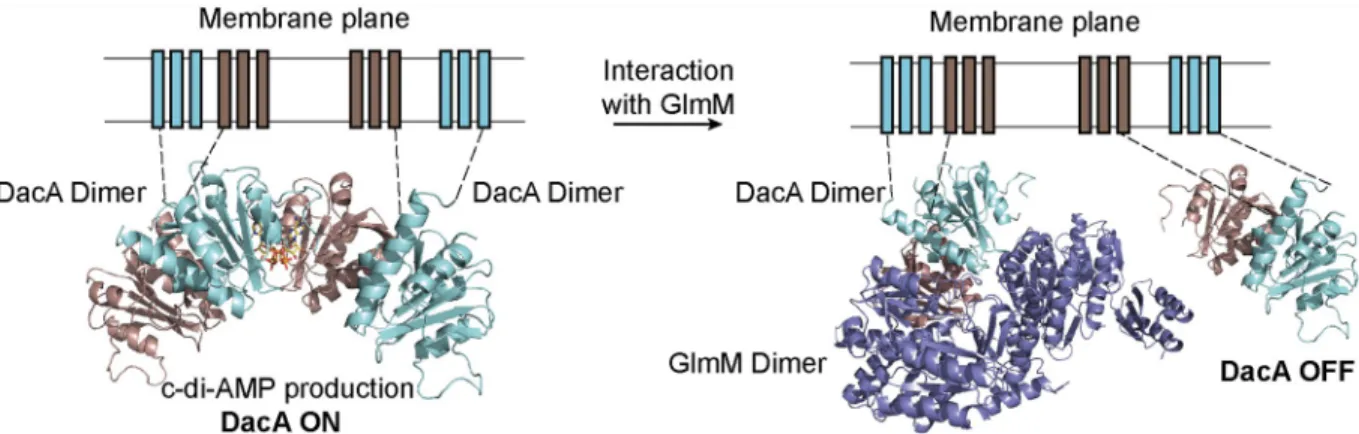
Fig 9. Proposed model of DacA inhibition by GlmM. (Left) For active c-di-AMP synthesis, two (or more) DacA dimers interact forming
catalytically active head-to-head oligomers. (Right) When GlmM binds to a DacA dimer, we speculate that this will prevent DacA from forming higher oligomers thereby turning off c-di-AMP production. GlmM dimer is colored in purple and a DacA dimer is colored in cyan (chain A) and brown (chain B). Bound ATP is colored in yellow. DacA transmembrane helices are modeled on the membrane plane and connected to the N-terminal helices of the DacACDdomain by dashed lines.
However, the data are of sufficient quality to construct models of the of DacACD-GlmM
com-plex. In our model, the DacACDactive sites in both protomers are capped by theα5helix of
GlmM and neighbouring residues (Fig 8D). Hence the interaction of the GlmM dimer with the DacACDdimer will prevent DacACDfrom forming higher oligomers required for activity.
In addition, the interaction of GlmM with DacACDcould potentially block the binding of
sub-strate to DacACD. Interestingly, positioned next to theα5helix in GlmM is residue F155,
which in a previous study was shown to modulate the GlmM-CdaA interaction inL. lactis
[17]. Our SAXS-based model provides now a molecular rational as to why a residue in this position could modulate complex formation and hence cyclase activity.
Within bacterial cells, the GlmM/DacA complex will likely be dynamic and in this manner allowing for c-di-AMP levels to be adjusted based on to environmental signals. It is conceiv-able that depending on the cellular levels of the substrate or product for GlmM (glucosamine-6-P and glucosamine-1-P, respectively), the ability of GlmM to interact with DacA changes and that in this manner c-di-AMP production is synchronized with the production of peptido-glycan precursors. GlmM functions in a pathway together with GlmS, which produces the GlmM substrate glucosamine-6-P and converts as part of this reaction glutamine to glutamate, and GlmU, which acts after GlmM and produces UDP-N-acetylglucosamine. It is also possible
that the GlmM/DacA interaction could be affected by the activity of GlmS and GlmU and availability of their substrates. This possibility is intriguing, as previous studies have revealed an impact of cellular glutamine, glutamate and UDP-N-acetylglucosamine levels on the cellular
c-di-AMP levels [17,18,50].
Taken together, our results provide the first structural characterization of the DacA/GlmM complex providing mechanistic insight into how c-di-AMP production is controlled directly at the synthesis level through the binding of GlmM to DacA. Additional work is needed to bet-ter understand the dynamics of this complex, in particular in the context of the bacbet-terial mem-brane and how this regulatory complex can be modulated by cellular or external stimuli.
Materials and methods
Bacterial strains and plasmid construction
All strains used in this study are listed inS5 Tableand primers used for plasmid construction are listed inS6 Table.E. coli strains were grown in Lysogenic broth (LB) supplemented when
appropriate with the antibiotics indicated inS5 Table. Protein expression was induced with 0.5 mM IPTG unless specified otherwise in the text or 0.2% arabinose. Plasmids for the expression of theS. aureus DacA and GlmM proteins were constructed as follows: The DNA fragments
coding for theS. aureus DacA catalytic domain starting from amino acid residue F101
(referred to as DacACDdomain) and GlmM were amplified by a PCR using LAC�genomic
DNA as template and primer pairs ANG1135/ANG1137 (for DacACD) or ANG2342/
ANG2343 (for GlmM). The primers for cloningglmM contained DNA sequences for
append-ing a C-terminal 6-His tag proceeded by a thrombin cleavage site. The PCR products were digested with NheI/EcoRI (for DacACD) or NcoI/XhoI (for GlmM-His) and then ligated with
plasmid pET28b, which has been digested with the same enzymes. The resulting plasmids pET28b-His-dacACDand pET28b-glmM-His were initially recovered in E. coli XL1-Blue
yield-ing strains ANG1858 and ANG3994 and subsequently introduced intoE. coli BL21 (DE3)
yielding the protein overexpression strains ANG1865 and ANG3997, respectively. Plasmids pET28b-His-dacACD-K and pET28b-His-dacACD-C for the expression of the DacACDvariants
with amino acid residues N166 and T172 replaced by lysines (DacACD-K variant) or cysteines
(DacACD-C variant) were constructed by Splicing Overhang Extensions (SOE) PCR. Primer
ANG2618 and ANG2619/ANG1137 (DacACD-C variant) were used for the first PCR. Next,
the respective PCR products were fused in a second PCR using primer ANG1135 and ANG1137. The resulting products were digested with NheI/EcoRI and ligated with pET28b that had been cut with the same enzymes. The resulting plasmids pET28b-His-dacACD-K and
pET28b-His-dacACD-C were initially recovered inE. coli XL1-Blue giving strains ANG4319
and ANG4321 and subsequently introduced inE. coli BL21 (DE3) yielding strains ANG4320
and ANG4322. For construction of plasmids pBAD33-dacA, pBAD33-dacA-ybbR and
pBAD33-dacA-ybbR-glmM the dacA, dacA-ybbR and dacA-ybbR-glmM regions were amplified
by PCR fromS. aureus LAC�chromosomal DNA using primer pairs ANG928/ANG2475,
ANG928/ANG2476 and ANG928/ANG2477, respectively. The PCR products were cut with KpnI and HindIII and ligated with plasmid pBAD33, which had been cut with the same enzymes. The resulting plasmids pBAD33-dacA, ybbR and pBAD33-dacA-ybbR-glmM were recovered in E. coli XL1-Blue yielding strains ANG4120, ANG4121 and
ANG4122. For construction of plasmid pBAD33-dacA-no-ybbR-glmM (strain ANG4263), the
YbbR start codon was mutated by SOE PCR. To this end, the front and back parts of the dacA-no-ybbR-glmM insert were amplified using plasmid pBAD33-dacA-ybbR-glmM as template
and primer pairs ANG1244/ANG2597 and ANG2598/ANG1245, respectively. The PCR frag-ments were fusing in a second PCR using primer pair ANG1244/ANG1245. The resulting product was digested with KpnI and HindIII and cloned into plasmid pBAD33 cut with the same enzymes. The sequences of inserts were verified by fluorescent automated sequencing at GATC Biotech. For the overexpression of theS. aureus DacA enzyme and His-tagged GlmM
proteins from different bacteria, the empty control plasmid pTrcHis60, as well as plasmids
pMLJ4 (GlmM-HisE. coli), pMLJ11 (GlmM-His S. aureus) or pMLD137 (GlmM-His P. aeru-ginosa) were introduced into E. coli strain XL1-Blue pBAD33-dacA, yielding strains ANG5174,
ANG5175, ANG5177 and ANG5178, respectively.
Protein expression, purification, and histidine-tag cleavage
One to two litre cultures ofE. coli strains BL21 (DE3) containing plasmids pET28b-His-dacACD(ANG1865), pET28b-His-dacACD-K (ANG4320), pET28b-His-dacACD-C (ANG4322) or pET28b-glmM-His (ANG3997) were grown in LB medium supplemented with kanamycin
(30μg/ml) at 37˚C with agitation. Once the cultures reached an A600of approximately 0.5–0.6,
protein expression was induced with 0.5 mM IPTG and the cultures were subsequently incu-bated for a further 3 hours at 37˚C. Cells were harvested by centrifugation, suspended in 20 ml of 50 mM Tris pH 7.5, 500 mM NaCl buffer and lysed by passing the cell suspensions twice through a French press cell at 1100 psi. For the purification of the His-DacACD/GlmM-His
complex, the appropriate cell lysates were mixed after lysis and incubated on ice for 10 min-utes. Next, the lysates were clarified by centrifugation at 20,000× g for 40 min and then loaded by gravity flow onto columns containing 4 ml Ni-NTA resin (Qiagen) equilibrated prior to use with 50 mM Tris pH 7.5, 500 mM NaCl buffer. The columns were washed with 30 ml of 50 mM Tris pH 7.5, 500 mM NaCl, then 30 ml of 50 mM Tris pH 7.5, 500 mM NaCl, 50 mM imidazole buffer and the proteins eluted in 5× 1 ml fractions with 50 mM Tris pH 7.5, 200 mM NaCl, 500 mM imidazole buffer. Elution fractions containing the purified protein or pro-tein complex were pooled and then loaded onto a Superdex 200 10/60 Hiload column (GE Healthcare), equilibrated with 30 mM Tris pH 7.5, 150 mM NaCl buffer. When the GlmM-His protein was purified alone, DTT was added to the purified protein at a final concentration of 1 mM after the size-exclusion chromatography step to maintain the protein in a soluble state. To cleave the His-tag from proteins, 20 U thrombin (Sigma) was added per 10 mg protein after the Ni-NTA column purification step and the mixture was incubated overnight at 4˚C with
agitation. The tag-less proteins were subsequently purified by size exclusion chromatography as described above. The purified proteins or complex were concentrated using 10 kDa cutoff Amicon centricons (Millipore). Protein concentration was measured either by determining the absorbance at 280 nm or by the Bradford method. For the investigation of the interaction between theS. aureus His-DacACDenzyme and theP. aurugniosa GlmM-His, one litre
cul-tures ofE. coli strain BL21 (DE3) containing plasmid pET28b-His-dacACD(ANG1865) was grown in LB medium supplemented with kanamycin (30μg/ml) at 37˚C with agitation. Once the culture reached an A600of 0.5, protein expression was induced with 0.5 mM IPTG and the
culture was subsequently incubated O/N at 16˚C. Strain XL1-Blue pMLD137 (GlmM-HisP. aeruginosa) was grown to an A600of 0.5 and protein expression was induced with 0.5 mM
IPTG and the culture subsequently incubated for an additional 4 hours at 37˚C. Cells were har-vested by centrifugation, suspended in 20 ml of 50 mM Tris pH 7.5, 500 mM NaCl buffer sup-plemented with 0.1% v/v ß-mercaptoethanol and complete EDTA free protease inhibitor (Roche) and lysed by sonication for 2 min at 15 sec on and 30 sec off intervals at 60% ampli-tude. The proteins and complex were purified by nickel affinity chromatography as described above but using only 2 ml of Ni-NTA resin and eluting the proteins in 2.5 ml of 50 mM Tris pH 7.5, 200 mM NaCl, 500 mM imidazole buffer supplemented with 0.1% v/v ß-mercap-toethanol. Next, 500μl of the eluate was separate on a Superdex 200 10/300GL column (GE Healthcare) using 30 mM Tris pH 7.5, 150 mM NaCl as running buffer. 0.5 ml fractions were collected, and aliquots of the indicated elution fractions separated on 12% PAA gels and pro-teins visualized by Coomassie staining.
SEC-MALS and native mass spectrometry analysis of the DacA
CD/GlmM
protein complex
100μl of the purified, tag-less DacACD/GlmM protein complex at 18 mg/ml was loaded onto a
Superdex 200 Increase 10/300 column (GE Healthcare) pre-equilibrated with 30 mM Tris pH 7.5, 150 mM NaCl buffer and run at 0.5 ml/min in the same buffer. The column was coupled to a MALS detector and refractometer and the UV absorbance, laser scattering and refractive index change were monitored throughout the run. The data were analysed using the ASTRA 6.0 software and fitted according to the Zimm model for static light scattering. The experiment was performed in duplicate and a representative graph is shown. Prior to the native mass spec-trometry experiment, the tag-less DacACD/GlmM was loaded onto a Superdex200 10/300
col-umn (GE Healthcare) pre-equilibrated with 200 mM ammonium acetate pH 7.5. Samples were further buffer exchanged with Amicon Ultra centrifugal filtration units (Merck Millipore) using 200 mM Ammonium Acetate (Fisher Scientific). Samples were diluted to 13.5μM. The samples were directly introduced into a first generation Waters Synapt QToF (Waters Corpo-ration, UK) using nanoelectrospray gold-coated borosilicate glass capillaries preparedin-house
[51]. Mass calibration was performed using 30 mg/mL Caesium Iodide (Fluka) solution. Machine parameters used were: capillary 1.4 kV, sampling cone 40 V, extraction cone 4 V, backing pressure 6.20 mbar, trap CE 15 eV, transfer CE 13 eV, bias 16 V, source wave velocity 300 ms-1, source wave height 0.2 V, trap wave velocity 300 ms-1, trap wave height 0.2 V, IMS wave velocity 260 ms-1, IMS wave height 8.0 V, transfer wave velocity 260 ms-1, transfer wave height 8.0 V. For collision cross section calculation calibrants were sprayed from 200mM Ammonium Acetate solution. The calibrants used were Cytochrome C (Calbiochem), β-Lacto-globulin (Sigma Aldrich), Bovine Serum Albumin (Sigma Aldrich), Concanavalin A (Sigma Aldrich), Serum P Albumin (Calbiochem) and Pyruvate Kinase (Sigma Aldrich). The CCS cal-ibration curve was produced using the method described by Thalassinoset al. [52], with a
calculated R2value of 0.9601. Spectra were analyzed using MassLynx v4.1 (Waters Corpora-tion, UK) and the program Amphitrite [53].
Protein crystallization, structure solution and analysis
The tag-less, nucleotide-free DacACDprotein and tag-less DacACD-C variant were crystallized
in 200 mM LiCl2, 100 mM Na-cacodylate pH 6.5, 30% PEG400, using a protein concentration
of 7.5 mg/ml. To obtain the ApCpp-bound DacACDcomplex structure, the tag-less DacACD
protein at a concentration of 7.5 mg/ml was incubated for 30 min on ice with 2 mM ApCpp (Jena Bioscience) and 2 mM MnCl2and the protein subsequently crystallized in 10 mM MgCl2,
50 mM MES pH 5.8, 0.2M KCl and 3% PEG8000 buffer. The tag-less GlmM protein was crystal-lized in the presence of 2 mM MgCl2and 2 mM GlcN-6P (Sigma) in 2.0 M sodium malonate
buffer. All proteins were crystallized by the vapour diffusion method. DacACD, DacACD-C and
GlmM crystals were frozen in liquid nitrogen without the addition of further cryo-protectants and datasets collected at the I03 Beamline at the Diamond Light Source (Harwell Campus, Did-cot, UK). Data integration was performed with DIALS [54], the reflections were scaled and merged with Aimless [55] and intensities were converted to structure factors using Ctruncate [56]. Analysis of the Matthews coefficient revealed that two protein molecules were present in the asymmetric unit for all proteins. The structures were solved by the molecular replacement method with Phaser [57] using theL. monocytogenes CdaA protein (PDB 4RV7) as a template
for DacACDor theB. anthracis GlmM protein (PDB 3PDK) as a template for the S. aureus
GlmM protein. Structure refinement was performed with Phenix [58] and model building with Coot [59]. Structure figures were created with Pymol and Chimera [43,60].
Affinity pull-down experiments
For the pull-down experiment, 50μM tag-less DacACDand 50μM GlmM-His protein were
mixed in 30 mM Tris pH 7.5, 150 mM NaCl buffer and incubated on ice for 10 min. Next, the protein mixture was applied by gravity flow to a column containing 1 ml of Ni-NTA equili-brated with 30 mM Tris pH 7.5, 150 mM NaCl buffer. As a control, 500μl of a 50 μM tag-less DacA protein solution in 30 mM Tris pH 7.5, 150 mM NaCl were also applied by gravity flow onto a column. The flow-through fractions were collected for subsequent SDS-PAGE analysis. The columns were then washed with 15 ml of 30 mM Tris pH 7.5, 150 mM NaCl, 10 mM imid-azole buffer and the wash fraction collected and proteins finally eluted with 2 ml 30 mM Tris pH 7.5, 150 mM NaCl, 500 mM imidazole buffer. Aliquots of the load, flow through, wash and elution fractions were run on a 12% SDS-PAGE gel and proteins visualized by Coomassie staining. The DacACD/GlmM-His pulldown experiment was performed in triplicate and a
rep-resentative result is shown; the control experiment using the DacACDprotein alone was
per-formed once.
DacA
CDenzyme activity assay
To assess the metal dependency of theS. aureus DacACDenzyme, enzyme reactions were set
up as follows: 2μl of 10X reaction buffer (400 mM Hepes pH 7, 1 M NaCl, 100 mM or 10 mM of MgCl2, CoCl2or MnCl2) was mixed on ice with 13.6μl of water and 2 μl of 1 mM ATP.
Next, 2μl of DacACDfrom a 50μM stock solution and 0.4 μl of α-P32-labelled ATP (Perkin
Elmer—3.3μM, 250 μCi) were then added to the sample yielding a final reaction volume of 20μl. The reactions were incubated at 37˚C for 4 h. Reactions were stopped by heating for 5 min at 95˚C and the conversion of ATP to c-di-AMP assessed by spotting 1μl of each reaction onto a TLC plate (Millipore). The nucleotides were separated using a 3.52 M (NH4)2SO4and
dried and the radioactive signal detected using a Typhoon FLA 700 imager. The bands corre-sponding to radioactive ATP and c-di-AMP were quantified using the ImageQuant software and the % ATP to c-di-AMP conversion calculated. As the highest enzyme activity was seen in the presence of MnCl2, all subsequent enzyme reactions were set up in reaction buffer
contain-ing a final concentration of 10 mM MnCl2. For the time course experiment, 100-μl reactions
were set and 10-μl aliquots removed at the indicated time point and stopped by heating. The activity of the DacACD-K and DacACD-C variants was assessed by setting up 20-μl reactions as
described above using a final protein concentration of 5μM and incubating the reactions for 4 h at 37˚C. To analyze the ATP conversion of DacACDwhen bound to GlmM, 2μl of GlmM
from a 100μM stock solution was added to a standard enzyme reaction yielding a DacACD:
GlmM molar ratio of 1:2 and the reactions were incubated for 4 h at 37˚C. To evaluate the impact of the addition of GlmM when added to an ongoing DacACDenzyme reaction, 2μl of
GlmM from a 100μM stock solution were added 30 min after initiating the reaction and the reaction was incubation for a further 2 h. As control, DacACDreactions set up in the absence
of GlmM were stopped after 30 min and 3 h. The enzyme assays were performed in triplicate and the average values and standard deviations plotted.
GlmM enzyme activity assay
The phosphoglucosamine mutase activity of GlmM was determined by using a coupled radio-active assay in which the GlcN-1-P synthesized from GlcN-6-P by GlmM was quantitatively converted toN-acetylglucosamine-1- phosphate (GlcNAc-1-P) in the presence of pure GlmU
enzyme [40]. The standard assay mixture (50μl) contained 50 mM Tris/HCl (pH 8), 3 mM MgCl2, 1 mM GlcN-6-P, 0.4 mM [14C]acetyl-CoA (1.9 GBq/mmol, 700 Bq), 0.7 mM
glucose-1,6-diphosphate, pureE. coli GlmU (30 ng) and S. aureus GlmM (10–50 ng) enzymes
(appro-priate dilutions in 20 mM phosphate buffer, pH 7.2, containing 0.5 mM MgCl2and 0.1%
2-mercaptoethanol). When the DacA protein was also included in the assay, pure GlmM and DacA proteins were first pre-incubated together with different ratios at 37˚C for 5 min before addition to the reaction mixture at t = 0. Then, mixtures were incubated at 37˚C for 30 min and the reactions were stopped by the addition of 10μl of glacial acetic acid. The radioactive substrate (acetyl-CoA) and product (GlcNAc-1-P) were separated by thin-layer chromatogra-phy (TLC) on pre-coated silica gel 60F254plates (Merck) using 1-propanol/ammonium
hydroxide/water (6:3:1, v/v) as the mobile phase (Rffactors of these compounds were 0.72 and 0.17, respectively). Radioactive spots were located and quantitated with a radioactivity scanner (Rita Star, Raytest Isotopenmeβgera¨te GmbH, Straubenhardt, Germany).
Assessment of protein stability by thermofluor analysis
To assess the protein stability, a thermofluor experiment was performed. Reactions were set up as follows: 2μl of either 10X enzyme reaction buffer (400 mM Hepes pH 7, 1 M NaCl, 100 mM MnCl2) or 10X protein purification buffer (300 mM Tris pH 7.5, 1.5M NaCl) were added to
the appropriate wells of a 96-well qRT-PCR plate and mixed with 15μl of H2O. Next, 2μl of
100μM DacACD, DacACD-C, DacACD-K (monomer) or DacACD-K (dimer) protein stock
solutions were added and finally 1μl of 100X Sypro orange was added to yield a total reaction volume of 20μl and a final concentration of Sypro orange of 5X. The reactions for each protein were set up in triplicate in a 96-well plate. An Applied Biosystems OneStepPlus real-time PCR system was used for the thermal unfolding reactions and the temperature was increased every 30 s by 1˚C from 25˚C to 95˚C and the fluorescence intensities measured. The data were ana-lyzed using the Applied Biosystems StepOne Plus software. Fluorescence readings of the tripli-cate samples were averaged and normalized for each protein as previously described [61] using