Early Growth Response protein 1 (Egr-1) expression by
Insulin-like growth factor 1 (IGF-1) involves MAPKs and
PKB pathways
par
Viktoria Youreva
Programme des Sciences Biomédicales Faculté de Médecine
Mémoire présenté à la Faculté des études supérieures en vue de l’obtention du grade de Maîtrise en Sciences (M.Sc)
en Sciences Biomédicales option Générale
July 2014
Faculté des études supérieures et postdoctorales
Ce mémoire intitulé :
Early Growth Response protein 1 (Egr-1) expression by
Insulin-like growth factor 1 (IGF-1) involves MAPKs and
PKB pathways
Présentée par : Viktoria Youreva
Évalué par un jury composé des personnes suivantes :
Dr Lise Coderre, président-rapporteur Dr Ashok K. Srivastava, directeur de recherche
Un remodelage vasculaire anormal est à la base de la pathogenèse des maladies cardio-vasculaires (MCV) telles que l’athérosclérose et l’hypertension. Des dysfonctionnements au niveau de la migration, l’hypertrophie et la prolifération des cellules musculaires lisses vasculaires (CMLV) sont des évènements cellulaires qui jouent un rôle primordial dans le remodelage vasculaire. L’insulin-like growth factor 1 (IGF-1), puissant facteur mitogène, contribue au développement des MCV, notamment via l’activation des protéines MAPK et PI3-K/PKB, composantes clés impliquées dans les voies de croissance cellulaire. Ces molécules sont également impliquées dans la modulation de l’expression de nombreux facteurs de transcription, incluant le facteur Egr-1. Egr-1 est régulé à la hausse dans différents types de maladies vasculaires impliquant les voies de signalisation de croissance et de stress oxydant qui par ailleurs peuvent être déclenchées par l’IGF-1. Cependant, la question d’une possible modulation de l’expression d’Egr-1 dans les CMLV demeure inabordée; plus spécifiquement, la caractérisation de la voie de signalisation reliant l’action d’IGF-1 à l’expression d’Egr-1 reste à établir. Dans cette optique, l’objectif de cette étude a été d’examiner l’implication de MAPK, PKB et des dérivés réactifs de l’oxygène (DRO) dans l’expression d’Egr-1 induite par l’IGF-1 dans les CMLV. L’IGF-1 a induit une augmentation marquée du niveau protéique de l’Egr-1 en fonction du temps et de la concentration utilisés. Cette augmentation a été inhibée en fonction des doses d’agents pharmacologiques qui ciblent les voies de signalisation de MAPK, PKB et DRO. De plus, l’expression du facteur de transcription, Egr-1, en réponse de l’IGF-1, a été atténuée suite à un blocage pharmacologique des processus cellulaires responsables de la synthèse d’ARN et de synthèse protéique. Pour conclure, on a démontré que l’IGF-1 stimule l’expression d’Egr-1 via les voies de signalisation, impliquant ERK1/2/JNK, PI3K/PKB. On a également proposé que les DRO jouent un rôle important dans ce processus. Dans l’ensemble, nous avons suggéré un nouveau mécanisme par lequel l’IGF-1 promeut la prolifération et l’hypertrophie cellulaire, processus à la base des anomalies vasculaires. Mots-clés : Egr-1, IGF-1, IGF-1R, ERK1/2, JNK, PKB, PI3-K, CMLV.
Aberrant vascular remodelling underlies the pathogenesis of major cardiovascular diseases (CVD), such as atherosclerosis and hypertension. Abnormal growth, migration and proliferation of vascular smooth muscle cells (VSMC) are believed to play a critical role in vascular remodelling. IGF-1, potent mitogen, is believed to contribute to the development of CVD through the hyperactivation of proliferative and growth promoting pathways including mitogen-activated protein kinase (MAPK) and protein kinase B (PKB) pathways. It has also been implicated in modulating the expression of multiple transcription factors, including the early growth response protein-1 (Egr-1). Egr-1 upregulation has been observed in different models of vascular diseases implicating growth and redox signalling such as observed in response to IGF-1. However, modulation of Egr-1 expression by IGF-1 in VSMC, more specifically the signaling pathways involved in this process remain poorly characterized. Therefore, in the present studies, we investigated the implication of MAPK, PKB and ROS in IGF-1-induce Egr-1 expression in VSMC. IGF-1 induced a marked increase in Egr-1 protein level in a time and dose-dependent fashion. This increase was dose dependently inhibited by different pharmacological inhibitors targeting MAPK, PKB and reactive oxygen species (ROS) generation. Furthermore, pharmacological inhibitors of RNA and protein synthesis also attenuated IGF-1-induce response on Egr-1 expression. In conclusion, we showed that IGF-1 stimulates the expression of Egr-1 through ERK1/2/JNK and PI3K/PKB. We also propose that ROS generation plays an important role in this response. Overall, we propose a novel mechanism through which IGF-1 may exert its deleterious responses in vascular abnormalities.
Résumé ... i
Abstract ... ii
Table des matières ... iii
Liste des figures ... v
Liste des abréviations, sigles et acronymes... vii
Remerciements ... x
CHAPITRE 1 ... 1
INTRODUCTION ... 1
1.1. IGFs system ... 3
1.2 IGFs protein and gene structure ... 3
1.3 IGF-1 synthesis and regulation ... 5
1.4 IGF-1 protein processing... 6
1.5 IGFBPs ... 7
1.6 IGF-1 receptor ... 8
1.7 IGF-1 signaling in vascular system ... 11
1.8 Biological actions of IGF-1 in vascular system ... 13
1.9 IGF-1 in cardiovascular pathophysiology ... 17
1.9.1 IGF-1 in atherosclerosis ... ...17
1.9.2 IGF-1 in hypertension ... 18
1.10 IGF-1 and transcriptional factors ... 19
1.11 Early growth response protein 1 (Egr-1) structure ... 19
1.12 Egr-1 expression and regulation... 21
1.13 Involvement of Egr-1 in cardiovascular pathophysiology ... 24
1.14 Goal of this study ... 26
CHAPITRE 2 ... 27
ARTICLE ... 27
Abstract ... 29
Introduction ... 30
Acknowledgments ... 41 Figure Legends ... 42 Reference List ... 59 CHAPITRE 3 ... 65 DISCUSSION GÉNÉRALE ... 65 CHAPITRE 4 ... 68 CONCLUSION ... 68 RÉFÉRENCES ... 71
CHAPITRE 1 - INTRODUCTION
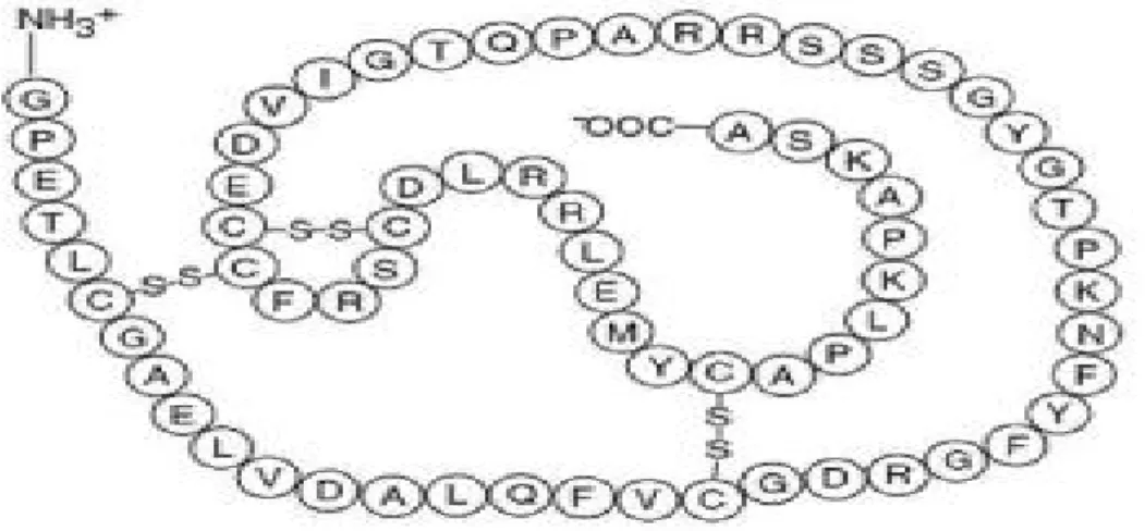
Figure 1.1 Structure of insulin-like growth factor-1 (IGF-1) amino acid sequence... 4
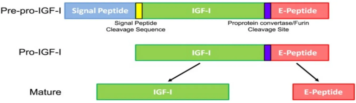
Figure 1.2 Insulin-like growth factor-1 (IGF-1) processing leading to mature IGF-1. ... 6
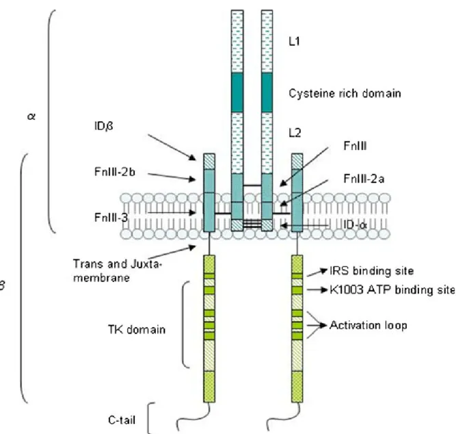
Figure 1.3 Schematic representation of Insulin-like growth fcator-1 (IGF-1). ... 10
Figure 1.4 Structure of inducible transcriptional factor Egr-1. ... 21
Figure 1.5 Regulatory regions of the human Egr-1 gene. ... 23
CHAPITRE 2 - ARTICLE
Figure 1. Insulin-like growth factor-1 (IGF-1) enhances early growth response protein 1 (Egr-1) expression in A10 vascular smooth muscle cells (VSMC) in a time and dose-dependent fashion………. 42Figure 2. Attenuation of Insulin-like growth factor-1 (IGF)-1-induced early growth response protein 1 (Egr-1) expression by pharmacological inhibitor AG1024 (IGF-1R-PTK inhibitor) in A10 vascular smooth muscle cells (VSMC) in a dose-dependent fashion ... 42
Figure 3. Attenuation of Insulin-like growth factor-1 (IGF)-1-induced early growth response protein 1 (Egr-1) expression by pharmacological inhibitors Actinomycine D (DNA synthesis inhibitor) and Cycloheximide (protein synthesis inhibitor) in A10 vascular smooth muscle cells (VSMC) in a dose-dependent fashion ... 43
Figure 4. Attenuation of Insulin-like growth factor-1 (IGF)-1-induced early growth response protein 1 (Egr-1) expression by pharmacological inhibitors PD98059 (MEK1/2 inhibitor) and SP600125 (JNK inhibitor) in A10 vascular smooth muscle cells (VSMC) in a dose-dependent fashion ... 43 Figure 5. Attenuation of Insulin-like growth factor-1 (IGF)-1-induced early growth
Figure 6. Attenuation of Insulin-like growth factor-1 (IGF)-1-induced early growth response protein 1 (Egr-1) expression by pharmacological inhibitor DPI (NADPH oxidase inhibitor) in A10 vascular smooth muscle cells (VSMC) in a dose-dependent fashion ... 44 Figure 7. Effect of the NAD(P)H oxidase inhibitor (DPI) on Insulin-like growth factor-1 (IGF-1)-induced PKB, ERK1/2 and JNK phosphorylation in A10 vascular smooth muscle cells (VSMC)
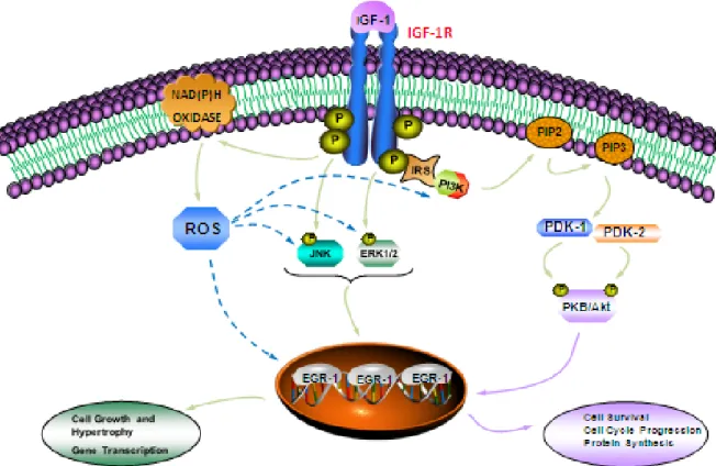
Figure 1.6 Schematic model of signaling pathways involved in IGF-1-induced Egr-1 expression in A10 vascular smooth muscle cells (VSMC) ... 70
ALS acid-labile subunit AP-1 activator protein 1
BAD Bcl-2 associated death promoter
Bcl-2 B-cell lymphoma 2
Ca2+ calcium
cAMP cyclic adenosine monophosphate CRE cAMP response element
CREB cAMP response element-binding protein
CVD cardiovascular disease
DBD DNA-binding domain
EBS Egr-1 binding sequence
EC endothelial cell
Egr-1 early growth response protein-1 Elk-1 ETS domain-containing protein Elk-1 eNOS endothelial nitric oxide synthase EPC endothelial progenitor cell ER endoplasmic reticulum
ERK1/2 extracellular signal-regulated kinase 1/2 FGF-2 fibroblast growth factor-2
FOXO forkhead box protein FnIII fibronectin type III domain GH growth hormone
GDP guanosine diphosphate
GPX-1 glutathione peroxidase-1
Grb2 growth factor receptor-bound protein 2 GSK-3 glycogen synthase kinase 3
GTP guanosine triphosphate HDAC1/2 histone deacetylases 1 and 2
factor type 2 receptor
IL-2 interleukin-2
iNOS inducible nitric oxide synthase
IGFBP 1-6 insulin-like growth factor binding proteins 1-6
IR insulin receptor
IRS insulin receptor substrate JNK 1/2/3 c-Jun N-terminal kinase 1/2/3
JAK/ STAT Janus kinase/signal transducer and activator of transcription LDL-C low-density lipoprotein-cholesterol
L-NAME Nω-nitro-L-arginine methyl ester
LDLR LDL receptor
MAPK mitogen activated protein kinase MCP-1 monocyte chemotactic protein-1 MEK mitogen extracellular regulated kinase M6P-R mannose-6-phosphate-receptor mTOR mammalian target of rapamycin
NADPH oxidase nicotamide adenine dinucleotide phosphate oxidase NAB 1/2 nerve growth factor-induced-A-binding protein 1/2 NGF neuronal growth factor
NGFI-A nerve growth factor inducible A NLS nuclear localization sequence NMR nuclear magnetic resonance
NO nitric oxide
NuRD nucleosomal remodelling and deacetylation complex oxLDL oxidized low-density lipoprotein
p70s6k p70 ribosomal S6 kinase
PDK phosphoinositide-dependent kinase
PI (3, 4,5)P3 phosphatidylinositol 3, 4, 5 tripshosphate
PKB protein kinase B
PKC protein kinase C
PTK protein tyrosine kinase R repressor domain
R-PTK receptor protein tyrosine kinase ROS reactive oxygen species
SAD strong activation domain Sap 1/2 SRF accessory protein 1/2 SAPK stress-activated protein kinase
SH2 src homology 2
Shc src homology collagen
SHR spontaneously hypertensive rat SMC smooth muscle cell
SOS son of seven less
SRE serum response element SRF serum response factor TNF-α tumour necrosis factor-α VSMC vascular smooth muscle cell WAD weak activation domain WKY Wistar-Kyoto
First and foremost, I would like to express my sincere gratitude and appreciations to my supervisor Dr Ashok K. Srivastava for his immeasurable support and encouragement through every step of this research project, his patience even when things were not going well and for all he has taught me. I would also like to thank him for accepting me in his lab as a summer intern and then let me pursue my M.Sc. studies. It is a privilege to work under his supervision. One could not wish for a better or friendlier supervisor.
Very special thanks go to my truly remarkable fellow labmate, Estelle Rolande Simo Cheyou, without whom this project would not have been possible, in every sense of the term. Thank you just doesn’t seem like enough. I am grateful for her constant encouragement, unconditional help, and innumerable contribution of her time in helping me to writing this memoir. She has provided me with much advices and good sense of humor over the past two years. Her true friendship and ability to create cool work atmosphere made a big difference in my life. I admire you Estelle!
A special mention must also be given to all of my previous labmates, especially Georgia Kapakos and George Vardatsikos for their constant help during my graduate training, for teaching me technical aspects of the science and help me to appreciate scientific research.
Last but not least, I want to say special thanks to my Mom and Dad, my brothers Vlad, Vladimir and Steve and my aunt Natalia for their unconditional love, support and encouragement in all my pursuits. I love you guys!
CHAPITRE 1
Cardiovascular diseases (CVD) are the primary cause of mortality and morbidity globally (1). The biology of CVD is very complex, however remodelling of blood vessels has been suggested to play an important role in the pathogenesis of CVD. Vascular smooth muscle cells (VSMC) are highly specialized cells that line the blood vessels and contribute to vessel contraction by regulating vascular tone and blood pressure (2, 3). Abnormal growth, migration and proliferation of VSMC are believed to be key events in vascular remodelling and have been implicated in the pathogenesis of vascular disease such as intimal hyperplasia, hypertension, atherosclerosis and restenosis (4, 5). During these pathophysiological states, there is an increased expression in activity of vasoactive peptides and growth factors such as insulin-like growth factor 1 (IGF-I) that are implicated to induce proliferation, hypertrophy, migration, apoptosis and differentiation in VSMC by structural alterations in the vessel wall (6-9). IGF-1 is a potent mitogen for VSMC and a potential pathogenic role of IGF-1/insulin-like growth factor 1 receptor (IGF-1R) system in vascular dysfunction has been suggested (10). A significantly higher level of IGF-1R and signaling has been reported in the VSMC isolated from the aorta of hypertensive animal models (11-13). In addition, dominant-negative or antisense oligonucleotides of IGF-1R were also shown to attenuate neointimal thickening in injured carotid arteries (13, 14). Thus, IGF-1/IGF-1R system via activation of growth promoting proliferative signaling pathways and gene expression has emerged as a key molecule involved in the pathogenesis of vascular disease. The following sections describe the structure and function of I as well as IGF-1-induced signaling pathways that trigger various responses of IGF-1 in VSMC.
1.1. IGFs system
IGFs play a fundamental role in the regulation of key cellular and physiological processes such as development, growth, cellular regulation and metabolism in mammals. Originally, IGFs were named sulphation factors by Salmon and Daughaday in 1957, because of their capacity to incorporate sulphate into cartilage (15). In 1972, a more intensive research of these factors proposed a new nomenclature, identifying two different types of peptides: somatomedin C and somatomedin A that mediate the effects of growth hormone (GH) (16). Finally, in 1978, Rinderknecht and Humbel found many resemblances between somatomedins and insulin in amino acid sequence and metabolic functions. Therefore, peptides were renamed IGF-1 and 2 (17). IGFs system consists of two ligands with similar structure: 1 and 2; three cell-membrane receptors 1R and IGF-2R and insulin receptor (IR); and a family of 6 high affinity insulin-like growth factor binding proteins (IGFBPs 1-6).
1.2 IGFs protein and gene structure
IGF-1 and IGF-2 each consist of a single chain of 70 and 67 amino acid polypeptides hormone with molecular weights of 7.6kDA and 7.4kDA respectively (Figure 1.1). Three dimensional structure of IGF-1 by nuclear magnetic resonance (NMR) and X-ray crystallography methods have been reported by several groups. However, IGF-2 structure has been less studied; therefore only two NMR structures have been reported to date. Three intramolecular disulphide bonds, formed by conserved cysteine residues, hold the IGFs structure (IGF-1 residues: Cys6-Cys48, Cys18-Cys61, Cys47-Cys52) together and consist of A, B, C and D domains. An A domain of 21 residues, contains alpha helix 2
(Ile43-Cys47 of IGF-1; Glu44-Phe48 of IGF-2) and alpha helix 3 (Leu54-Glu58 of IGF-1; Ala54-Tyr59 of IGF-2) joined by a loop, and B domain of 29 residues, with an extended N-terminal coil followed by a tight turn and a central alpha helix1 (Gly8-Cys18 of IGF-1; Gly10-Val20 of IGF-2), both forming hydrophobic core. IGFs possess the C peptide region of 12 amino acids that is an active part of the IGFs and an 8-amino acid carboxyterminal D domain (17, 18).
IGF-1 is the product of Igf1 gene that is situated on the long arm of chromosome 12. It is a single copy gene consisting of 2 precursor peptides, 153-amino acid IGF-1A and 195-amino acid IGF-1B. Two separate mRNAs produced by alternative splicing of the primary gene transcript are necessary to synthesise these 2 peptides. On the other hand, IGF-2 gene is found on chromosome 11. The 180-amino acid IGF-2 preprohormone contains a carboxy-terminal peptide of 89 amino acids and a signal peptide of 24 amino acids, both of which are cleaved post-translationaly to produce 67-amino acid plasma protein (19).
Figure 1.1 Structure of insulin-like growth factor-1 (IGF-1) amino acid sequence. IGF-1 is a single copy of 70 amino acid cyclic protein with three disulphide bridges linked by cysteine residues. (Based on ref. (20)).
1.3 IGF-1 synthesis and regulation
Igf-1 gene consists of 6 exons and 5 introns. IGF-1 synthesis occurs primarily in the liver and its plasma concentration is regulated by liver GH; however the ability of GH to stimulate IGF-1 is dependent on nutritional status. Once synthesised and released in the circulation, IGF-1 can be found in a free form or mainly bound to IGFBPs. Genetic factors and age are also important determinants that control the variability of serum IGF-1 concentrations. Other hormones, including cortisol, oestradiol, thyroxine and testosterone interact with GH in regulating hepatic IGF-1 synthesis (21). It has been shown that IGF-1 synthesis also occurs in numerous peripheral tissues, such as skeletal muscle, bone and cartilage, acting locally as autocrine/paracrine or endocrine factors that circulate in the plasma in order to regulate cell growth (22). Normal plasma IGF-1 concentration is maintained through the negative-feedback mechanism by supressing GH synthesis in the pituitary gland. Two promoters P1, (expressed in all tissues) and P2 (primarily expressed in the liver), initiate the IGF-1 mRNA transcription containing exon 1 and exon 2, respectively (23).
In addition, in cardiovascular system, IGF-1 is also secreted and synthesized by vascular cells. There is evidence of three IGF-1 mRNA transcripts sized 8.2, 1.7, and 0.9-1.2kb that have been localized in smooth muscle cells of adult rat aorta and two IGF-1 transcripts of 2.1 and 1.6 kb expressed in aortic endothelial cells. IGF-1 is also secreted by both endothelial cells (EC) and VSMC and regulated by growth factors, vasoactive peptides and hormones (24, 25).
1.4 IGF-1 protein processing
IGF-1 processing leading to mature IGF-1 protein begins with IGF-1 gene translation into a Pre-pro-IGF-1 precursor protein, which consists of a signal peptide, signal peptide cleavage site, mature IGF-1, pro-protein convertase cleavage site and C-terminal E-peptide extension (26) (Figure 1.2). During translation in the endoplasmic reticulum (ER), the N-terminal signal peptide is cleaved, resulting in pro-IGF-1 polypeptide (mature IGF-1 and E-peptide) that can be differently processed before secretion. This processing includes Pro-IGF-1 intracellular protease cleavage separating the mature IGF-1 for secretion from free E-peptide, maintenance of pro-IGF-1 to be secreted without cleavage, or N-glycosylation in the E-peptide, and then secretion. Thus, there are three different forms of IGF-1 protein, which are: mature IGF-1, glycosylated-pro-IGF-1 and nonglycosylated-pro-IGF-1 (27, 28).
Figure 1.2 Insulin-like growth factor-1 (IGF-1) processing leading to mature IGF-1. The Igf1 gene is first translated into a Pre-pro-IGF-1 precursor protein that includes a signal peptide, signal peptide cleavage site, IGF-1, pro-protein convertase site, and E-peptide. During translation, the signal peptide is removed from the remaining protein, now called Pro-IGF-1. Further protease cleavage separates the mature IGF-1 from free E-peptide. While mature IGF-1 has many accepted growth effects on a wide variety of cells and tissues, the purpose and actions of the E-peptide are relatively unknown. (Brisson BK., and Barton ER.,: Front Endocrinol. 27: 42, 2013).
1.5 IGFBPs
The level of free IGFs in the circulation is regulated by a family of IGFBPs, consisting of six members (IGFBP-1 to IGFBP-6) and found in all extracellular biological fluids. IGFBPs act as transporters of IGF, therefore prolonging their half-life and control free IGF availability in the circulation. IGFBPs also act as inhibitors by sequestrating IGF from its receptor and as a promoter by increasing IGF-IGFR interaction (29). Other important role of IGFBPs independent of IGF-binding, include their direct association with many extracellular and cell surface molecules (30). In extracellular fluids, 99% of IGF-1 is bound with high affinity to IGFBPs and only less than 1% of total amount of IGF is in an unbound form (31). In tissues, IGF-1 and IGF-2 form binary complexes with different type of IGFBPs. In the circulation, IGFBPs are found as ternary complexes of 150 kDa consisting of IGF, predominantly of IGFBP-3 or a fraction of IGFBP-5 and an 85 kDa liver-derived glycoprotein (acid-labile subunit, ALS). The ternary complex occurs in the liver where the IGF-1 and ALS are derived from the hepatocyte and IGFBP-3 is produced in the Kupffer cells (32, 33). These complexes prolong the circulating IGF-1 half-life, from less than 15 min up to 16 hours. They have limited ability to cross the capillary endothelial barrier, therefore maintaining a large circulating reservoir of IGF which can be then available to the tissues (34). In contrast to ternary complexes, some members of IGFBP family (IGFBP-1,-2,-4 and -5) also form binary complexes of 50kDa in plasma that are incompletely saturated with IGF, therefore permeable to vascular endothelium (29). It has been shown that post-translational IGFBP modifications, including proteolysis, phosphorylation and glycosylation, have the ability to alter the IGF-binding affinity (35).
IGFBPs share a highly conserved structure consisting of three domains: N-terminal (80-93 amino acid residues) and C-terminal domains (65 amino acid residues), both cysteine rich, and the highly variable central domain (55 amino acid residues). The N-terminal and C-terminal domains participate in IGF-binding, whereas, the central domain is unique to all IGFBPs. It acts as a hinge between N- and C-terminal domains where occurs most of post-translational modifications and proteolytic cleavage, resulting in loss of IGF-binding (32). In addition to IGFBP similarities, some structural differences have been described. It appears that only eight disulphide bonds are present in IGFBP-6 compared with nine in the other members of IGFBPs family (32).
1.6 IGF-1 receptor
IGF-1 exerts its biological actions through its interaction with a specific high affinity IGF-1R (25). IGF-1R is a 1367 amino acids heterotetrameric cell-surface glycoprotein that is synthesized as a 30 residue single polypeptide chain preproreceptor and is cleaved post translationally to pro-receptor. The proreceptor forming disulphide-linked dimers is then glycosylated, folded and dimerized by chaperones, calnexin and calreticulin (36) and transported to the Golgi apparatus to yield mature receptor consisting of two alpha (1-707 residues) and beta subunits (712-1337 residues) (37). The mature IGF-1R has a similar biochemical structure as IR and belongs to a large family of receptor-protein tyrosine kinases (R-PTK). IGF-1R is composed of 3 domains: the N-terminal extracellular hormone-binding domain, a transmembrane region and the C-terminal intracellular domain with intrinsic kinase activity and regulatory residues (38). The alpha subunit and 194 residues of beta subunit are located in N-terminal part of IGF-1R and include 11 potential
N-linked glycosylation sites (Asn-X-Ser/Thr motifs) and also consist of two homologous leucine-rich domains (L1 and L2) separated by a cysteine-rich region (39). The beta subunit comprise three fibronectin type III domains (FnIII-1 residues 461-579, FnIII-2 580-798 and FnIII-3 799-901) (40), followed by tyrosine kinase catalytic domain (973-1255 residues). The tyrosine kinase domain is flanked by 2 regulatory regions: a juxtamembrane site (Asn-Pro-X-Tyr motifs) involved in docking of insulin receptor substrates (IRS), Src homology collagen (Shc) and internalization of the receptor (41), and also a 108-residue carboxy-terminal tail (Gly-X-Gly-X-X-Gly motifs) that contains the phosphotyrosine binding sites, therefore contributing to IGF-1R activation (41, 42) (Figure 1.3).
In addition to IGF-1, IGF-2 exerts its physiological effects by binding to IGF-2R, also called mannose-6-phosphate-receptor (M6P-R). IGF-2R is a single cell-surface receptor with no PTK activity, containing 4 structural domains: the amino-terminal signal sequence, the extracytoplasmic domain, the transmembrane region and the carboxy-terminal tail (43). The IGF-2R participates in clearing IGF-2 from the cell surface by internalization and lysosomal degradation, therefore decreasing its mitogenic effects (29). It also has been shown that IGF-2R exists as a cleaved form in circulation, therefore is able to act as an IGFBP for IGF-2 protein (44).
Figure 1.3 Schematic representation of Insulin-like growth fcator-1 (IGF-1).
Secondary structure of IGF-1R demonstrates the location of different domains in α and β subunits that are linked with disulphide bonds. (Adapted from Gatenby et al: Pflugers Arch 465: 1065-1074, 2013).
1.7 IGF-1 signaling in vascular system
IGF-1 exerts its physiological effects through the activation of IGF-1R via multiple signaling cascades, including the mitogen activated protein kinase (MAPK) and phophatidyl-inositol 3-kinase/protein kinase B (PI3-K/PKB) cascade (45). The activation of these pathways is implicated in many cellular processes such as cell growth, proliferation, migration and survival of VSMC (25, 46, 47). IGF-1 binding to the extracellular IGF-1R alpha subunit results in conformational changes, leading to subsequent trans-autophophorylation of intrinsic tyrosine residues, Tyr 1131, 1135 and 1136 of IGF-1R beta-subunit, evoking an increased tyrosine kinase activity of the receptor (48). The first step in signal transduction by IGF-1R is the phosphorylation of several cytosolic receptor substrates, including IRS 1-4 and adaptor protein Shc, that serve as docking sites for Src homology 2 (SH2) domain-containing proteins, such as growth factor receptor-binding protein 2 (Grb-2), guanine nucleotide exchange factor son of sevenless (SOS), Nck and p85 regulatory subunit of PI3-K (49).
One of signaling cascades that emerges following IGF-1R activation involves MAPK pathway. MAPK are a group of serine-threonine kinases that has been closely associated with cell migration, proliferation and differentiation and also plays an important role in apoptosis (50). In mammalian cells, 5 members of the MAPK family have been described, including extracellular signal regulated kinases 1 and 2 (ERK1/2), also known as p42/44 as MAPK, Jun N-terminal kinase 1, 2 and 3 (JNK1/2/3), also called stress-activated protein kinase (SAPK), p38, ERK5 and ERK7 (51, 52). The most studied members of MAPK class to date are ERK1/2, JNK and p38α/β/γ/δ (48). IGF-1-induced ERK1/2 activation is initiated by the recruitment of Grb-2/SOS complex on the phosphorylated IRS
or adaptor protein Shc leading to the activation of guanosine triphosphate (GTP)-binding protein Ras, by exchanging the small guanosine diphosphate (GDP) to GTP. This process initiates sequential phopsphorylation by recruiting serine-threonine kinase Raf (A-Raf, B-Raf and C-B-Raf) to the membrane, after activating MAP kinase kinase 1 and 2 (MEK1 and MEK2) and MAP kinase (ERK1/2) in Thr and Tyr within a conserved Thr-Glu-Tyr motif in their activation loop (49, 53, 54). Activation of ERK1/2 can either phosphorylate and stimulate a number of cytosolic proteins, or translocate to the nucleus and activate several transcription factors, such as ETS domain-containing protein Elk-1 (Elk1), SRF, ATF2 and Jun implicated in immediate early gene transcription (55).
A second well-known pathway that is triggered following IGF-1R activation in vascular system involves another SH2 domain-containing protein, the p85 regulatory subunit of PI3-K (56-58). Interaction of p85 with the phosphorylated IRS leads to the activation of catalytic subunit of PI3-K, p110. Activated PI3-K catalyzes the conversion of phosphatidylinositol 4, 5-bisphosphate (PI(4,5)P2) to phosphatidylinositol 3, 4,
5-trisphosphate (PI(3, 4,5)P3), which is a key signaling molecule and an important second
messenger that recruits cytosolic proteins with pleckstrin-homology (PH) domain such as serine-threonine PKB and phosphoinositide-dependent kinase 1 and 2 (PDK1/2) (59). PKB is phosphorylated in the activation loop of its catalytic domain on Thr308 and in the C-terminal domain on Ser473 by PDK1/2 respectively (60, 61). PKB has multiple downstream targets, such as mammalian target of rapamycin (mTOR) implicated in protein synthesis, p70 ribosomal S6 kinase (p70s6k), forkhead box protein (FOXO), glycogen
synthase kinase-3 (GSK-3) and also triggers anti-apoptotic effects trough the phosphorylation of Bcl-2-associated death promoter (Bad) (62, 63).
It has been also shown that IGF-1R can signal via Janus kinases-1/2/3/signal transducers and activators of transcription (JAK-1,2,3/STAT) pathway, activated by cytokines and growth factors, depending on the cell type (64). Cell proliferation, differentiation, death and embryonic development are the cellular events that are regulated by the JAK/STAT pathway. The activated IGF-1R/JAK complex serves as a docking site for the recruitment of specific SH-2 domain-containing STAT proteins, resulting in STAT homodimerization and translocation to the nucleus acting as transcription factors (65).
1.8 Biological actions of IGF-1 in vascular system
In vasculature, IGF-1 exerts pleiotropic actions. In fact, a number of studies supported the concept that IGF-1 is a potent mitogen that plays a permissive role in vascular dysfunction, because of its pro-atherogenetic properties that contribute to VSMC proliferation and migration, thereby promoting neointima formation (66, 67). IGF-1 has also been shown to enhance chemotactic macrophage migration (68) and promote cell adherent molecule expression leading to the development of cardiovascular disease, such as atherosclerosis (69, 70). However, IGF-1 has also been suggested to serve as a vascular protective factor. There is increasing evidence suggesting a role of IGF-1 in endothelial repair and regulation of vascular tone under pathological conditions. IGF-1 has been shown to induce vasorelaxation through the regulation of nitric oxide (NO) (an important vasoprotective factor in cardiovascular homeostasis) production via the stimulation of inducible NO synthase (iNOS) in VSMC as well as the activation of endothelial nitric oxide synthase (eNOS) in EC (71). This hypothesis has been supported by the F. Perticone group, where endothelial dysfunction, as illustrated by aberrant vasodilatory properties, was
associated with low IGF-1 levels (72). In fact, IGF-1 supplementation in diabetic patients, characterized by impaired vascular tone, improves vasodilatory responses mainly by increasing eNOS activity (69). Several in vivo and in vitro studies have shown that in the presence of low levels of IGF-1, the contracted aortic rings stimulated with norepinephrine undergo vasorelaxation, whereas, the high levels of IGF-1 are able to induce a complete vasodilation in coronary artery (73). Experiments using a pharmacological inhibitor of NO synthase, Nω-nitro-L-arginine methyl ester (L-NAME), has attributed the vasodilatory
properties of IGF-1 to its ability to induce NO production, suggesting that IGF-1 acts on vascular tone via endothelial NO stimulation (74, 75). In EC, recent publications have demonstrated that IGF-1 increased eNOS dependent NO production through the activation of PI3K/PKB pathways involving Ser1177 residue phosphorylation (69, 76). Moreover, in healthy human vessels, IGF-1 infusion has also been found to induce vasorelaxation through the activation of potassium channels that leads to a decrease in intracellular calcium (77). The contribution of IGF-1 in NO production has also been observed in endothelial progenitor cells (EPC) know to be important for endothelial repair. In fact, following IGF-1 administration in human subjects, an increased number of EPC was observed and associated with NO bioavailability (78). In addition to its effects on vascular tone, IGF-1 plays an important role in arterial tone and vascular resistance by increasing blood flow and lowering blood pressure. In vivo studies, Pete G., et al have shown that exogenous IGF-1 administration in Wistar rats resulted in a decrease of blood pressure and an increase of blood flow (75). The role of endogenous IGF-1 has also been documented in cardiovascular physiology. In 4 month old liver-derived IGF-1 knock-out (LI-IGF-1-/-)
mice model that is characterized with low serum IGF-1 level; systolic blood pressure was significantly increased (79).
IGF-1 is commonly considered as a potent mitogen with growth promoting and proliferative properties on VSMC that contribute in the process of restenosis after balloon injury and activation of IGF-1R triggers signaling pathways leading to VSMC apoptosis. In fact, the group of Lim has shown that dominant negative IGF-1R inhibits VSMC proliferation and neointimal formation through its proapoptotic actions in rat injured carotid arteries (14). Increased VSMC apoptosis contribute to the structural degeneration of media layer and therefore, leads to intimal thickening (80). Some studies suggest that poor expression of IGF-1 and IGF-1R in intimal layer would likely contribute to an excessive loss of VSMC, which is considered as a hallmark of advanced destabilized atherosclerotic plaques (81, 82). Conversely, it has also been postulated that high levels of IGF-1 contributes to the structural integrity of the fibrous cap and by acting as a potent survival factor preventing apoptosis of VSMC. VSMC accumulation in return may act as a vascular repair mechanism. Therefore, this protective role of IGF-1 may be beneficial for stabilization of the atherosclerotic plaque in the early stage of atherosclerosis (83, 84).
It has been also documented that in vascular system, IGF-1 can exert anti-inflammatory and antioxidant properties by mainly modulating cytokine responses, as well as by upregulating eNOS activity. It has been shown that the infusion of IGF-1 in the aorta of ApoE-nulle mice reduced macrophage infiltration within atherosclerotic lesions, decreased aortic expression of pro-inflammatory cytokines and increased levels of circulating ECP. Furthermore, IGF-1 suppressed vascular superoxide levels via an increase in eNOS activity and NO bioavailability in the same model (85, 86). In vitro study, using
cultured human aortic EC, has also reported that IGF-1 has potent endothelial antioxidant effects on both basal and oxidized LDL-induced reactive oxygen species (ROS) generation. This activity is mainly mediated through the upregulation of glutathione peroxidase-1 (GPX-1) expression and its activity (87). Yet, in contrast to the anti-inflammatory and antioxidant effects of IGF-1, a large body of evidence suggests that IGF-1 has pro-inflammatory activity. For instance, in vitro study, using monocytes/macrophages, IGF-1 has been shown to induce the release of pro-inflammatory cytokines, such as tumour necrosis factor-α (TNF-α), that may play a role in plaque growth and disruption and also to enhance chemotactic macrophage migration in atherosclerosis (88). In addition, it has been shown that IGF-1 and ROS generation co-ordinately enhance VSMC growth leading to atherosclerotic process. This is demonstrated by the ability of IGF-1 to increase ROS generation in VSMC and possibly amplify its activity via ROS activation (89, 90). To further clarify IGF-1 beneficial or harmful actions on inflammatory responses, oxidative stress, atherosclerotic plaque development and endothelial dysfunction in vascular system, additional studies are needed.
IGF-1 has been also shown to play an important role in the regulation of cardiac function. IGF-1 and its receptor are found in fetal and adult rat cardiomyocytes where they play a critical role during embryological development. In cultured rat cardiomyocytes, IGF-1 has been shown to directly induce cardiac hypertrophy through PI3-K/ERK pathways. Activation of these intracellular signaling cascades may be directly associated with elevation of intracellular calcium (Ca2+) concentration, therefore may induce myocardial
of contractile proteins such as actin, myosin light chain-2, troponin I, β-myosin heavy chain and skeletal α-actin in neonatal rat cardiac myocytes (92). IGF-1 also play a role in decreasing protein degradation and participates in early neonatal cardiomyocyte proliferation and maturation (93). In myocardial ischemia followed by reperfusion injury, administration of IGF-1 in animals seems to be cardioprotective; it improves cardiac performance during reperfusion and improves post-ischemic recovery in hypertrophied hearts (94). IGF-1 may also act as an anti-apoptotic factor by decreasing the number of apoptotic cardiomyocytes through the activation of B-cell lymphoma 2 (Bcl-2) pathways (93). The loss of cardiomyocytes results in heart failure (93).
1.9 IGF-1 in cardiovascular pathophysiology
In the vasculature, a significant decrease in IGF-1 level could be beneficial in certain pathological states, including the early stage of atherosclerotic plaque formation. However, over the last years, numerous studies have correlated low circulating IGF-1 levels with an increased risk for cardiovascular disease, such as atherosclerosis, coronary artery diseases, ischemic heart disease and hypertension (71, 95-98).
1.9.1 IGF-1 in atherosclerosis
Atherosclerosis is an inflammatory process, where VSMC proliferation and migration to the intima layer is a major pathological vascular action resulting in neointima formation and therefore development of atherosclerotic lesions (6). Evidence supporting a role for IGF-1 in the pathogenesis of atherosclerosis comes from the data supporting that IGF-1 and its receptor expression are markedly increased in human atherosclerotic plaques,
including VSMC (99). Accumulating evidence indicates that IGF-1 has been considered to contribute to the development of atherosclerosis via its ability to stimulate VSMC migration and proliferation (81). In fact, studies exploring mechanical injury models, using different inhibitors, have shown that a decrease of IGF-1or IGF-1 signaling in VSMC is associated with reduced neointimal area size, suggesting the IGF-1 role in promoting vascular hyperplasia. In particular, targeted overexpression of IGF-1 in VSMC from carotid arteries increases neointimal formation (100). Che and collegues have demonstrated that IGF-1 enhanced basal TNF-α production and mRNA expression in cultured, bovine EC, suggesting a potential pro-inflammatory role of IGF-1 in early atherosclerotic lesions (101). Additionally, in atherosclerotic macrophages, IGF-1 promotes excess LDL uptake and cholesterol esterification as well as release of inflammatory cytokines and enhances their chemotactic macrophages migration (68, 69).
1.9.2 IGF-1 in hypertension
Several studies have investigated vascular effects of IGF-1 in hypertension (6). In fact, increased IGF-1 mRNA expression has been found in rat models of hypertrophying aorta, urinary bladder as well as portal vein (102). In addition, in a model of abdominal aortic coarctation hypertension, an increased IGF-1 mRNA and protein expression were detected (103), while a significantly elevated IGFBP-4 mRNA has been shown (104). In spontaneously hypertensive rats (SHR), impaired IGF-1 vasorelaxant properties were detected in norepinephrine pre-contracted thoracic aortic rings compared to normotensive (WKY) rings (73). Further evidence demonstrated a significant increase in arterial pressure in homozygous mice with a site-specific mutation in IGF-1 exon 3 (105), suggesting the
important role of IGF-1 in the regulation of blood pressure. Moreover, Tivesten and colleagues have observed an increase in peripheral resistance and systolic blood pressure in liver-specific IGF-1 knockout mice, suggesting IGF-1 involvement in regulation of blood pressure (79). Studies performed in patients with essential hypertension report elevated circulating levels of IGF-1, suggesting its involvement in pathophysiology of hypertension (81, 106).
1.10 IGF-1 and transcriptional factors
As described above, IGF-1, participates in progression of vascular remodelling through the activation of growth promoting and proliferative signaling pathways. Activation of multiple transcriptional factors plays an important role in vascular remodelling and IGF-1 has been shown to induce the expression of several genes in non-vascular and non-vascular systems (107, 108). Among these transcriptional factors, the early growth response protein-1 (Egr-1) has been suggested to play an important role in regulating the transcription of several genes implicated in the development of vascular disease (109).
1.11 Early growth response protein 1 (Egr-1) structure
The early growth response protein (Egr) belongs to a group of DNA-binding zinc finger proteins that act as nuclear effectors of extracellular signals. Egr-1 was first discovered in a model for neuronal differentiation following the stimulation of PC-12 cells with neuronal growth factor (NGF) and consequently was assigned as nerve growth factor inducible A (NGFI-A) (110). Later, other laboratories also reported the existence of this
gene and variously named as zif268 (111), Krox24 (112) and TIS8 (113). Finally, during studies with human fibroblasts, Sukhatme and colleagues named this phosphoprotein, belonging to the C2H2 type zinc finger transcriptional factor, as Egr-1 (114). Mature Egr-1
is a 80-82 kDa nuclear protein consisting of 533 amino acids in length. The Egr-1 gene is located on human chromosome 5q23-q31 and composed of two exons and one intron (115). Transcriptional factors are characterized by a modular structure, in other words, different functions, including DNA-binding, repression or transactivation of transcription can be attributed to different regions within the protein. The C-terminal region is rich in serine, threonine and proline, and the N-terminal contains only serine and threonine. The encoded Egr-1 protein consists of functionally independent domains that regulate Egr-1 transcription factor activity (Figure 1.4). The central DNA-binding domain (DBD), located between amino acids 332-416, consists of three zinc finger motives of the cysteine-histidine subtype (116). The zinc finger domain is represented as an alpha helix and an antiparallel beta-pleated sheet. A bipartite nuclear localization sequence (NLS) of 15 amino acids on DBD N-terminal flanking region is also present in the structure of Egr-1 protein (117). Structural studies have determined a N-terminal strong activation domain (SAD) stretching from amino acids 3-281 and a weak activation domain (WAD) at the C-terminal (118). Additionally, a repressor domain (R), consisting of a conserved region of 34 amino acids residues, has been mapped between the activation and DNA-binding domain and acts as a binding region for the transcriptional co-repressor proteins nerve growth factor-induced-A-binding proteins 1 and 2 (NAB1 and NAB2) (119, 120). Egr-1 regulates the transcription of target genes by binding with high affinity via its zinc finger motives to the GC-rich DNA sequence 5’-GCGGGGGCG-3’ which is found in multiple target gene promoters, leading
to changes in target-gene expression (121), and therefore named Egr-1 binding sequence (EBS) (122). Following DNA binding, Egr-1 activates or represses gene transcription.
Figure 1.4 Structure of inducible transcriptional factor Egr-1.
A: Shematic representation of Egr-1 gene. Egr-1 consists of 5 independent domains, such as strong activation domain (SAD) promoting Egr-1 activity, a repressordomain (R), a nuclear localization site (NLS), a DNA-bindingdomain (DBD) containing 3 zinc fingers of the Cis2 Hist2 type and a weak activation domain (WAD) at the C-terminal (Gashleretal., 1993). (Based on ref (119)).
1.12 Egr-1 expression and regulation
Egr-1 transactivation is increased through the interaction of transcriptional transactivators, including cyclic adenosine monophosphate (cAMP) response element-binding protein (CREB), also known as CBP, and p300, and the activation region of Egr-1 (123). However, transcriptional activity of Egr-1 can also be negatively regulated by nuclear co-repressors NAB1 and NAB2 that bind to the inhibitory domain of Egr-1. In fact, deletion of repressor site induce a significant increase in Egr-1 transcriptional activity and overexpression of NAB1 resulted in its complete blockage (124). NAB1 is constitutively expressed at low level in most cell types, whereas NAB2 acts as an important factor induced by the same stimuli as Egr-1 and its expression is more
tissue-selective. Therefore, NAB2 participates in a negative feedback by downregulating Egr-1 activity (124). These co-repressors block the Egr-1-transcriptional activity via recruitment of the inhibitory nucleosomal remodelling and deacetylation (NuRD) complex, containing histone deacetylases 1 and 2 (HDAC1/2), to the Egr-1 promoter (125). The NAB2 expression is itself controlled by Egr-1, thereby enabling Egr-1 to regulate its own biological activity to avoid over-transactivation of target gene expression (126).
As mentioned earlier, the regulation of Egr-1 is very complex. Numerous studies have investigated and characterized several functional motifs on Egr-1 promoter (Figure 5). Egr-1 promoter contains two proximal and three distal serum response elements (SRE) with their adjacent Ets binding sites next to the 5’ TATA-box (127). SRE activity is mediated by two transcriptional factors, such as the serum response factor (SRF) and the ternary complex factor containing ETS domain-containing protein Elk-1 (Elk1), SRF accessory protein-1 or 2 (Sap1 or Sap2) proteins. In order to exhibit the biological activity, these two transcriptional factors bind to SRE. Elk1 protein is phosphorylated by MAPKs (JNK and ERK) leading to increased DNA binding, ternary complex formation as well as SRE-mediated transcriptional activation (128). Two cAMP response elements (CRE) are also located on the regulatory region of the human Egr-1 promoter. It has been shown that CRE may regulate Egr-1 gene transcription through the activation of SAPK2 signaling cascade (129). Furthermore, the Egr-1 promoter consists of two gene-specific activator protein 1 (Sp1) binding sites, activator protein (AP1) and the EBS (130). Interestingly, Egr-1 protein can downregulate the transcription of its own gene
via the EBS sequence (120). In addition, to transcriptional regulation, Egr-1 expression can also be controlled by post-translational modifications (122, 131). Acetylation, for instance, is one of Egr-1 post-translational modifications resulting in decreased Egr-1 activity (132); however, phosphorylation (133) and sumoylation (134) are also reported to induce an increase in its expression.
Egr-1 is expressed in different cell types. Among vascular cells that express Egr-1 are endothelial cells, smooth muscle cells (SMC), fibroblast and leukocytes (124). Egr-1 is activated in responses to a large number of stimuli, including growth factors hormones, neurotransmitters, shear stress and hypoxia. Following cell activation, Egr-1 is expressed primarily in the nucleus and has been shown to regulate the transcription of several genes implicated in the pathogenesis of vascular disease (135).
Figure 1.5 Regulatory regions of the human Egr-1 gene.
The transcriptional start is indicated by the arrow. The 5’-flanking region contains a TATA box, several serum-response elements (SRE), a cyclic AMP response element (CRE), and proposed binding sites for transcription factors Sp1 and NF-kB. Moreover, Egr-1 binds to its own promoter via the EBS site. (Based on ref (120)).
1.13 Involvement of Egr-1 in cardiovascular pathophysiology
Egr-1 has an extremely low or undetectable expression in the normal artery wall, but has been shown to be highly expressed in response to acute injury, growth factors, shear stress and hypoxia (124). Several studies have revealed an involvement of Egr-1 in wound healing, VSMC growth, migration, proliferation and intimal thickening (109, 124, 136). A large body of literature demonstrates an upregulation of Egr-1 expression at the endothelial wound edge following balloon catheter injury of the rat aorta (137). Santiago and colleagues provided the first direct evidence for the involvement of Egr-1 in wound healing through the local release of endogenous fibroblast growth factor-2 (FGF-2) by a paracrine activation of MAPKs signaling (138).
Using a variety of gene-silencing approaches, it has been also shown that Egr-1 contributes to SMC growth and intimal thickening in response to vascular injury. In fact, a growing body of evidence suggests that, during neointima formation in rat carotid arteries and in post-stent placement in pig coronary arteries, SMC proliferation and regrowth can be blocked by Egr-1 inhibition with antisense oligonucleotides (139-141). In accordance with these observations, in vitro study also demonstrated inhibition of neointimal formation in a rat model of balloon injury using a novel DNA enzyme ED5 that degrades Egr-1 mRNA (142, 143). In addition, significant reduction of neointima formation by at least 50% was observed in vein grafts of Egr-1 knockout mice compared to wild-type mice (144).
Other studies provided further evidence for a role of Egr-1 in the development of vascular disease. In fact, Egr-1 expression was found to be significantly increased in atherosclerotic vessels compared to normal ones, where 5-fold higher expression of Egr-1
in human carotid lesions compared to adjacent tunica media was reported (145). It was suggested from these studies that the increased expression of Egr-1 in the atherosclerotic lesions was due to high level of Egr-1-target proinflammatory genes, such as TNFα, interleukine-2 (IL-2) and monocyte chemotactic protein-1 (MCP-1) that initiate early-stage atherosclerotic lesions (145). Another study in cells has also revealed increased Egr-1 protein expression in human atherosclerotic lesions and tunica media (146). Furthermore, during hypercholesterolemia, elevated Egr-1 expression was also found in SMC of atherosclerotic lesions of fat fed low density lipoprotein receptor (LDLR)-null mice after 5 weeks, and was progressively increased at 10 to 20 weeks (145). Similar observations were also made in apoE-deficient mice, suggesting an important role of Egr-1 in atherogenesis and lesion progression in human and mouse (147). Ghazvini-Boroujerdi and colleagues have also demonstrated an increased expression of Egr-1 in the pathogenesis of calcific aortic valve disease (148). It has been shown that Egr-1 is able to regulate multiple proinflammatory events that contribute to atherosclerotic process. For example, Egr-1 is found in inflammatory cells, such as CD68+ macrophages, of atherosclerosis lesions in
fructose-fed LDLR deficient mice (animal model for atherosclerosis) (149). The ability of Egr-1 to induce an inflammatory response may be one of the mechanisms for its pro-atherosclerotic effects.
The implication of modified lipoproteins, a key factor in promoting atherogenic events, on Egr-1 expression has also been investigated. Experimental evidence suggested that oxidized lipoproteins such as oxidized low-density lipoprotein (oxLDL) upregulate Egr-1 expression via protein kinase C (PKC)/ERK pathways (150). PKC isoforms are
important upstream regulators of Egr-1 in response to vascular stress. PKCβ-mediated upregulation of Egr-1 results in vascular migration, proliferation and inflammation. In human VSMC, PKCβII pathway is crucial for LDL-induced cell proliferation (151). In fact, an in vivo study, has demonstrated that atherosclerotic lesions were significantly decreased in mice lacking both apoE and PKCβII compared to apoE-null mice (152). Collectively, these studies support the central role of Egr-1 in the development of vascular diseases and its involvement in a several signaling pathways.
1.14 Goal of this study
IGF-1 activates the expression of several genes linked to transcriptional factors involved in cell cycle control and inflammation. Egr-1 is a key transcriptional factor that plays an important role in vascular biology. Egr-1 has been shown to mediate gene transcription and its expression is upregulated in cardiovascular pathology, such as atherosclerosis, cardiac hypertrophy and ischemia. Moreover, Egr-1 expression is highly elevated in VSMC following vascular injury and has been suggested to regulate VSMC growth and intimal thickening. IGF-1 is a potent mitogen that has been implicated in pathophysiology of CVD through the activation of growth promoting and proliferative signaling pathways, such as MAPK and PI3-K/PKB in VSMC. However, no detailed studies on IGF-1-induced Egr-1 expression exist and the role of MAPK and PI3-K/PKB signaling pathways remains unexplored. Therefore, the goal of the present study is to investigate the involvement of MAPK, PI3-K/PKB signaling systems in IGF-1-induced Egr-1 expression.
CHAPITRE 2
ARTICLE
Early Growth Response protein 1 (Egr-1) expression by Insulin-like growth
factor 1 (IGF-1) involves MAPKs and PKB pathways
Early Growth Response protein 1 (Egr-1) expression by Insulin-like growth factor 1 (IGF-1) involves MAPKs and PKB pathways
Viktoria Youreva and Ashok K. Srivastava
Laboratory of Cell Signaling, Research Center, Centre hospitalier de l’Université de Montréal (CHUM) –CRCHUM and Department of Medicine, Université de Montréal, Montréal, Quebec, H2X 0A9, Canada
Address of correspondence: Ashok K. Srivastava, Ph.D CRCHUM Tour Viger 900 rue St-Denis, R08-326 Montreal, Québec, H2X 0A9 Canada Tel:514-890-800 ext. 23604 Fax:514-412-7648
Abstract
Egr-1 is a transcription factor that plays an important role in vascular biology. Egr-1 expression has been shown to be upregulated in cardiovascular disease (CVD). IGF-1, a potent mitogen and vasoactive factor, is believed to contribute to the development of CVD, such as atherosclerosis, through the hyperactivation of mitogenic and growth promoting pathways, including mitogen-activated protein kinase (MAPK) and protein kinase B (PKB) pathways, as well as regulation of multiple transcription factors. Reactive oxygen species (ROS) have been shown to mediate the effects of IGF-1 and is believed to contribute to the pathogenesis of vascular abnormalities. We have previously shown that IGF-1 can induce the expression of Egr-1 in VSMC; however, the signaling pathways involved in this process remain unexplored. Therefore, we have investigated the involvement of MAPK, PKB and ROS in IGF-1-induced Egr-1 expression. Treatment of VSMC with IGF-1 enhanced Egr-1 protein levels in a time and dose-dependent fashion. Pharmacological blockade of RNA synthesis and protein synthesis by Actinomycine D and Cycloheximide, respectively, attenuated IGF-1-induced Egr-1 expression. AG1024, a selective pharmacological inhibitor of IGF-1R, also attenuated IGF-1-induced Egr-1 expression. Moreover, PD98059 and SP600125, two selective inhibitors of MEK/ERK1/2 and JNK family of MAPKs, respectively, significantly decreased IGF-1-stimulated increase in Egr-1 expression in VSMC. In addition, pharmacological blockade of PI3K/PKB pathways by Wortmannin/SC-66 respectively, also attenuated IGF-1-induced Egr-1 expression. DPI (Diphenyleneiodonium), an NAD(P)H oxidase inhibitor, also blocked the Egr-1 expression in response to IGF-1. In summary, these data demonstrate that ERK1/2/JNK, PI3K/PKB and ROS-dependant signaling events play a critical role in IGF-1 induced expression of Egr-1 in VSMC (Supported by CIHR grants).
Introduction
Aberrant proliferation, migration and growth of vascular smooth muscle cells (VSMC) are believed to be key events in vascular remodelling and contribute to the pathogenesis of vascular abnormalities such as atherosclerosis, hypertension and intimal thickening (1, 2). The involvement of growth factors and vasoactive peptides such as endothelin-1 and angiotensin-II in these processes has been well documented in the past decades. We have previously shown an involvement of insulin-like growth factor-1 receptor (IGF-1R) transactivation in triggering ET-1 and Ang-II-induced mitogenic and proliferative responses in VSMC (3, 4). IGF-1, a potent mitogen and vasoactive factor is known to induce structural alterations in the vessel wall by its ability to trigger proliferation, migration, hypertrophy and apoptosis of VSMC (1, 5-7). These cellular responses are induced through the activation of its tyrosine kinase receptor, IGF-1R (8) that is highly expressed in VSMC (9, 10). The signaling pathways induced by IGF-1R activation include mitogen-activated protein kinase (MAPK) and phosphatidylinositol 3-kinase/protein kinase B (PI3-K/PKB) (5, 10-12) that lead to the regulation of the expression and the activity of multiple transcriptional factors and genes involved in vascular remodelling (13). The contribution of IGF-1R receptor signaling in vascular pathologies has been suggested and is supported by studies showing a significantly higher level of IGF-1 R concomitantly with increased signaling in VSMC isolated from the aorta of hypertensive animal models (14-16). In addition, dominant-negative or antisense oligonucleotides of IGF-1R were also reported to attenuate neointimal thickening in injured carotid arteries (16, 17).
Early growth response protein 1 (Egr-1) is a zinc finger transcription factor, that is activated in responses to a large number of stimuli, including growth factors, vasoactive peptides, shear stress and hypoxia (13, 18-20). Egr-1 regulates the transcription of several genes implicated in the pathogenesis of vascular disease. It has been implicated in the development of atherosclerotic lesions and plays critical roles in regulating neointimal thickening in response to vascular injury and VSMC abnormal growth (21-23). Egr-1 expression is highly upregulated in atherosclerotic vessels as compared to normal vessels (24). In injured arteries, Egr-1 expression has also been shown to be rapidly and transiently increased in both endothelial cells and VSMC (25). In parallel, studies have demonstrated the inhibitory effect of Egr-1 knockout during neointima formation in rat carotid arteries as well as in vein graft, suggesting its critical role in vessel damage (13, 26). We have recently demonstrated that Egr-1 expression is upregulated in response to IGF-1 in VSMC (16, 19). However, the signaling pathways implicated in IGF-1-induced Egr-1 expression in VSMC remain poorly characterized. In this study, we have investigated the involvement of MAPK and PI3-K/PKB signaling pathway, as well as ROS generation in transducing the effect of IGF-1 on Egr-1 expression in VSMC.
Materials and Methods
Antibodies and reagents:
IGF-1 was obtained from Peprotech Inc and cell culture reagents were procured from Invitrogen Corp. (Grand Island, New York). AG1024 (IGF-1R inhibitor), PD98059 (MEK inhibitor), SP600125 (JNK inhibitor), Wortmannin (PI3-K inhibitor), SC-66 (AKT inhibitor XVIII) and DPI (NAD(P)H oxidase inhibitor) were purchased from Calbiochem (San Diego, California). Phospho-Ser473-specific-PKB, anti-PKB, phospho- Thr183/Tyr185
-specific-JNK, anti-JNK, Egr-1, β-tubulin and anti-rabbit antibodies were from Cell Signaling (Beverly, Massachusetts, USA), and phospho-Thr202/Tyr204-specific-ERK1/2
horseradish peroxidase-conjugated goat anti-mouse immunoglobulin antibodies were from Santa Cruz Biotechnology (Santa Cruz, Calif.). The enhanced chemiluminescence (ECL) detection system kit was procured from Perkin Elmer (Montreal, QC).
Cell culture:
A-10 VSMC derived from thoracic aorta of embryonic rat were maintained in culture with Dulbecco’s modified Eagle’s medium (DMEM) containing 10% fetal bovine serum (FBS) and 1% penicillin/streptomycin, in a humidified atmosphere of 5% CO2 exchange at 37oC.
They were sub-cultured twice a week by harvesting them with trypsin/ethylenediaminetetraacetic acid (EDTA). The cells were grown to 80-90% confluence in 60-mm culture plates and incubated in serum-free DMEM 5 hours prior to treatment.
Cell lysis and Immunoblotting:
Confluent, serum-starved quiescent A-10 VSMC were pre-treated in the presence of various agents for 30 min, followed by stimulation with 6.5nmol/L IGF-1 for 1 h. Then cells were washed twice with ice-cold PBS and lysed in 200μl of buffer (25 nmol/L Tris-HCl, pH 7.5. 25 mmol/L NaCl, 1 mmol/L Na orthovanadate, 10 mmol/L Na fluoride, 10 mmol/L Na pyrophosphate, 2 mmol/L benzamidine, 2 mmol/L ethylenebis (oxyethylenenitrilo)-tetraacetic acid (EGTA), 2 mmol/L ethylenediamine-tetraacetic acid (EDTA), 1 mmol/L phenylmethylsulphonyl fluoride (PMSF), 10 μg/ml aprotinin, 1% Triton X-100, 0.1% sodium dodecyl sulfate (SDS) and 0.5 μg/ml leuleptin) on ice. The cell lysates were centrifuged and clarified by centrifugation at 12000g for 10 min at 4oC.
Protein concentrations were determined by Bradford assay. Equal amounts of protein were subjected to either 7.5% or 10% sodium dodecyl sulphate polyacrylamide gel electrophoresis (SDS-PAGE), transferred to polyvinylidine difluoride (PVDF) membranes (Millipore, USA) and incubated overnight at 4oC with respective primary antibodies Egr-1
(1:1000), and β-tubulin (1:8000). The antigen-antibody complex was detected by horseradish peroxidase-conjugated secondary antibody (1:4000), and protein bands were visualized by ECL. The same blots were subsequently reprobed with corresponding total antibodies to detect the total amount of these proteins. The intensity of the bands was quantified by densitometric analysis using Quantity One Bio-Rad Corp. imaging and Graphpad Prism 5 (San Diego, CA, USA) software programs.
Preparation of the nuclear fraction:
Cells incubated in the absence or presence of various agents were washed twice with ice-cold PBS and lysed in 200μl of buffer solution containing 10 mmol/L Hepes, 10 mmol/L KCl, 0.1 mmol/L EDTA, 0.1 mmol/L EGTA, 1 mmol/L PMSF, 1 mmol/L protease cocktail inhibitor and 1 mmol/L Na orthovanadate. The cells lysates were vortexed for 10 s at highest setting after adding NP40 10% detergent and were centrifuged at 18327g for 4 min at 4oC. The pellet was resuspended in 60 μL of buffer containing 10 mmol/L Hepes, 400
mmol/L NaCl, 0.1 mmol/L EDTA, 0.1 mmol/L EGTA, 1 mmol/L PMSF, 1 mmol/L protease cocktail inhibitor and 1 mmol/L Na orthovanadate. The suspension was sonicated for 6 cycles at 10 s per cycle with 30 s intervals between each cycle and then centrifuged at 18327g for 5 min at 4oC. The pellet was discarded and the supernatant, which corresponds
to the nuclear fraction, was collected. The protein content in the nuclear fraction was determined by Bradford assay and equal amounts of protein were subjected to SDS-PAGE and immunoblotted using Egr-1 antibodies.
Statistics:
Statistical analysis was performed by one-way, standard analysis of variance (ANOVA) followed by a Tukey post hoc test. All data are reported as means ± SE. The differences between means were considered significant at P < 0.05.
Results
IGF-1 induces rapid increase in Egr-1 expression in dose and time-dependent fashion through the IGF-1R in A10 VSMC
Egr-1, a zinc finger transcription factor, plays a critical role in cardiovascular pathobiology (28). However, the effect of IGF-1 on Egr-1 expression in VSMC has not been characterized in details; therefore, we investigated the response of IGF-1 on Egr-1 expression in A10 aortic VSMC. As shown in Fig. 1A, treatment of VSMC with IGF-1 induced the expression of Egr-1 in a dose-dependent manner and a significant increase in Egr-1 level was detected at 0.65 nmol/L IGF-1, the lowest dose tasted. Further increasing the IGF-1 concentration led to a more robust increase in Egr-1 expression and at 6.5 nmol/L, there was about 25 fold increase in Egr-1 expression compared to control cells. Moreover, IGF-1-induced expression of Egr-1 was time-dependent; however, Egr-1 expression could be detected only after the stimulation of cells for 60 min, which was significantly reduced after the treatment for 90 min (Fig. 1B). Therefore, all further experiments were carried out with cells treated with 6.5 nmol/L of IGF-1 for 60 min. In order to confirm the involvement of IGF-1R in IGF-1-induced increase in Egr-1 expression, we used AG1024, a highly specific inhibitor that blocks IGF-1R tyrosine kinase activity. As depicted in Fig.2, a marked inhibition of Egr-1 expression was observed in cells treated with 10μM AG1024 compared to VSMC treated with IGF-1 alone. These results demonstrate that IGF-1 acts through its IGF-1R to upregulate Egr-1 expression in VSMC.
IGF-1 upregulates Egr-1 in A10 VSMC
Growth factors modulate protein expression by acting either on translational process leading to protein synthesis or by acting at the transcriptional level leading to changes in mRNA synthesis. In order to test whether the upregulation of Egr-1 in response to IGF-1 is the result of enhanced mRNA or protein synthesis, we used Actinomycin D (A) and Cycloheximide (B); two compounds that specifically target and inhibit RNA synthesis by complexing with DNA via deoxyguanosine residues thereby preventing the elongation of RNA chains (29) and protrin synthesis by blocking translation of mRNA on cytosolic 80S ribosomes (30), respectively. As shown in Fig. 3A, treatment of VSMC with Actinomycin D, prior to stimulation with IGF-1, resulted in a dose-dependent inhibition of Egr-1 expression. Similarly, Cycloheximide (Fig. 3B), a known inhibitor of protein synthesis, also markedly attenuated IGF-1-induced Egr-1 expression in VSMC by about 60% at 100ng/ml; however it appeared to be slightly less potent than the inhibitory effect of Actinomycin D on IGF-1-induced Egr-1 expression.
IGF-1-induced Egr-1 expression via MAPKs pathways in A10 VSMC
Since IGF-1 has been shown to activate MAPK signaling pathways (5, 10) and because MAPK pathways are known to regulate Egr-1 expression in different cell types of non-vascular origin (31, 32), we hypothesized that IGF-1 upregulates Egr-1 expression through MAPK signaling cascades. To confirm this and in order to identify which of these MAPKs are implicated in IGF-1-induced Egr-1 expression, PD98059 and SP600125 two specific pharmacological inhibitors of MEK/ERK1/2 and JNK pathways respectively were
used. As shown in Fig.4A, Egr-1 expression was strongly enhanced by IGF-1, and PD98059 treatment dose-dependently attenuated this effect, with a marked attenuation of about 80% observed at 5 μM and almost complete inhibition of Egr-1 expression was detected at 10 μM. In addition, pre-treatment of A10 VSMC with SP600125 prior to stimulation with IGF-1 also dose-dependently decreased Egr-1 expression (Fig.4B). This indicated the involvement of both MEK/ERK and JNK pathways in IGF-1-induced Egr-1 expression in VSMC.
Activation of PI3-K/PKB pathways is required for IGF-1-induced Egr-1 expression in A10 VSMC
The implication of PI3-K/PKB pathways in triggering some signaling responses of IGF-1 has been well documented (11, 33). In addition, PKB pathway is known to modulate the protein synthesis and activity of the transcriptional factors through its multiple downstream targets, including p70S6K and elF4E in multiple cell types (34). However, involvement of the PKB in mediating IGF-1-induced Egr-1 expression in VSMC has not been examined. Therefore, to investigate whether PI3-K/PKB pathway participates in IGF-1-induced Egr-1 expression, we used Wortmannin (PI3-K inhibitor) (35) and SC-66 (PKB inhibitor) (36). As shown in Fig.5A, 100nM of Wortmannin caused a significant reduction in Egr-1 expression induced by IGF-1. Consistent with the results shown in Fig.5A, pre-treatment of A10 VSMC with SC-66 also dose-dependently inhibited IGF-1-induced Egr-1 expression. In fact, a significant inhibitory effect of SC-66 could be detected at 1 μM, representing 40% of inhibition; however, higher concentrations of this antagonist exerted a
more robust inhibitory effect and almost completely attenuated the Egr-1 expression induced by IGF-1 (Fig.5B). These data demonstrates that IGF-1-induced Egr-1 expression requires PI3-K/PKB pathways in VSMC.
Activation of ROS pathways is required for IGF-1-induced signaling and Egr-1 expression in A10 VSMC
ROS are believed to contribute to the pathogenesis of vascular abnormalities. IGF-1 has been reported to mediate many of its responses through the activation of NAD(P)H oxidase derived generation of ROS (37, 38). However, a role of ROS in IGF-1-induced Egr-1 expression in VSMC remains unexplored, therefore by using diphenyleneiodonium (DPI), a NAD(P)H oxidase inhibitor, we investigated whether ROS is involved in Egr-1 expression in response to IGF-1. As depicted in Fig.6. DPI pre-treatment markedly reduced IGF-1-induced Egr-1 expression in VSMC. Interestingly, DPI treatment of VSMC also resulted in the attenuation of IGF-1 induced phosphorylation of PKB (Fig. 7A), ERK1/2 (Fig. 7B), and JNK (Fig. 7C) in VSMC. These data confirm the involvement of ROS generation in IGF-induced signaling, as well as in IGF-1-IGF-induced Egr-1 expression in A10 VSMC.