Publisher’s version / Version de l'éditeur:
Proceedings of SPIE, 7906, 2011
READ THESE TERMS AND CONDITIONS CAREFULLY BEFORE USING THIS WEBSITE.
https://nrc-publications.canada.ca/eng/copyright
Vous avez des questions? Nous pouvons vous aider. Pour communiquer directement avec un auteur, consultez la
première page de la revue dans laquelle son article a été publié afin de trouver ses coordonnées. Si vous n’arrivez pas à les repérer, communiquez avec nous à [email protected].
Questions? Contact the NRC Publications Archive team at
[email protected]. If you wish to email the authors directly, please see the first page of the publication for their contact information.
This publication could be one of several versions: author’s original, accepted manuscript or the publisher’s version. / La version de cette publication peut être l’une des suivantes : la version prépublication de l’auteur, la version acceptée du manuscrit ou la version de l’éditeur.
For the publisher’s version, please access the DOI link below./ Pour consulter la version de l’éditeur, utilisez le lien DOI ci-dessous.
https://doi.org/10.1117/12.875912
Access and use of this website and the material on it are subject to the Terms and Conditions set forth at
Poly(vinyl alcohol) cryogel, multi-layer artery phantoms for optical coherence tomography
Bisaillon, Charles-Étienne; Campbell, Gordon; Pazos, Valérie; Lamouche, Guy
https://publications-cnrc.canada.ca/fra/droits
L’accès à ce site Web et l’utilisation de son contenu sont assujettis aux conditions présentées dans le site LISEZ CES CONDITIONS ATTENTIVEMENT AVANT D’UTILISER CE SITE WEB.
NRC Publications Record / Notice d'Archives des publications de CNRC:
https://nrc-publications.canada.ca/eng/view/object/?id=e8206bbf-1aa1-4490-b2f7-e179f18283c0 https://publications-cnrc.canada.ca/fra/voir/objet/?id=e8206bbf-1aa1-4490-b2f7-e179f18283c0
Poly(vinyl alcohol) cryogel, multi-layer artery phantoms for optical
coherence tomography
Charles-Etienne Bisaillon1, Gordon Campbell2, Valérie Pazos1, Guy Lamouche1
1
Industrial Materials Institute, National Research Council Canada, Boucherville, Québec.
2
Industrial Materials Institute, National Research Council Canada, London, Ontario.
ABSTRACT
In this paper, we present recent development of phantoms of coronary arteries with representative mechanical properties. The phantoms were made of poly(vinyl alcohol) cryogel (PVA-C). Multilayer phantoms were fabricated by an overmoulding process. The optical properties are adjusted in each layer by the different number of freeze-thaw cycles in combination with additives. The mechanical properties of the multilayer phantoms are characterized, and various means for improving the strain hardening are investigated.
Keywords: Optical coherence tomography, phantoms, poly (vinyl alcohol), arteries.
1. INTRODUCTION
Over the last few years, we have developed a method for the fabrication of artery phantoms for intravascular optical coherence tomography (IV-OCT) [1]. Using different mixtures of alumina and carbon black in silicone, the resulting materials mimic the amplitude and total attenuation of the OCT signal from the different artery layers. We then construct the phantoms in the form of multilayer tubes. With small modifications allowing the addition of various inclusions, this method has evolved into a very versatile process to fabricate complex phantoms of pathological arteries [2]. The phantoms can also mimic the elastic behavior of porcine arteries by adjusting the formulation of the silicone used as the matrix material. Unfortunately, this only applies to small deformations. For higher deformations, the elastic behavior of the silicone remains linear, whereas arteries show high non-linearity. When a certain deformation is reached, the stress within the tissue starts increasing more rapidly with further deformation. This effect is called the strain hardening.
Phantoms that mimic both the OCT signature and the non-linear mechanical behavior of arteries up to large deformations would help the development and testing of different applications such as OCT elastography or OCT monitoring of angioplasty procedures.
We previously reported preliminary results for a method in which poly(vinyl alcohol) cryogel (PVA-C) is used as a matrix material to fabricate artery phantoms with strain hardening [3]. We presented relations describing the amplitude and the total attenuation of the OCT signal with variation of the number of freeze-thaw cycles (FTCs) used to obtain the cryogels. Since the number of FTCs can only take discrete values, more freedom on these properties can be gained by the use of additives. Characterization of the amplitude and of the total attenuation of the OCT signal was performed on slab-shaped samples of PVA-C with varying concentrations of different additives. Alumina was mainly used to increase the amplitude, and block printing ink was mainly used to increase the total attenuation of the OCT signal. Mechanical characterization was also performed on slab-shaped
samples. It showed that PVA-C did present the desired strain hardening, but not to the extent found in porcine coronary arteries.
In this paper, we present progress made toward the realization of artery phantoms that exhibit strain hardening with PVA-C. We first present the fabrication method and resulting OCT images of multilayer tubular PVA-C phantoms. We then present the mechanical characterization of the multilayer phantoms, and the mechanical characterization of samples made with different strategies to increase the strain hardening.
2. ARTERY-LIKE PHANTOMS 2.1 Method
In order to fabricate multilayered and tubular phantoms of PVA-C, we use a process of overmoulding. A stainless steel (SS) shaft serves as a mandrel to shape de light of the phantom; a set of SS tubes, with different inner diameters, serves as moulds to form the outer surface of each layer. First, the shaft and the smallest tube are mounted and aligned concentrically in polycarbonate end housings. The gap (about 300 microns) between the shaft and the tube is then filled by injecting a solution of PVA in water, the PVA hydrogel (PVA-H). Once filled, the assembly is put in an environmental chamber for at least one controlled FTC. After the FTC, the outer tube is replaced with a larger diameter tube, and the process is repeated until the desired number of layers (three in the case of artery phantoms) is reached. Fig 1 shows a photograph of the shaft, inserted in a tube, and the housings.
Shaft (I.D.)
Tube (O.D.)
In previous work, we fabricated slab-shaped samples using different numbers of FTCs and adding different concentrations of alumina and block printing ink. OCT measurements were performed on the samples to characterize the dependency of the amplitude and of the total attenuation of the OCT signal to these parameters. Other parameters that influence cryogel properties, like the concentration of PVA in the hydrogel, the temperatures reached during the FTCs, the rate of variation between those temperatures, etc, were kept constant. The concentration of PVA in hydrogels was 10% and one FTC was defined as sequential cooling, then warming, between -20 and +20 °C at a rate of 0.1
o
C/min, with a one hour hold at -20 oC.
Multilayer phantoms are fabricated by the overmoulding process described above, which causes the number of FTCs to be incremented between each layer, starting from the inner layer. The phantoms are fabricated with three mixtures of PVA-H with different concentrations of alumina and ink. The concentration of PVA in the hydrogel and the definition of the FTCs remains the same as before for all samples. For the first phantoms we fabricated, the formulation of each layer was based on the previous characterization experiments and on target values of the amplitude and total attenuation of each artery layer. However, the resulting phantoms did not have the expected contrast typical of arteries in OCT images. This contrast can be qualitatively described as well delineated signal rich, signal poor and signal rich layers that respectively correspond to the intima, the media, and the adventitia.
Other phantoms were fabricated with revised formulations to improve the contrast. After the overmoulding process, the three layers of these phantoms had the following formulations:
• Layer 1: No additives, 4 FTCs, • Layer 2: No additives, 2 FTCs,
• Layer 3: 20 mg/ml of alumina, 1 FTC.
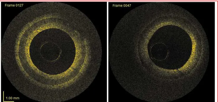
The multilayer phantoms were imaged with our custom built IV-OCT system. The system has been described in a previous publication [4]. Fig. 2 shows two images from an OCT pullback acquired on one of these phantoms. The image on the left shows the rich-poor-rich signal expected from the formulations used for each layer. There is also very good delineation between all the layers. However, taken in a different region of the sample, the image on the right does not present the same delineation between layer 1 and 2. There is also a loss of visibility of layer 3, as its inner boundary is well observed, but its outer boundary is not. Regions giving images similar to the image on the right were frequently observed in the phantoms.
Fig. 2: Images taken from a pullback in a multilayer PVA-C phantom. The left image shows very good contrast, but in the right image, the contrast between the first and second layer is different, and
the visibility of the third layer is decreased.
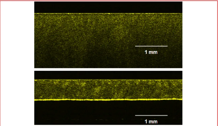
The reasons for the loss of contrast between layers 1 and 2 in some regions were investigated through several experiments. In one of these experiments, we investigated the effect of the difference in thickness between slab-shaped samples and multilayer artery phantoms on the resulting cryogel. Fig. 3 shows OCT images of two PVA-C samples. Both were made of 10% PVA-H without additives, both went through 2 FTCs, and both were made in flat SS moulds. However, one was made with 4 mm spacing between plates, and the other was made with 0.380 mm spacing. It can be observed that in the 4 mm sample, the scattering is more homogeneous than in the 0.380 mm sample, where several clusters of high signal can be observed. This loss of uniformity observed in thinner samples is believed to be responsible for the lack of contrast observed in the multilayer sample of Fig. 2.
In future work, in order to avoid losing the contrast between layers due to small thicknesses, we will change the overmoulding process for an alternate process where all layers are exposed to the same number of FTCs. The difference in optical properties in each layer would then only be provided by additives, which will insure good contrast and delineation. We also expect this alternate process to improve the uniformity of the samples. The three layers would be exposed to FTCs together, with the phantom at is final thickness through all the FTCs, instead of starting with very thin layer, and then successively building the other layers.
Fig. 3: Top: OCT image of a 4 mm thick sample. Bottom: OCT image of a 0.380 mm thick sample. Both sample are 10% PVA – 2 FTCs gels.
3. INCREASED STRAIN HARDENING
Uniaxial tensile tests were performed on different strip samples to characterize their mechanical properties using a standard mechanical tester (ElectroForce 3200, EnduraTEC, Bose).
3.1 Multilayer phantom
The mechanical characterization of the multilayer phantom from Fig. 2 is presented in Fig. 4. The phantom is compared to a strip of silicone with the formulation used in our usual method. It is also compared to target values of human artery layers extracted from the literature. Holzapfel et al. performed tensile tests in both the axial and the circumferential directions of the three layers of 13 human coronary artery segments [5]. They fitted an anisotropic strain energy function to the stress – stretch data obtained in both directions and averaged the fit parameters. In Fig. 4, we present the stress – stretch curves from the intima and the media layers stretched in the circumferential direction. These results show that the mechanical properties have a lot of variation between the layers, but also that the strain hardening in arteries is much greater than in the multilayer PVA-C phantom. In following development, we investigated strategies to increase the strain hardening to better mimic the properties of human arteries. Two strategies are presented in the next subsections.
Fig. 4: Tensile test results for the multilayer PVA-C phantom compared to results for silicone and human media and intima from literature.
3.2 FTC – stretch – FTC
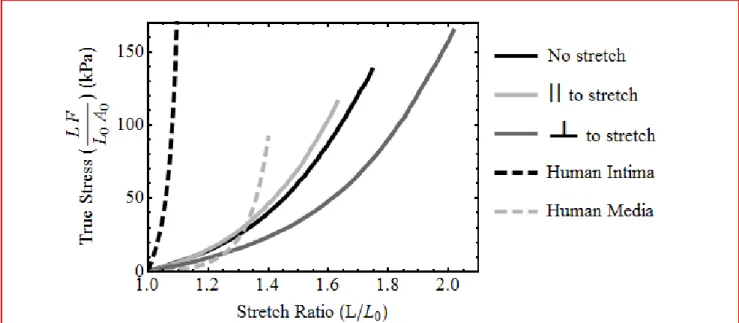
In a first attempt at improving the strain hardening, we stretched a cryogel sample, and held it at constant extension while performing the FTCs. It was hypothesized that extending the solidified PVA-C specimen would stretch and align the polymer chains/entanglements. By exposing the sample to additional FTCs, we expected to partially preserve that alignment which should stiffen the specimen. To test that concept, a slab shape 10% PVA – 1 FTC was stretched to a ratio of 1.5, and then submitted to one additional FTC. Tensile tests were then performed in the directions parallel and perpendicular to the stretch axis. The results are compared to a 10% PVA – 2 FTC that was not stretched and to human intima and human media from literature in Fig. 5. The results of the tensile test in the parallel axis showed a modest increase in strain hardening. However, the results of tensile test in the perpendicular direction showed a noticeable decrease of the overall stiffness. Therefore, that particular method did not give the expected effect and the hypothesis was disproved.
Fig. 5: Tensile test results for the FTC-stretch-FTC trial. Extension is performed in both the parallel and perpendicular directions to the stretch axis The results are ploted with a normal 10% PVA – 2
FTCs without stretch, and with human intima and human media to compare the results.
3.3 Mesh
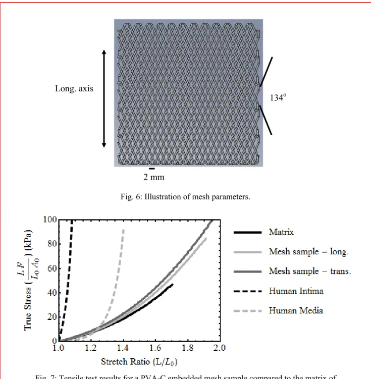
A second concept was tested to improve the strain hardening of the phantoms. This concept is to embed a mesh of stiff PVA-C in a matrix of softer PVA-C. A setup consisting of a pneumatic dispenser, a 3D motion system, and a cooling chamber was built. The pneumatic dispenser extrudes 10% PVA-H in form of fibers that are drawn into a mesh by the 3D motion system. The cooling chamber freezes the PVA-H in place. The solidified mesh is exposed to additional FTCs, and then embedded in a covering of 10% PVA-H for additional FTCs. As proof of that concept, slab shaped specimens were produced with different fiber sizes and spacing. One of the specimens was made with 10% PVA-H fibers of 1 mm diameter, and 2 mm spacing between fibers. The angle between the fibers is 134o. This value is based on the parameter φ, averaged for the adventitia, in the paper from Holzapfel et al. [5]. It represents the angle between symmetrically aligned fibers in the model of fiber reinforced composite material they use to describe the mechanical behavior of arteries. The mesh parameters are illustrated in Fig. 6. The frozen mesh was exposed to 5 FTCs, and then embedded in 10% PVA-H (2 mm thickness). The whole sample was finally exposed to another 2 FTCs. Fig. 7 shows the results of tensile tests performed on the mesh specimen, as well as on a portion of the matrix without mesh. The results are also compared to human intima and human media. The tests performed in the longitudinal and the transverse axis of the mesh show a small increase in strain hardening compared to the surrounding matrix. However, it is still very different from target curves for human intima and human media. Nevertheless, we believe that this particular avenue of development deserves further attention since it is inspired by the actual fibrous structure of arteries. There are many parameters that can be adjusted to further improve the strain hardening, like the mesh stiffness, the fiber size, the spacing of fibers, the angle of fiber orientation, etc. We are currently further investigating this topic.
Fig. 6: Illustration of mesh parameters.
Fig. 7: Tensile test results for a PVA-C embedded mesh sample compared to the matrix of surrounding PVA-C and to human intima and human media.
4. CONCLUSION
In summary, we have presented a method for the fabrication of multilayer tubular PVA-C phantoms. The OCT imaging of the phantoms showed the three rich – poor – rich signal layers typical of arteries. However, the homogeneity of the phantoms requires further improvement by the development of an alternate fabrication process where all layers are exposed to a constant number of FTCs. Mechanical characterization of the phantoms showed that non-linear strain hardening is
134o Long. axis
artery layers. An interesting avenue of research to fabricate more realistic phantoms is to embed a stiff mesh, into, and adhered to, a softer matrix creating a fiber-reinforced composite construct. This avenue is based on the actual structure of the native artery that has a preferred orientation of the fiber components, embedded in a polyglycans matrix.
ACKNOWLEDGEMENTS
The authors would like to express their appreciation to Bill Wells for fabricating the PVA-C specimens.
REFERENCES
[1] C.-E. Bisaillon, M.-M. Lanthier, M. L. Dufour et al., "Durable coronary artery phantoms for optical coherence tomography," Proc. SPIE. 7161, 71612E-10 (2009).
[2] C.-E. Bisaillon, M. L. Dufour, and G. Lamouche, "Durable phantoms of atherosclerotic arteries for optical coherence tomography," Proc. SPIE. 7548, 75483G-4 (2010).
[3] C.-E. Bisaillon, G. Campbell, C. de Grandpre et al., "Multilayer tubular phantoms for optical coherence tomography," Proc. SPIE. 7567, 75670I-6 (2010).
[4] G. Lamouche, M. Dufour, M. Hewko et al., “Intravascular optical coherence tomography on a beating heart model,” Journal of Biomedical Optics, 15(4), 046023-7 (2010).
[5] G. A. Holzapfel, G. Sommer, C. T. Gasser et al., “Determination of layer-specific mechanical properties of human coronary arteries with nonatherosclerotic intimal thickening and related constitutive modeling,” Am. J. Physiol Heart Circ Physiol, 289, H2048-H2058 (2005).