HAL Id: inserm-02495599
https://www.hal.inserm.fr/inserm-02495599
Submitted on 2 Mar 2020
HAL is a multi-disciplinary open access
archive for the deposit and dissemination of
sci-entific research documents, whether they are
pub-lished or not. The documents may come from
teaching and research institutions in France or
abroad, or from public or private research centers.
L’archive ouverte pluridisciplinaire HAL, est
destinée au dépôt et à la diffusion de documents
scientifiques de niveau recherche, publiés ou non,
émanant des établissements d’enseignement et de
recherche français ou étrangers, des laboratoires
publics ou privés.
a mouse model of Birk-Barel intellectual disability
syndrome
Alexis Cooper, Tamer Butto, Niklas Hammer, Somanath Jagannath, Desiree
Fend-Guella, Junaid Akhtar, Konstantin Radyushkin, Florian Lesage, Jennifer
Winter, Susanne Strand, et al.
To cite this version:
Alexis Cooper, Tamer Butto, Niklas Hammer, Somanath Jagannath, Desiree Fend-Guella, et al..
Inhi-bition of histone deacetylation rescues phenotype in a mouse model of Birk-Barel intellectual disability
syndrome. Nature Communications, Nature Publishing Group, 2020, 11 (1), pp.480.
�10.1038/s41467-019-13918-4�. �inserm-02495599�
Inhibition of histone deacetylation rescues
phenotype in a mouse model of Birk-Barel
intellectual disability syndrome
Alexis Cooper
1
, Tamer Butto
2,10
, Niklas Hammer
3,10
, Somanath Jagannath
3
, Desiree Lucia Fend-Guella
1
,
Junaid Akhtar
2
, Konstantin Radyushkin
4
, Florian Lesage
5
, Jennifer Winter
1,4
, Susanne Strand
6
,
Jochen Roeper
3,11
, Ulrich Zechner
1,7,11
* & Susann Schweiger
1,4,8,9,11
*
Mutations in the actively expressed, maternal allele of the imprinted KCNK9 gene cause
Birk-Barel intellectual disability syndrome (BBIDS). Using a BBIDS mouse model, we identify here
a partial rescue of the BBIDS-like behavioral and neuronal phenotypes mediated via residual
expression from the paternal Kcnk9 (Kcnk9
pat) allele. We further demonstrate that the
second-generation HDAC inhibitor CI-994 induces enhanced expression from the paternally
silenced Kcnk9 allele and leads to a full rescue of the behavioral phenotype suggesting CI-994
as a promising molecule for BBIDS therapy. Thus, these
findings suggest a potential approach
to improve cognitive dysfunction in a mouse model of an imprinting disorder.
https://doi.org/10.1038/s41467-019-13918-4
OPEN
1Institute of Human Genetics, University Medical Center of the Johannes Gutenberg University Mainz, Langenbeckstr. 1, 55131 Mainz, Germany.2Institute for
Developmental Biology and Neurobiology, Johannes Gutenberg University Mainz, Staudingerweg 9, 55128 Mainz, Germany.3Institute of Neurophysiology, Goethe University Frankfurt, Theodor-Stern-Kai 7, 60590 Frankfurt, Germany.4Focus Program Translational Neuroscience, Center for Rare Diseases, University Medical Center of the Johannes Gutenberg University Mainz, Langenbeckstr. 1, 55131 Mainz, Germany.5Université Côte d’Azur, INSERM, Centre National de la Recherche Scientifique, Institut de Pharmacologie Moléculaire et Cellulaire, Labex ICST, 660, route des Lucioles Sophia Antipolis, 06560 Valbonne, France.6Department of Internal Medicine I, University Medical Center of the Johannes Gutenberg University Mainz, Obere Zahlbacher Straße 63, 55131 Mainz, Germany.7Senckenberg Center of Human Genetics, Weismüllerstraße 50, 60314 Frankfurt, Germany.8Center for Orphan Diseases of the Central Nervous System, University Medical Center of the Johannes Gutenberg University Mainz, Langenbeckstr. 1, 55131 Mainz, Germany.9German Resilience Centre, University Medical Center of the Johannes Gutenberg University Mainz, Langenbeckstr. 1, 55131
Mainz, Germany.10These authors contributed equally: Tamer Butto, Niklas Hammer11These authors jointly supervised this work: Jochen Roeper, Ulrich
Zechner, Susann Schweiger *email:u.zechner@senckenberg-humangenetik.de;susann.schweiger@unimedizin-mainz.de
123456789
B
irk-Barel intellectual disability (ID) dysmorphism
syn-drome (BBIDS or KCNK9 imprinting synsyn-drome, OMIM
612292
) is typically associated with congenital central
hypotonia, developmental delay, intellectual disability, severe
feeding problems, and hyperactivity
1,2. The disease is inherited
autosomal dominantly with maternal-only transmission
1, as the
KCNK9 gene is embryonically paternally silenced (imprinted) in
man and mouse. It encodes the potassium channel subunit
TASK3, which dimerizes to form two-pore domain potassium
(K2P) leak channels
3. The mouse Kcnk9 gene maps to an
imprinted cluster on mouse chromosome 15 together with further
imprinted genes, i.e., the brain-specific maternally expressed
genes Ago2, Chrac1, and Trappc9 and the paternally expressed
Peg13 gene
4.
Kcnk9 mRNA expression is widespread in the central nervous
system
5,6; in rodents with notably high levels in cerebellar granule
neurons, the locus coeruleus (LC), the dorsal raphe nuclei,
hip-pocampal CA1 and CA3 pyramidal neurons, and several
hypo-thalamic nuclei
7,8. Homozygous deletion of Kcnk9 in the mouse
(Kcnk9KO
hom) reduces the resting potassium conductance and
enhances spike
firing accommodation in adult cerebellar granule
neurons
9. Kcnk9KO
hommice further display increased nocturnal
motor activity, cognitive deficits, as well as a reduced sensitivity to
inhalation anesthetics and the cannabinoid receptor agonist
WIN55212-2 mesylate
10–12. More recently, RNAi-based
knock-down of Kcnk9 and expression of a dominant-negative mutant
KCNK9, which had been associated with the human disease
phenotype were shown to impair neuronal migration during
mouse cortical development
13. However, the phenotype of mice
with heterozygous deletion of the active maternal Kcnk9 allele
(Kcnk9KO
mat) thus mimicking BBIDS has not yet been
characterized.
DNA methylation is well established as a key player in
imprinting regulation by multiple studies, which characterized
the kinetics and mechanisms of methylation reprogramming at
imprinting control regions (ICRs). The critical involvement of
post-translational histone modifications in transcriptional
reg-ulation, in general, is also well accepted, but much less explored
for ICRs
14. It is assumed that an interplay between DNA
methylation and histone acetylation is essential for proper erasure
and resetting of imprints in the germline as well as selective
imprint maintenance during postzygotic reprogramming
15. A
differentially methylated region (DMR) which is methylated on
the maternal allele and assumed to be involved in Kcnk9 gene
regulation has been only identified in the promoter region of
Peg13
16,17. Promoter CpG islands of Kcnk9 are unmethylated, but
display high levels of active histone H3 lysine 4 monomethylation
(H3K4me1) and histone H3 lysine 27 acetylation (H3K27ac)
chromatin marks in brain tissues
4.
Here, we characterize the behavioral and neuronal phenotype
of mice with heterozygous deletion of the active maternal Kcnk9
allele (Kcnk9KO
mat). We demonstrate partial behavioral rescue in
these animals compared with full knockout animals and show
that epigenetic manipulation stimulates Kcnk9
patexpression
sufficiently, to rescue the behavioral phenotypes, thereby opening
new avenues for treatment of cognitive dysfunctions in BBIDS.
Results
Deletion of
Kcnk9 leads to impaired behavior. To assess
behavioral deficits along the BBIDS phenotype in Kcnk9KO mice,
we performed in vivo experiments in adult wild type (WT) as well
as Kcnk9KO
matand Kcnk9KO
hommice with a deletion of Kcnk9
exon 2 as previously described
18(Fig.
1
a). Following the strictly
monoallelic expression pattern of Kcnk9 in mouse brain (<1%
paternal expression)
3, we expected largely concordant phenotypes
in mice carrying a deletion of both Kcnk9 alleles (Kcnk9KO
hom)
or only the maternal Kcnk9 allele (Kcnk9KO
mat), respectively.
No differences in behavior between Kcnk9KO
hom,
Kcnk9KO-mat, and WT littermates were seen in elevated plus maze and open
field testing for anxiety (Supplementary Fig. 1a, b) and in the
rotarod-test for motor coordination (Supplementary Fig. 1c).
Given the described intellectual disability in BBIDS patients, we
then examined whether working memory was affected in
Kcnk9KO mice. Spontaneous alternation relies on the natural
tendency of rodents to explore a novel environment and can be
quantified using a Y-maze task. The ability to remember the
immediately preceding choice is considered an indicator for
active working memory. In the Y-maze, spontaneous alternation
was defined as consecutive entries into all three arms without
revisiting an arm. The average percentage of spontaneous
alternation made by Kcnk9KO
hom, Kcnk9KO
mat, and WT mice
across a 10-min trial was analyzed and revealed a significant
reduction of spontaneous alternation by about 10% in both
Kcnk9KO
homand Kcnk9KO
matmice compared with WT
littermates (Fig.
1
b). There was no significant difference in the
extent of the working memory impairment between Kcnk9KO
homand Kcnk9KO
matmice (Fig.
1
b). Our
findings indicate an
impaired working memory in Kcnk9KO
homand Kcnk9KO
matmice, which is consistent with previously reported observations in
Kcnk9KO
hommice
10.
Circadian rhythms in mammals are endogenously coordinated
oscillations of biological parameters such as the sleep/wake cycles
with an overall period length of about 24 h
19. KCNK9 channels
are expected to contribute to the in vivo electrical activity of
neurons in those brain regions associated with the regulation of
circadian rhythms and arousal
10–12,20. Spontaneous motor
activity analysis during light (resting) and dark (active) phase
was performed, resembling day and night in diurnal humans,
respectively. A significantly increased overall locomotor activity
during the dark phase was found in Kcnk9KO
homand
Kcnk9KO
matmice compared with WT controls, describing an
exaggerated nocturnal activity in Kcnk9KO animals (Fig.
1
c),
which is in consensus with previous observations
10. Surprisingly,
an intermediate phenotype for dark phase activity was observed
in the Kcnk9KO
matanimals with statistically significant
differ-ences to both WT and Kcnk9KO
homanimals. No significant
differences between genotypes were observed in the overall
locomotor activity during the light phase (Fig.
1
c, Supplementary
Table 1).
Together, these data demonstrate that the loss of Kcnk9 in mice
results in the impairment of behavioral parameters resembling
components of the BBIDS phenotype. Interestingly, we found an
intermediate phenotype for nocturnal locomotor activity in
animals with a loss of only the actively expressed maternal allele
(Kcnk9KO
mat) suggesting the involvement of the silenced
paternal allele.
Non-canonical imprinting of
Kcnk9 in the mouse brain. In
human and mouse brain KCNK9 was reported to be
mono-allelically expressed from the maternal allele
3,4while the paternal
allele is silenced. To elucidate if the intermediate phenotype of
Kcnk9KO
matanimals was due to residual expression from the
paternal allele, we analyzed the parent-of-origin allele-specific
expression pattern of Kcnk9 in different brain regions of F1
hybrid animals from crosses between C57BL/6 (B6) and Mus
musculus castaneus (Cast/Ei) mouse strains [(C57BL/6xCast/Ei)
F1]
3. As expected, we observed a predominant expression of the
maternal Kcnk9 allele in all analyzed brain regions (Fig.
1
d).
However, we also detected significant expression from the
repressed paternal allele, which represented 1–14% of all
100,000
****
**
*
**
*
a
b
d
c
Y-maze Day 1 5 6 7 8 Circadian rhythm WT Kcnk9KOmat Kcnk9KOhom WT 20% C57BL/6J Cast/Ei 16% 12% 100% 0% 10% 20% 30% 40% 50% 60% 70% 80% 90% 100% 0% 10% Cerebellum Pons Olfactor y b ulb Cor tex HippocampusHypothalam us StriatumMidbr ain Medulla Locus coer uleus Cerebellum Pons Olfactor y bulb Cor tex HippocampusHypothalam us Striatum Midbr ain Medulla Locus coer uleus 20% 30% 40% 50% 60% 70% 80% 90% 8% 4% 0% Maternal PaternalKcnk9KOmat Kcnk9KOhom
80,000 Distance tr a v elled [cm] 60,000 100 P ercent alter nation (%) 75 50 25 40,000 20,000 0
Light phase Dark phase
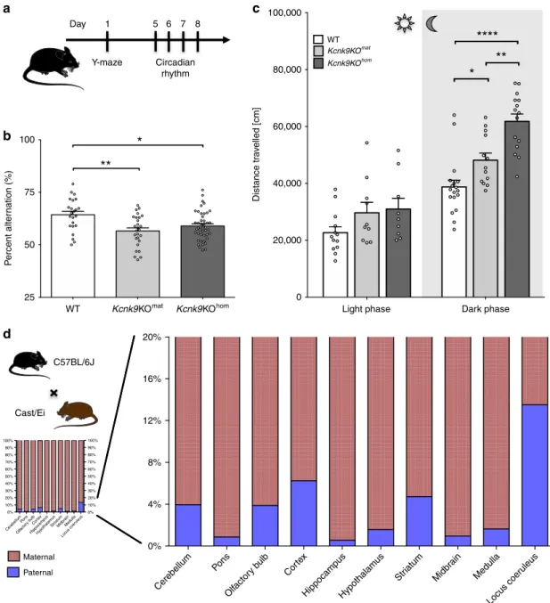
Fig. 1 Deletion of non-canonical imprintedKcnk9 gene leads to impaired behavior of Kcnk9KO mice. a Schematic representation of the sequence of mouse behavioral experiments.b Y-maze percentage alternation analysis of WT (n= 23), Kcnk9KOmat(n= 27), and Kcnk9KOhom(n= 44) mice. A
spontaneous alternation was defined as consecutive entries into all three arms without revisiting an arm. Kcnk9KO mice display a significant decrease in percentage alteration compared with WT mice. One-way ANOVA: F(2, 91)= 7.261, P = 0.0012; followed by Bonferroni’s multiple comparison post hoc test, *P < 0.05, **P < 0.01.c Total locomotor activity (distance traveled in the home cage) in light (12 h, sun symbol)/dark (12 h, moon symbol) phase. The left section shows no difference in distance traveled in the light phase of WT (n= 13), Kcnk9KOmat(n= 10) and Kcnk9KOhom(n= 9) mice. One-way
ANOVA: F(2, 29)= 2.281, P = 0.1203. The right section depicts the nocturnal activity of WT (n = 18), Kcnk9KOmat(n= 13) and Kcnk9KOhom(n= 15) mice.
Kcnk9KOmatand Kcnk9KOhommice display significantly increased nocturnal hyperactivity compared with WT littermates with activity scores of
Kcnk9KOmatmice intermediate between Kcnk9KOhomand WT mice. One-way ANOVA: F(2, 43)= 22.70, P < 0.0001; followed by Bonferroni’s multiple
comparison post hoc test, *P < 0.05, **P < 0.01, ****P < 0.0001.b, c Behavioral experiments were performed in seven independent sessions. d Non-canonical Kcnk9 imprinting in (C57BL/6xCast/Ei)F1 hybrid mice. Quantification of Allele-Specific Expression by Pyrosequencing (QUASEP) of several brain regions from (C57BL/6xCast/Ei)F1 hybrid mice; maternal allele (red) and paternal allele (blue). Cerebellum n= 14, pons n = 10, olfactory bulb n = 12, cortex n= 14, hippocampus n = 14, hypothalamus n = 9, striatum n = 5, midbrain n = 5, medulla n = 5, and locus coeruleus n = 4; n = biologically independent samples.a–d Data are means ± 1 SEM (standard error of the mean). Statistical analyses and approaches are provided in Supplementary Table 1. Source data are provided as a Source Datafile. The mouse images in this figure were created using Servier Medical Art templates, which are licensed under a Creative Commons Attribution 3.0 Unported License;https://smart.servier.com.
transcripts depending on the brain region analyzed (Fig.
1
d,
suppl. Fig. 2c). Highest paternal expression was observed in the
LC (Fig.
1
d). Accuracy of the LC tissue-punches was
demon-strated through elevated gene expression levels of tyrosine
hydrolase (TH) in LC samples (Supplementary Fig. 2a, b).
These data demonstrate a brain region-specific leakiness of the
imprint on the Kcnk9
patallele with a peak in the LC. This
leakiness might be responsible for the observed intermediate
phenotype in Kcnk9KO
matanimals and suggested a particular
importance of LC Kcnk9 expression for locomotor activity during
the active (dark) phase.
Kcnk9 knockdown in the LC induces elevated nocturnal
activity. To test the role of functional Kcnk9 expression in LC
neurons for the control of nocturnal activity, we bilaterally
infused AAV vectors for expression of eGFP and shRNAmir
sequences, either scrambled
shRNAmir-scrambled-EF1a-eGFP) or specifically targeting Kcnk9 mRNA
(pAAV-Syn-shRNAmir-Kcnk9-EF1a-eGFP), into the LC of WT mice under
stereotactic control (Fig.
2
a). Efficiency of the used shRNA
sequence was pre-tested in vitro (Fig.
2
b) and validated by
RT-qPCR from mouse brain tissue (Fig.
2
c). Targeting efficiency and
selectivity of stereotactically guided infusion was demonstrated by
eGFP immunohistochemistry (Fig.
2
a). Twenty-one days after
injection, animals were tested for circadian activity and working
memory. A significant and selective increase of nocturnal activity
was detected in the shRNAmir-Kcnk9 injected animals compared
with age-matched control animals injected with the scrambled
shRNAmir virus (Fig.
2
d). These results demonstrated that
altering local expression of Kcnk9 in the LC was sufficient to
selectively affect dark-phase activity in mice. Furthermore, it
identified LC as an important neural hub for mediating the
behavioral effects of altered Kcnk9 expression, which warranted
further mechanistic analysis of these neurons. Interestingly, a
clear trend toward impaired working memory was also observed
in shRNAmir-Kcnk9 injected animals (Fig.
2
e), suggesting that
Kcnk9 expression also controls working memory-related activity
of LC neurons.
Increased dark-phase LC pacemaking activity in
Kcnk9KO
hombut not
Kcnk9KO
matmice. Electrical activity of LC neurons
drives periods of wakefulness and arousal
21–23. Indeed, selective
optogenetic activation of LC neurons revealed their causal role in
sleep-to-wake transitions and locomotor arousal by
demonstrat-ing that sustained 3-Hz LC neuronal stimulation enhanced
spontaneous locomotion (total track length) by about 50%
24. As
we detected a similar increase of spontaneous locomotion during
the active (dark) phase of Kcnk9KO
hommice compared with WT
controls, we reasoned that KCNK9-dependent differences in LC
pacemaking activity might contribute to this phenotype.
By recording from synaptically isolated (including inhibition of
somatodendritic alpha2-autoreceptors), spontaneously active LC
neurons in brainstem slices from adult mice, we observed a dark
phase-selective, about 70% increase of pacemaker frequency in
LC neurons from Kcnk9KO
hommice (Fig.
3
a–d). In contrast, no
significant dark-phase increase of LC pacemaker frequency was
observed in WT animals (Fig.
3
c, d). These results demonstrate
that some degree of KCNK9 channel expression is necessary to
selectively dampen enhanced pacemaker activity during the dark
phase, which is present in Kcnk9KO
hommice. Interestingly, LC
neurons from Kcnk9KO
matanimals displayed no significant
increase in pacemaker activity in the dark phase (Fig.
3
c, d), but a
significant difference in pacemaker activity in the dark was
observed between Kcnk9KO
homand Kcnk9KO
matanimals. This
indicated that a certain level of functional expression of KCNK9
channels from the Kcnk9
patallele in LC is present in Kcnk9KO
matanimals to prevent the full nocturnal in vitro pacemaker
frequency increase. This level of Kcnk9 expression might at the
same time not be sufficient to dampen in vivo LC activity, which
is also driven by synaptic inputs from neuronal networks, in line
with the observed intermediate behavioral phenotype regarding
dark-phase locomotion in Kcnk9KO
matmice. In line with this
argument, we show below that boosting further expression of
KCNK9 channel subunits from paternal alleles indeed also
restores the WT motor activity phenotype.
CI-994 activates the paternally repressed
Kcnk9 allele. The
paternally inherited Kcnk9/KCNK9 gene is epigenetically silenced
yet structurally unimpaired. Our behavioral and gene expression
experiments suggest a contribution of paternally expressed Kcnk9
to the intermediate phenotype of the Kcnk9KO
matmice and the
possibility of upregulation of the paternal allele in the case of loss
of the maternal allele. We, therefore, speculated that exogenous
application of epigenetic modulators might alter the structure at
the Kcnk9 promoter region and result in further derepression of
the Kcnk9
patallele. We hypothesized that this could fully
com-pensate for the loss of the maternal allele and rescue the
BBIDS-like phenotype of Kcnk9KO
matmice.
To investigate the paternally derived Kcnk9 transcript levels
after epigenetic drug treatment, we isolated E14 mouse primary
cortical neurons (mPCNs) from WT and Kcnk9KO
matanimals
from crosses between WT males and Kcnk9KO
homfemales
(Fig.
4
a). We then chose six different compounds representing
different classes of epigenetic modulators for treatment (Fig.
4
b).
Murine PCNs were treated for 3 days with the epigenetic
modulators and Kcnk9 expression was analyzed by RT-qPCR.
mPCNs treated with DZNep (20 µM), SAHA (30 µM), VPA (5
mM), and CI-994 (40 µM) exhibited a significantly increased
Kcnk9
patexpression (RT-qPCR) compared with Kcnk9KO
matmPCNs treated with dimethyl sulfoxide (DMSO) (control
vehicle) (Fig.
4
c). By contrast, treatment with the compounds
Zebularine and C646 did not show any differential Kcnk9
expression in Kcnk9KO
matmPCNs compared with control
conditions (Supplementary Fig. 3a).
For further investigations, we focused on the benzamide-based
second-generation histone deacetylase inhibitor (HDACi) CI-994,
a selective inhibitor of class I HDACs, which is a potent inhibitor
of HDAC1 and 3 isoenzymes relative to HDAC6 and HDAC8
25.
CI-994 showed the most efficient upregulation of Kcnk9
patexpression in Kcnk9KO
matmPCNs (Fig.
4
c). Furthermore, a
previous study reported that intraperitoneal administration of
CI-994 in WT mice resulted in long-lasting CI-CI-994 levels in the brain
without affecting the overall behavioral phenotype of mice
26.
The effect of CI-994 in Kcnk9KO
matmPCNs was further tested
in a dose-response experiment. Kcnk9KO
matmPCNs were treated
with increasing concentrations of CI-994 for 24 h, and Kcnk9
expression was analyzed using RT-qPCR in comparison to
DMSO-treated Kcnk9KO
matand WT control cells (Fig.
4
d). As
expected, the Kcnk9
patexpression in DMSO-treated Kcnk9KO
matmPCNs was significantly reduced compared with that of WT
mPCNs. After CI-994 treatment of Kcnk9KO
matmPCNs, we
observed a strong linear correlation between the increase of
Kcnk9
patexpression and CI-994 dosage suggesting a specific,
dose-dependent effect of CI-994. Notably, Kcnk9 expression in
80 µM CI-994-treated Kcnk9KO
matmPCNs exceeded that of
DMSO-treated WT mPCNs in vitro (Fig.
4
d). Treatment of
Kcnk9KO
matmPCNs with 20 µM CI-994 showed a consistent
increase of Kcnk9
patexpression even after 10 days suggesting a
prolonged and stable in vivo effectiveness (Fig.
4
e). Cell viability
analysis after treatment of Kcnk9KO
matmPCNs with 10 or 20 µM
CI-994 did not reveal any toxic effect (Supplementary Fig. 4c).
Intriguingly, CI-994 treatment did not affect the expression of
other nearby imprinted genes within the imprinted cluster on
mouse chromosome 15 in vitro (Supplementary Fig. 3b).
CI-994 rescues the behavioral phenotype of
Kcnk9KO
matani-mals. The detection of Kcnk9
patallele expression in several brain
regions, prominently among them the LC, as well as the
observation of intermediate phenotypes in Kcnk9KO
matanimals
led us to hypothesize that epigenetic manipulation could further
stimulate paternal gene expression and thereby boost the
phe-notypical rescue.
For testing, either DMSO (100%) or CI-994 (30 mg/kg of body
weight in 100% DMSO) were injected daily over 14 days in the
peritoneum (Fig.
5
a). After injections, Kcnk9
patexpression was
induced up to ~3-fold compared with DMSO-treated controls in
several brain regions including the cerebellum, hippocampus,
150,000 100,000 50,000 0 0 0 PFC Hippocampus LC 1 1 2 ** 2 100 1525 100%
a
b
50% 25% 8% 11% 7% 16%3% 6% 4% 5% 6% 0% 1 50 µm 1000 µm 2 3 4 5 6 7 8 9 10 100% NT 1 ID 21mer sequence Position on mRNA Region shRNA (shmir) 2 3 4 5 6 7 8 9 10 ORF pAAV-Syn-shRNAmir-Kcnk9-EF1a-eGFP pAAV-Syn-shRNAmir-scrambled-EF1a-eGFP ORF ORF ORF ORF ORF ORF ORF ORF ORF 1526 1563 1653 1881 1959 1986 2076 2082 2205 P e rcent alter nation (%) Distance tr a v elled (cm) Relativ e Kcnk9 e xpression F o ld change e x pression (T yrosine h y dro xylase/I mpa2) F old change e xpression (Kcnk9/Impa2 ) 0.0797 75 50 25 Kcnk9 knoc k do wn Scr amb led control Kcnk9 knoc k do wn Scr amb led control Kcnk9 knoc k do wn Scr amb led control 200 µmc
d
e
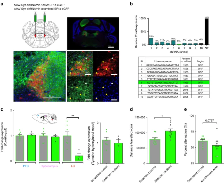
*Fig. 2Kcnk9 knockdown in the locus coeruleus induces elevated nocturnal activity. a Bilateral virus injection with pAAV-Syn-shRNAmir-Kcnk9-EF1a-eGFP (KD) or Syn-shRNAmir-scrambled-EF1a-pAAV-Syn-shRNAmir-Kcnk9-EF1a-eGFP (SC) in the locus coeruleus (LC): Top left: schematic coronal section of the mouse brain at the position −5.4 relative to bregma43; red triangles represent the LC; top right: Immunofluorescence staining of a coronal section. Scale represents 1000 µm (4×). Bottom left: Images of the LC. Scale represents 200µm (20×); bottom right: Magnification of LC cells. Scale represents 50 µm (60×), majority of tyrosine hydroxylase (TH; red) expressing cells co express GFP (green) (yellow arrows), while also GFP negative TH expressing cells could be observed (white arrow). Blue, DAPI; green, GFP; red, TH (TH serves as a norepinephrine marker).b Cell-based quantification of Kcnk9 gene knockdown (Kcnk9 KD) using Kcnk9-specific shRNAs by RT-qPCR compared with negative control (NT). Validated shRNAs with KD > 80%: 2, 3, 4, 5, 6, 7, 8, 9, and 10. ShRNA 6 was utilized for AAV generation in 293T cells. Arithmetic means of Kcnk9 expression of presented IDs were provided by Sirion Biotech.c Left: Kcnk9 knockdown efficiency validation in vivo. Kcnk9 expression analysis in the prefrontal cortex (PFC, blue circle, SC n = 8, KD n = 4), hippocampus (pink circle, SC n = 7, KD n= 4) and LC (red circle, SC n = 7, KD n = 4) by RT-qPCR revealed a significant down-regulation of Kcnk9 gene expression in the LC of Kcnk9 KD mice compared with mice injected with scrambled controls. Mann–Whitney U: P = 0.004. Right: Tyrosine hydroxylase (TH) RT-qPCR expression analysis in the LC to validate accuracy of tissue collection. LC samples of Kcnk9 KD (n= 4) and scrambled controls (n = 7) exhibit similar TH levels demonstrating sample collection accuracy. n= biologically independent mice. d Total locomotor activity in dark (12 h) phase, 21 days after AAV-injections. Mice injected with pAAV-Syn-shRNAmir-Kcnk9-EF1a-eGFP (n= 10) display increased nocturnal activity compared with pAAV-Syn-shRNAmir-scambled-EF1a-eGFP (n = 9) injected controls. Mann–Whitney U: P = 0.0101. e Y-maze percentage alternation analysis reveals tendency toward impaired working memory in Kcnk9 KD mice (n= 10) compared with age-matched controls (n = 10); n = biologically independent mice. Mann–Whitney U: P = 0.0797. Values are means ± SEM. Statistical analyses and approaches are provided in Supplementary Table 1. Source data are provided as a Source Datafile.
pons, hypothalamus, olfactory bulb, and the LC (Fig.
5
b).
Importantly, this drug-induced rescue also included brain regions
like the hippocampus, where only a small degree (<1%) of
spontaneous expression from paternal alleles was observed in the
Kcnk9KO
matmice. In an allele-specific assay using WT (C57BL/
6xCast/Ei)F1 hybrids, we found that CI-994 affects specifically
expression of the Kcnk9
patallele (Supplementary Fig. 4b). Only a
very weak upregulation of global (maternal plus paternal) Kcnk9
expression was seen in WT animals after CI-994 treatment
(Fig.
5
c, Supplementary Fig. 4c). Importantly, and in line with the
in vitro observations, expression of only Kcnk9 but no other of
the genes located within the imprinting cluster on mouse
chromosome 15 was found to be increased after CI-994 injection
in vivo (Fig.
5
d).
To assess whether CI-994-mediated enhancement of Kcnk9
expression also promotes behavioral recovery, WT, Kcnk9KO
mat,
and Kcnk9KO
hommice were subjected to behavioral testing after
treatment with either DMSO (100%) or CI-994 (30 mg/kg of body
weight). In the Y-maze task, CI-994 treatment led to a significant
increase of spontaneous alternation in the Kcnk9KO
matmice
compared with DMSO-treated Kcnk9KO
matcontrol mice
(Fig.
5
e). Post hoc testing for multiple comparisons showed no
difference between CI-994-treated Kcnk9KO
matanimals and
either DMSO- or CI-994-treated WT animals (Fig.
5
e). WT mice
treated with either DMSO or CI-994 did not exhibit deficits in
working memory and significant differences in the Y-maze. By
contrast, no increase of spontaneous alternation was seen in the
CI-994 treated Kcnk9KO
homanimals suggesting that the paternal
allele silenced in the Kcnk9KO
matanimals carries the effect of the
treatment (Supplementary Fig. 5a).
We then asked whether CI-994 treatment also alters nocturnal
activity in Kcnk9KO
mator in Kcnk9KO
hommice. Indeed,
Kcnk9KO
matmice showed a significant decrease in horizontal
nocturnal activity during the 12-h dark phase after CI-994
treatment (Fig.
5
f). Again, post hoc analysis of multiple
comparisons showed no significant difference between CI-994
-treated Kcnk9KO
matand DMSO- or CI-994-treated WT animals
suggesting full rescue of the nocturnal hyperactivity phenotype in
the Kcnk9KO
matanimals. No drug effect was observed in
Kcnk9KO
homanimals and total locomotor activity in the light
phase was not altered by CI-994 treatment irrespective of the
genotype of the animals (Fig.
5
f, Supplementary Fig. 5b).
CI-994 interferes with H3K27 acetylation at the
Kcnk9 locus.
Imprinting of the Kcnk9/KCNK9 gene is thought to be regulated
by a maternally methylated germline differentially methylated
region (DMR) in the promoter of the Peg13/PEG13 gene
3.
12 WT**
*
Locus coeruleusa
b
TH Neurobiotin Merge 500 µm 50 µm WT — dark phase Light phase Mean frequency (Hz) 10 mV 1 s Dark phase Kcnk9KOmatKcnk9KOmat — dark phase
Kcnk9KOhom — dark phase
Kcnk9KOhom 10 8 6 4 2 0
c
d
Fig. 3 Electrophysiological analysis of LC neurons. a Schematic overview of the locus coeruleus (LC) and its position in the adult mouse brain, bregma −5.40 and −5.52 (the coordinates were according to the mouse brain atlas43). Green triangles represent the LC.b Confocal images showing TH-positive
signal (green), neurobiotin-positive signal (red) and a merge of both signals (yellow). Upper images show an overview of the LC at a low-magnification (×4) and lower images display signals of analyzed single-cell at high-magnification (×60 oil). c Electrophysiological traces of single cells in the night phase of WT, Kcnk9KOmatand Kcnk9KOhomanimals. Recordings from synaptically isolated spontaneously active LC neurons reveal increased pacemaker
frequency in LC neurons from Kcnk9KOhommice during dark phase.d Scatter dot-plot of mean frequencies including all analyzed cells in the day and night
phase of WT (light n= 46, N = 5, dark n = 23, N = 3), Kcnk9KOmat(light n= 41, N = 5, dark n = 26, N = 4) and Kcnk9KOhom(light n= 22, N = 3, dark n =
30, N= 5) animals. Ordinary one-way ANOVA with Bonferroni’s multiple comparison post hoc test, *P < 0.05, **P < 0.01. Values are means ± SEM. N = number of mice, n= total number of cells. Statistical analyses and approaches are provided in Supplementary Table 1. Source data are provided as a Source Datafile.
Acetylation of histone 3 lysine 27 (H3K27ac), monomethylation
of histone 3 lysine 4 (H3K4me1) as well as DNA methylation at
the maternally methylated DMR in the human PEG13 promoter
region have been suggested to control human KCNK9 expression
4(Fig.
6
a, Supplementary Fig. 6a-d). CI-994 is a second-generation
class I histone deacetylase inhibitor inhibiting HDACs1 and 3
with high specificity
25. To clarify the epigenetic mechanism
underlying the induction of the Kcnk9
patallele through CI-994 we
investigated the methylation status of two subregions of the
Peg13-DMR (Peg13 DMR1 and Peg13 DMR2) in adult
hippo-campus and LC of DMSO- and CI-994-treated Kcnk9KO
matmice
using bisulfite pyrosequencing. Methylation levels of about
40–50% typical for imprinted gene DMRs in somatic cells
were determined for both DMRs in the two brain regions
of Kcnk9KO
matmice. These methylation levels were not
significantly altered by CI-994 treatment of Kcnk9KO
matmice
(Supplementary Fig. 6c, d). Our results indicate that the
upre-gulation of Kcnk9 mRNA in various brain regions of Kcnk9KO
matmice after CI-994 treatment was independent of the DNA
methylation status at the Peg13-DMR.
The human PEG13-DMR has been further demonstrated to
bind CTCF-cohesin which conveys chromatin looping between a
brain-specific enhancer region, marked by H3K27ac and
H3K4me1, and the PEG13 and KCNK9 promoters to supposedly
control brain-specific KCNK9 expression
4. To determine the
effect of CI-994 on the Kcnk9 locus in the hippocampus and LC
of Kcnk9KO
matmice, we performed Chromatin
immunoprecipi-tation (ChIP)-qPCR for H3K27ac and H3K4me1 marks following
CI-994 or vehicle treatment. Using several public mouse brain
ChIP-seq datasets, we did not
find an orthologous region in the
8
a
c
b
e
d
6 4 2 E14 Cortical neurons A B C Neuron seeding 4 days Drug Inhibition Histone acetyltransferase Histone deacetylase Histone deacetylase Histone deacetylase DNA methyltransferase Histone methyltransferase C646 CI-994 Dznep SAHA VPA Zebularine 1–3 days RT-qPCR Cycle number Fluorescence 6 4 3 2 1 0 5 –5 Isolation of E14 primary cortical neuronsCI-994/ DMSO –2 0 Harvest sells Medium change Medium change Harvest sells 1 Treatment RT-qPCR 4 10 4 3 2 1 0 5 0 DMSO DMSO DMSO CI-994 20 µM Day 1 Day 10 DMSO WT 4 µM 20 µM 40 µM 80 µM VPA DZnep SAHA CI994
Kcnk9KOmat Kcnk9KOmat Log 2 FC ( Kcnk9 /Impa2 ) Log 2 FC ( Kcnk9 /Impa2 ) F o ld change Kcnk9 e x pression 1 2 3 4
mouse displaying a brain-specific enhancer chromatin signature
similar to that identified at the human imprinted domain on
chromosome 8q24. Thus, we focused our analysis on the
promoter and intronic region of the Kcnk9 locus, which are
enriched for both marks in the mouse brain ChIP-seq datasets
(Fig.
6
a). Due to the small size of the LC, we opted to use a novel
low-input ChIP-method
27for both regions (LC and
hippocam-pus) and validated this by conventional ChIP-qPCR in the
hippocampus (Supplementary Fig. 7b). Upon CI-994 treatment,
we observed a significant more than two-fold increase in
H3K27ac depositions at the two Kcnk9 regions both in the
hippocampus and LC of Kcnk9KO
matmice (Fig.
6
b). The levels of
H3K27ac were also increased in the hippocampus of WT
littermates however the increase was not as prominent as in the
Kcnk9KO
matanimals (Supplementary Fig. 7i, j). An intergenic
region, lacking H3K27ac or H3K4me1 modifications, was used as
negative control (Supplementary Fig. 8c-j). Looking at
allele-specific H3K27ac deposition in the hippocampus of (C57BL/
6xCast/Ei)F1 hybrid animals using two different SNPs in intron 1
of Kcnk9, we found that, as expected, the maternal allele had a
higher degree of H3K27ac association than the paternal allele.
CI-994 treatment of Kcnk9KO
matmice, however, led to a higher
degree of H3K27ac deposition at the paternal than at the
maternal Kcnk9 allele (Fig.
6
c).
In addition, we tested the deposition of H3K4me1 both in the
LC and hippocampus to assess if histone acetylation inhibition
leads to additional changes in other histone marks. Treatment
with CI-994 led to only a slight, but not significant increase of
H3K4me1 deposition at the Kcnk9 promoter region
(Supplemen-tary Fig. 7f-h). Our
findings indicate that CI-994 treatment affects
histone acetylation consistently at the promoter and intronic
regions of Kcnk9 of the investigated brain tissues and has
diverging effects on H3K4me1 deposition at the investigated
Kcnk9 locus.
Taken together, our
findings demonstrate that the paternal
allele of Kcnk9 is not fully silenced in the brain, further
derepressed in the case of loss of the maternal allele and
substantially activated upon exogenous epigenetic modulation.
Our data provide evidence for a promising therapeutic effect of
the class I HDAC inhibitor CI-994, in a mouse model for BBIDS
with maternal loss of Kcnk9.
Discussion
Kcnk9/KCNK9 is an imprinted gene in mouse and man that is
expressed from the maternal allele. Pathogenic variants
present on only the maternal allele, therefore, cause disease in
patients
1,2. In a study comparing full Kcnk9 knockout animals
(Kcnk9KO
hom)
with
maternal
Kcnk9
knockout
animals
(Kcnk9KO
mat) and WT littermates in a behavioral battery, we
surprisingly found an intermediate phenotype in nocturnal
locomotor activity in Kcnk9KO
matanimals. Furthermore,
elec-trophysiological analysis of tyrosine hydroxylase (TH)-positive
neurons in the LC, a region involved in regulation of arousal and
locomotor activity in mice, displayed significantly increased
spontaneous
firing frequencies only during the active dark phase
in full knock-outs in contrast to WT, while it was fully rescued in
the Kcnk9KO
matanimals. Allele-specific expression analysis of
Kcnk9 mRNA in (C57BL/6JxCast/Ei)F1 hybrid animals revealed
significant residual paternal expression of up to 14% of total
Kcnk9 expression in the LC in the Kcnk9KO
matanimals. Daily i.p.
injection of the second-generation HDAC inhibitor CI-994 could
increase this expression up to three-fold and resulted in a full
rescue of the behavioral phenotypes of Kcnk9KO
matanimals.
We show here that an increase in dark-phase locomotion in the
Kcnk9KO
homanimals is associated with an increase of in vitro
pacemaker frequency in the LC. Deletion of the active maternal
Kcnk9 allele in the mouse is supposed to lead to a phenotype
comparable to the full knockout. Surprisingly, however, our study
identified an intermediate behavioral phenotype in nocturnal
(hyper)activity in the Kcnk9KO
matanimals, as well as a fully
rescued cellular phenotype (LC
firing frequency) (Figs.
1
c,
3
).
Extended allele-specific expression analysis using (C57BL/
6JxCast/Ei)F1 mouse hybrids demonstrated residual Kcnk9
expression from the paternal allele in several brain regions with a
significant peak in the LC of 14% of total Kcnk9 expression
coming from the paternal allele.
The LC plays an essential role in arousal and sleep-wake
transitions
28and, as the origin of noradrenergic neurons
pro-jecting into the hippocampus and the prefrontal cortex, is also
involved in the regulation of long-term and working memory,
two essential building blocks of higher cognition/brain
func-tions
29. In a knockdown experiment using shRNAmir containing
AAVs targeting the Kcnk9 mRNA specifically injected into the LC
we found a similar behavior phenotype in circadian activity as
seen in the full and maternal Kcnk9 knockout animals
demon-strating an essential and causal contribution of Kcnk9 expression
within the LC in the control of nocturnal locomotor activity.
Interestingly, our electrophysiological data show full rescue of
in vitro pacemaker
firing frequency through Kcnk9
patexpression
in the LC in Kcnk9KO
matanimals on the one hand and only a
partial rescue of dark-phase activity on the other. This might
indicate different levels of Kcnk9 expression in order to control
on one hand isolated pacemaker function, which typically
oper-ates with very small ionic currents of a few picoamps, compared
Fig. 4 Identification of epigenetic modulators upregulating Kcnk9patexpression in mPCNs. a Workflow of murine primary cortical neurons (mPCN)
isolation, drug treatment, and RT-qPCR analysis.b Epigenetic modulators used for treatment of mPCNs (left column) with enzymatic activities inhibited by them (right column).c Significant upregulation of paternal allele-derived Kcnk9 mRNA in DZnep-, SAHA-, VPA- and CI-994-treated E14 Kcnk9KOmat
mPCNs compared with DMSO-treated Kcnk9KOmatmPCNs detected by RT-qPCR (3-day treatment, normalization to Impa2, DMSO n= 7 cultures/group,
DZnep (20µM) SAHA (30 µM), VPA (5 mM) and CI-994 (40 µM) n = 3 cultures/group). One-way ANOVA: F(5, 22) = 75.51, P < 0.0001; followed by Bonferroni’s multiple comparison post hoc test to Kcnk9KOmatDMSO control, ****P < 0.0001.d Upregulation of paternal allele-derived Kcnk9 mRNA after
treatment of Kcnk9KOmatmPCNs with different CI-994 concentrations compared with paternal allele-derived Kcnk9 mRNA of DMSO-treated Kcnk9KOmat
mPCNs and Kcnk9 mRNA of DMSO-treated WT mPCNs detected by RT-qPCR (1 day treatment, normalization to Impa2, Kcnk9KOmatDMSO n= 9; 4 µM n
= 2; 20–80 µM n = 4 and WT DMSO n = 5 cultures/group). One-way ANOVA: F(5, 22) = 75.51, P < 0.0001; corrected with Bonferroni’s multiple comparison post hoc test to Kcnk9KOmatDMSO mPCNs, ***P < 0.001****, P < 0.0001,c, d performed in 2–4 independent experiments. e Duration of Kcnk9
derepression in vitro. Workflow is illustrated in schematical representation (left). After 1 and 10 days, Kcnk9 is significantly upregulated after CI-994 treatment in mPCN compared with DMSO controls. One day DMSO n= 2, 1 day CI-994 n = 3, day 10 DMSO n = 6, day 10 CI-994; n = 4; n = samples (3–5 wells/sample; 6 embryos pooled). Day 1: P = 0.0297, day 10: P = 0.0001, Mann–Whitney U. c–e Values are means ± SEM. Statistical analyses and approaches are provided in Supplementary Table 1. Source data are provided as a Source Datafile. Images displaying the mouse, the embryo and the neurons in thisfigure were created using Servier Medical Art templates, which are licensed under a Creative Commons Attribution 3.0 Unported License;
* *** **** *** *** **** ** ** ** * ** **** Cerebellum Cor tex Hippocampus Pons Medulla Olfactor y bulb Locus coer uleus
Caudal pontine reticular f
ormation Prefrontal cor tex Hypothalam us 7 6 Log 2 FC e xpression ( Kcnk9 /Impa2 ) 5 3 4 2 1 0
DMSO CI994 DMSO CI994 WT Kcnk9KOmat
Daily i.p. injection
a
c
b
5 4 4 1 2 2 0 3 6 6 7 8 Brain removal Circadian rhythm Y-maze 8 9 10 11 12 13 14 14+ 10 Kcnk9 e xpression nor maliz ed to DMSO treatment 80,000 60,000 40,000 20,000 0Light phase Dark phase
WT DMSO Distance tr a v elled [cm] P ercent alter nation (%) WT CI-994 Kcnk9KOmat DMSO
Kcnk9KOmat CI-994
DMSO
Eif2c2 Chrac1 Peg13 Trappc9 Kcnk9
d
CI994 (30 mg/kg) WT Kcnk9KOmat 100 4 3 2 1 Kcnk9 fold change e xpression 0 75 25DMSO CI-994 DMSO CI-994
50
e
f
** **
Fig. 5 Effects of CI-994 histone deacetylase inhibitor treatment in vivo. a Experimental design of the mouse study. CI-994 or DMSO was intraperitoneally injected.b Significant upregulation of Kcnk9 after CI-994 treatment in several brain regions normalized to DMSO treatment, Mann–Whitney U test. Regions from left to right, DMSO/CI-994, n= 12/13; 6/9; 12/14; 7/8; 9/12; 6/6; 9/9; 6/6; 7/8 and 7/4. c Upregulation of Kcnk9 in the cerebellum of CI-994-treated Kcnk9KOmatmice (n= 13), but not of CI-994-treated WT mice (n = 12), each compared with DMSO-treated Kcnk9KOmat(n= 12) and WT mice (n = 9).
Two-way ANOVA and subsequent Bonferroni’s multiple comparisons test, ****P < 0.0001 (all comparisons except DMSO:WT vs. CI-994:WT). d RT-qPCR expression analysis of known genes in the imprinting cluster on mouse chromosome 15. CI-994 treatment (30 mg CI-994/kg body weight) did not affect expression of Trappc9, Peg13, Chrac1, and Eif2c2 in the hippocampus of Kcnk9KOmatmice. Significant increase of Kcnk9 expression after CI-994 treatment, n = 5, P = 0.0079,
Mann–Whitney U test. e Y-maze percentage alteration was examined for DMSO-treated WT mice (n = 20), CI-994-treated WT mice (n = 18), DMSO-treated Kcnk9KOmatmice (n= 21) and CI-994 treated Kcnk9KOmat(n= 24) mice. CI-994 or DMSO-treated WT mice and CI-994-treated Kcnk9KOmatmice did not
exhibit deficits in working memory. CI-994-treated Kcnk9KOmatmice showed a significant increase in percent spontaneous alteration compared with
DMSO-treated Kcnk9KOmatmice. Two-way ANOVA, Tukey post hoc test, *P < 0.05, **P < 0.01.f Distance traveled in the home cage in light (12 h) phase (left section)
and the dark (12 h) phase (right section). No differences between DMSO-treated WT (n= 20), CI-994-treated WT (n = 10), DMSO-treated Kcnk9KOmat(n= 8)
and CI-994-treated Kcnk9KOmat(n= 9) mice in the distance traveled in the light phase were detected. DMSO-treated Kcnk9KOmat(n= 12) mice displayed a
significant increase of nocturnal activity compared with DMSO-treated WT mice (n = 24) and CI-994-treated Kcnk9KOmat(n= 13) mice as well as a visible, but
not significant increase of nocturnal activity compared with CI-994-treated WT mice (n = 15). Two-way ANOVA, Bonferroni’s post hoc test, **P < 0.01. b–f n = sample or data arising from biologically independent animals.e, f Behavioral experiments were performed in nine independent sessions. b–e Values are means ± SEM (±indicates the standard error). Statistical analyses and approaches are provided in Supplementary Table 1. Source data are provided as a Source Datafile. The mouse image in thisfigure was created using Servier Medical Art templates, which are licensed under a Creative Commons Attribution 3.0 Unported License;
with in vivo function for behavioral control, where neurons are
embedded in active networks and larger currents are necessary to
alter their
firing patterns. Our data suggest that the influence of
KCNK9 on pacemaker frequency and potentially in vivo activity
could be feasible as a therapeutical strategy for attention deficit
and hyperactivity disorder (ADHD). However, future studies
should directly record from LC neurons in vivo in awake
behaving mice as well as from Kcnk9KO
matand Kcnk9KO
homanimals to better define the functional role of KCNK9/TASK3
channels.
Nocturnal activity values (measured as distance traveled, in cm)
of WT mice showed considerable variability between experiments.
This variability may be attributed to various factors such as
dif-ferent treatments (naïve vs. injected) of the analyzed mice as well
as different timepoints and environmental conditions at which the
experiments were performed. It is well-known, that animal
behavior heavily relies on environmental conditions within animal
facilities and that these conditions can slightly change with time
30.
For example, in our animal facility, maintenance work was carried
out in the time between the circadian locomotor activity
experiments for the
first manuscript version and those including
treatment of Kcnk9KO
hommice with CI-994 for the revised
manuscript version. In light of these limitations, we strongly argue
that a comparison of nocturnal activity values makes only sense
within the same experiment but not across the different
experi-ments performed.
A recent study has suggested histone methylation as a potential
therapeutic target for another imprinting disorder, the
Prader-Willi syndrome (PWS)
31. The authors showed that UNC0642, a
selective inhibitor of euchromatic histone-lysine
N-methyl-transferase-2, derepressed the maternal copies of paternally
expressed PWS candidate genes and promoted the growth and
survival of mouse pups with a paternal deletion of the PWS
region without altering DNA methylation at the PWS-ICR. We
show here that derepression of the paternal allele in Kcnk9KO
matanimals leads to a significant increase in Kcnk9 expression in
selected brain regions and that this is enough to substantially
influence the behavioral phenotype in domains likely to be
regulated by LC activity. Furthermore, we found that the
second-generation HDAC inhibitor CI-994 is another attractive option
Kcnk9 intron (potential enhancer)Kcnk9 exon 1 500 bases Chr15 AS-ChlP assay rs240891437 rs47242380 Kcnk9 promoter H3K27ac/H3 binding in LC (F old change to v eh) H3K27ac f o ld enr ichment in hippocampus (Nor m aliz ed to v eh) Kcnk9 promoter Kcnk9 intron Kcnk9 promoter Kcnk9 intron DMSO DMSO 5 * * 4 3 2 1 0 5 Cerebellum H3K4me3 Cerebellum H3K4me1 Cerebellum H3K27a 4 3 2 1 0 CI-994 CI-994
DMSO CI-994 DMSO CI-994
a
b
100 rs240891437 rs47242380 H3K27ac in % nor maliz ed to input 80 60 40 20 0 WT DMSOWT Cl-994 kcnk9K Omat DMSO kcnk9 KO mat Cl-994 WT DMSOWT Cl-994 kcnk9K Omat DMSO kcnk9 KO mat Cl-994c
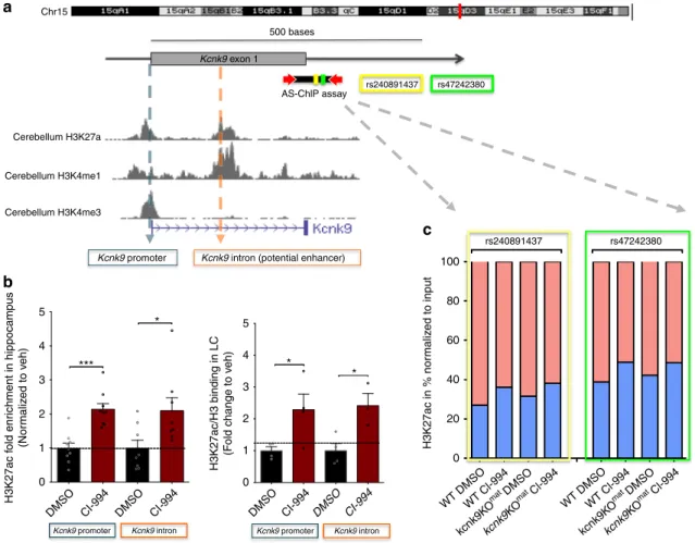
* *** Kcnk9 intron9 Kcnk9 promoter9 Kcnk9 intron9 Kcnk9 promoter9Fig. 6 Treatment with CI-994 affects H3K27 acetylation at theKcnk9 locus. a Schematic presentation of the Kcnk9 locus on distal mouse chromosome 15. The murine Kcnk9 gene is shown with the corresponding H3K27ac, H3K4me1 and H3K4me3 peaks in mouse cerebellum (UCSC Genome Browser on Mouse July 2007 (NCBI37/mm9) Assembly).b Increased deposition of H3K27ac marks at the promoter and intronic region of Kcnk9 in hippocampus and locus coeruleus (LC) in Kcnk9KOmatmice following treatment with CI-994 (normalized to veh). Hippocampus (left): in Kcnk9 promoter, DMSO (n= 9) vs.
CI-994 (n= 9), P = 0.0001; in Kcnk9 intron, DMSO (n = 8) vs. CI-994 (n = 8), P = 0.0270. LC (right) in Kcnk9 promoter, DMSO (n = 4) vs. CI-994 (n = 4), P= 0.0450; in Kcnk9 intron, DMSO (n = 4) vs. CI-994 (n = 3), P = 0.0202. Values are means ± SEM, *P ≤ 0.05, ***P < 0.001, by Student’s t test. c Chromatin Immunoprecipitation followed by pyrosequencing reveals allele-specific chromatin deposition of H3K27ac. Two SNPs (rs240891437 and rs47242380) in the intronic region of the Kcnk9 in the hippocampus of (C57BL/6xCast/Ei)F1 hybrid mice were analyzed; maternal allele (red) and paternal allele (blue). Untreated state (DMSO) reveals higher acetylation levels in maternal allele compared with the paternal allele. The paternal allele exhibits higher H3K27 acetylation enrichment after CI-994 treatment compared with the maternal allele. WT:DMSO n= 3, WT:CI-994 n = 4, Kcnk9KOmat:DMSO
n = 6, and Kcnk9KOmat:CI-994 n= 4. b, c n = biologically independent animals. Data generated in two independent experiments. Statistical analyses and
for treatment of imprinting disorders such as BBIDS. CI-994
treatment of Kcnk9KO
matmice as a mouse model of BBIDS
substantially activated the repressed paternal allele of Kcnk9 in
several brain areas resulting in a significant increase in Kcnk9
expression. This led to the full rescue of specific domains of brain
function impaired in BBIDS. Our data for the
first time shows
successful phenotypic rescue of impaired brain function in a
mouse model for an imprinting disorder using an epigenetic
modulator.
Similar to the
findings in the UNC6042-treated mouse model of
PWS
31, activation of the Kcnk9
patallele was not accompanied by
changes of DNA methylation at the Peg13 DMR, which is assumed
to play a key role in regulation of Kcnk9 imprinting also in the
mouse
3. Notably, the bisulfite technique did not allow us to
dis-criminate between 5-methylcytosine and 5-hydroxymethylcytosine
levels possibly affected by CI-994. Though, our data strongly argue
that CI-994-mediated derepression of the Kcnk9
patallele is
pri-marily caused by an increase of H3K27ac at regulatory elements in
the promotor and intronic region of the Kcnk9 gene. However, the
treatment did not display significant differences in H3K27ac
deposition between the LC and hippocampus tissues suggesting a
region-independent effect of the HDAC inhibitor. Such effects
have commonly been reported in multiple genome-wide studies
investigating the effect of HDAC inhibitors
32. Interestingly, CI-994
as a histone deacetylase inhibitor did not only selectively increase
H3K27ac but also slightly increased H3K4me1 at the Kcnk9
pro-moter region. This is not unexpected since crosstalk mechanisms
between histone-modifying enzymes often affect the binding and
activity of further histone-modifying enzymes. For instance,
treatments with HDAC inhibitors were reported to affect globally
the histone methylation marks specifically of H3K4me1
32. These
observations are repeatedly described in the literature suggesting a
broad effect of HDAC inhibitors on chromatin structure and
transcription
33,34.
In general, epigenetic modulation is supposed to rather
unspecifically target numerous genes and their expression in the
genome. Side effects of all sorts including tumor development
and metabolic dysregulation are believed to be likely with such
substances. Also, interference with brain function leading to
cognitive impairment and mental dysfunction cannot be
excluded. However, valproic acid, a
first generation HDAC
inhibitor has been used in anti-epileptic therapy for decades
without severe tumorigenic or metabolic long-term effects.
CI-994 is a second-generation, specific class I HDAC inhibitor.
Although a relatively broad effect of CI-994 on gene expression
has been demonstrated in vivo, it is being used in several clinical
trials for anti-cancer therapy having proven inhibitory potency
of epithelial-mesenchymal transition processes
35. Accumulating
evidence also suggests that CI-994 provides neuroprotective
effects in the central nervous system and cell survival in vitro
36.
Regarding brain function in WT mice no effect of CI-994
treatment has been seen in the open
field
26. In our experiments,
no significant influence of two weeks CI-994 injection was seen
on working memory or circadian locomotor activity in WT
animals. Furthermore, an increase of Kcnk9
patexpression after
CI-994 injections was only seen in selected brain regions, but
not in, e.g., cortex or prefrontal cortex, and mainly in the
maternal knockout animals (only very weakly in WT animals).
Allele-specific ChIP-qPCR of (C57BL/6JxCast/Ei)F1 hybrids
showed that the CI-994 effect is more pronounced on the
paternal allele (Fig.
6
c). This leads to a visible increase in
expression of the paternal but not the maternal allele
(Supple-mentary Fig. 4a) and might be due to the high acetylation status
of the active maternal allele. Furthermore, CI-994 treatment did
not affect the expression of other nearby imprinted genes within
the imprinted cluster on mouse chromosome 15 in vitro and
in vivo. Taken together these data suggest a rather specific
epigenetic action of CI-994 in Kcnk9KO
matanimals, which
might make CI-994 safe for usage in BBIDS patients. However,
so far all CI-994 related studies have been carried out in vitro or
in adult animals. If used in patients, early treatment during
development might be more efficient. In order to make this
possible, further studies on CI-994 specificity and possible
adverse and teratogenic effects in young mice have to be
conducted.
In summary, we show here that brain region-specific
upregu-lation of the paternally silenced Kcnk9 allele particularly in the LC
of Kcnk9KO
matanimals modulates nocturnal hyperactivity, a
central phenotype of Kcnk9 knockout animals. We further show
that the class I HDAC inhibitor CI-994 leads to a significant
additional increase of Kcnk9
patexpression in several brain regions
and fully rescues behavioral alterations in Kcnk9KO
matanimals.
This rescue is associated with an increase of H3K27 histone
acetylation at the promotor and intronic region of the Kcnk9 gene
but not with changes in DNA methylation at the differentially
methylated region in the Peg13/Kcnk9 locus. Our data suggest
epigenetic modulation by CI-994 as a promising therapeutic
strategy in patients with BBIDS and, more generally, derepression
of imprinted gene alleles as a sustainable approach for the
treatment of imprinting disorders.
Methods
Mice. All experimental procedures were performed in accordance with institu-tional animal welfare guidelines and were approved by the ethical committee of the state government of Rhineland-Palatinate, Germany (ID: 23 177-07/G 17-1-022). Kcnk9KOhommice were provided by Florian Lesage, Institut de Pharmacologie
Moléculaire et Cellulaire, Valbonne, France. The gene targeting strategy of Kcnk9KOhommice was based on a cre-mediated deletion of exon 2 encoding pore
domains P1 and P2, transmembrane domains M2–M4 as well as the cytoplasmic C-terminus18. Breeding of two heterozygous animals revealed homozygous, het-erozygous and WT animals at a proportion of 1:2:1. WT littermates were used as controls. WT and heterozygous Kcnk9KO mice with inactivation of the maternal Kcnk9 allele (Kcnk9KOmat) were obtained by crossing male WT mice with female
Kcnk9KOmatmice. Finally, breeding of homozygous female and heterozygous male
mice resulted in Kcnk9KOmatand Kcnk9KOhommice. The mice were kept under
specific-pathogen-free (SPF) conditions on a 12 h light/12 h darkness cycle in standard polystyrene cages with free access to water and food. Using tail tip DNA, mice were genotyped by assessing exon 2 excision using the primersflanking this region (F, 5′-TGCGAGCTTCAGAGAGAGGATG-3′ and R, 5′-ATGCTC-TAATCTCCAGTCTG-3′) producing fragments for WT and mutant alleles. Additional primers within exon 2 were applied to generate a control product for the WT allele (F, 5′-CACCACGCCATGTACTTCCT-3′ and R, 5′-GGACCG-GAAGTAGGTGTTCC-3′). Male mice at 8–10 weeks of age were used for expression analysis and to investigate the behavioral phenotype with and without drug treatment. Allele-specific expression analyses were carried out with total RNA from F1 offspring derived from crosses between female WT C57BL/6J or Kcnk9KOhommice and male WT Mus musculus castaneus (Cast/Ei) mice.
Behavioral testing. Littermate WT and Kcnk9KO mice were tested in 4–8 cohorts of mice, with testing beginning at 8–10 weeks of age. All experimenters were blinded to the genotype of the mice throughout the studies and behavioral analyses. Immediately after the behavioral tests, animals were sacrificed and whole brains were rapidly removed and incubated in RNAlater® (Sigma) for subsequent expression analysis.
Drug administration in vivo. Nine weeks old male Kcnk9KOmatand WT mice
were injected intraperitoneally once daily with CI-994 (ApexBio) or dimethyl sulfoxide (DMSO) (control vehicle) for 7 consecutive days before behavior experiments were initiated (Fig.5a). On days of testing, mice were injected 2 h before behavior experiments. A single injection contained either 20 µl DMSO or 20 µl CI-994 (35 mg/kg) dissolved in 100 % DMSO.
Toxicity/viability test in CI-994 treated mPCNs. mPCNs were washed with PBS andfixed with 4% paraformaldehyde for 2 h. Subsequently, cells were incubated for 20 min in 0.4% Triton X-100 in PBS at 37 °C and further rinsed three times with PBS. After blocking with 10% sheep serum at 37 °C for 1 h, cells were washed for a further three times with PBS. Next, neurons were incubated overnight at 4 °C with anti-neuron-specific nuclear protein NeuN monoclonal antibody (cat. # ab104225; dilution 1:200 in PBS), followed byfive washes with PBS for 5 min each. The