O R I G I N A L A R T I C L E
N-acetylcysteine treatment ameliorates the skeletal
phenotype of a mouse model of diastrophic dysplasia
Luca Monti
1
, Chiara Paganini
1
, Silvia Lecci
1
, Fabio De Leonardis
1
, Eric Hay
3
,
Martine Cohen-Solal
3
, Simona Villani
2
, Andrea Superti-Furga
4
,
Ruggero Tenni
1
, Antonella Forlino
1
and Antonio Rossi
1,
*
1
Department of Molecular Medicine, Unit of Biochemistry and
2Department of Public Health, Experimental and
Forensic Medicine, Unit of Biostatistics and Clinical Epidemiology, University of Pavia, 27100 Pavia, Italy,
3Inserm
U1132 and University Paris 7, Hopital Lariboisiere, 75010 Paris, France and
4Department of Pediatrics, Lausanne
University Hospital, University of Lausanne, Lausanne 1011, Switzerland
*To whom correspondence should be addressed at: Dipartimento di Medicina Molecolare, Unità di Biochimica‘Alessandro Castellani’, Via Taramelli, 3/B, I-27100 Pavia, Italy. Tel: +39 0382987229; Fax: +39 0382423108; Email: antrossi@unipv.it
Abstract
Diastrophic dysplasia (DTD) is a recessive chondrodysplasia caused by mutations in SLC26A2, a cell membrane sulfate–chloride antiporter. Sulfate uptake impairment results in low cytosolic sulfate, leading to cartilage proteoglycan (PG) undersulfation. In this work, we used the dtd mouse model to study the role of N-acetyl--cysteine (NAC), a well-known drug with antioxidant properties, as an intracellular sulfate source for macromolecular sulfation. Because of the important pre-natal phase of skeletal development and growth, we administered 30 g/l NAC in the drinking water to pregnant mice to explore a possible
transplacental effect on the fetuses. When cartilage PG sulfation was evaluated by high-performance liquid chromatography disaccharide analysis in dtd newborn mice, a marked increase in PG sulfation was observed in newborns from NAC-treated pregnancies when compared with the placebo group. Morphometric studies of the femur, tibia and ilium after skeletal staining with alcian blue and alizarin red indicated a partial rescue of abnormal bone morphology in dtd newborns from treated females, compared with pups from untreated females. The beneficial effect of increased macromolecular sulfation was confirmed by chondrocyte proliferation studies in cryosections of the tibial epiphysis by proliferating cell nuclear antigen
immunohistochemistry: the percentage of proliferating cells, significantly reduced in the placebo group, reached normal values in dtd newborns from NAC-treated females. In conclusion, NAC is a useful source of sulfate for macromolecular sulfation in vivo when extracellular sulfate supply is reduced, confirming the potential of therapeutic approaches with thiol compounds to improve skeletal deformity and short stature in human DTD and related disorders.
Introduction
The diastrophic dysplasia sulfate transporter (DTDST, also known as SLC26A2) is a sulfate–chloride antiporter expressed in a wide range of tissues, which allows recruitment of sulfate from the extracellular space into the cytoplasm (1). Functional impairment of SLC26A2 causes reduced sulfate uptake leading to low levels of intracellular sulfate, particularly in cells with a high synthetic
requirement of sulfate (2). A reduction in the intracellular sulfate pool causes the synthesis and secretion of undersulfated proteo-glycans (PGs), and this in turn affects the composition, architec-ture, signaling and mechanical properties of the extracellular matrix (3,4). As the cartilage extracellular matrix contains high amount of sulfated PGs, the main consequences of PG undersulfa-tion affect cartilage development, homeostasis and structure,
Received: December 22, 2014. Revised and Accepted: July 15, 2015
© The Author 2015. Published by Oxford University Press. All rights reserved. For Permissions, please email: journals.permissions@oup.com doi: 10.1093/hmg/ddv289
Advance Access Publication Date: 23 July 2015 Original Article
resulting in the onset of chondrodysplastic phenotypes. Thus, mutations in the SLC26A2 gene cause a family of recessive osteo-chondrodysplasias which include, in the order of decreasing severity, achondrogenesis type 1B (ACG1B, MIM no. 600972), atelosteogenesis type 2 (AO2, MIM no. 256050), DTD (MIM no. 222600) and recessive multiple epiphyseal dysplasia (EDM4, MIM no. 226900). The different clinical phenotypes are related to the residual function of the sulfate transporter and, consequently, to the level of PG undersulfation in patients’ cartilage (2,5,6).
DTD is a non-lethal skeletal dysplasia,first described by Lamy and Maroteaux (7) in 1960, characterized by growth retardation, skeletal dysplasia, progressive kyphoscoliosis of the spine, typ-ical hand and foot deformities, joint contractures and reduced viability (8). DTD patients have marked physical difficulties that affect common activities of daily living, their work activities, par-ticipation in society and theirfinancial situation; all these diffi-culties and the lack of a decisive non-invasive therapy seriously worsen the quality of life of the patients (9). Currently, no decisive therapies are available for DTD as well as for most skeletal disor-ders; patients are treated mainly by orthopedic interventions and physiotherapy in order to reduce skeletal deformities and to maintain articular mobility (10,11).
We demonstrated previously a correlation between PG under-sulfation and the severity of the clinical phenotypes of SLC26A2-related disorders (2). A lower level of PG undersulfation and a less-compromised sulfate transporter function detected in SLC26A2 patients’ cartilage and fibroblasts are associated with less severe clinical phenotypes (2). Thesefindings suggest that increasing PG sulfation in the cartilage of DTD patients might result in an amelioration of the clinical phenotype.
Studies performed in vitro and in vivo have pointed out the potential relevance of sulfur-containing amino acids and their derivatives, such as N-acetyl--cysteine (NAC; C5H9NO3S,
Pub-Chem Compound CID 12035), as sources of intracellular sulfate, through their catabolism, when extracellular sulfate concentra-tion is low or when its uptake is impaired such as in SLC26A2-re-lated chondrodysplasias (12–14). In particular, NAC has been proposed as an alternative sulfation source as cysteine is poorly soluble, its sulfhydryl group easily oxidizes and its concentration is tightly regulated by the liver in order to keep its intracellular concentration below the threshold of cytotoxicity (15). NAC is a FDA-approved drug largely used in clinics with minimal side ef-fects. This drug wasfirst reported to have clinical benefits in the early 1960s, when it was used as an effective mucolytic agent in patients with cysticfibrosis through the reduction of disulfide bonds in glycoproteins of the mucus (16). Then it has been used in several disorders with different etiologies such as paracetamol (acetaminophen) overdose, pulmonaryfibrosis, nephropathy, cardiovascular disease, diabetes and cancer (17). Furthermore, NAC protects cells from oxidative stress through its ability to bind free radicals, such as the hydroxyl radical and H2O2(18).
Moreover, NAC metabolism to cysteine allows the maintenance of an adequate intracellular concentration of glutathione, the main antioxidant molecule in cells (19). Due to its antioxidant properties, NAC is also used in neurodegenerative diseases (20). Some years ago, we generated a mouse model of DTD (dtd mouse) by knocking in the Slc26a2 mouse gene a mutation previ-ously identified in a DTD patient (21). The dtd mouse shows growth retardation and reduced bone growth, shortening of the long bones, thoracic kyphosis causing deformities of the rib cage with reduced volume and hip dysplasia, thereby recapitulating es-sential aspects of the DTD phenotype in humans (21). Reduced sulfate uptake was demonstrated in chondrocyte, osteoblast and fibroblast cultures, and sulfation studies revealed undersulfation
of chondroitin sulfate PGs in cartilage and bone. The phenotypic similarities between mutant mice and human DTD make this mouse strain a good model for the study of SLC26A2-related disor-ders and a useful tool to develop new treatments and test candi-date pharmacological compounds.
We already assessed the potential value of NAC to ameliorate cartilage PG sulfation in dtd mice in vivo (14). In that work, newborn dtd mice were treated for 1 week with daily hypodermic injections of NAC, and then cartilage PG sulfation was measured in the cartil-age from treated mice versus the placebo group. A mild but signifi-cant increase in PG sulfation was observed in dtd mice treated with NAC due to its catabolism. Pharmacokinetic experiments showed that NAC was rapidly removed from the bloodstream within 8 h and that this rapid removal may be a hindrance to its pharmaco-logical effects on the correction of macromolecular sulfation (14). These results suggest that NAC efficacy might be even higher if the drug concentration in the plasma would be more constant.
On the basis of these observations, in this work we evaluated the contribution of NAC as a potential intracellular sulfate source for cartilage PG sulfation, as well as its ability to ameliorate the skeletal phenotype of dtd mice by administering NAC in the drink-ing water to pregnant mice from 0.5 day post-coitum until delivery. Once demonstrated that NAC given to pregnant mice crosses the placenta, we characterized the phenotype of newborns from trea-ted mice by cartilage PG sulfation, morphological studies of the skeleton by differential staining with alcian blue and alizarin red and by radiography and chondrocyte proliferation by immunohis-tochemistry. Experimental data demonstrated that the cartilage PG sulfation increase in dtd fetuses treated with the drug resulted in an amelioration of long bone morphology and cartilage histology.
Results
NAC enters the bloodstream of pregnant mice and by placental transfer reaches fetuses
Pregnant wild-type mice were treated for the whole pregnancy with free access to 30 g/l NAC, pH 7.0–7.4, in place of drinking water used in the placebo group. To evaluate NAC intestinal ab-sorption, the plasma NAC concentration was measured at the 17th day of pregnancy by reverse-phase high-performance liquid chromatography (HPLC) after pre-columnfluorescence derivati-zation using 7-fluorobenzo-2-oxa-1,3-diazole-4-sulfonic acid ammonium salt (SBD-F) (Supplementary Material, Fig. S1). A con-centration of 1.0 ± 0.6 µg NAC/ml plasma (n = 5) was detected in treated females, indicating that the drug administered orally was absorbed in the intestine. In the same animals, NAC placental transfer to fetuses was demonstrated: a concentration of 0.7 ± 0.3 µg NAC/g of fetus weight (n = 16) was detected in homogenates of fetuses from treated pregnant females. Overall, these data indi-cated that NAC given orally to pregnant mice can cross placenta and reaches fetuses; conversely, NAC was not detected in fetus homogenates from untreated pregnant mice, as already observed in plasma from untreated pregnant mice.
NAC increases sulfation of cartilage PGs in newborn dtd mice
To assess the effect of NAC treatment on macromolecular sulfa-tion in newborn dtd and wild-type mice, heterozygous females were mated to heterozygous males and pregnant animals were treated for the whole pregnancy with 30 g/l NAC in the drinking water. Newborn mice were sacrificed, and the femoral head cartilage was harvested. For sulfation analysis of chondroitin
sulfates, glycosaminoglycans (GAGs) were recovered from cartil-age by digestion with papain, followed by cetylpyridinium chlor-ide precipitation. After hyaluronic acid removal, purified GAGs were digested with both chondroitinase ABC and ACII, and released disaccharides were analyzed by HPLC followed by detec-tion at 232 nm (Supplementary Material, Fig. S2).
The amount of the non-sulfated disaccharide (ΔDi-0S) relative to the total amount of disaccharides (ΔDi-0S, ΔDi-4S and ΔDi-6S) significantly varied among groups (Kruskal–Wallis test = 36.22, P = 0.0001). In dtd mice from treated females, a relevant decrease (P < 0.001) with respect to dtd mice from untreated females was detected (Fig.1). NAC does not affect PG sulfation in wild-type newborns: the relative amount ofΔDi-0S was similar in wild-type pups from treated and untreated pregnant females. More-over, the amount of 4-O-sulfated disaccharides (ΔDi-4S) and 6-O-sulfated disaccharides (ΔDi-6S), relative to the total amount of disaccharides (ΔDi-0S, ΔDi-4S and ΔDi-6S), showed a hetero-genic distribution across groups (F = 47.58, P < 0.001 and F = 5.45, P = 0.0025, respectively) (Fig.1). RegardingΔDi-4S, in dtd mice from treated females, a significant improvement was observed with respect to dtd pups from untreated ones (P < 0.001) even if the amount was again lower than that in wild-type newborns from untreated (P = 0.002) and treated (P < 0.001) females. Overall, these results showed a marked increase in PG sulfation in new-born mutant mice from females treated with NAC compared with untreated ones, even if macromolecular sulfation did not reach normal values. Regarding ΔDi-6S, an increase in dtd newborns from treated mice with respect to dtd from untreated females was evidenced (P = 0.011), and the relative amount was similar to that in wild-type newborns (Fig.1).
In addition, the relative amounts ofΔDi-4S and ΔDi-6S in wild-type newborns from treated and untreated females were similar,
indicating that NAC did not affect sulfation on C4 and C6 of the hexosamine moiety and that NAC contribution to PG sulfation was not relevant in wild-type mice as sulfate came mainly from the extracellular environment because of the SLC26A2.
NAC treatment ameliorates the skeletal phenotype of dtd newborns
The skeleton of newborn mice from both NAC-treated and un-treated pregnant females was studied by differential staining with alcian blue and alizarin red to stain cartilage and bone, re-spectively. Several deformities were evident in the skeleton of dtd newborns from untreated mice compared with wild-type lit-termates: tibiae were curved, tibiae and femurs were shorter and ilia were shorter and thicker. Conversely, in dtd newborn mice from females treated with NAC, an amelioration toward a normal bone morphology was observed, including longer and straighter tibiae, longer femurs and longer and thinner ilia compared with dtd pups from untreated females (Fig.2).
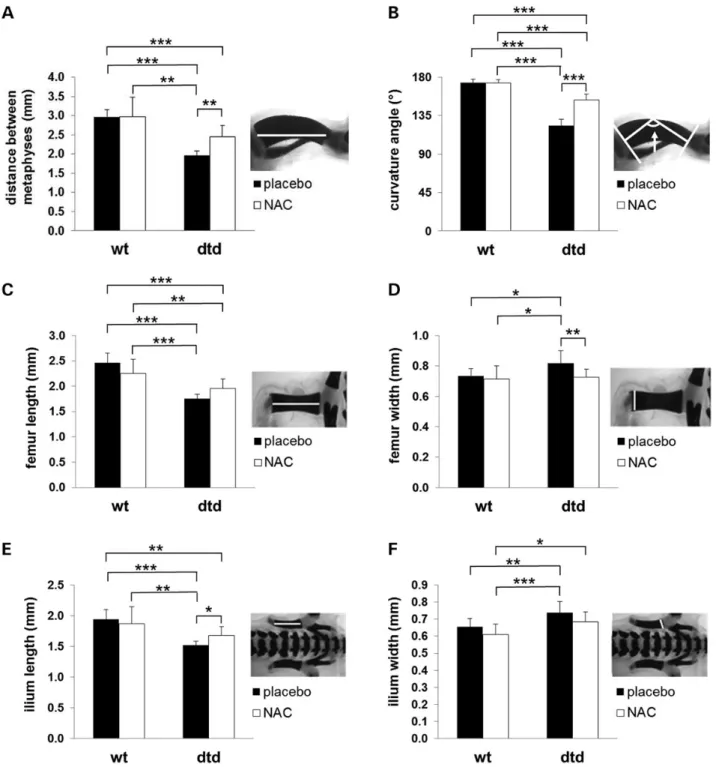
In skeleton preparation stained with alcian blue and alizarin red, morphometric analysis of tibiae, femurs and hips, performed by two different blinded observers, confirmed the skeletal pheno-type amelioration observed in dtd pups from females treated with the drug, compared with mutant newborns from untreated
Figure 1. NAC increases PG sulfation of femoral head cartilage in newborn dtd mice. Cartilage PG sulfation from the femoral head was measured by HPLC disaccharide analysis of chondroitin sulfate GAGs after digestion with chondroitinase ABC and ACII on the basis of the relative amount of the non-sulfated disaccharide (ΔDi-0S), the 4-O-non-sulfated (ΔDi-4S) disaccharide and the 6-O-sulfated (ΔDi-6S) disaccharide. In dtd newborns from females treated with NAC, a decrease in the relative amount ofΔDi-0S and a corresponding increase in the mono-sulfated disaccharides (ΔDi-4S and ΔDi-6S) were observed compared with dtd newborns from untreated females indicating a significant increase of PG sulfation, even if they do not reach the normal range. NAC does not affect PG sulfation in wild-type mice; in fact, no differences in the relative amounts of non-sulfated (ΔDi-0S) and monosulfated (ΔDi-4S and ΔDi-6S) disaccharides were observed in cartilage from newborn wild-type mice born from females treated with NAC, compared with the placebo group. Error bars indicate mean ± SD (n = 14); *P < 0.05; **P < 0.01 and ***P < 0.001. Representative HPLCs of this analysis are reported in Supplementary Material, Figure S2.
Figure 2. NAC ameliorates the skeletal phenotype of dtd newborns from females treated with the drug. The skeletons of newborns mice were studied by differential staining with alcian blue and alizarin red. Thefigure focuses on the morphology of tibia, femur and hip. Several deformities were evident in the skeleton of dtd versus wild-type newborns from untreated females: tibiae were curved, tibiae and femurs were shorter and ilia (black arrows) were shorter and thicker. Conversely, in dtd newborn mice from females treated with NAC, an amelioration toward a normal bone morphology was observed, including longer and straighter tibiae, longer femurs and longer and thinner ilia, compared with dtd pups from untreated females. Morphometric analysis is reported in Figure 3. Scale bar: 1 mm. Radiographs confirm the skeletal phenotype amelioration of dtd newborn mice from females treated with NAC. X-ray of the hind limbs showed that in dtd newborn mice from treated females, the tibiae and femurs were more similar to their normal morphology compared with dtd newborn mice from untreated females: tibiae were less curved and longer and femurs were longer and thinner. No differences in wild-type newborn mice from treated or untreated females were observed.
females. In the tibia, the distance between metaphysis (Kruskal– Wallis test = 27.6, P < 0.001) and curvature angle (F = 162.5, P < 0.001) showed a significant difference among the four groups (Fig.3A and B). A relevant improvement in the distance between metaphysis and a decrease in the curvature angle of the diaph-ysis were observed in the tibiae of dtd newborns from treated
mice with respect to dtd pups from untreated females (P = 0.003 and P < 0.001, respectively). Similarly, femur length and width were significantly different among groups (F = 26.4, P < 0.0001 and F = 5.1, P = 0.0039, respectively). Even if femur length amelio-rated in dtd pups from treated mice with respect to dtd newborns from untreated females (Fig.3C), the improvement did not reach
Figure 3. Morphometric analysis of tibia, femur and ilium demonstrates the skeletal phenotype amelioration of dtd newborn mice from females treated with NAC. Morphometric studies were performed by observers masked to treatment and genotype in the skeletal preparations stained with alcian blue and alizarin red shown in Figure2by considering the mineralized part of each bone (white bar in each picture). Bone measurements in dtd newborn mice from females treated with NAC versus the placebo group showed an amelioration toward the normal bone morphology. In fact in the tibia (A), an increase in the distance between the metaphysis and (B) a decrease in the curvature angle were observed; in the femur (C), a trend toward the increase of its length and (D) a decrease in its width were detected; in the ilium (E), an increase in the length and (F) a decrease in its width were found. No differences were observed in wild-type newborn mice from NAC-treated females and untreated ones. Statistical comparison of the dtd pups from treated females compared with wild-type pups from untreated females demonstrated a rescue to normal values of the femur width and ilium width, whereas the other parameters approached the wild-type range. Error bars indicate mean ± SD (n = 7); *P < 0.05; **P < 0.01 and ***P < 0.001.
a statistical relevance (P = 0.13); the length in dtd newborns from treated females was still significantly lower than that in wild-type pups from untreated and treated females (P < 0.001 and P < 0.01, respectively). The dtd NAC newborns did not show a dif-ferent femur width with respect to wild-type pups from placebo-treated females (P > 0.90), but a relevant reduction with respect to dtd pups from untreated females (P = 0.007) (Fig.3D).
Also morphometry of the hips was different among groups (Fig. 3E, Kruskal–Wallis test = 26.4, P < 0.0001; Fig. 3F, F = 8.1, P = 0.0002). In particular, a significant increase in the ilium length in dtd pups from treated mice with respect to those from untreated (P = 0.014) and a decrease in the ilium width, however not relevant, were found (P = 0.15).
Thesefindings support the hypothesis that NAC supplemen-tation ameliorates the skeletal phenotype of dtd pups toward the normal bone morphology.
The skeletal phenotype amelioration observed by alcian blue and alizarin red staining was further confirmed by X-ray studies of the hind limbs of newborn dtd and wild-type mice from both treated and untreated females; in newborn dtd mice from fe-males treated with NAC versus the untreated group, tibiae and femurs were less curved and similar to wild-type ones (Fig.2).
To assess whether NAC ameliorates bone mineralization, bone mineral density (BMD) was measured at the total body with a GE Lunar Piximus. BMD of newborn dtd mice from untreated females was similar to that of wild-type mice (0.018 ± 0.004 versus 0.017 ± 0.001 mg/cm2, n = 8); in dtd newborns from treated females,
BMD was not affected by NAC (0.019 ± 0.003, n = 8).
NAC treatment improves chondrocyte proliferation in tibia epiphysis of newborn dtd mice
In previous studies, we demonstrated reduced chondrocyte pro-liferation in dtd mice compared with wild-type littermates. This observation was made in chondrocyte cultures (21), in the fem-oral head of newborn mice by proliferating cell nuclear antigen (PCNA) immunohistochemistry (3) and in the proliferative zone of the tibia growth plate of P21 animals (22). The increase in PG sulfation toward normal values and the skeleton amelioration due to NAC suggested an impact also on chondrocyte prolifer-ation. In order to study chondrocyte proliferation, PCNA immu-nohistochemistry was performed on 7 µm thick cryosections of the proximal epiphysis of the tibiae from wild-type and dtd new-born mice from both treated with the drug or untreated pregnant females. Tibia cryosections were incubated with an anti-PCNA antibody and counterstained with hematoxylin (Fig.4A). Then, the number of proliferating chondrocytes (PCNA-positive cells) relative to the total chondrocyte population was measured in a fixed area (100 000 µm2, 400 µm × 250 µm rectangle) of the tibia
epiphysis and tibia growth plate (Fig.4B). The percentage of pro-liferating chondrocytes of the proximal epiphysis and growth plate was significantly different among the four groups (Krus-kal–Wallis test = 23.69, P = 0.0001 and Krus(Krus-kal–Wallis test = 22.37, P = 0.0001, respectively). Interestingly, the percentage of prolifer-ating chondrocytes of the proximal epiphysis in dtd newborns from treated females was not different from that in wild-type pups (P > 0.90) and significantly higher than that in dtd new-borns from untreated females (P < 0.001) (Fig.4B), whereas the percentage of proliferating chondrocytes in dtd pups from un-treated females was significantly reduced than that in wild-type pups from untreated (P < 0.001) and treated (P = 0.002) mice. In dtd mice born from untreated females, the percentage of pro-liferating chondrocytes in the tibia growth plate was significantly lower than that in wild-type newborns from untreated (P < 0.001)
and treated females (P = 0.002). As observed in proliferating chon-drocytes of proximal epiphysis, the percentage of proliferating chondrocytes in the growth plate of dtd newborns from females treated with NAC was similar to that in wild-type pups (P > 0.90), but it was statistically higher than that in dtd mice born from un-treated females (P = 0.0012) (Fig.4B). In wild-type littermates, con-sistent with no differences detected in PG sulfation and skeletal phenotype after NAC treatment, also chondrocyte proliferation was not affected both in the epiphysis and in the growth plate.
Figure 4. Chondrocyte proliferation is increased in the epiphysis of dtd newborns from NAC-treated females. (A) PCNA immunostaining of proximal tibia epiphysis cryosections obtained from wild-type and dtd newborns from females both untreated and treated with NAC. Nuclei of PCNA-positive cells are brown; sections were counterstained with hematoxylin. Scale bar: 200 µm. The areas enclosed in the red and black boxes (epiphysis and growth plate, respectively) are shown at higher magnification. Scale bar: 50 µm. (B) In a fixed area [100 000 µm2, 400 µm × 250 µm white dashed rectangle in (A)] of tibia epiphysis
and growth plate, the number of PCNA-positive cells relative to the total cell population was determined. The percentage of proliferating cells in dtd newborn mice from females treated with NAC increased compared with dtd newborns from untreated females and reached normal values when compared with wild-type newborns. In wild-type newborns, chondrocyte proliferation was not affected by NAC. Error bars indicate mean ± SD (n = 5) and an average of four sections per animal was considered; **P < 0.01 and ***P < 0.001.
Cartilage PG sulfation increase in newborn dtd mice by NAC matches increased GAG sulfation of urine
Urinary GAGs come from physiological PG metabolism linked to remodeling of extracellular matrix components; thus, urinary GAG concentration and GAG species have been used as biomar-kers for specific disorders such as mucopolysaccharidosis (23). We previously reported that GAG urine from dtd animals at P30 and P60 was undersulfated, compared with wild-type littermates (24). Thus, to further support cartilage PG sulfation increase in dtd newborns from pregnant mice treated with NAC, sulfation of urin-ary chondroitin sulfate GAGs was measured. For this purpose, urine from urinary bladders of dtd and wild-type newborns from treated or untreated females was collected, and GAG sulfation was measured after chondroitinase digestion, disaccharide label-ing with 2-aminoacridone and HPLC analysis followed by fluores-cence detection (Supplementary Material, Fig. S3). PG sulfation was significantly different between groups (Kruskal–Wallis test = 17.85, P < 0.0001) The amount of non-sulfated disaccharide (ΔDi-0S) relative to the total amount of disaccharides (ΔDi-0S, ΔDi-4S and ΔDi-6S) in the urine of dtd newborns from females treated with NAC significantly decreased compared with urine of dtd newborns from untreated females (P = 0.0012) (Fig.5), even if it did not reach wild-type values (P < 0.001). TheΔDi-0S decrease was paralleled by a significant increase in ΔDi-4S in the urine of dtd newborns from treated mice versus dtd newborns from untreated mice (P < 0.01), even if it did not reach wild-type values (P < 0.01). These data con-firmed cartilage PG sulfation analysis, as GAGs from urine of dtd newborns from mice treated with NAC were less undersulfated compared with dtd newborns from untreated mice. As already ob-served in cartilage sulfation studies, urinary GAG sulfation was not affected by NAC in wild-type littermates.
Discussion
Mutations in the SLC26A2 gene cause a spectrum of skeletal phe-notypes which includes, in the order of decreasing severity,
ACG1B, lethal before or shortly after birth, AO2, lethal after birth, DTD, a severe chondrodysplasia compatible with life and EDM4, a mild chondrodysplasia with minor radiological changes and clubfoot.
DTD is an autosomal recessive disorder that affects cartilage and bone development. Although skeletal problems are already present at birth, abnormal growth continues post-natally. Now-adays, no pharmacological, cell or gene therapy is available for DTD; patients take advantage of physiotherapy to relieve pain and manage disabilities. Moreover, orthopedic surgery corrects severe deformities such as scoliosis corrected by arthrodesis, or both hip osteoarthritis and knee instability and degeneration cor-rected by arthroplasty (11,25); however, all these remedies do not eradicate deformities, which are likely to occur again.
The disease is caused by mutations in the SLC26A2 gene en-coding for a sulfate–chloride antiporter of the cell membrane and results in sulfate uptake impairment. This sulfate transporter is ubiquitous, but the consequences of its functional impairment affect tissues with a high demand for sulfate, above all cartilage, as cartilage extracellular matrix is rich in sulfated PGs. Thus, muta-tions in the SLC26A2 gene cause cartilage PG undersulfation.
Previous studies using cartilage biopsies,fibroblast and chon-drocyte cultures from patients with ACG1B, AO2 and DTD demon-strated a direct correlation between the severity of the clinical phenotype and the degree of functional impairment of the sulfate transporter and cartilage PG sulfation. The decrease in cartilage PG sulfation is associated with more severe clinical phenotypes (2); thisfinding suggests the chance to ameliorate the clinical pheno-type of DTD patients by increasing PG sulfation.
In order to overcome the sulfate transport defect and to improve cartilage PG sulfation, alternative drug strategies targeted at in-creasing cytosolic sulfate can be considered. It has been reported that some SLC26A2 mutations cause protein misfolding, resulting in a defective SLC26A2 membrane localization (6,26); thus a thera-peutic option might consider the use of chemical chaperones in order to restore the correct folding and trafficking of the mutant sul-fate transporter. In fact, the combination of structure-guided cor-rectors (chemical chaperones) represents an effective therapeutic approach for cysticfibrosis in patients bearing the ΔF508 mutation in the cysticfibrosis transmembrane conductance regulator, which impairs its folding, plasma membrane expression, function and stability (27). However, chemical chaperones would not be useful for mutations impairing SLC26A2 transporter activity without af-fecting its folding and/or targeting to the plasma membrane.
Inorganic sulfate in the cytoplasm may be derived either from the extracellularfluids or from the catabolism of sulfur-contain-ing amino acids (cysteine and methionine) and other thiols. The latter pathway can provide cytoplasmic sulfate for macromolecu-lar sulfation, independently of the nature of the SLC26A2 muta-tion. We have previously demonstrated that sulfate recruitment from sulfur-containing amino acids is active in cultured fibroblasts and chondrocytes from SLC26A2 patients, and this pathway, at least in vitro, can partially compensate the lack of sul-fate caused by the reduced uptake function of the mutant sulsul-fate transporter (12,13). Moreover, using the dtd mouse, we demon-strated that the same pathway is active also in cartilage in vivo (14). In that study, the hypodermic injection of NAC to newborn dtd mice for 1 week resulted in a mild but significant increase of PG sulfation due to NAC catabolism. However, pharmacoki-netic experiments showed that NAC was removed from the bloodstream of treated mice within 8 h, indicating that its mild contribution to macromolecular sulfation might be due to the fast drug turnover. These results suggest that NAC efficacy might be improved by maintaining its plasma level constant.
Figure 5. Urine GAG sulfation analysis in newborn dtd mice from NAC-treated females confirms cartilage PG sulfation increase. GAGs in the urine were purified by cetylpyridinium chloride precipitation, and sulfation was measured by HPLC disaccharide analysis after pre-column derivatization with 2-aminoacridone (Supplementary Material, Fig. S3). Results showed a significant decrease in the relative amount of the non-sulfated disaccharide (ΔDi-0S) and a significant increase in the amount of ΔDi-4S in newborn dtd mice from pregnant mice treated with NAC compared with dtd newborn from the placebo group, indicating an increase in urinary GAG sulfation that paralleled cartilage PG sulfation increase. As expected, regardless of NAC treatment, urinary GAG sulfation was not affected in wild-type newborns from both NAC-treated and untreated females. Error bars indicate mean ± SD (n = 3; each sample comes from pooling four urinary bladders); **P < 0.01 and ***P < 0.001.
For this purpose, NAC plasma levels were kept stable by ad-ministration of the drug in the drinking water. However, this treatment can be considered in adult mutant mice or at least in animals at the weaning age (P21), when unfortunately severe skeletal defects are already present. For this reason, in order to treat mutant mice in the very early steps of the onset of the skel-etal phenotype, we treated pregnant females for the whole preg-nancy and we evaluated drug efficacy in newborn mice. This approach was valuable as skeletogenesis starts in the fetal period and the dtd phenotype is already present in mutant mice at birth (21) and in patients as well (28).
By treating wild-type female mice with free access to 30 g/l NAC in the drinking water during pregnancy, we demonstrated that NAC was present both in female plasma and in fetuses con-firming placental transfer of NAC to fetuses as previously re-ported in humans (29) and suggesting the opportunity to treat fetuses by treating pregnant mouse females.
Once assessed the effectiveness of the administration method, the potential efficacy of NAC was studied in newborn wild-type and dtd mice from females treated with NAC or the placebo (drink-ing water) by biochemical, morphological and immunohisto-chemical studies of the skeleton. HPLC analysis demonstrated a marked increase of cartilage PG sulfation in the femoral head of mutant mice born from females treated with NAC, compared with the ones treated with the placebo. Even if the PG sulfation in-crease did not reach normal values after NAC treatment, the par-tial rescue of macromolecular sulfation was translated also at the morphological level; in fact, when the skeletons of dtd newborns from mice treated with NAC were compared with those of dtd newborns from the placebo group, there was a clear amelioration toward the normal bone morphology: bones of dtd newborns from the treated group were longer and straighter when compared with untreated dtd mice. This amelioration was confirmed by morpho-metric studies of tibiae, femurs and ilia. The BMD was not different in newborn dtd mice compared with wild-type animals and was not affected by NAC, suggesting that the drug has no impact on the mineralization process.
In previous studies, we demonstrated both in vitro- and in vivo-reduced chondrocyte proliferation in mutant mice is caused by defects in Ihh and Wnt signaling due to cartilage PG undersulfa-tion (3,21,22,30). In fact, Ihh as well as other growth factors bind to sulfated GAG chains; the extent of binding and thus diffusion in the extracellular matrix relies on the degree of PG sulfation (31). Thus, in order to determine whether NAC treatment affects also chondrocyte proliferation due to the recovery of PG sulfation, PCNA immunostaining was performed on tibia cryosections of dtd and wild-type newborns from both treated and untreated fe-males. A reduced number of proliferating cells was detected in the epiphysis and in the growth plate of untreated mutant new-born mice compared with wild-type mice, whereas in treated dtd newborn mice, chondrocyte proliferation reached normal values. Compared to our previous work in which NAC was injected daily (14), the different administration method was crucial in order to guarantee a constant concentration of NAC in the bloodstream of pregnant females, resulting in a greater PG sulfation effect in new-born dtd mice that was translated also in an amelioration of the skeleton at the morphological level. A dose-ranging study per-formed on C57Bl/6 females demonstrated that the NAC concentra-tion used in this study (30 g/l in the drinking water) can be halved without affecting the plasma drug concentration.
The choice of NAC with respect to other sulfur-containing amino acids such as cysteine comes from the observation that cysteine has lower solubility in water; in addition, NAC is a FDA-approved drug largely used in clinics with minimal side
effects (32). In this work, the therapeutic role of NAC relies on its capacity to provide, through its catabolism, cytoplasmic sul-fate for macromolecular sulfation, as demonstrated by PG sulfa-tion increase in cartilage and urine. This is a novel not yet considered property of this molecule; in fact as discussed previ-ously, NAC is a well-known drug used for its antioxidant proper-ties, reduction of disulfide bonds and serving as a precursor of cysteine for glutathione synthesis (17).
In developing a therapy, it is mandatory to set up reliable non-invasive protocols in order to evaluate the effectiveness of the treatment in patients. This is not trivial when considering skeletal disorders as non-invasive methods to evaluate the cartilage and bone phenotype in patients rely mainly on X-rays, computed tom-ography and magnetic resonance imaging. GAGs from urine have been investigated in many disorders including mucopolysacchar-idoses, kidney disorders, diabetes and leukemia (33–37). In add-ition, GAG sulfation was altered in the urine of a patient with spondyloepiphyseal dysplasia, Omani type, a recessive chondro-dysplasia with a defect in GAG sulfation (38). We demonstrated previously that GAG sulfation in the urine of 1- and 2-month-old dtd mice was reduced compared with age-matched wild-type an-imals, suggesting that urine GAG analysis is potentially relevant as a biomarker to follow the disease course (24). On the basis of these data, we measured the sulfation of urinary GAGs in newborns from females treated with NAC versus the untreated group. Simi-larly to results reported in cartilage, in dtd newborns from treated pregnant mice, a decrease in the relative amount of the non-sul-fated disaccharide (ΔDi-0S) and an increase in the relative amount of the 4-O-sulfated disaccharide (ΔDi-4S) compared with untreated mutant mice were observed. These data suggested that urine GAG sulfation analysis could be a non-invasive biomarker in order to check the efficacy of the treatment.
In conclusion, we have demonstrated at the biochemical and morphological levels that NAC, through its catabolism, is a useful source of sulfate for macromolecular sulfation in the dtd mouse, an animal model of DTD. By comparison to the previous drug ad-ministration method that considered daily hypodermic injection (14), we have observed that it is crucial for the treatment to pro-vide tissues with a steady supply of the drug byfixing its plasma concentration. Studies on new drug delivery systems that allow the regular release of NAC are under investigation in order to treat dtd mice even after birth and to pave the way on a potential NAC pharmacological therapy to ameliorate this painful chon-drodysplasia and thus the quality of life of DTD patients.
Materials and Methods
Mouse care and drug treatment
The dtd mouse is a knock-in for the c.1184C>T transition causing an A386V substitution in the eighth transmembrane domain of the Slc26a2 gene product, which strongly reduces the activity of the transporter (21). In this study, wild-type and homozygous mutant mice (dtd) with a C57Bl/6J×129/SV background were used. Genotyp-ing to distGenotyp-inguish mutant from heterozygous and wild-type animals was performed by polymerase chain reaction, using genomic DNA extracted from mouse tail clips as described previously (21).
Animals were bred with free access to water and standard pelleted food. Care and use of mice for the drug treatment de-scribed in this study were in compliance with relevant animal welfare institutional guidelines, and protocols were approved by the Animal Care and Use Committee of the University of Pavia. In order to achieve an oral dosage of 4 g/kg body weight/day of NAC, a solution 30 g/l NAC (Sigma-Aldrich, St Louis, MO, USA),
brought to pH 7–7.4 with NaOH, was made fresh every day and ad-ministered to mice in place of drinking water, used in the placebo group. This concentration was based on average water consump-tion per mouse (6-month-old; 30 g body weight) of 4 ml/day measured in our animal facility.
In order to evaluate NAC intestinal absorption in pregnant mice treated with the oral solution of the drug, the plasma NAC concentration from wild-type pregnant mice was measured by reverse-phase HPLC after pre-columnfluorescence derivatiza-tion using SBD-F (Sigma-Aldrich). The treatment with the drug lasted from 0.5 day post-coitum up to the 17th day of pregnancy; then pregnant mice were sacrificed and their blood was collected and centrifuged to get plasma. As shown in Supplementary Ma-terial Figure S1B, a peak corresponding to NAC was present in chromatograms from HPLC thiol analysis of treated pregnant mouse plasma, whereas the drug was not present in chromato-grams from untreated pregnant mouse plasma (Supplementary Material, Fig. S1A). A concentration of 1.0 ± 0.6 µg NAC/ml plasma (n = 5) was detected in treated females, indicating that the drug administered orally was absorbed in the intestine.
NAC placental transfer to fetuses was evaluated by HPLC thiol analysis in fetus homogenates, from wild-type pregnant females sacrificed after 17 days of treatment with 30 g/l NAC in the drink-ing water. Fetuses were homogenized immediately after their withdrawal, and NAC concentration in homogenates was mea-sured by reverse-phase HPLC. A concentration of 0.7 ± 0.3 µg NAC/g of fetus weight (n = 16) was detected in homogenates of fe-tuses from treated pregnant females (Supplementary Material, Fig. S1D). Thus, these data indicated that NAC given orally to pregnant mice crosses placenta and reaches fetuses; conversely, NAC was not detected in fetus homogenates from untreated pregnant mice, as already observed in plasma from untreated pregnant mice (Supplementary Material, Fig. S1C).
Subsequently for a dose-ranging study, two groups of C57Bl/ 6JOla-Hsd females (Harlan Laboratories) were treated for 17 days with oral doses of 4 and 2 g/kg body weight/day of NAC, re-spectively; after 17 days of drug administration, the NAC plasma concentration was similar to the plasma drug concentration of NAC-treated pregnant mice used in this study. Interestingly, the same NAC plasma concentration was observed with oral doses of 4 and 2 g/kg body weight/day [1.6 ± 0.7 and 1.9 ± 0.8 µg NAC/ml plasma, respectively (n = 5)].
HPLC thiol analysis in mouse plasma and fetuses
NAC concentration was determined in mouse plasma and fetus homogenates. Female mice were sacrificed at 17 days of pregnancy, 500 µl of blood was collected and 10 µl of 0.5 ethylenediaminete-traacetic acid (EDTA), pH 8.00, was added. Then samples were cen-trifuged at 3000g for 15 min to recover the plasma that was used for NAC analysis by HPLC with pre-column derivatization.
Fetuses at embryonic day 17.5 were collected and homoge-nized in 500 µl of phosphate-buffered saline using an Ultra-Turrax disperser (IKA–Werke, Staufen, Germany) and then centrifuged at 13 000g for 5 min at 4°C; supernatants were used for NAC analysis by pre-column derivatization.
Samples derivatization was performed according to Ferin et al. (39). For analysis of total NAC, 50 µl (70 pmol) of cysteamine (Sigma-Aldrich) as internal standard and 15 µl of 10% (v/v) tribu-tylphosphine (Sigma-Aldrich) in dimetil formamide (Sigma-Al-drich) were added to 100 µl of sample to reduce disulfide bonds and incubated for 30 min at 4°C. Then, 150 µl of 10% (w/v) tri-chloroacetic acid (Sigma-Aldrich) and 1 m EDTA were added to precipitate proteins and samples were centrifuged at 15 000g
for 10 min. An aliquot of 60 µl of the supernatant was derivatized with 120 µl of 125 m potassium borate (Sigma-Aldrich), pH 10.5, 4 m EDTA and 60 µl of 1 mg/ml SBD-F (Sigma-Aldrich) in 125 m potassium borate, pH 9.5, at 60°C for 60 min in the dark. Samples were kept in the dark at 4°C until analysis.
For HPLC analysis, a binary pump system (1525µ Binary HPLC Pump, Waters, Milford, MA, USA) coupled to afluorescence de-tector (2475 Multyλ Fluorescence Detector, Waters) set at λex
385 nm andλem515 nm was used. Chromatography was carried
out at room temperature with a LichroCART 250-4 Superspher 100 RP18 (4 × 250 mm) column (Merck, Darmstadt, Germany) and LiChrospher 100 RP18 (4 × 25 mm) (Merck) as pre-column. Mobile phases were 0.1 KH2PO4(Merck), pH 3.5 (Buffer A) and
0.1 KH2PO4(Merck), pH 3.5 in 50:50 (v/v) H2O and acetonitrile
(Merck) (Buffer B). The mobile phaseflow rate was 0.8 ml/min, and elution was performed with a linear gradient of 0–20% Buffer B in 30 min.
HPLC analysis of GAG sulfation in femoral head cartilage
For cartilage disaccharide analysis, cartilage from the femoral heads of newborn mice was carefully isolated under a dissection microscope (Leica Microsystems, Milano, Italy) and processed as described previously (21). To recover GAGs, tissue specimens were digested with 10 U of papain (Sigma-Aldrich) in 0.1 so-dium acetate, pH 5.6, 5 m EDTA and 5 m cysteine at 65°C for 24 h. After papain denaturation at 100°C, released GAGs were re-covered by precipitation with 1% (w/v) cetylpyridinium chloride (final concentration). The precipitate was washed three times with 10% (w/v) potassium acetate in 96% ethanol and with 96% ethanol, respectively, dried and solubilized in hyaluronidase buf-fer (20 m sodium acetate, pH 6.0, 75 m NaCl). Hyaluronic acid was removed by digestion with 4 U of Streptomyces hyaluronidase (Seikagaku Corp., Tokyo, Japan) at 60°C overnight, followed by ultrafiltration with Ultrafree—0.5 Centrifugal filter units (nominal molecular weight limit 5K, Merck). Purified GAGs were digested with 30 mU of both chondroitinase ABC (Seikagaku Corp.) and chondroitinase ACII (Seikagaku Corp.) in 30 m sodium acetate, 30 m Tris-acetate, pH 7.35, at 37°C overnight. Undigested pro-ducts were removed by precipitation with 4 volumes of 96% etha-nol and storage at−20°C overnight, followed by centrifugation. Disaccharides in the supernatants were evaporated, solubilized in water and injected in a Supelcosil LC-SAX1 (Sigma-Aldrich) HPLC column (4.6 × 250 mm), using a gradient of 5–400 m KH2PO4(Merck), pH 4.5, at aflow rate of 1 ml/min and room
tem-perature (2). The elution profile was measured at 232 nm with a diode array detector (2998 PDA Detector, Waters).
Analysis of the skeleton and morphometric analyses
Skeletal characterization of newborn mice was performed by double staining with alcian blue and alizarin red, as described previously (21). Briefly, newborns were sacrificed, dissected to re-move skin and internal organs,fixed in 96% ethanol and defatted in acetone for 2 weeks. The preparations were then stained with a solution containing 1 volume of 0.3% (w/v) Alcian Blue 8GX (Sigma-Aldrich) in 70% ethanol, 1 volume of 0.1% Alizarin Red S (Sigma-Aldrich) in 96% ethanol, 1 volume of acetic acid and 17 vo-lumes of 70% ethanol, for 3 days at 37°C. After staining, the speci-mens were cleared in 1% KOH in glycerol (80:20, 60:40, 40:60 and 20:80) until complete tissue clarification, changing the solution every 2–3 days; then skeletal preparations were stored in 100% glycerol.
Skeletal preparations were photographed on a Leica M165 FC stereomicroscope connected to a Leica DFC425 C digital camera; morphometric measurements were performed by two different observers blinded to treatment and genotype using the LAS 4.5 software (Leica Microsystems).
Radiography and BMD
Radiographies of the hind limbs were performed in a Faxitron MX-20 cabinet X-ray system (Faxitron Bioptics, Tucson, AZ, USA). The exposure was set at 33 kV for 20 s with 5-fold magnifi-cation; high resolution X-ray mammographyfilms and intensify-ing screens (Kodak, Rochester, Madison, NY, USA) were used.
BMD was measured at the total body using a GE Lunar Piximus (GE Healthcare, WI, USA); data are expressed as mg/cm2. PCNA immunostaining
Tibiae of wild-type and dtd newborn mice were dissected, cleaned from the surrounding soft tissue, embedded in optimal cutting temperature (Sakura, Leiden, The Netherlands), immedi-ately frozen in dry ice and stored at−80°C. About 7 μm thick sec-tions were cut parallel to the long axis of the tibia using a Leica CM 1850 UV cryostat (Leica Microsystems). For the detection of proliferating cells in epiphyseal tibia cryosections, a PCNA immu-nohistochemistry assay was used (PCNA Staining Kit, Life Tech-nologies, Carlsbad, CA, USA), according to the manufacturer’s instructions. Images were collected using a Leica DM 5500B microscope connected to a Leica DFC 480 digital camera through the LAS 4.5 software (Leica Microsystems). The number of prolif-erating chondrocytes (PCNA-positive cells) was determined in a fixed area (100 000 µm2, 400 µm × 250 µm rectangle) of the tibia
epiphysis and growth plate.
HPLC analysis of GAG sulfation in mouse urine
GAG sulfation in the urine was measured by disaccharide analysis using HPLC withfluorescence detection. For this purpose, 100 µl of urine was pooled from four newborn mice urinary bladders and centrifuged at 13 000g for 5 min to clarify samples. Supernatants were collected and 1/50 (v/v) of 10% (w/v) cetylpyridinium chloride was added and incubated overnight at 4°C. Samples were then centrifuged for 15 min at 13 000g at 4°C, and pellets were washed three times with 500 µl of 10% (w/v) potassium acetate in 96% ethanol and with 500 µl of 96% ethanol, respectively. Pellets were air-dried and subsequently resuspended in 200 µl of 0.1 ammo-nium acetate buffer, pH 7.35, containing 30 mU of chondroitinase ABC (Seikagaku Corp.) and 30 mU of chondroitinase ACII (Seikaga-ku Corp.) and incubated at 37°C overnight. Samples were then cleared by centrifugation, and the supernatants were lyophilized. The lyophilizates were dissolved in 40 µl of 12.5 m 2-aminoacri-done (Life Technologies) in 85:15 (v/v) dimethyl sulfoxide: glacial acetic acid and incubated in the dark for 15 min before adding 40 µl of 1.25 NaBH3CN (Sigma-Aldrich), followed by incubation
at 37°C overnight in the dark.
HPLC was carried out with a C18 column (Prontosil 120-3-C18-ace-EPS 3.0, 4.6 × 200 mm, Bischoff, Leonberg, Germany) at room temperature. Mobile phases were 0.1 ammonium acetate (Sigma-Aldrich), pH 8.0 (Buffer A) and 60:40 (v/v) acetonitrile (Merck): 0.1 ammonium acetate (Sigma-Aldrich), pH 8.0 (Buffer B). The mobile phaseflow rate was 1 ml/min and the following elution program was used: 3 min 100% Buffer A, linear gradient to 25% Buffer B in 6 min and linear gradient to 41% Buffer B in 30 min. Eluates were monitored using afluorescence detector
(2475 Multyλ Fluorescence Detector, Waters), with excitation and emission wavelengths of 425 and 525 nm, respectively.
Statistical analysis
Quantitative data are summarized as mean ± standard deviation (SD). Differences between groups (newborn wild-type from fe-males treated with placebo, newborn wild-type from fefe-males treated with NAC, newborn dtd from females treated with pla-cebo and newborn dtd from females treated with NAC) were eval-uated using parametric analysis of variance (ANOVA) or the analogous non-parametric test (Kruskal–Wallis test) when the assumptions for ANOVA were not verified. If a significant differ-ence was found, multiple comparison test with Bonferroni cor-rection for the number of contrasts investigated (n = 6) was applied. After a significant Kruskal–Wallis test, Mann–Whitney test was used in the post hoc analyses.
A P-value less than 0.05 was considered significant. In the multiple comparison test, P < 0.0083 was a marker of significant contrast; however, thefinal values were reported at P < 0.05.
All the analyses were conducted using STATA 12®software.
Supplementary Material
Supplementary Material is available at HMG online.
Acknowledgement
The authors sincerely thank Dr Marcella Facchini for helpful sug-gestions on administration of N-acetylcysteine to mice. Conflict of Interest statement. None declared
Funding
The research leading to these results has received funding from the European Community’s Seventh Framework Programme under grant agreement no. 602300 (SYBIL), Telethon-Italy (grant no. GGP06076) and Fondazione CARIPLO (grant no. 2011-0270).
References
1. Hästbacka, J., de la Chapelle, A., Mahtani, M.M., Clines, G., Reeve Daly, M.P., Daly, M., Hamilton, B.A., Kusumi, K., Trivedi, B., Weaver, A. et al. (1994) The diastrophic dysplasia gene encodes a novel sulfate transporter: positional cloning by fine-structure linkage disequilibrium mapping. Cell, 78, 1073–1087.
2. Rossi, A., Kaitila, I., Wilcox, W.R., Rimoin, D.L., Steinmann, B., Cetta, G. and Superti-Furga, A. (1998) Proteoglycan sulfation in cartilage and cell cultures from patients with sulfate trans-porter chondrodysplasias: relationship to clinical severity and indications on the role of intracellular sulfate production. Matrix Biol., 17, 361–369.
3. Mertz, E.L., Facchini, M., Pham, A.T., Gualeni, B., De Leonardis, F., Rossi, A. and Forlino, A. (2012) Matrix disruptions, growth, and degradation of cartilage with impaired sulfation. J. Biol. Chem., 287, 22030–22042.
4. Kvist, A.J., Johnson, A.E., Morgelin, M., Gustafsson, E., Bengts-son, E., Lindblom, K., Aszodi, A., Fassler, R., Sasaki, T., Timpl, R. et al. (2006) Chondroitin sulfate perlecan enhances collagen fibril formation. Implications for perlecan chondrodyspla-sias. J. Biol. Chem., 281, 33127–33139.
5. Karniski, L.P. (2001) Mutations in the diastrophic dysplasia sulfate transporter (DTDST) gene: correlation between sulfate transport activity and chondrodysplasia phenotype. Hum. Mol. Genet., 10, 1485–1490.
6. Karniski, L.P. (2004) Functional expression and cellular distri-bution of diastrophic dysplasia sulfate transporter (DTDST) gene mutations in HEK cells. Hum. Mol. Genet., 13, 2165–2171. 7. Lamy, M. and Maroteaux, P. (1960) [Diastrophic nanism].
Presse Med., 68, 1977–1980.
8. Horton, W.A., Rimoin, D.L., Lachman, R.S., Skovby, F., Hollis-ter, D.W., Spranger, J., Scott, C.I. and Hall, J.G. (1978) The phenotypic variability of diastrophic dysplasia. J. Pediatr., 93, 609–613.
9. Kruger, L., Pohjolainen, T., Kaitila, I., Kautiainen, H., Arkela-Kautiainen, M. and Hurri, H. (2013) Health-related quality of life and socioeconomic situation among diastrophic dyspla-sia patients in Finland. J. Rehabil. Med., 45, 308–313.
10. Jalanko, T., Remes, V., Peltonen, J., Poussa, M. and Helenius, I. (2009) Treatment of spinal deformities in patients with dia-strophic dysplasia: a long-term, population based, retro-spective outcome study. Spine (Phila Pa 1976), 34, 2151–2157. 11. Helenius, I., Remes, V., Tallroth, K., Peltonen, J., Poussa, M.
and Paavilainen, T. (2003) Total hip arthroplasty in dia-strophic dysplasia. J. Bone Joint Surg. Am., 85-A, 441–447. 12. Rossi, A., Cetta, A., Piazza, R., Bonaventure, J., Steinmann, B.
and Superti-Furga, A. (2003) In vitro proteoglycan sulfation de-rived from sulfhydryl compounds in sulfate transporter chondrodysplasias. Pediatr. Pathol. Mol. Med., 22, 311–321. 13. Rossi, A., Bonaventure, J., Delezoide, A.L., Superti-Furga, A.
and Cetta, G. (1997) Undersulfation of cartilage proteoglycans ex vivo and increased contribution of amino acid sulfur to sul-fation in vitro in McAlister dysplasia/atelosteogenesis type 2. Eur. J. Biochem., 248, 741–747.
14. Pecora, F., Gualeni, B., Forlino, A., Superti-Furga, A., Tenni, R., Cetta, G. and Rossi, A. (2006) In vivo contribution of amino acid sulfur to cartilage proteoglycan sulfation. Biochem. J., 398, 509–514.
15. Dominy, J.E. Jr, Hirschberger, L.L., Coloso, R.M. and Stipanuk, M.H. (2006) Regulation of cysteine dioxygenase degradation is mediated by intracellular cysteine levels and the ubiquitin-26 S proteasome system in the living rat. Biochem. J., 394, 267–273.
16. Rushworth, G.F. and Megson, I.L. (2014) Existing and potential therapeutic uses for N-acetylcysteine: the need for conver-sion to intracellular glutathione for antioxidant benefits. Pharmacol. Ther., 141, 150–159.
17. Samuni, Y., Goldstein, S., Dean, O.M. and Berk, M. (2013) The chemistry and biological activities of N-acetylcysteine. Biochim. Biophys. Acta, 1830, 4117–4129.
18. Aruoma, O.I., Halliwell, B., Hoey, B.M. and Butler, J. (1989) The antioxidant action of N-acetylcysteine: its reaction with hydrogen peroxide, hydroxyl radical, superoxide, and hypo-chlorous acid. Free Radic. Biol. Med., 6, 593–597.
19. Kerksick, C. and Willoughby, D. (2005) The antioxidant role of glutathione and N-acetyl-cysteine supplements and exer-cise-induced oxidative stress. J. Int. Soc. Sports Nutr., 2, 38–44. 20. Bavarsad Shahripour, R., Harrigan, M.R. and Alexandrov, A.V. (2014) N-acetylcysteine (NAC) in neurological disorders: me-chanisms of action and therapeutic opportunities. Brain Behav., 4, 108–122.
21. Forlino, A., Piazza, R., Tiveron, C., Della Torre, S., Tatangelo, L., Bonafe, L., Gualeni, B., Romano, A., Pecora, F., Superti-Furga, A. et al. (2005) A diastrophic dysplasia sulfate transporter (SLC26A2) mutant mouse: morphological and biochemical
characterization of the resulting chondrodysplasia pheno-type. Hum. Mol. Genet., 14, 859–871.
22. Gualeni, B., Facchini, M., De Leonardis, F., Tenni, R., Cetta, G., Viola, M., Passi, A., Superti-Furga, A., Forlino, A. and Rossi, A. (2010) Defective proteoglycan sulfation of the growth plate zones causes reduced chondrocyte proliferation via an al-tered Indian hedgehog signalling. Matrix Biol., 29, 453–460. 23. Komosinska-Vassev, K., Blat, D., Olczyk, P., Szeremeta, A.,
Jura-Poltorak, A., Winsz-Szczotka, K., Klimek, K. and Olczyk, K. (2014) Urinary glycosaminoglycan (uGAG) excretion in healthy pediatric and adolescent population. Clin. Biochem., 9120, 00406–00408.
24. Karousou, E., Asimakopoulou, A., Monti, L., Zafeiropoulou, V., Afratis, N., Gartaganis, P., Rossi, A., Passi, A. and Karamanos, N.K. (2014) FACE analysis as a fast and reliable methodology to monitor the sulfation and total amount of chondroitin sul-fate in biological samples of clinical importance. Molecules, 19, 7959–7980.
25. Helenius, I., Remes, V., Lohman, M., Tallroth, K., Poussa, M., Helenius, M. and Paavilainen, T. (2003) Total knee arthro-plasty in patients with diastrophic dysplasia. J. Bone Joint Surg. Am., 85-A, 2097–2102.
26. Maeda, K., Miyamoto, Y., Sawai, H., Karniski, L.P., Nakashima, E., Nishimura, G. and Ikegawa, S. (2006) A compound hetero-zygote harboring novel and recurrent DTDST mutations with intermediate phenotype between atelosteogenesis type II and diastrophic dysplasia. Am. J. Med. Genet. A, 140, 1143–1147.
27. Okiyoneda, T., Veit, G., Dekkers, J.F., Bagdany, M., Soya, N., Xu, H., Roldan, A., Verkman, A.S., Kurth, M., Simon, A. et al. (2013) Mechanism-based corrector combination restores DeltaF508-CFTR folding and function. Nat. Chem. Biol., 9, 444–454. 28. Canto, M.J., Buixeda, M., Palau, J. and Ojeda, F. (2007) Early
ultrasonographic diagnosis of diastrophic dysplasia at 12 weeks of gestation in a fetus without previous family history. Prenat. Diagn., 27, 976–978.
29. Wiest, D.B., Chang, E., Fanning, D., Garner, S., Cox, T. and Jen-kins, D.D. (2014) Antenatal pharmacokinetics and placental transfer of N-acetylcysteine in chorioamnionitis for fetal neuroprotection. J. Pediatr., 165, 672–677.e2.
30. De Leonardis, F., Monti, L., Gualeni, B., Tenni, R., Forlino, A. and Rossi, A. (2014) Altered signaling in the G1 phase deregu-lates chondrocyte growth in a mouse model with proteogly-can undersulfation. J. Cell. Biochem., 115, 1779–1786.
31. Cortes, M., Baria, A.T. and Schwartz, N.B. (2009) Sulfation of chondroitin sulfate proteoglycans is necessary for proper Indian hedgehog signaling in the developing growth plate. Development, 136, 1697–1706.
32. Atkuri, K.R., Mantovani, J.J. and Herzenberg, L.A. (2007) N-acetylcysteine—a safe antidote for cysteine/glutathione deficiency. Curr. Opin. Pharmacol., 7, 355–359.
33. Stone, J.E. (1998) Urine analysis in the diagnosis of mucopoly-saccharide disorders. Ann. Clin. Biochem., 35, 207–225. 34. Mitsuhashi, H., Tsukada, Y., Ono, K., Yano, S. and Naruse, T.
(1993) Urine glycosaminoglycans and heparan sulfate excre-tions in adult patients with glomerular diseases. Clin. Nephrol., 39, 231–238.
35. Sindelka, G., Skrha, J., Stibor, V. and Stolba, P. (1993) Glycosa-minoglycans in urine of type 1 diabetic patients. Sb Lek., 94, 77–80.
36. Mavrikakis, M.E., Kontoyannis, D., Karli, J., Kittas, C., Giagia-kou, E., Moulopoulou, A. and Koutras, D.A. (1989) Glycosami-noglycans in urine, articular and periarticular tissues in streptozotocin diabetes in rats. Endocrinol. Exp., 23, 295–304.
37. Luikart, S.D., Fosdick, L., Ogle, K.M., Peterson, B.A. and Bloom-field, C.D. (1989) Serum and urine glycosaminoglycans in myeloid leukemia and myelodysplasia. Leukemia, 3, 48–50. 38. Thiele, H., Sakano, M., Kitagawa, H., Sugahara, K., Rajab, A.,
Hohne, W., Ritter, H., Leschik, G., Nurnberg, P. and Mundlos, S. (2004) Loss of chondroitin 6-O-sulfotransferase-1 function results in severe human chondrodysplasia with progressive
spinal involvement. Proc. Natl Acad. Sci. USA, 101, 10155– 10160.
39. Ferin, R., Pavao, M.L. and Baptista, J. (2012) Methodology for a rapid and simultaneous determination of total cysteine, homocysteine, cysteinylglycine and glutathione in plasma by isocratic RP-HPLC. J. Chromatogr. B Anal. Technol. Biomed. Life Sci., 911, 15–20.