Characterization and application of type VI-B RNA-targeting CRISPR systems
by
David Benjamin Turitz Cox B.S. Biology
Stanford University
Submitted to the Department of Biology in Partial Fulfillment of the Requirements for the Degree of
Doctor of Philosophy at the
Massachusetts Institute of Technology June 2018
( 2018 David Benjamin Turitz Cox. All rights reserved
The author hereby grants to MIT permission to reproduce and distribute publicly paper and electronic copies of this thesis document in whole or in part in any medium now
known or hereafter created.
Signature of Author:
Signature redacted
Department of Biology March 1 2 th 2018
Certified by:
Signature redacted
Fengthang Associate Professor of Biological Engineering and Brain and Cognitive Sciences Thesis Supervisor Accepted by: MASS-AC-UISETTS INSTITUTE OF TECHNOLOGY
APR 23 2018
Signature redacted
/711
.0 Amy Ke a( g Professor of Biology Chairman, Committee for Graduate StudentsCharacterization and application of type VI-B RNA-targeting CRISPR systems
by
David Benjamin Turitz Cox
Submitted to the Department of Biology on March 12"', 2018 in Partial Fulfillment of the
Requirements for the Degree of Doctor of Philosophy at the Massachusetts Institute of Technology
Abstract:
The ability to modify nucleic acids is critical for establishing the role of genetic and transcribed elements in mediating biological phenotypes. Manipulating endogenous DNA sequences in eukaryotic genomes has been greatly aided by the advent of genome editing technologies that utilize programmable nucleases. DNA nucleases derived from class 2 CRISPR systems, which provide adaptive immunity in prokaryotes through cleavage of nucleic acids using a single, multi-domain, RNA-guided endonuclease, have been
particularly useful in this regard because they enable targeting of new sites through simple Watson-Crick base pairing rules. Recent computational studies have uncovered the existence of predicted RNA-targeting class 2 CRISPR systems, suggesting that the power of genome editing techniques might be extended to the level of transcripts.
In this thesis, I present work describing the discovery and characterization of a new RNA-targeting class 2 CRISPR system: type VI-B. Using a combination of biochemistry and bacterial genetics, we demonstrated that the predicted nuclease of the VI-B system, Casl3b, is an RNA-guided RNase, whose activity can be modulated by the csx genes that often appear in genetic proximity to casl3b.
Next, we characterized the behavior of Casl3b and the related enzymes Casl3a and Casl3c in mammalian cells, identifying orthologs of Casl3a and Casl3b with specific RNA interference activity in mammalian cells.
Finally, we showed that catalytically inactive versions of a Casl3b ortholog can direct adenosine-to-inosine deaminase activity to transcripts in human cells when fused to the catalytic domain of ADAR2. Using structure-guided mutagenesis, we created a high-specificity version of this system that can be utilized in research or potentially therapeutic contexts. The description of a Casl3b ortholog that can be used to knockdown or recruit
RNA-modifying domains to transcripts in mammalian cells suggests the utility of this technology to interrogate and modify transcript function in diverse contexts.
Thesis Supervisor: Feng Zhang
Title: Associate Professor, Departments of Brain and Cognitive Sciences and Biological Engineering, MIT.
Acknowledgments
Deciding to join the lab of my thesis advisor, Feng Zhang, was one of the best decisions I've made in life. Scientifically, Feng has taught me how to think clearly about problems and what it takes to bring a scientific vision to life. More importantly, I've learned from Feng how to be both a strong and kind person; that excellence does not have to come at the expense of others, or oneself. His deep passion for doing great science will always be an inspiration to me. I can't think of someone else that I would rather have had to advise me during this critical period of my academic and personal development.
The Zhang lab is composed of an extremely talented group of individuals. I am grateful to all of them for providing a stimulating academic environment and friendship during graduate school. I would like to specifically thank my co-authors with whom much of this work was done: Neena Pyzocha, Aaron Smargon, Omar Abudayyeh and Jonathan Gootenberg. Erin Blackwell, Rich Belliveau and Rhiannon MacCrae have been
immeasurably helpful in keeping the lab running smoothly and being a source of support for me during challenging times-thank you.
I am grateful to my committee for their support and advice throughout my
graduate career: David Sabatini, David Bartel and David Liu. It is truly remarkable to be able to assemble three of the top scientists in the world to evaluate your work and give you thoughtful advice, and I am indebted to them for their time and effort spent on my young career.
I am also a part of other academic programs in the Boston area: HST and the MD-PhD program of Harvard Medical School. I would like to thank them for their continued support.
My close friends from college and childhood have been a tremendous source of
support for me during graduate school: to Christian, Jake, Jonathan, Chou, Pradeep, Rohan, Girmay, Brett and Aaron, it's all love. I've made a lot of new friends during my time in Boston/Cambridge, there are too many to name here, but I want to say thank you for your friendship. I'd also like to thank my pick-up basketball friends, whose competitive spirit is always refreshing to be around.
Finally, I'd like to thank my family. To my parents, Carl and Gilda, thank you for raising me and loving me always. To my brothers Evan and Matt, thank you for making me who I am. And to my newest (and some soon-to-be) family members, Laura, Maddy,
Ag, and Oliver, I'm so happy you guys are a part of my life. Also, to my future in-laws,
Candi and Michael Men, thank you for welcoming me into your family. Last, I would like to thank my fiance, Clara. There are some things too precious to put on paper. How much I love you is one of those things. I can't wait to start our life together!
Table of Contents
A b stra c t ... 3
Acknowledgem ents...4
Chapter 1: Introduction
In tro d u ctio n ... 7
Developm ent of genom e editing techniques for eukaryotes...7 CRISPR-Cas systems: RNA guided endonucleases that mediate adaptive immunity in p ro k a ry o tes...13
Genom e editing using class 2 CRISPR system s... 17
Extending genom e editing techniques to RNA ... 19
N o te ... 2 2
R efere n c es ... 2 3 Chapter 2: Casl3b is a Type VI-B CRISPR-associated RNA-Guided RNase differentially regulated by accessory proteins Csx27 and Csx28
A b stra ct ... 3 3 In tro d u ctio n ... 34 R e su lts ... 3 4 D iscu ssio n ... 7 4 Author Contributions...76 R eferen ces ... 7 7
Supplem entary Tables...80 M aterials and M ethods...81
Chapter 3: RNA Editing with CRISPR-Cas13
A b stra ct ... 9 9
In tro d u ctio n ... 9 9
R e su lts ... 1 0 2
D iscu ssio n ... 16 2
Author contributions...165 Supplem entary text...168 M aterials and M ethods...168
R eferen ces...18 0
Supplem entary Tables...184
Chapter 4: Discussion and future directions
What is the molecular basis for Csx27 and Csx28 modulation of Casl3b activity...213
What is the molecular basis for non-specific RNase activity by Cas13 enzymes?... 214
Why do Cas13 enzymes fail to exhibit off-target RNase activity in mammalian cells?...216
Improving CRISPR-Cas13 RNA base editing...217
Transcriptome editing applications of Cas13 in mammalian cells...219
C o n clu sio n ... 2 2 0 References...221
Introduction
Understanding the relationship between genes and phenotypes is a long-standing goal of biological research. Establishing these relationships requires characterizing
phenotypes empirically and manipulating genetic substrates directly to establish causality. Although the former has been the subject of study for centuries, the latter has lagged behind. Efforts to link gene sequences to phenotypes have been recently aided by the advent of next-generation sequencing technologies (1-3), leading to the creation of large gene-variant datasets, from which phenotypic relationships can be hypothesized. Increased sequencing power has been particularly valuable for human genetics, where mapping of the human genome (4, 5) in combination with sequencing data from patients has established the role of genetic variation in human health: of the 20,000 genes in the human genome, approximately 3,800 have a mutation that is currently linked to disease (6).
However, despite increased powers of observation, the ability to make genetic perturbations to establish the causality and mechanism of potential disease-causing
mutations, or to treat disease, has grown more slowly. Additionally, with the appreciation of the diverse roles that RNA can play in mediating disease (7-9), there is a growing need for the ability to make perturbations directly to transcripts. Below, I review the
development of technologies that allow for targeted, programmable perturbations of nucleic acids in complex eukaryotic genomes, beginning with methods to make changes to
DNA sequence, followed by a description of CRISPR-Cas systems that have improved
these techniques, and conclude with a description of RNA-targeting CRISPR-Cas systems that hold similar promise for manipulating transcripts.
Development of genome editing techniques for eukaryotes
Genome editing is broadly defined as the creation of targeted sequence changes at defined positions in cellular genomic DNA. Eukaryotic genome editing derives from seminal studies in yeast, where it was found that introducing an exogenous DNA vector
containing homology to a genomic locus led to the incorporation of vector DNA into the native locus (10-13). This phenomenon required Rad52 (13), suggesting that this process was possibly mitotic homology-directed repair (HDR), a process normally utilized to repair
DNA double-strand breaks (DSBs) during DNA replication. This led to the
'gene-targeting' method, where a targeted genetic change could be introduced into by creating a targeting vector with the desired mutation flanked by homologous sequences to the
genomic locus (11).
Gene-targeting was extended to mammalian pluripotent cells in a series of
landmark studies by initiated by Mario Cappechi, leading to the creation of animal models with targeted genetic mutations, enhancing our understanding of mammalian gene
function, and raising the possibility of manipulating endogenous genetic sequences for therapy (14-18). Despite the utility of gene-targeting techniques, the frequencies of successful gene-targeting events in mammalian cells by introduction of a vector encoding the desired change were low (1 in 10 or less); and thus the use of creative selection strategies were often required to identify clones carrying the desired modification (17, 19).
Although selection strategies were refined and improved, it was clear that a more efficient method for targeted genetic manipulation was needed to increase frequencies for more rapid generation of animal models and to explore the possibility of this approach for genetic therapy.
Studies of DSB repair pointed towards a strategy to improve the rate of HDR and thus gene-targeting, eventually leading to the next generation of genome editing
technologies. In yeast, it was observed that insertion of the target site of the naturally occurring endonucleases HO and Isce-I into ectopic locations stimulated HDR at target sites (20-23). Similarly, studies of the mobile P element of Drosophila, which is thought to create DSBs during mobilization, showed that introduction of synthetic DNA molecules with homology to the P-element excision site resulted in their incorporation (24). Jasin and colleagues also provided evidence that DSBs were recombinogenic in mammalian cells,
8
by introducing yeast nuclease target sites into extrachromosomal substrates or endogenous
loci and demonstrating that cleavage of these elements enhanced recombination rates (25-27).
By demonstrating that DSBs could be used to stimulate HDR at defined locations
in eukaryotic genomes, these studies implied that creating DSBs near desired sites of gene-targeting might improve the efficiency of modification. Creating targeted DSBs in
mammalian cells would also provide an additional functionality to gene-editing technologies. DSBs in mammalian cells can also be resolved by non-homologous
mechanisms, especially in the absence of a homologous template, often through the Non-homologous end joining (NHEJ) pathway (28-30) (Figure 1). NHEJ resolution of DSBs typically produces a heterogeneous mixture of insertions or deletions (indels) upon repeated activation by a nuclease (31), leading to inactivating frameshift mutations if targeted to protein coding sequences. Thus, the ability to create targeted DSBs would provide two advantages for genetic perturbation of endogenous genes: introduction of precise mutations at increased frequencies via HDR, and imprecise, likely disabling genetic perturbations via NHEJ (Figure 1). Testing whether these predicted benefits of targeted
DSB formation were true necessitated a technology that could introduce DSBs at
DNA double stranded break 4099)
A
Nonmiornologous Homology- dirocted 3
enOd-tfonng repair (H0R) genornic 5 M:. .3 DNA 3 regair 53 lemplate 3" premnature InOWt mutation stop codon
_'_. ::3 precise gene eadtig
Figure 1: Schematic of the major DNA DSB repair pathways in mammalian cells,
adapted from Hsu et. al. (32).
The first successful approach to this problem was to combine the sequence-agnostic nuclease domain of the Fok I type II restriction endonuclease with modular, sequence-specific C2H2 zinc finger DNA binding domains (Figure 2A) (33-35). C2H2 zinc fingers
(ZFs) are a class of DNA binding domains found in certain eukaryotic transcription factors
(36, 37). Structural studies of naturally occurring tandem ZFs suggested that each 30
amino-acid finger coordinates one Zn* atom using two cysteine and histidine residues, adopts a 3 1 a conformation and contacts 3 base-pairs of DNA via major groove interactions (38). Importantly, the fingers appeared to bind each 3 base-pair target site independently using side-chain interactions from the a helix. The modular structure of ZFs suggested that individual fingers could be arbitrarily combined in a new order to specify target sequences. Initial evidence supporting the possibility of designer DNA
nucleases was from in vitro studies by Chandrasegaran, Berg and colleagues,
demonstrating that fusion of the Fok I nuclease domain to synthetic ZFs resulted in DSBs controlled by predicted ZF specificities (33, 39). These proteins, termed zinc finger nucleases (ZFNs) were shown to operate most efficiently with two ZFNs being bound adjacent to a target sequence, likely due to the requirement for Fok I dimerization for cleavage (35, 40). ZFNs were soon shown to promote genome editing in Xenopus laevis oocytes (35), in vivo in D. melanogaster (31) and in human cells (41) . The studies of
ZFN activity in vivo confirmed the predicted outcomes of targeted DSB creation at
chromosomal loci: targeted mutagenesis in the absence of a homologous template (31), and increased rates of HDR at the DSB site in the presence of a gene-targeting vector (35, 41).
These seminal studies established the field of genome editing by increasing the rates and ease of endogenous genetic modification, enabling selection-free genetic perturbations. Furthermore, by creating a synthetic protein with separable nuclease and DNA binding domains, it was possible to replace the nuclease activity with other effector domains, such as transcriptional effector domains (42) and chromatin modifying enzymes (43). This array of tools added new modalities for testing and manipulating endogenous gene function.
A B
IIIITOI___ I F
ZFs TALE
Figure 2: Schematic representation of ZF (A) and TALE (B) based nucleases for genome
editing. By attaching tandem repeats of ZF or TALE modules that bind to specific DNA sequences to the sequence agnostic Fok I DNA nuclease domain, depicted in brown,
site-specific DNA cleavage can be achieved. Fok I operates most effectively as a dimer,
requiring two ZF/TALE-Fok I molecules to be bound near a target DNA sequence for effective cleavage.
Optimizations to initial ZFN designs improved genome editing capabilities. Construction of longer ZF arrays allowed for recognition of target sites long enough to specify unique target sites in complex mammalian genomes (44, 45) and creation of Fok I
obligate heterodimers minimized the risk of off-target mutagenic events (46, 47).
However, targeting ZFs to new sequences remained a challenge. The modular structure of ZFs suggested that evolving individual fingers to recognize each of the 64 possible 3-base combinations and combining them in an appropriate order could create Zs with arbitrary specificity. Although conceptually appealing, evolution of individual finger specificity proved challenging and some ZF domains were found not to be strictly modular, making design of new ZFNs a non-intuitive, labor-intensive task (48, 49), a restriction that has limited their scalability. Nevertheless, ZFNs established the power of genome editing techniques for mammalian cells, showing the first possibility of its potential for therapeutics and genetics.
The challenge of designing ZFs with altered specificity highlighted the need for genome editing technologies with better modularity and a predictable nucleic-acid
recognition code. Transcription activator like effectors (TALEs) derived from the bacterial plant pathogen Xanthomonas sp. provided these qualities and improved early editing technologies significantly. TALEs are proteins naturally secreted by Xanthomonas that modulate host gene expression to promote successful colonization (50-53). The DNA-binding domains of TALEs are composed of an array of 33 or 34 amino-acid long peptide repeats, each of which recognizes a single-base pair using two hypervariable amino acids, known as the repeat-variable di-residues (RVDs) (54), leading to a simple correspondence between amino acid sequence and base-recognition (55, 56). Likely due to their natural existence as long arrays of repeat monomers, TALEs proved to be more modular than Zs and capable of recognizing target sites that could specify unique sequences in complex
genomes. TALEs were combined with the Fok I nuclease domain to make TALE nucleases (TALENs), which were shown to mediate genome editing outcomes with rates comparable to ZFNs in eukaryotic cells (Figure 2B) (57-63). The relative simplicity of identifying TALEs with new target specificities lead to significant excitement about their utility in the academic community.
TALEs still presented a technical challenge, however: the highly repetitive nature of the monomeric repeat domains made molecular cloning of new constructs challenging. New cloning methods partially addressed this issue (64, 65), but genome editing
technologies that could be more easily be programmed to target new sequences were still needed. The development of RNA-guided genome editing by reconstituting the function of CRISPR-Cas systems in heterologous contexts overcame this challenge, leading to
widespread use of genome editing technologies.
CRISPR-Cas systems: RNA-guided endonucleases that mediate adaptive immunity in prokaryotes
Clustered regularly interspaced short palindromic repeat (CRISPR) loci function as adaptive immune systems in prokaryotes (66-73). The study of CRISPR biology began unexpectedly in 1987 when Nakata and colleagues described a series of five highly homologous, 29-nt repeat sequences (direct-repeats) separated by four 32-nt
non-homologous sequences (spacers) in their study sequencing the iap gene from Escherichia
coli, the first example of a CRISPR array that would eventually be shown to mediate
RNA-guided immunity (74). Additional sequencing studies uncovered genetic elements with similar structures in other bacteria and archaea (70-72), eventually leading to their recognition as a distinct family of prokaryotic repeat elements and a unified name for such systems: CRISPR (75). It was soon appreciated that CRISPR arrays did not exist as individual elements, but were often associated with adjacent ORFs predicted to encode
products that interacted with nucleic acids, termed CRISPR-associated (Cas) genes, which together form CRISPR systems (Figure 3) (75).
cas genes
CRISPR array
direct repeat
spacer
(DR)
Figure 3: Schematic representation of CRISPR loci, which are composed of cas genes in proximity to a CRISPR array containing constant direct repeat sequences separated by hypervariable spacer sequences.
The function of these loci remained mysterious until bioinformatics studies lead to the discovery that spacers within the CRISPR array were homologous to genetic elements of extrachromosomal origin (e.g. bacteriophages and conjugative plasmids), raising the possibility of CRISPR as a genetic defense system (68, 69, 73). In 2007, Barrangou and colleagues provided direct experimental evidence that CRISPR loci encoded genetic immunity to mobile genetic elements (66). Using a naturally occurring CRISPR locus from Streptococcus thermophilus, it was shown that: (i) CRISPR loci integrated new spacers with homology to infecting bacteriophages in response to infection, (ii) spacer sequence content dictated bacteriophage resistance and (iii) these phenomena genetically required neighboring cas genes. Detailed biochemical work soon provided greater molecular detail on the mechanism by which spacer sequences mediated immunity. Using a naturally occurring CRISPR system from E. coli, Van der Oost and colleagues showed that the transcribed CRISPR array was processed into smaller units containing individual spacers and partial DRs by Cas proteins in vitro (67). Furthermore, once processed, the
individual spacer containing mature CRISPR RNAs (crRNAs) remained bound to the Cas enzyme complex. The Cas enzyme complex that bound the crRNAs was genetically necessary for interference, supporting a model of RNA-guided immunity. In this work, the authors also created synthetic CRISPR arrays, to re-direct immunity against the dsDNA lambda (X ) phage. CRISPR arrays predicted to generate crRNAs that hybridized to both the template and non-template strand of the virus mediated immunity, showing for the first time that CRISPR activity could be programmed, and likely acted on DNA.
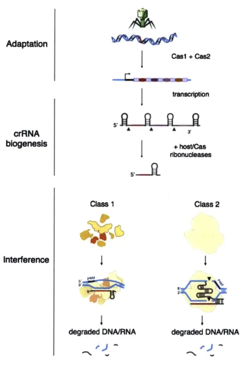
It is now understood that adaptive immunity at CRISPR loci occurs in a three-step process (Figure 4). First, invading nucleic acids are inserted into the CRISPR array in a process called adaptation (66). Second, the CRISPR array is transcribed and
processed into crRNAs containing single spacers in a process called crRNA biogenesis (67,
76, 77). Finally, interference against foreign genetic elements is mediated through the
action of Cas nucleases, which in complex with crRNAs find and cleave their targets through Watson-Crick basepairing of the Cas ribonucleoprotein complex with target sequences (67, 78-80).
Although adaptation, crRNA biogenesis and interference are common to all CRISPR systems, the specific mechanisms by which individual loci execute crRNA
biogenesis and interference can vary significantly. These mechanistic differences have been used to designate the class and type of the CRISPR system in question.
CRISPR systems are broadly divided into two classes based on the number of cas
genes that mediate interference and can be further sub-divided into types and subtypes on the basis of signature cas gene content, sequence homology, and locus architecture (81).
Class 1 systems utilize multiple cas gene products assembled in complexes to degrade target substrates, whereas class 2 systems utilize a single, large, multi-domain protein to achieve interference (Figure 4) (78, 82, 83). The simplicity of class 2 systems, where the specificity of a single nuclease can be reprogrammed through changing a short sequence of RNA, has facilitated their development into genome editing technologies.
Adaptation crRNA biogenesis
A
I1
Cal + Cas2Itranscription
5,, s3' Class 1 degraded DNA/RNA Class 2 degraded DNA/RNAFigure 4: Mechanisms of nucleic-acid interference by CRISPR systems. Casi and Cas2, sometimes in combination with other Cas proteins mediate insertion of DNA derived from mobile genetic elements into the CRISPR array, in a process called adaptation. The CRISPR array is transcribed as a long primary transcript and then processed into individual spacer-containing units (crRNAs) by Cas ribonucleases that occasionally are
16
aided by host enzymes. crRNAs direct Cas nuclease activity through a complementarity-dependent mechanism. Class 1 CRISPR systems utilize multi-protein, Cas nuclease containing complexes for target cleavage, whereas class 2 systems utilize a Cas single nuclease for nucleic-acid interference. Adapted from Hsu et. al. (32).
Genome editing using class 2 CRISPR systems
Class 2 DNA-targeting CRISPR systems are advantageous compared to other genome editing tools because they provide a method of targeting DSBs to endogenous loci through simple Watson-Crick base pairing rules. The first effector domain from a class 2 CRISPR system to be harnessed for genome editing in mammalian cells was Cas9 from type II CRISPR loci (Figure 5) (84, 85). Cas9 endonuclease activity is dependent on two non-coding RNAs: the crRNA, which directs cleavage specificity and a trans-activating CRISPR RNA (tracrRNA) that promotes crRNA maturation and is also required for cleavage (Figure 5A) (86). Studies showed that in vitro (87, 88) and in prokaryotic cells
(78, 83, 86), crRNA, tracrRNA and Cas9 are necessary for cleavage of complementary
dsDNA targets, suggesting that reconstitution of Cas9 activity in a heterologous system could be achieved by expressing Cas9, the tracrRNA and a targeting crRNA. Notably, it was also shown that the crRNA and tracrRNA could be fused into a single RNA molecule that mediated in vitro cleavage, potentially reducing the complexity of the system further
A
Cas9 crRNA tracrRNA
spacer DR
5''
B
RuvC
HNH
Figure 5: (A) Schematic representation of Cas9, crRNA and tracrRNA that together
form a ribonucleoprotein complex with DNA cleavage activity. (B) The HNH and RuvC
nuclease domains of Cas9 cleave the complementary and non-complementary DNA strands
of a crRNA-specified target site adjacent to a compatible PAM, creating a DSB.
Recognition of a target sequence by Cas9 also requires a compatible
protospacer-adjacent motif (PAM) (68, 89-91), a short nucleotide sequence immediately adjacent to
the target that Cas9 recognizes through protein-DNA contacts (92). PAM recognition initiates unwinding of the nearby dsDNA by Cas9, allowing for crRNA hybridization (93). Once the crRNA is hybridized to the target sequence, Cas9 uses two separate DNA nuclease domains, HNH and RuvC, to cleave the complementary and non-complementary
DNA strands, respectively, 3 nucleotides from the PAM, creating a DSB within the
crRNA:target heteroduplex (Figure 5B) (87, 88).
The targeting mechanism of Cas9 suggested this system could be utilized for editing complex mammalian genomes: the PAM of Cas9 is short (-3-5 bp) (90, 91),
allowing for flexible target site selection, and the spacer sequence of the mature crRNA is sufficiently long (-20 nt) (86) to specify unique target sites. DNA endonuclease activity of Cas9 from Streptococcus pyogenes SF370 was reconstituted in mammalian cells in 2013, defining a new class of RNA-guided genome editing tools (84, 85).
Creating DSBs with Cas9 through Watson-Crick basepairing rules allowed for simple, more predictable targeting of genomic sequences and the widespread adoption of Cas9 as a genome editing tool. The flexible nature of Cas9 targeting has led to the development of new genetic-screening methodologies for human cells (94-96), the development of catalytically inactive variants that can be combined with other effector domains (97-99) and paved the way for additional RNA-guided genome editing
technologies based on novel class 2 CRISPR effectors.
Extending genome editing techniques to RNA
Initial studies of class 2 CRISPR systems suggested that these loci exclusively encoded DNA nucleases, an idea that has been challenged by recent computational studies.
By extending the types of features used to search for new CRISPR loci, novel class 2
systems predicted to encode single-effector, RNA-guided, RNA-targeting Cas nucleases were discovered (Figure 6) (100). The RNA-targeting ability of these loci is predicted due to the absence of DNA targeting domains and the presence of conserved Higher
Eukaryotes and Prokaryotes Nucleotide-binding (HEPN) motifs, which mediate RNA degradation in a variety of contexts (101).
Class 2 systems with predicted DNA targeting (types 11, V)
HEPN HEPN
-- [I'.T
casl3a (c2c2) casi cas2 CRISPR
HEPN HEPN
casl3c (c2c7) CRISPR
HEPN HEPN HEPN
cas13b (c2c6) CRISPR
TM HEPN HEPN
casl3b(c2c6) CRISPR
Figure 6: The phylogenetic relationship of class 2 CRISPR encoding predicted DNA nucleases are collapsed.
VI-A@ VI-Ce VI-B1lO VI-B2@ systems. Leptotrichia shahii B031_RS0110445 Fusobacterium perfoetens T364_RS0105110 Prevotella buccae HMPREF6485_RS00335 Bergeyella zoohelcum HM PREF9699_02005
All class 2 systems
The discovery of putative class 2 RNA-targeting CRISPR systems opens the possibility of extending the simplicity of RNA-guided genome editing to transcripts. Transcriptome editing would ideally apply the functionalities of genome editing to RNA, with simple methodologies to both knockdown and modify the function of transcripts. Powerful technologies for suppressing the function of transcripts in eukaryotic cells exist, including RNA interference (RNAi) (102, 103) and anti-sense oligonucleotides (ASOs)
(104, 105), which trigger enzymatic degradation by endogenous enzymes (RNAi and ASOs) or sterically occlude functional RNA motifs (ASOs) to prevent RNA function (106).
Technologies that can modify the function of transcripts have been more challenging to develop. These techniques generally mimic the concept of TALE and ZF-based effectors for DNA applications: fusing tandem repeats of a modular RNA-binding protein domain that recognizes RNA bases through protein contacts, to RNA-modifying domains of known function (107). The RNA-binding domain commonly used for this application is the
pumilio repeat (PUF) domain, which structural studies suggest may recognize single RNA
bases as part of a tandem array (108, 109). Similar to ZFs, developing PUFs with arbitrary specificity has proven challenging, possibly due to unanticipated complexity of the binding code (110, 111), limiting the prospects of recruiting RNA-modifying domains to endogenous transcripts.
Development of class 2 RNA-targeting CRISPR systems for transcriptome engineering could enable a simple method for modifying transcript function through recruitment of RNA-modifying enzymes fused to Cas effectors by Watson-Crick basepairing rules.
Class 2 CRISPR systems containing HEPN domains have been designated as type VI and further divided into subtypes A, B and C (Figure 6) (112). The RNA-targeting ability of type VI-A CRISPR systems has been recently confirmed and shown to be catalytically mediated by conserved residues in the HEPN domain of the Casl3a protein encoded by these loci (113). Casl3a cleaves ssRNA targets in a crRNA-dependent manner that requires complementarity with the target, suggesting the possibility of CRISPR-based transcriptome engineering.
At the outset of this thesis, the existence and function of type VI-B and VI-C CRISPR systems were unknown. The work presented in this thesis seeks to characterize novel type VI CRISPR systems and test their utility for transcriptome editing applications in mammalian cells. There are three specific questions addressed in the work presented:
(i) Do other type VI CRISPR systems exist?
By computationally mining publicly available microbial genomes using a novel
CRISPR discovery pipeline, we identified type VI-B CRISPR loci, which encode a putative
RNA-guided RNAse, Casl3b.
(ii) How do type VI-B CRISPR loci function?
Using in vitro assays and reconstitution of native VI-B loci in heterologous prokaryotic expression systems, we provide evidence for a model in which a
ssRNA targets. We show genetically that the phenomenon of RNA interference by VI-B systems in vivo can be repressed or enhanced by the expression of csx27 or csx28,
respectively, small effector proteins that naturally co-occur with Casl3b in type VI-B loci.
(iii) Can nucleases from type VI CRISPR systems be used for transcriptome
editing applications in mammalian cells?
To test for the ability of type VI nucleases (Cas13 a/b/c) to suppress transcript
function, we reconstituted their nuclease activity in mammalian cells. To address whether Cas13 nucleases could be used to modify transcript function, we created catalytically inactive variants of Casl3b (dCasl3b) from Prevotella sp. P5-125 fused to the catalytic domain of adenosine deaminase acting on RNA (ADAR) enzymes, showing that dCasl3b could specifically direct the enzymatic activity of the ADAR deaminase domains to target transcripts.
The first two questions are addressed in the 2nd chapter of this thesis, and the final
question in the 3rd chapter.
Note: A portion of the description of CRISPR-Cas systems in this introduction was adapted form my preliminary exam proposal.
References
1. P. Nyren, A. Lundin, Enzymatic method for continuous monitoring of
inorganic pyrophosphate synthesis. Analytical biochemistry 151, 504 (Dec, 1985).
2. E. D. Hyman, A new method of sequencing DNA. Analytical biochemistry
174, 423 (Nov 1, 1988).
3. J. Shendure et al., Accurate multiplex polony sequencing of an evolved
bacterial genome. Science 309, 1728 (Sep 9, 2005).
4. E. S. Lander et al., Initial sequencing and analysis of the human genome. Nature 409, 860 (Feb 15, 2001).
5. J. C. Venter et al., The sequence of the human genome. Science 291, 1304 (Feb 16, 2001).
6. OMIM Gene Map Statistics. (2018).
7. T. A. Cooper, L. Wan, G. Dreyfuss, RNA and disease. Cell 136, 777 (Feb
20, 2009).
8. D. S. Cabianca et al., A long ncRNA links copy number variation to a polycomb/trithorax epigenetic switch in FSHD muscular dystrophy. Cell 149, 819 (May 11, 2012).
9. A. Jain, R. D. Vale, RNA phase transitions in repeat expansion disorders. Nature 546, 243 (Jun 8, 2017).
10. A. Hinnen, J. B. Hicks, G. R. Fink, Transformation of yeast. Proceedings of the National Academy of Sciences of the United States of America 75, 1929 (Apr, 1978).
11. R. J. Rothstein, One-step gene disruption in yeast. Methods in enzymology
101, 202 (1983).
12. J. W. Szostak, R. Wu, Insertion of a genetic marker into the ribosomal DNA of yeast. Plasmid 2, 536 (Oct, 1979).
13. T. L. Orr-Weaver, J. W. Szostak, R. J. Rothstein, Yeast transformation: a
model system for the study of recombination. Proceedings of the National Academy of Sciences of the United States of America 78, 6354 (Oct, 1981).
14. K. Folger, K. Thomas, M. R. Capecchi, Analysis of homologous
recombination in cultured mammalian cells. Cold Spring Harbor symposia on quantitative biology 49, 123 (1984).
15. K. R. Thomas, K. R. Folger, M. R. Capecchi, High frequency targeting of genes to specific sites in the mammalian genome. Cell 44, 419 (Feb 14, 1986).
16. K. R. Thomas, M. R. Capecchi, Site-directed mutagenesis by gene targeting
in mouse embryo-derived stem cells. Cell 51, 503 (Nov 6, 1987).
17. M. R. Capecchi, Altering the genome by homologous recombination. Science
244, 1288 (Jun 16, 1989).
18. 0. Smithies, R. G. Gregg, S. S. Boggs, M. A. Koralewski, R. S.
Kucherlapati, Insertion of DNA sequences into the human chromosomal beta-globin locus by homologous recombination. Nature 317, 230 (Sep 19-25, 1985).
19. S. Thompson, A. R. Clarke, A. M. Pow, M. L. Hooper, D. W. Melton, Germ
line transmission and expression of a corrected HPRT gene produced by gene targeting in embryonic stem cells. Cell 56, 313 (Jan 27, 1989). 20. N. Rudin, E. Sugarman, J. E. Haber, Genetic and physical analysis of
double-strand break repair and recombination in Saccharomyces cerevisiae. Genetics 122, 519 (Jul, 1989).
21. J. A. Nickoloff, E. Y. Chen, F. Heffron, A 24-base-pair DNA sequence from the MAT locus stimulates intergenic recombination in yeast. Proceedings of
the National Academy of Sciences of the United States of America 83, 7831 (Oct, 1986).
22. A. Ray, I. Siddiqi, A. L. Kolodkin, F. W. Stahl, Intra-chromosomal gene conversion induced by a DNA double-strand break in Saccharomyces cerevisiae. Journal of molecular biology 201, 247 (May 20, 1988).
23. A. Plessis, A. Perrin, J. E. Haber, B. Dujon, Site-specific recombination determined by I-SceI, a mitochondrial group I intron-encoded endonuclease expressed in the yeast nucleus. Genetics 130, 451 (Mar, 1992).
24. N. Nassif, J. Penney, S. Pal, W. R. Engels, G. B. Gloor, Efficient copying of nonhomologous sequences from ectopic sites via P-element-induced gap repair. Molecular and cellular biology 14, 1613 (Mar, 1994).
25. P. Rouet, F. Smih, M. Jasin, Introduction of double-strand breaks into the
genome of mouse cells by expression of a rare-cutting endonuclease. Molecular and cellular biology 14, 8096 (Dec, 1994).
26. P. Rouet, F. Smih, M. Jasin, Expression of a site-specific endonuclease
stimulates homologous recombination in mammalian cells. Proceedings of the National Academy of Sciences of the United States of America 91, 6064 (Jun 21, 1994).
27. F. Smih, P. Rouet, P. J. Romanienko, M. Jasin, Double-strand breaks at the target locus stimulate gene targeting in embryonic stem cells. Nucleic
acids research 23, 5012 (Dec 25, 1995).
28. P. A. Jeggo, DNA breakage and repair. Advances in genetics 38, 185
(1998).
29. J. H. Wilson, P. B. Berget, J. M. Pipas, Somatic cells efficiently join
unrelated DNA segments end-to-end. Molecular and cellular biology 2, 1258 (Oct, 1982).
30. J. K. Moore, J. E. Haber, Cell cycle and genetic requirements of two pathways of nonhomologous end-joining repair of double-strand breaks in Saccharomyces cerevisiae. Molecular and cellular biology 16, 2164 (May,
1996).
31. M. Bibikova, M. Golic, K. G. Golic, D. Carroll, Targeted chromosomal
cleavage and mutagenesis in Drosophila using zinc-finger nucleases. Genetics 161, 1169 (Jul, 2002).
32. P. D. Hsu, E. S. Lander, F. Zhang, Development and applications of
CRISPR-Cas9 for genome engineering. Cell 157, 1262 (Jun 5, 2014).
33. Y. G. Kim, J. Cha, S. Chandrasegaran, Hybrid restriction enzymes: zinc
finger fusions to Fok I cleavage domain. Proceedings of the National
Academy of Sciences of the United States of America 93, 1156 (Feb 6,
1996).
34. L. Li, L. P. Wu, S. Chandrasegaran, Functional domains in Fok I restriction
endonuclease. Proceedings of the National Academy of Sciences of the United States of America 89, 4275 (May 15, 1992).
35. M. Bibikova et al., Stimulation of homologous recombination through
targeted cleavage by chimeric nucleases. Molecular and cellular biology 21, 289 (Jan, 2001).
36. G. P. Diakun, L. Fairall, A. Klug, EXAFS study of the zinc-binding sites in the protein transcription factor IIIA. Nature 324, 698 (Dec 18-31, 1986). 37. J. M. Berg, Proposed structure for the zinc-binding domains from
transcription factor IIIA and related proteins. Proceedings of the National Academy of Sciences of the United States of America 85, 99 (Jan, 1988). 38. N. P. Pavletich, C. 0. Pabo, Zinc finger-DNA recognition: crystal structure
of a Zif268-DNA complex at 2.1 A. Science 252, 809 (May 10, 1991). 39. J. R. Desjarlais, J. M. Berg, Toward rules relating zinc finger protein
sequences and DNA binding site preferences. Proceedings of the National Academy of Sciences of the United States of America 89, 7345 (Aug 15, 1992).
40. J. Smith et al., Requirements for double-strand cleavage by chimeric
restriction enzymes with zinc finger DNA-recognition domains. Nucleic acids research 28, 3361 (Sep 1, 2000).
41. M. H. Porteus, D. Baltimore, Chimeric nucleases stimulate gene targeting in
human cells. Science 300, 763 (May 2, 2003).
42. Y. Choo, I. Sanchez-Garcia, A. Klug, In vivo repression by a site-specific
DNA-binding protein designed against an oncogenic sequence. Nature 372, 642 (Dec 15, 1994).
43. A. G. Rivenbark et al., Epigenetic reprogramming of cancer cells via targeted DNA methylation. Epigenetics 7, 350 (Apr, 2012).
44.
Q.
Liu, D. J. Segal, J. B. Ghiara, C. F. Barbas, 3rd, Design of polydactylzinc-finger proteins for unique addressing within complex genomes. Proceedings of the National Academy of Sciences of the United States of America 94, 5525 (May 27, 1997).
45. R. R. Beerli, C. F. Barbas, 3rd, Engineering polydactyl zinc-finger
transcription factors. Nature biotechnology 20, 135 (Feb, 2002).
46. J. C. Miller et al., An improved zinc-finger nuclease architecture for highly specific genome editing. Nature biotechnology 25, 778 (Jul, 2007).
47. M. Szczepek et al., Structure-based redesign of the dimerization interface
reduces the toxicity of zinc-finger nucleases. Nature biotechnology 25, 786 (Jul, 2007).
48. C. L. Ramirez et al., Unexpected failure rates for modular assembly of engineered zinc fingers. Nature methods 5, 374 (May, 2008).
49. J. C. Miller, C. 0. Pabo, Rearrangement of side-chains in a Zif268 mutant highlights the complexities of zinc finger-DNA recognition. Journal of molecular biology 313, 309 (Oct 19, 2001).
50. K. Gu et al., R gene expression induced by a type-III effector triggers
disease resistance in rice. Nature 435, 1122 (Jun 23, 2005).
51. S. Kay, S. Hahn, E. Marois, G. Hause, U. Bonas, A bacterial effector acts as a plant transcription factor and induces a cell size regulator. Science 318, 648 (Oct 26, 2007).
52. J. Boch, U. Bonas, Xanthomonas AvrBs3 family-type III effectors: discovery and function. Annual review of phytopathology 48, 419 (2010).
53. A. J. Bogdanove, S. Schornack, T. Lahaye, TAL effectors: finding plant genes for disease and defense. Current opinion in plant biology 13, 394 (Aug, 2010).
54. C. M. Hopkins, F. F. White, S. H. Choi, A. Guo, J. E. Leach, Identification of a family of avirulence genes from Xanthomonas oryzae pv. oryzae.
Molecular plant-microbe interactions : MPMI 5, 451 (Nov-Dec, 1992).
55. J. Boch et al., Breaking the code of DNA binding specificity of TAL-type III
effectors. Science 326, 1509 (Dec 11, 2009).
56. M. J. Moscou, A. J. Bogdanove, A simple cipher governs DNA recognition
by TAL effectors. Science 326, 1501 (Dec 11, 2009).
57. M. Christian et al., Targeting DNA double-strand breaks with TAL effector
nucleases. Genetics 186, 757 (Oct, 2010).
58. T. Li et al., TAL nucleases (TALNs): hybrid proteins composed of TAL
effectors and FokI DNA-cleavage domain. Nucleic acids research 39, 359 (Jan, 2011).
59. T. Li et al., Modularly assembled designer TAL effector nucleases for
targeted gene knockout and gene replacement in eukaryotes. Nucleic acids
research 39, 6315 (Aug, 2011).
60. J. C. Miller et al., A TALE nuclease architecture for efficient genome editing. Nature biotechnology 29, 143 (Feb, 2011).
61. P. Huang et al., Heritable gene targeting in zebrafish using customized
TALENs. Nature biotechnology 29, 699 (Aug 5, 2011).
62. L. Tesson et al., Knockout rats generated by embryo microinjection of
TALENs. Nature biotechnology 29, 695 (Aug 5, 2011).
63. D. Hockemeyer et al., Genetic engineering of human pluripotent cells using TALE nucleases. Nature biotechnology 29, 731 (Jul 7, 2011).
64. T. Cermak et al., Efficient design and assembly of custom TALEN and
other TAL effector-based constructs for DNA targeting. Nucleic acids
research 39, e82 (Jul, 2011).
65. F. Zhang et al., Efficient construction of sequence-specific TAL effectors for
modulating mammalian transcription. Nature biotechnology 29, 149 (Feb, 2011).
66. R. Barrangou et al., CRISPR provides acquired resistance against viruses in
prokaryotes. Science 315, 1709 (Mar 23, 2007).
67. S. J. Brouns et al., Small CRISPR RNAs guide antiviral defense in prokaryotes. Science 321, 960 (Aug 15, 2008).
68. A. Bolotin,- B. Quinquis, A. Sorokin, S. D. Ehrlich, Clustered regularly interspaced short palindrome repeats (CRISPRs) have spacers of extrachromosomal origin. Microbiology 151, 2551 (Aug, 2005).
69. F. J. Mojica, C. Diez-Villasenor, J. Garcia-Martinez, E. Soria, Intervening sequences of regularly spaced prokaryotic repeats derive from foreign genetic
elements. Journal of molecular evolution 60, 174 (Feb, 2005).
70. F. J. Mojica, C. Diez-Villasenor, E. Soria, G. Juez, Biological significance of
a family of regularly spaced repeats in the genomes of Archaea, Bacteria
and mitochondria. Molecular microbiology 36, 244 (Apr, 2000).
71. F. J. Mojica, C. Ferrer, G. Juez, F. Rodriguez-Valera, Long stretches of
short tandem repeats are present in the largest replicons of the Archaea Haloferax mediterranei and Haloferax volcanii and could be involved in replicon partitioning. Molecular microbiology 17, 85 (Jul, 1995).
72. F. J. Mojica, G. Juez, F. Rodriguez-Valera, Transcription at different
salinities of Haloferax mediterranei sequences adjacent to partially modified
PstI sites. Molecular microbiology 9, 613 (Aug, 1993).
73. C. Pourcel, G. Salvignol, G. Vergnaud, CRISPR elements in Yersinia pestis
acquire new repeats by preferential uptake of bacteriophage DNA, and provide additional tools for evolutionary studies. Microbiology 151, 653
(Mar, 2005).
74. Y. Ishino, H. Shinagawa, K. Makino, M. Amemura, A. Nakata, Nucleotide
sequence of the iap gene, responsible for alkaline phosphatase isozyme conversion in Escherichia coli, and identification of the gene product.
Journal of bacteriology 169, 5429 (Dec, 1987).
75. R. Jansen, J. D. Embden, W. Gaastra, L. M. Schouls, Identification of genes
that are associated with DNA repeats in prokaryotes. Molecular
microbiology 43, 1565 (Mar, 2002).
76. T. H. Tang et al., Identification of 86 candidates for small non-messenger
RNAs from the archaeon Archaeoglobus fulgidus. Proceedings of the
National Academy of Sciences of the United States of America 99, 7536
(May 28, 2002).
77. J. Carte, R. Wang, H. Li, R. M. Terns, M. P. Terns, Cas6 is an endoribonuclease that generates guide RNAs for invader defense in prokaryotes. Genes & development 22, 3489 (Dec 15, 2008).
78. J. E. Garneau et al., The CRISPR/Cas bacterial immune system cleaves
bacteriophage and plasmid DNA. Nature 468, 67 (Nov 4, 2010).
79. L. A. Marraffini, E. J. Sontheimer, CRISPR interference limits horizontal
gene transfer in staphylococci by targeting DNA. Science 322, 1843 (Dec
19, 2008).
80. B. Wiedenheft et al., RNA-guided complex from a bacterial immune system
enhances target recognition through seed sequence interactions. Proceedings
of the National Academy of Sciences of the United States of America 108, 10092 (Jun 21, 2011).
81. K. S. Makarova et al., An updated evolutionary classification of
CRISPR-Cas systems. Nature reviews. Microbiology 13, 722 (Nov, 2015).
82. K. S. Makarova et al., An updated evolutionary classification of
CRISPR-Cas systems. Nature reviews. Microbiology, (Sep 28, 2015).
83. R. Sapranauskas et al., The Streptococcus thermophilus CRISPR/Cas
system provides immunity in Escherichia coli. Nucleic acids research 39, 9275 (Nov, 2011).
84. L. Cong et al., Multiplex genome engineering using CRISPR/Cas systems.
Science 339, 819 (Feb 15, 2013).
85. P. Mali et al., RNA-guided human genome engineering via Cas9. Science
339, 823 (Feb 15, 2013).
86. E. Deltcheva et al., CRISPR RNA maturation by trans-encoded small RNA
and host factor RNase III. Nature 471, 602 (Mar 31, 2011).
87. M. Jinek et al., A programmable dual-RNA-guided DNA endonuclease in
adaptive bacterial immunity. Science 337, 816 (Aug 17, 2012).
88. G. Gasiunas, R. Barrangou, P. Horvath, V. Siksnys, Cas9-crRNA
ribonucleoprotein complex mediates specific DNA cleavage for adaptive immunity in bacteria. Proceedings of the National Academy of Sciences of the United States of America 109, E2579 (Sep 25, 2012).
89. H. Deveau et al., Phage response to CRISPR-encoded resistance in
Streptococcus thermophilus. Journal of bacteriology 190, 1390 (Feb, 2008).
90. P. Horvath et al., Diversity, activity, and evolution of CRISPR loci in
Streptococcus thermophilus. Journal of bacteriology 190, 1401 (Feb, 2008).
91. F. J. Mojica, C. Diez-Villasenor, J. Garcia-Martinez, C. Almendros, Short
motif sequences determine the targets of the prokaryotic CRISPR defence system. Microbiology 155, 733 (Mar, 2009).
92. H. Nishimasu et al., Crystal structure of Cas9 in complex with guide RNA
and target DNA. Cell 156, 935 (Feb 27, 2014).
93. S. H. Sternberg, S. Redding, M. Jinek, E. C. Greene, J. A. Doudna, DNA interrogation by the CRISPR RNA-guided endonuclease Cas9. Nature 507, 62 (Mar 6, 2014).
94. 0. Shalem et al., Genome-scale CRISPR-Cas9 knockout screening in human
cells. Science 343, 84 (Jan 3, 2014).
95. T. Wang, J. J. Wei, D. M. Sabatini, E. S. Lander, Genetic screens in human
cells using the CRISPR-Cas9 system. Science 343, 80 (Jan 3, 2014).
96: S. Konermann et al., Genome-scale transcriptional activation by an engineered CRISPR-Cas9 complex. Nature 517, 583 (Jan 29, 2015).
97. S. Konermann et al., Optical control of mammalian endogenous
transcription and epigenetic states. Nature 500, 472 (Aug 22, 2013).
98. P. Mali et al., CAS9 transcriptional activators for target specificity
screening and paired nickases for cooperative genome engineering. Nature biotechnology 31, 833 (Sep, 2013).
99. I. B. Hilton et al., Epigenome editing by a CRISPR-Cas9-based
acetyltransferase activates genes from promoters and enhancers. Nature biotechnology 33, 510 (May, 2015).
100. S. Shmakov et al., Discovery and Functional Characterization of Diverse Class 2 CRISPR-Cas Systems. Molecular cell 60, 385 (Nov 5, 2015).
101. V. Anantharaman, K. S. Makarova, A. M. Burroughs, E. V. Koonin, L.
Aravind, Comprehensive analysis of the HEPN superfamily: identification of novel roles in intra-genomic conflicts, defense, pathogenesis and RNA
processing. Biology direct 8, 15 (2013).
102. A. Fire et al., Potent and specific genetic interference by double-stranded
RNA in Caenorhabditis elegans. Nature 391, 806 (Feb 19, 1998).
103. S. M. Elbashir et al., Duplexes of 21-nucleotide RNAs mediate RNA
interference in cultured mammalian cells. Nature 411, 494 (May 24, 2001).
104. P. C. Zamecnik, M. L. Stephenson, Inhibition of Rous sarcoma virus
replication and cell transformation by a specific oligodeoxynucleotide. Proceedings of the National Academy of Sciences of the United States of America 75, 280 (Jan, 1978).
105. M. L. Stephenson, P. C. Zamecnik, Inhibition of Rous sarcoma viral RNA
translation by a specific oligodeoxyribonucleotide. Proceedings of the National Academy of Sciences of the United States of America 75, 285 (Jan, 1978).
106. A. Khvorova, J. K. Watts, The chemical evolution of oligonucleotide
therapies of clinical utility. Nature biotechnology 35, 238 (Mar, 2017). 107. J. P. Mackay, J. Font, D. J. Segal, The prospects for designer
single-stranded RNA-binding proteins. Nature structural & molecular biology 18, 256 (Mar, 2011).
108. X. Wang, J. McLachlan, P. D. Zamore, T. M. Hall, Modular recognition of RNA by a human pumilio-homology domain. Cell 110, 501 (Aug 23, 2002). 109. T. A. Edwards, S. E. Pyle, R. P. Wharton, A. K. Aggarwal, Structure of
Pumilio reveals similarity between RNA and peptide binding motifs. Cell 105, 281 (Apr 20, 2001).
30
110. M. T. Miller, J. J. Higgin, T. M. Hall, Basis of altered RNA-binding specificity by PUF proteins revealed by crystal structures of yeast Puf4p.
Nature structural & molecular biology 15, 397 (Apr, 2008).
111. Y. Wang, L. Opperman, M. Wickens, T. M. Hall, Structural basis for specific recognition of multiple mRNA targets by a PUF regulatory protein. Proceedings of the National Academy of Sciences of the United States of
America 106, 20186 (Dec 1, 2009).
112. A. S. Sergey Shmakov, David Scott, David Cox, Neena Pyzocha, Winston
Yan, Omar 0. Abudayyeh, Jonathan S. Gootenberg, Kira S. Makarova, Yuri I. Wolf, Konstantin Severinov, Feng Zhang, Eugene V. Koonin2, Diversity and evolution of Class 2 CRISPR-Cas systems Nature Reviews Microbiology, (2016).
113. 0. 0. Abudayyeh et al., C2c2 is a single-component programmable
RNA-guided RNA-targeting CRISPR effector. Science 353, aaf5573 (Aug 05,
Chapter 2
Adapted from the following article, which appears in Molecular Cell:
Casl3b is a Type VI-B CRISPR-associated RNA-Guided RNase differentially regulated by accessory proteins Csx27 and Csx28
Aaron A. Smargon",2
,4 1 0 David B.T. Cox' 2,3,5,6,10, Neena K. Pyzocha" 2 3 ,,0
Kaijie Zheng1', Ian M. Slaymaker2 , Jonathan S. Gootenberg', Omar A.
Abudayyehl2 3 ,
Patrick Essletzbichler' 3 , Sergey Shmakov,9, Kira S. Makarova9, Eugene V. Koonin9,
and Feng Zhang 2 3,1 t
1 Broad Institute of MIT and Harvard
Cambridge, Massachusetts 02142, USA
2 McGovern Institute for Brain Research at MIT
Cambridge, Massachusetts 02139, USA
3 Departments of Brain and Cognitive Science and Biological Engineering
Massachusetts Institute of Technology, Cambridge, MA 02139, USA
4 Department of Electrical Engineering and Computer Science
Massachusetts Institute of Technology, Cambridge, Massachusetts 02139, USA
5 Harvard-MIT Division of Health Sciences and Technology, Harvard Medical School,
Boston, Massachusetts 02115, USA
6Department of Biology, Massachusetts Institute of Technology, Cambridge,
Massachusetts 02139, USA
7 Department of Systems Biology, Harvard Medical School, Boston, MA 02115, USA
8 Skolkovo Institute of Science and Technology, Skolkovo 143025, Russia
' National Center for Biotechnology Information National Library of Medicine, National Institutes of Health
Bethesda, MD 20894, USA
1 Co-first author
" Lead Contact
f Correspondence should be addressed to z hangvbroadinstitute.org.
Abstract
CRISPR-Cas adaptive immune systems defend microbes against foreign nucleic acids via RNA-guided endonucleases. Using a computational sequence database mining approach, we identify two Class 2 CRISPR-Cas systems (subtype VI-B) that lack Casi and Cas2 and encompass a single large effector protein, Casl3b, along with one of two previously uncharacterized associated proteins, Csx27 or Csx28. We establish that these CRISPR-Cas systems can achieve RNA interference when heterologously expressed. Through a combination of biochemical and genetic experiments, we show that Casl3b processes its own CRISPR array with short and long direct repeats, cleaves target RNA, and exhibits collateral RNase activity. Using an E. coli essential gene screen, we demonstrate that Casl3b has a double-sided protospacer-flanking sequence and elucidate RNA secondary structure requirements for targeting. We also find that Csx27 represses, whereas Csx28 enhances, Casl3b-mediated RNA interference. Characterization of these CRISPR systems creates opportunities to develop tools to manipulate and monitor cellular transcripts.
INTRODUCTION
CRISPR-Cas (clustered regularly interspaced short palindromic repeats and CRISPR-associated proteins) systems are divided into two classes, Class 1 systems, which utilize multiple Cas proteins and CRISPR RNA (crRNA) to form an effector complex, and the more compact Class 2 systems, which employ a large, single effector with crRNA to mediate interference (Makarova et al., 2015). CRISPR-Cas systems display a wide evolutionary diversity, involving distinct protein complexes and different modes of operation, including the ability to target RNA (Abudayyeh et al., 2016; East-Seletsky et al., 2016; Hale et al., 2009; Jiang et al., 2016; Staals et al., 2013; Staals et al., 2014; Tamulaitis et al., 2014).
Computational sequence database mining for diverse CRISPR-Cas systems has been carried out by searching microbial genomic sequences for loci harboring the casi gene, the most highly conserved cas gene involved in the adaptation phase of CRISPR immunity (Marraffini, 2015). Among other findings, this approach led to the discovery of the Class 2 subtype VI-A system with its signature effector Casl3a (previously known as C2c2), which targets RNA (Abudayyeh et al., 2016; East-Seletsky et al., 2016; Shmakov et al., 2015). Since distinct variants of functional Class 1 CRISPR systems have been characterized that lack casi (Makarova et al., 2015), we sought to identify Class 2 CRISPR-Cas systems lacking casi by modifying the computational discovery pipeline so that it is not seeded on Casi. Here we report the characterization of a Class 2 subtype, VI-B, which was discovered through this computational approach, and demonstrate that the VI-B effector, Casl3b, is an RNA-guided RNase.
RESULTS