THE JOURNAL OF INFECTIOUS DISEASES • VOL. 149, NO. 4 • APRIL 1984 © 1984 by The University of Chicago. All rights reserved. 0022-1899/84/4904-0008$01.00
Granulocyte Neutral Proteases and Pseudomonas Elastase as Possible Causes of
Airway Damage in Patients with Cystic Fibrosis
Susanne Suter, Urs B. Schaad, Laurent Roux, Urs E. Nydegger, and Francis A. Waldvogel
From the Infectious Disease Division. Department of Medicine. University Hospital. 1211 Geneva 4; and the
Departments of Pediatrics. Universities of Geneva and Bern. Switzerland
We studied the possible role of granulocyte neutral proteases as mediators of airway destruction in patients with cystic fibrosis (CF) who were infected with Pseudomonas aeruginosa. We measured the enzymatic activities of bronchial secretions on purified radioactively labeled complement component three (C3), elastin, and a granulocyte elas-tase-specific substrate. Bronchial secretions from 18 patients with CF who were infected withP aeruginosa had a significantly higher mean value for C3 cleaving, elastolytic, and granulocyte elastase-like activity than did two control groups. High enzymatic activities were observed in patients with CF who have advanced bronchial disease (that had been determined by a clinical scoring system). Kinetics of proteolysis of radioactively labeled C3 and inhibition profiles of the activities of the three enzymatic activities studied sug-gest that they are mainly derived from granulocytes. In addition, 20 of 31 strains of P aeruginosa isolated from patients with CF inactivated purified o l-antiprotease in vitro. We postulate that granulocyte neutral proteases and P aeruginosa may act syner-gistically in the airways of patients with CF and may contribute to the destruction of elastin and inactivation of C3.
Cystic fibrosis is a fatal hereditary disease, in which the leading cause of death is respiratory failure [I]. Patients with cystic fibrosis (CF) usually have a long history of purulent bronchitis leading to progressive destruction of small bronchioles and followed by involvement of the large airways [2]. The pathogens most frequently associated with these respiratory-tract infections are Staphylococ-cus aureus and Pseudomonas aeruginosa [2, 3]. Clinical and pathological observations suggest that progressive airway destruction is accelerated during infections with P aeruginosa [2]. However, little information is available regarding the mech-anisms involved in progressive bronchiolar and bronchial damage and the persistence ofP
aerugi-nosa in bronchial secretions.
Bronchial secretions from patients with CF
con-Received for publication July 21, 1983, and in revised form November 18, 1983.
Part of this work was supported by grant no. 3.836.0.79 from the Swiss Research Foundation.
We thank Anneliese Kahr and Sassi Dharan for technical assistance, W. Ruch for advice and suggestions, Dr. P. Naftolin and Dr. J.-D. Vassalli for reviewing the manuscript, and Francoise Michaud for secretarial assistance.
Please address requests for reprints to Dr. Susanne Suter, Infectious Disease Division, University Hospital, 1211 Geneva 4, Switzerland.
523
tain numerous PMNs [2] that might contribute to airway damage by release of neutral proteases such as elastase, collagenase, cathepsin G, and others [4, 5]. These enzymes are indeed able to destroy im-portant structural proteins of the lung and its air-ways such as elastin, collagen, and proteoglycans [4, 5] in vitro; in addition, they can inactivate im-portant opsonins such as complement component C3 [6-8] and IgG and IgM immunoglobulins [9-11].Itis also well established that they are released extracellularly during phagocytosis [12]. Further-more, P aeruginosa may secrete an elastase that destroys the two main inhibitors protecting the lung and its airways from the activity of PMN neutral proteases: al-antiprotease [13-16] and the bronchial mucosal proteinase inhibitor [17]. In addition, this bacterial elastase is also active on C3 [18] and elastin [15].
We therefore measured the enzymatic activities of bronchial secretions from patients with CF (who were infected with P aeruginosa) on purified human C3, bovine elastin, and a granulocyte elas-tase-specific substrate. Two groups of patients with bronchial secretions rich in PMNs (one group with and the other without bacterial infection of the tracheobronchial tree) and a granular enzyme preparation isolated from human PMNs were used as controls.
Table 1. Description of patients with cystic fibrosis. Clinical scores
Patient Age Status Pulmonary Bacteriology no. (years) and activity findings of sputum
1 18 4 4-5 4 2 18 4-5 4 4 3 14 2 3 4 4 20 3-4 4 4 5 11 2 3 4 6 9 2-3 4 4 7 14 2-3 3 3-4 8 18 2 2-3 3 9 6 1-2 3 3-4 10 20 3-4 4 4 11 14 4 4 4 12 10 2 3 3-4 13 18 2-3 3 4 14 6 2 3 3 15 5 2 2 3 16 5 2 3 4 17 16 4-5 5 5 18 6 4 4 4
NOTE. The scores of the patients were determined accord-ing to the scoraccord-ing system of Kraemer et al. [23] by one of the authors (U .B.S.) unaware of the results of the activities mea-sured in bronchial secretions.
Patients and Methods
Group 1. Twenty-four samples of bronchial secretions were collected after chest physiotherapy
from 18 patients with CF, all of whom had P
aeru-ginosainfections (table 1). The patients were either attending cystic fibrosis clinics or were inpatients at the children's hospitals, University of Bern or Geneva, Switzerland. The diagnosis of cystic fibrosis had been established on clinical features of the disease and confirmed by sweat sodium and chloride concentrations above 70 meq/l. The clin-ical and pulmonary conditions of the patients were determined by one of the authors who was un-aware of the laboratory results and were recorded via a scoring system outlined previously [19].
All but two patients were on similar antibiotic treatment when the secretions were collected. The mean age of the patients was 13 years (range, 5-20 years).
Group 2. Twenty samples of bronchial secre-tions, which fulfilled the quality criteria estab-lished by Murray and Washington [20], were col-lected from 20 patients with acute exacerbations of chronic bronchitis, as documented by chronic cough, purulent sputum, positive chest findings, and roentgenograms. In nine patients bronchiec-tases had been documented by bronchography or
bronchoscopy. Bronchial lavage fluid was obtained from six patients by bronchoscopy on the same day that the sputum samples were obtained. Pathogens
isolated from sputum included: Haemophilus
influenzae [18], Streptococcus pneumoniae [8], Escherichia coli [1], Proteus vulgaris [1], S aureus
[5], and Aspergillus [1]. Three patients had mixed
infections. Eighteen patients had a history of smoking. The mean age of the patients was 50 years (range, 9-62 years).
Group 3. Ten samples of bronchial secretions were obtained as morning specimens from 10 pa-tients who had no evidence of acute or chronic
respiratory-tract infection. Diagnosis included:
acute asthma (2), pulmonary embolism (1),
em-physema with chronic respiratory failure (1),
chronic heart failure (3), bronchial carcinoma (1),
and metastatic cancer of the lung (1). All patients
produced sputum that was negative when cultured for pathogens. The mean age of the patients was 68 years (range, 22-90 years).
Processing of bronchial secretions and of P aeruginosa isolates. Bacterial cultures and
identifications were performed according to well-established methods. Leukocyte counts in bron-chial secretions were evaluated semiquantitatively [20], by a technician unaware of the study results. The secretions were mixed with a half volume of sterile isotonic saline and centrifuged within 1 hr at 1,000 g until the supernatant was clear. The supernatants were frozen at - 70 C until tested. Protein concentration was determined by a stan-dard method (Bio-Rad, Richmond, Calif).
Thirty-one strains ofP
aeruginosa
were isolatedfrom the bronchial secretions of patients with CF
who were infected withP
aeruginosa;
the cultureswere maintained in skim milk at - 20C,.
Microbiological techniques. In order to test the
P
aeruginosa
strains for elastase productionvquan-titative cultures were performed as follows: 0.1 ml of an overnight culture was added to 5 ml of Mueller-Hinton broth and incubated in a shaking waterbath at 37 C for 24 hr. The size of the inocu-lum and the final bacterial concentration were determined by making plates of serial.dilutions on Mueller-Hinton agar and counting the colony forming units. The 24-hr cultures were centrifuged in an Eppendorf centrifuge at 8,000 g for 10 min (Eppendorf, Hamburg, FRO). The supernatants were frozen at - 70 C in small aliquots until used.Purification of human C3, labeling of C3 with
1251 and preparation of
125I-labeled C3-Sepharose. C3 was purified to homogeneity from human
Proteases in CF bronchial secretions
plasma with the use of the method of Tack and
Prahl [22]. It was labeled with 1251 (Amersham
International, Amersham, England), by means of
the chloramine T method [23]. Approximately 1 X
106 counts per minute (cpm) were incorporated
into 1 /Ag of purified C3. Labeled C3 was either used directly or it was coupled to CnBr-activated
Sepharose 4B (Pharmacia Fine Chemicals,
Uppsala, Sweden) by a standard method.
Assessment of calcium- and magnesium-inde-pendent C3-cleaving activity. In order to rule out C3-cleaving activity deriving from complement convertases, these experiments were performed in the presence of EDTA (20 mmol final concentra-tion). 1251-labeled C3-Sepharose beads
correspond-ing to 2 X 10⁵ cpm were suspended in 150 /AI of
calcium- and magnesium-free Dulbecco's PBS (pH 7.4) and incubated with 50 /AI of bronchial secre-tions that had been previously mixed with EDTA (20 mmol final concentration), with granular
enzymes, or with P aeruginosa culture
super-natants at 37 C for 5 hr. 1251-labeled C3 fragments were separated from 1251-labeled C3-Sepharose
beads by centrifugation at 8,000 g for 5 min.
Cleavage of C3 was expressed as the percentage of cpm in the supernatant divided by the cpm of the total sample. Spontaneous breakdown of 1251_
labeled C3 incubated alone was 1.2070 ± 0.2%; this
background was subtracted from the results.
Demonstration of proteolysis of 125I-labeled C3 by polyacrylamide gel electrophoresis [24]. The kinetics of proteolysis and the pattern of break-down products of 1251-labeled C3 generated by the bronchial secretions from a patient with CF and a
P aeruginosa infection were compared with those
obtained from the culture supernatant of a strain ofP aeruginosa from the same patient, and with
those observed when granular enzymes were used. Four microliters of a bronchial secretion, 34 /AI of granular enzymes and 240 /AI of the supernatant
from a P aeruginosa culture were incubated with
labeled C3 corresponding to 5 X 10⁶ cpm in
'Iris-NaCI buffer (pH 7.4) in a total volume of 600 /AI. Aliquots of 25 /AI were withdrawn after 0, 2, 5, 8, 13, 20, 30, 60, 120, and 300 minutes of incubation at 37 C and mixed with 12.5 /AI of SDS sample buf-fer (1% 2-mercaptoethanol, 2% SDS, 80 mmol Tris-HCI pH 6.8, 10% glycerol, and 0.05% bromo-phenol-blue) and heated at 100 C for 3 min. The samples were run on discontinuous slab gels with 5% (wt/vol) acrylamide in the stacking gel and 12.5% (wt/vol) acrylamide in the resolving gel. Proteins were revealed by autoradiography with a
525
Kodak X-omat S film with exposure at - 70 C for 3 hr. Monitoring of molecular weights was accom-plished with the use of Sendai virus proteins [25], which were labeled with [35S]methionine as marker proteins with known molecular weights. Controls included 1251-labeled C3 incubated both alone and
with saliva from two patients with CF andP
aeru-ginosa infections and from one normal individual. Preparation of granular enzymes from human PMNs. Granular enzymes were prepared from PMNs collected from fresh heparinized blood of normal adult volunteers, according to the method of Baggiolini [21] and modified as previously described [8]. They had an elastolytic activity of 17.4 UE and granulocyte elastase-like activity of 0.59 UTGP (see below for definition of units).
Assessment of elastolytic activity. Elastolytic activity was measured as described by Hornebeck [26] with purified bovine elastin (Sigma Co, St.
Louis) radioactively labeled with 3H (New
England Nuclear Corp, Boston). Ten
micro-liters of the test sample were incubated with 500 /Ag of 3H-Iabeled elastin suspended in 1 ml of Tris (0.1
M,pH 8.2) containing Brij 35 (0.01 %) and sodium
azide (0.02%), for 16 hr at 37 C. The samples were
then centrifuged at 3,300g for 10 min. The
radio-activity of 100fJI of the supernatants was measured and used to calculate the amount of solubilized elastin. One milligram of completely solubilized 3H-Iabeled elastin corresponded to 145,000 cpm. Elastolytic activity (UE) was expressed as the number of milligrams of labeled elastin solubilized by 1 ml of enzyme solution.
Measurement of granulocyte elastase-like
activ-ity. Granulocyte elastase-like activity was
mea-sured as previously described [8] with the use of
synthetic Sue-Ala-Ala-Pro-Val-7-amino-4-methyl
coumarin (TGP 928; a gift from M. Baggiolini, Wander Research Institute, Bern, Switzerland) as a substrate. The sequence Ala-Ala-Pro-Val has been shown to be a specific substrate for granulocyte elastase [27].
The enzymatic activity was expressed in UTGP
=
micromoles of 7-amino-5-methyl-coumarinsolubilized per minute by 1 ml of enzyme solution
(extinction coefficient at 360 nm
=
12,300).Paer-uginosa culture supernatants, Mueller-Hinton broth, saliva from two patients with CF and from one normal individual had no activity on this substrate.
Inactivation of partially purified o-l antipro-tease by supernatants of P aeruginosa cultures. e-I
first, inhibition of 50 IJI of granular enzyme by increasing concentrations of partially purified
0'-1-antiprotease (Sigma) was measured; a 100% inhi-bition was reached by a concentration of 10IJgof o-I antiprotease (in 10 IJI). Thereafter, samples of 30 IJI of supernatants ofP aeruginosacultures were incubated with 60 IJI of Dulbecco's PBS (pH 7.4) containing 60IJgofo-I anti protease at 37 C for 24 hr, and the inhibitory activity of 15 IJI of this incu-bation mixture was tested on 50 IJI of granular enzyme. o-I antiprotease incubated alone at 37 C for 24 hr kept its full inhibitory capacity on gran-ular enzymes; Mueller-Hinton broth had no effect on either granular enzymes or TGP 928.
Inhibition experiments. Phenylmethylsulfonyl-fluoride (PMSF, Sigma) [28] was used at 2 mmol final concentration, Ac-(Ala)4-chloromethylke-tone at 1 mmol (kindly provided by Dr. J. Powers, Georgia Institute of Technology, Atlanta) [29], partially purified o-I antiprotease at 10 IJmol, and Zincov [30] at 500 IJmol. The results were expressed as percent inhibition of the enzymatic activity of a sample. With Ac-(Ala)4-chloromethylketone the samples were preincubated for 60 min, but with the other inhibitors the incubation was only for 5 min.
Statistics. The results were expressed as mean
± SEM. For comparison of groups, the
Mann-Whitney U test or Spearman's correlation coeffi-cient were used. Enzymatic activities within each group were compared by linear regression analysis by the method of least squares.
Results
Leukocyte quantitation and protein concentra-tions of bronchial secreconcentra-tions. Stained smears showed comparable mean leukocyte scores: group 1,2.3 ± 0.2 (range, 1-3); group 2,2.7 ± 0.2 (range, 2-3); group 3, 2.1 ± 0.1 (range, 2-3). The three groups of bronchial secretions had a significantly different mean protein concentration: group 1, 6.3
± 0.9gil and group 2, 5.1 ± 0.5gil were signifi-cantly higher than group 3, 2.7 ± 0.6gil (P
<
.01).Proteolytic and esterolytic activities in bronchial secretions (figure 1): 125I-labeled C3-c1eaving activity. In group 1 the mean C3-cleaving activity was 48.8010 ± 2.5%, a value significantly higher than that of group 2, which was 15.4% ± 3.5% (P
<
.001) and of group 3, which was7.6010 ± 0.9%(P
<
.001). The difference between groups 2 and 3 was also significant (P<
.001). The same held true when the results were expressed per gram of protein.Elastolytic activity. The mean elastolytic
activ-125 I-C3
Granulocyte Elastolytic elastase-like
cleaving activity activity activity
% UE UTGP 60 2 1.5 40 1 1 20 0.5 0 0 0
*
Group II III II III II III
Figure 1. Three enzyme activities were measured in bronchial secretions from three groups of patients:Group 1, patients with CF infected withP aeruginosa; group 2, patients with exacerbated chronic bronchitis; group 3, patients with various noninfectious conditions, but with significant numbers of granulocytes in their bronchial secretions. C3-cleaving activity was determined by measuring the radioactivity of 12sI-Iabeled C3 fragments released into the supernatant from 12sI-Iabeled C3-Sepharose after incubation with 50~lof the bronchial secretion for 5 hr at 37 C. The results were expressed as the percentage of radioactivity in the supernatant divided by the radioactivity of the total sample. Elastolytic activity(UE)was expressed as milligrams of 3H-Iabeled elastin hydrolyzed by1ml of enzyme solution after 16hr of incubation at 37 C. Granulocyte elastase-like activity was measured as UTGP
=
micromoles of 7-amino-5-methyl-coumarin released/min per milliliter of enzyme solution.Proteases in CF bronchial secretions 527
Figure 2. The results of enzymatic activities of the 24 hr culture supernatants of 31 strains ofP aeruginosa iso-lated from the patients with CF (group 1)on the substrates J25I-labeled C3-Sepharose and 3H-labeled elastin were determined in vitro as described for figure 1. Group A represents 20 strains ofP aeruginosa inactivating o-I anti protease as described in material and methods and group B, 11 strains without effect on o-I antiprotease. Group A strains were considered as elastase-producing strains on the basis of their activity on the three substrates e-I antiprotease, 125I-labeled C3-Sepharose, and 3H-labeled elastin [18-21].
ity of group 1 was 1.9 ± 0.3 VE compared with 0.54 ± 0.09 (P
<
.05) in group 2 and 0.04 ± 0.02(P
<
.001) in group 3. The difference between groups 2 and 3 was also significant(P<
.01). These differences were also significant when expressed per gram of protein.Granulocyte elastase-like activity. The mean granulocyte elastase-like activity was strikingly higher in group 1 than in group 2 bronchial secre-tions: 1.39 ± 0.56 VTGP vs. 0.12 ± 0.05 UTGP (P
<
.001). Group 3 samples had no detectable granu-locyte elastase-like activity, as was the case in saliva obtained from two patients with CF andP aerugi-nosa infections and one normal individual.In group 1 patients, high clinical scores (which indicated poor pulmonary condition) correlated with high 1251-labeled C3-cleaving activitytr, =
.62, P
<
.01), elastolytic activity (rs=
.81, P<
.001), and granulocyte elastase-like activity(r, = .73, P
<
.01).Since the activities of the three enzymes mea-sured could possibly be attributed to the presence of PMN neutral proteases [6-8, 29], correlations between enzymatic activities within each group were calculated. Group 1: a linear correlation was found between 125I-labeled C3-cleaving and gran-ulocyte elastase-like activity (r = .49, P
<
.025)and between elastolytic and granulocyte elastase-like activity(r = .89, P
<
.001). Group 2: a linear correlation was observed between 125I-labeled C3-cleaving and granulocyte elastase-like activity (r= .48,P
<
.025), between elastolytic and granulo-cyte elastase-like activity (r = .56, P<
.01), and between C3-cleaving and elastolytic activity(r = .49, P
<
.025).Heating of both bronchial secretion samples from three patients with CF and granular enzymes showed that incubation at 56 C for 30 min inacti-vated only 10% of the 1251-labeled C3-cleaving activity of these samples, a finding consistent with the heat resistance of PMN elastase found by others [31).
Proteolytic activities of P aeruginosa isolates from patients with CF (figure2). The mean bac-terial concentration of quantitative P aeruginosa
cultures was 3.9 ± 0.4
x
108 cfu/ml. The culturesupernatants were tested for P aeruginosaelastase by measuring their activity on three substrates known to be cleaved by P aeruginosaelastase in vitro: e-I anti protease [15, 16], 1251-labeled C3 [14], and 3H-Iabeled elastin [13]. Twenty of the 31 strains of P aeruginosa were shown to inactivate o-I anti protease in vitro, as measured by a func-tional assay: 30fJIof the culture supernatants of 20
P aeruginosa strains inactivated 60fJg of e-I anti-protease completely. The 20 strains inactivating e-I antiprotease in vitro (figure 2, group A strains) also had a significantly higher 1251-labeled C3-cleaving activity (29.6010 ± 3.3010) and elastolytic activity (1.45 ± 0.31 VE) than did the 11 remain-ing strains (figure 2, group B strains). C3-cleavremain-ing activity of these strains was 7.6% ± 1% and elasto-lytic activity 0.24 ± 0.05VE(P
<
.(01). In group A strains there was a linear correlation between 1251_ labeled C3-cleaving and elastolytic activity (r=
.88,P<
.001). Finally, there was no correlation between mucoid appearance and enzyme secretion of a particular strain; nine group A strains and four group B strains were mucoid.Comparison ofproteolytic and esterolytic activ-ities. With these results at hand, we tried to deter-mine whether the' levels of enzymatic activity measured in secretions from patients with CF were mainly due to enzymes originating from PMNs or from Pseudomonas strains. To make these deter-minations, we used time-course and biochemical patterns of 1251-labeled C3 proteolysis as well as inhibition experiments for differentiation.
Time course and pattern of proteolysis of1251_
B A El astcl vt lc activity B A 125I-C3 cleaving activity
o
Group % 40 20INCUBATION TIME (minutes)
o
2 5 8 13 203060120300 Zo
~ w a:o
w ~en 0-1 < ~ o Zo
a: men
w :E > N Z W a: < -I ::J Z<
a: C) ~ Z ~en<
<z Za: OW :EQ. O::J cen ::J w wa: en::J Q.!3 ::J u 0 . - ~B F-·,,,,tt,IIit.,.
-
-_.
.~.- ••I,••.
f • •' .-...
••••
-
--
-labeled C3. Time course and pattern of proteoly-sis of 125I-labeled C3 induced by a bronchial secre-tion from a patient with CF, by the P aeruginosa
culture supernatant of the elastase-producing strain from the same patient, and by the granular enzyme preparation were studied in detail as shown in figure 3. Identical patterns of breakdown of 125I-labeled C3 by the bronchial secretion and by the granular enzyme preparation can be recog-nized: both preparations caused a breakdown of the alpha chain of 125I-labeled C3 into fragments with apparent molecular weights of113
x
103and100
x
103 and later, 32x
103, 25
x
10\ and 18x
103• In contrast, incubation of 125I-labeled C3
with the culture supernatant of the elastase-pro-ducing strain ofP aeruginosa led to a single cleav-age of the alpha chain with generation of an alpha fragment of an apparent molecular weight of 110
x
103and later, two small breakdown products of16
x
103 and 17x
103• Limited fragmentation of
the alpha chain by too Iowan elastase concentra-tion in the supernatant of the P aeruginosaculture was excluded by testing the culture supernatant undiluted, whereas the bronchial secretion and the granular enzyme preparation were assayed at dilu-tions of 1:30and 1:5,respectively. 125I-labeledC3
incubated alone at 37 C for 5hr showed no evi-dence of breakdown of either the alpha- or the beta-chain. Saliva from two patients with CF and
P aeruginosainfections and from one normal indi-vidual had no effect on 125I-labeled C3.
Figure 3. Four microliters of a cystic fibrosis bronchial secretion, 34 JAI of granular enzymes, and 240 JAI of the culture supernatant of the elastase-producing strain of
Paeruginosa from the same patient were incubated at 37 C with 12sI-labeled C3 at a concentration correspond-ing to 5 x 10⁶cpm in a total volume of 600 JAI (Tris-NaCI buffer, pH 7.4). Aliquots of 25 JAI were withdrawn at various periods of incubation indicated in the figure. The timed samples were mixed with SDS sample buffer, heated at 100 C for 3 min, and the resulting breakdown of 12sI-labeled C3 examined on a 12.5010 polyacrylamide gel. The arrows ex and(Jdesignate the alpha- and beta-chain of 12sI-labeled C3; BF corresponds to the buffer front. The observed proteolysis is restricted to the alpha-chain and the pattern of breakdown is similar for the bronchial secretion and for granular enzymes resulting in alpha-fragments with apparent molecular weights of 100x 103and 113x 103for the early incubation periods
and small fragments of apparent molecular weights of 3? x 103, 25X 103 , and18x 103at 5 hr of incubation.
Incubation with thePaeruginosaculture supernatant lead to only one apparent cleavage of the alpha-chain with a fragment of apparently 110 x 103and two small
frag-Discussion
In this study we found that bronchial secretions from patients with cystic fibrosis (CF) who had
P aeruginosa infections (group 1) had strikingly higher enzymatic activities on radioactively labeled
C3and elastin, as well as on a granulocyte elastase-specific peptide substrate, than did the bronchial secretions of two control groups, i.e., patients with chronic bronchitis (group 2) and patients without infection (group 3; figure 1). Most strains of
P aeruginosa isolated from the patients with CF secreted bacterial elastase in vitro (figure 2), as determined by the proteolytic activity of their cul-ture supernatants on purified e-l antiprotease, C3,
ments of 16x 103and 17x 103•Molecular weights were
monitored with Sendai virus proteins as marker proteins [25]; the marker proteins are not shown on the figure.
Proteases in CF bronchial secretions
and elastin, all three of which are substrates for this enzyme [13-15, 18]. Each patient was infected with at least one elastase-producing strain of
P aeruginosa. The following arguments suggest that the proteolytic activities measured in bron-chial secretions from patients with CF most likely derived from granulocyte neutral proteases, i.e., elastase and possibly cathepsin G. First, the time-course of the appearance and the size of break-down products of 125I-Iabeled C3 were identical when labeled C3 was incubated with bronchial
secretions from patients with CF and P aeruginosa
infection or with granular enzymes (figure 3), but it was clearly different when 125I-labeled C3 was
incubated with a P
aeruginosa
culturesuperna-tant. Second, PMSF (an inhibitor of serine-pro-teases [28J) and Ac-(Ala)4-chloromethylketone (a granulocyte elastase-specific inhibitor [28]) sub-stantially suppressed 125I-labeled C3-c1eaving ac-tivity of both bronchial secretions from a patient
with CF who had P
aeruginosa
infections andgranular enzymes, but not of supernatants from
P
aeruginosa
cultures (table 2). The contribution of complement convertases to 125I-Iabeled C3 break-down was excluded by measuring 125I-labeled C3-cleaving activity in the presence of EillA. Among other proteases possibly involved in C3 cleavage by bronchial secretions from patients with CF who had Paeruginosa
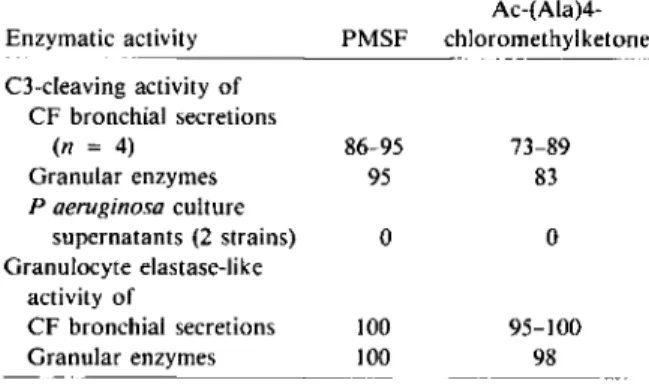
infections, collagenase isin-Table 2. Range of inhibition ofI25I-Iabeled C3-cleaving
and granulocyte elastase-like activity by PMSF and Ac-(Ala)4-chloromethylketone.
-Inhibition(070)by inhibitors* Ac-(Ala)4-Enzymatic activity PMSF chloromethylketone C3-cleaving activity of CF bronchial secretions (n = 4) 86-95 73-89 Granular enzymes 95 83 P aeruginosa culture supernatants (2 strains) 0 0 Granulocyte elastase-like activity of CF bronchial secretions 100 95-100 Granular enzymes 100 98
NOTE. Granular enzymes and Paeruginosaculture super-natants were tested in triplicate. Inhibition is expressed as% =
100 (enzymatic activity with the inhibitor ) - enzymatic activity without the inhibitor x 100 . * The final concentration of the inhibitors was as follows: PMSF=2.5 mmol, Ac-(Ala)4-chloromethylketone=1 mmol.
529
hibited by EDTA [28] and is also not inhibited by the two inhibitors used in the experiment.
Third, three elastases may contribute to mea-sured elastolytic activity in bronchial secretions of
patients with CF: (1) granulocyte elastase [32], a
serine protease inhibited by PMSF [28], Ac-(Ala)4-chloromethylketone [27], and its physiological inhibitor e-I antiprotease [16]; (2) pseudomonas elastase, a zinc-metalloprotease [14, 18] inhibited
by Zincov [30]; and (3) macrophage elastase [32],
which is a metalloprotease inhibited bya-2
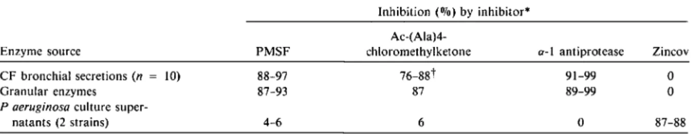
macro-globulin, but not by e-I antiprotease [31]. Our inhibition experiments show that the elastolytic activity of bronchial secretions from patients with CF was inhibited by the three inhibitors of granu-locyte elastase, but not by the inhibitor of pseudo-monas elastase (table 3). Macrophage elastase also has access to the lung [32], but this enzyme would not have been inhibited by the inhibitors used [31]. The only other enzyme also known to have some elastolytic properties [33] and similar inhibition patterns would be cathepsin G [28].
Fourth, although the substrate we used for measuring granulocyte elastase-like activity is specific for granulocyte elastase in purified systems [27], its specificity cannot be fully demonstrated in biological fluids. However, the complete inhibition of granulocyte elastase-like activity by PMSF and Ac-(Ala)4-chloromethylketone further character-ized this activity as a serine protease such as elas-tase. In addition, the linear correlation found be-tween C3-c1eaving, elastolytic, and granulocyte elastase-like activity of bronchial secretions from patients with CF suggest that the three activities
derive from the same group of enzymes. Itshould
also be noted that when granulocyte elastase is
complexed with a-2 macroglobulin, its second less
important inhibitor in vivo [34], the complex enzyme-inhibitor still has some activity on small peptide substrates similar to the one we used [35], but not on elastin. This may explain why granulo-cyte elastase-like activity of bronchial secretions from patients with CF compared with their elasto-lytic activity was so much higher than granulocyte elastase-like activity compared with elastolytic activity of granular enzymes.
Several aspects of our experimental design and clinical material must be considered. Collecting bronchial secretions from children is difficult; thus, we used adults as control patients. Bronchial
Table 3. Range of inhibition of elastolytic activity by various inhibitors.
-Inhibition(010)by inhibitor*
Ac-(Ala)4-Enzyme source PMSF chloromethylketone o-I anti protease Zincov
CF bronchial secretions(n = 10) 88-97 76-88t 91-99 0
Granular enzymes 87-93 87 89-99 0
P aeruginosaculture
super-natants (2 strains) 4-6 6 0 87-88
- _ . _ . _ - - -
-NOTE. Granular enzymes and P aeruginosa culture supernatants were tested in triplicate. Inhibition is expressed as Oli ( enzymatic activity with the inhibitor
00)
o= 100 - _enzymatic activity without the inhibitor x.1 .
*The final concentration of the inhibitors was as follows: PMSF = 2 mmol, Ac-(Ala)4-chloromethylketone 1 mmol, o-I antiprotease = 10 IAmol, and Zincov = 500 IAmol.
t n = 3.
P
aeruginosa
infections were obtained by chest physiotherapy, since, in our centers, it is considered ethically unjustified to submit patients with CF to bronchial lavages, which have not demonstrated therapeutic value. Bronchial lavage fluids could be obtained from six of our control patients with chronic bronchitis on the same day as sputum samples. Proteolytic activities showed good corre-lation in the six sample pairs. Despite extensive research, we were unable to find more than three patients with CF who were not harboring small quantities ofPaeruginosa;
these patients were the ideal control group. All three patients were under seven years of age, but had proteolytic activities comparable to adult patients with chronic bron-chitis.In our group of patients with CF, high clinical scores, indicating poor pulmonary condition, correlated with high proteolytic activities in bron-chial secretions. This finding may reflect a high degree of chronic infection leading to accumu-lation of larger numbers of PMNs or inactivation of the two most important inhibitors of gran-ulocyte elastase in the lung by longstanding coloni-zation or infection with P aeruginosa. Indeed,
P
aeruginosa
elastase is known to inactivate both the bronchial mucosal proteinase inhibitor pro-tecting the airways [36] and a-I antiprotease [16], and each patient with CF was infected with at least one elastase-producing strain. Although we cannot link this in vitro demonstration with inactivation of inhibitors in vivo, the nearly complete sup-pression of elastolytic activity of CF bronchial secretions by purified a-I antiprotease in vitro indi-cates that in vivo the inhibitor potential for PMN neutral proteases was too.Bronchial secretions from patients with CF also had high proteolytic activity on 125I-Iabeled C3, an activity most likely originating from PMN neutral proteases. Since granulocyte elastase in vitro not only inactivates C3 [6, 7] but also IgG and IgM [10, 11], it is conceivable that free granulocyte elastase in vivo leads to functional inactivation of these opsonins and thereby favors the persistence of microorganisms, a well known characteristic of
P
aeruginosa
infections in these patients [2, 3]. From our observations we conclude that the high proteolytic activities measured in CF bron-chial secretions most likely reflect the activity of PMN neutral proteases, especially elastase and cathepsin G. This free proteolytic activity may be caused by either an excess of proteolytic enzymes or a lowered local inhibitor potential, the latter being induced by Paeruginosa
elastase. PMN neutral proteases may also inactivate opsonins in the bronchial tree of these patients and therefore contribute to persistence of microorganisms. In a prospective study, we currently follow these prote-olytic activities of bronchial secretions from pa-tients with CF who had Paeruginosa
infections before and after iv antibiotic therapy againstP
aeruginosa
and investigate the efficacy of syn-thetic inhibitors of granulocyte elastase on these proteolytic activities. Such inhibitors may be considered as possible adjuncts to antimicrobial therapy in patients with CF.References
1. Sant'Agnese PAD. Cystic fibrosis. In: Vaughan VC, McKay RJ, Behrman RE eds. Textbook of pediatrics. 11th ed. Philadelphia: WB Saunders Company, 1979:1988-2001
Pro/eases in CF bronchial secretions
2. Pennington JE, Wolff SM; Puziss M. Summary of a work-shop on infections in patients with cystic fibrosis. J Infect Dis 1979;140:252-6
3. Marks MI. The pathogenesis and treatment of pulmonary infections in patients with cystic fibrosis. J Pediatr 1981; 98:173-9
4. Janoff A, Sloan B, Weinbaum G, Damiano V, Sandhaus RA, Elias J, Kimbel R. Experimental emphysema in-duced with purified human neutrophil elastase: tissue localization of the instilled protease. Am Rev Respir Dis 1977;115:461-78
5. Sandberg LB, Soskel NT, Leslie JG. Elastin structure, bio-synthesis and relation to disease states. N Engl J Med 1981;304:566-79
6. Taylor JC, Crawford IP, Hugli TE. Limited degradation of the third component (C3) of human complement by human leukocyte elastase (HLE): partial characteri-zation of C3 fragments. Biochemistry 1977:16:3390-6 7. Johnson U, Ohlsson K, Olsson I. Effects of granulocyte
neutral proteases on complement components. Scand J Immunol 1976;5:421-6
8. Suter S, Nydegger UE, Roux L, Waldvogel FA. Cleavage of C3 by neutral proteases from granulocytes in pleural empyema. J Infect Dis 1981;144:499-508
9. Lew DP, Despont J-P, Perrin LH, Aguado M-T, Lambert PH, Waldvogel FA. Demonstration of a local exhaustion of complement components and of an enzymatic degra-dation of immunoglobulins in pleural empyema: a possi-ble factor favouring the persistence of local bacterial in-fections. Clin Exp Immunol 1980;42:506-14
10. Prince HE, Folds JD, Spitznagel JK. Proteolysis of human IgG by human polymorphonuclear leucocyte elastase produces an Fe fragment with in vitro biological activity. Clin Exp Immunol 1979;37:162-8
11. Prince HE, Folds JD, Spitznagel JK. Interaction of human polymorphonuclear leukocyte elastase with human IgM. In vitro production of an Fabu-like fragment. Mol Immunol 1979;16:301-6
12. Stossel TP. Phagocytosis (second of three parts). N Engl J Med 1974;290:774-80
13. Moskowitz RW, Heinrich G. Bacterial inactivation of human serum alpha-l antitrypsin. J Lab Clin Med 1971; 77:777-85
14. Morihara K, Tsuzuki H, Oda K. Protease and elastase of Pseudomonas aeruginosa: inactivation of human plasma ai-proteinase inhibitor. Infect Immun 1979;24: 188-93
15. Morihara K. Production of elastase and proteinase by Pseudomonas aeruginosa.J Bacteriol 1964;88:745-57 16. Gadek JE, Fells GA, Zimmermann RL, Rennard SI,
Crystal RG. Anti-elastase of the human alveolar struc-tures. Implications for the protease-antiprotease theory of emphysema. J Clin Invest 1981;68:889-98
17. Johnson DA, Carter-Hamm B, Dralle WM. Inactivation of human bronchial mucosal proteinase inhibitor by Pseu-domonas aeruginosaelastase. Am Rev Respir Dis 1982; 126:1070-3
18. Schultz DR, Miller KD. Elastase ofPseudomonas aerugi-nosa:inactivation of complement components and com-plement-derived chemotactic and phagocytic factors. Infect Immun 1974;10:128-35
531
19. Kraemer R, Rudeberg A, Kliiy M, Rossi E. Relationship be-tween clinical conditions, radiographic findings and pul-monary functions in patients with cystic fibrosis. Helv Paediatr Acta 1979;34:417-28
20. Murray PR, Washington JA II. Microscopic and bacterio-logic analysis of expectorated sputum. Mayo Clinic Proc 1975;50:339-44
21. Baggiolini M. The isolation of granules from neutrophile polymorphonuclear leukocytes (PMNs). Methods Enzy-mol 1974;31:345-53
22. Tack BF, Prahl JW. Third component of human comple-ment: purification from plasma and physiochemical characterization. Biochemistry 1976;15:4513-21 23. McConahey PJ, Dixon FJ. A method of trace iodination of
proteins for immunologic studies. Int Arch Allergy Appl Immunol 1966;29:185-9
24. Laemmli UK. Cleavage of structural proteins during the as-sembly of the head of bacteriophage T4. Nature 1970; 227:680-5
25. Wagner RR, Prevec L, Brown F, Summers OF, Sokol F, MacLeod R. Classification of rhabdovirus proteins: a proposal. J Virol 1972;10:1228-30
26. Hornebeck W, Schnebli HP. Effect of different elastase inhibitors on leukocyte elastase pre-adsorbed to elastin. Hoppe-Seylers Z Physiol Chern 1982;363:455-8 27. Nakajima K, Powers JC, Ashe BM, Zimmermann M.
Mapping the extended substrate binding site of cathepsin G and human leukocyte elastase. Studies with peptide substrates related to the ai-protease inhibitor reactive site. J BioI Chern 1979;254:4027-31
28. Schmidt W. Neutrale Proteasen aus menschlichen Leu-kozyten: Isolierung, Charakterisierung und biologische Wirkungen. Inaugural-Dissertation Fachbereich Chemie der Philipps-Universitat Marburg/Lahn, Federal Repub-lic of Germany, 1975:1-66
29. Powers JC, Gupton BF, Harley AD, Nishino N, Whitley RJ. Specificity of porcine pancreatic elastase, human leukocyte elastase and cathepsin G: inhibition with pep-tide chloromethyl ketones. Biochim Biophys Acta 1977; 485:156-66
30. Hudgin RL, Charleson SE, Zimmermann M, Mumford R, Wood PL. Enkephalinases: selective peptide inhibitors. Life Sci 1981;29:2593-2601
31. Hornebeck W, Bellon G, Brechemier 0, Godeau G, Robert L. Control of elastic tissue destruction by elastase inhibi-tors. Progr Clin BioI Res 1981;54:233-46
32. Cohen AB. Potential adverse effects of lung macrophages and neutrophils. Fed Proc 1979;38:2644-7
33. Reilly CF, Travis J. The degradation of human lung elastin by neutrophil proteinases. Biochim Biophys Acta 1980; 621:147-57
34. Ohlsson K. ai-antitrypsin and a2-macroglobulin. Interac-tions with human neutrophil collagenase and elastase. Ann NY Acad Sci .1975;256:409-19
35. Harpel PC, Human alpha 2-macroglobulin. Methods Enzymol 1976;45:639-52
36. Hochstrasser K, Albrecht GJ, Schonberger OL, Rasche B, Lempart K. An elastase-specific inhibitor from human bronchial mucus: isolation and characterization. Hoppe-Seylers Z Physiol Chern 1981;362:1369-75