Cocoa polyphenols suppress TNF-a-induced vascular endothelial growth
factor expression by inhibiting phosphoinositide 3-kinase (PI3K) and
mitogen-activated protein kinase kinase-1 (MEK1) activities in mouse epidermal cells
Jong-Eun Kim
1, Joe Eun Son
1, Sung Keun Jung
1,2, Nam Joo Kang
2,3, Chang Yong Lee
4, Ki Won Lee
2*
and Hyong Joo Lee
1*
1Department of Agricultural Biotechnology, Seoul National University, Seoul, Republic of Korea
2Department of Bioscience and Biotechnology, Bio/Molecular Informatics Center, Konkuk University, Seoul, Republic of Korea 3School of Applied Biosciences, Kyungpook National University, Daegue 702-701, Republic of Korea
4
Department of Food Science and Technology, Cornell University, Geneva, NY, USA
(Received 1 December 2009 – Revised 22 March 2010 – Accepted 25 March 2010 – First published online 16 June 2010)
Cocoa polyphenols have antioxidant and anti-inflammatory effects. TNF-a is a pro-inflammatory cytokine that has a vital role in the pathogenesis of inflammatory diseases such as cancer and psoriasis. Vascular endothelial growth factor (VEGF) expression is associated with tumorigenesis, CVD, rheumatoid arthritis and psoriasis. We tested whether cocoa polyphenol extract (CPE) inhibited TNF-a-induced VEGF expression in promotion-sensitive JB6 mouse epidermal cells. CPE significantly inhibited TNF-a-induced up-regulation of VEGF via reducing TNF-a-induced activation of the nuclear transcription factors activator protein-1 (AP-1) and NF-kB, which are key regulators of VEGF expression. CPE also inhibited TNF-a-induced phosphorylation of protein kinase B (Akt) and extracellular signal-regulated kinase. CPE blocked activation of their downstream kinases, p70 kDa ribosomal protein S6 kinase and p90 kDa ribosomal protein S6 kinase. CPE suppressed phosphoinositide 3-kinase (PI3K) activity via binding PI3K directly. CPE did not affect TNF-a-induced phosphorylation of mitogen-activated protein kinase kinase-1 (MEK1) but suppressed TNF-a-induced MEK1 activity. Collectively, these results indicate that CPE reduced TNF-a-induced up-regulation of VEGF by directly inhibiting PI3K and MEK1 activities, which may contribute to its chemopreventive potential.
Cocoa polyphenols: Phosphoinositide 3-kinase: Mitogen-activated protein kinase kinase-1: TNF-a: Vascular endothelial growth factor
TNF-a is a 17 kDa protein consisting of 156 amino acids which acts as a cellular mediator provoking inflammation, pro-liferation, tumorigenesis and even apoptosis(1). This cytokine plays a key role in the pathogenesis of diseases such as cancer, rheumatoid arthritis, Crohn’s disease, diabetes and psoriasis(2). TNF-a binds with TNF receptor I (p55 receptor) or II (p75 receptor) and associates with adaptor proteins to trigger downstream kinases such as phosphoinositide 3-kinase (PI3K) – protein kinase B (Akt) and mitogen-activated protein kinases. Many inflammatory proteins are expressed through activation of transcription factors such as activator protein-1 (AP-1) and NF-kB by upstream signalling. Therefore, inhibition of the TNF-a signalling pathway may help prevent these diseases(3).
Vascular endothelial growth factor (VEGF) is a multi-functional cytokine that is a key regulator of angiogenesis(4). VEGF expression is associated with the pathology of
tumorigenesis, CVD, rheumatoid arthritis and psoriasis. Tumour cells require blood vessels because of nutrient and oxygen deficiency, and VEGF promotes tumour cell survival via angiogenesis. VEGF is involved in tumour progression by controlling angiogenesis as well as tumour promotion. The Ha-ras gene mutation in a chemical-induced two-stage carcinogenesis model has been shown to enhance VEGF expression and benign papilloma development(5). Therefore, inhibition of VEGF expression might be a promising approach to protect against carcinogenesis.
Cocoa contains high amounts of polyphenols(6) and contributes to a healthy life through strong antioxidant and anti-inflammatory activities and a increase of NO bioavailabil-ity(7,8). Recently, the health benefits of dark chocolate with reduced energy and an increased proportion of cocoa were revealed(9). Our previous studies show that cocoa contains more polyphenols and a higher antioxidant capacity than tea
* Corresponding authors: Dr Ki Won Lee, fax þ 82 2 3436 6178, email [email protected]; Dr Hyong Joo Lee, fax þ 82 2 873 5095, email [email protected] Abbreviations:Akt, protein kinase B; AP-1, activator protein-1; CPE, cocoa polyphenol extract; DTT, dithiothreitol; EGTA, ethylene-glycol-bis(a-aminoethyl)-N,N,N0,N0-tetra-acetic acid; ERK, extracellular signal-regulated kinase; FBS, fetal bovine serum; JNK, c-Jun N-terminal kinase; MEK1, mitogen-activated protein-kinase kinase-1; MEM, Eagle’s minimum essential medium; MKK4, mitogen-activated protein kinase kinase 4; MMP-2, matrix metalloproteinase-2; p70S6K, p70 kDa ribosomal protein S6 kinase; p-, phosphorylated; Pþ
, promotion-sensitive; PI3K, phosphoinositide 3-kinase; PMSF, phenylmethanesulfonyl fluoride; TPA, 12-O-tetradecanoylphorbol-13-acetate; Tris, 2-amino-2-hydroxymethyl-propane-1,3-diol; VEGF, vascular endothelial growth factor.
qThe Authors 2010
British
Journal
of
and red wine(10). It exerted protective effects against H2O2 -induced inhibition of gap junction intercellular communication in rat epithelial cells(11) and cocoa polyphenol extract (CPE) inhibited phorbol ester-induced inflammation through suppres-sion of cyclo-oxygenase-2 expressuppres-sion in mouse skin(12). Recently, we reported that CPE inhibits thrombin-induced expression and activation of matrix metalloproteinase-2 (MMP-2) in vascular smooth muscle cells and phorbol ester-induced neoplastic transformation of promotion-sensitive (Pþ) JB6 mouse epidermal cells through direct inhibition of mitogen-activated protein kinase kinase-1 (MEK1)(13,14). However, the chemopreventive effects of cocoa involve mechanisms besides the inhibition of MEK1. Here, we tested whether cocoa polyphenols inhibit TNF-a-induced VEGF expression and we suggested a novel molecular target of CPE, PI3K.
Materials and methods Chemicals
Eagle’s minimum essential medium (MEM), gentamicin, fetal bovine serum (FBS) and L-glutamine were purchased from GIBCO BRL (Carlsbad, CA, USA). TNF-a was purchased from ProSpec-Tany TechnoGene (Rehovot, Israel). The ELISA kit for VEGF was purchased from R&D Systems (Minneapolis, MN, USA). The antibodies against phosphorylated (p-) MEK (Ser217/221), total extracellular signal-regulated kinase (ERK), p-p90 kDa ribosomal S6 kinases (p90RSK; Thr359/Ser363), total p90RSK, p-p70 kDa ribosomal protein S6 kinase (p70S6K; Thr389) and total p70S6K were obtained from Cell Signaling Biotechnology (Beverly, MA, USA). p-Akt (Ser 473), p-ERK (Thr202/ Tyr204), total ERK and total MEK1 were purchased from Santa Cruz Biotechnology (Santa Cruz, CA, USA). The MEK1 activity assay kit and PI3K active protein were purchased from Upstate Biotechnology (Lake Placid, NY, USA). CNBr-Sepharose 4B, glutathione-Sepharose 4B and [g-32P]ATP were from GE Healthcare (Piscataway, NJ, USA), and the protein assay kit was purchased from Bio-Rad Laboratories (Hercules, CA, USA). G418 and the lucifer-ase assay substrate were from Promega (Madison, WI, USA).
Cell culture
The JB6 Pþ mouse epidermal cell lines were cultured in monolayers in 5 % FBS – MEM, 1000 units of penicillin and 1 mg/ml of streptomycin at 378C in a 5 % CO2 incubator. The JB6 mouse epidermal cell line stably transfected with dominant negative p85 (DN-p85), NF-kB, and AP-1 luciferase reporter plasmid was a gift from Dr Zigang Dong(15) and maintained in 5 % FBS – MEM and 200 mg/ml G418.
Preparation of cocoa polyphenol extract
Cocoa polyphenols were extracted from commercial cocoa powder (50 g) produced by LOTTE Confectionery Co. Ltd (Seoul, Korea) with 500 ml of 50 % (v/v) aqueous ethanol under reflux for 6 h. After the extraction, the solution was filtered twice to collect the extract. The collected cocoa extract was loaded onto a styrene-based adsorption resin column
(60 £ 450 mm, HP-20; Mitsubishi Chemical Co., Tokyo, Japan), washed with 20 % (v/v) aqueous ethanol, and then eluted with 60 % (v/v) aqueous ethanol. The eluted CPE was concentrated at 508C under reduced pressure, frozen, and dried.
Determination of vascular endothelial growth factor production
JB6 Pþ cells (5 £ 103) were cultured in ninety-six wells and incubated for 48 h. After incubation, the cells were pretreated with CPE at the indicated concentrations for 1 h before incu-bation with TNF-a (4 ng/ml) for 18 h, and then the culture medium was harvested. VEGF was quantified according to the manufacturer’s instructions.
Measurement of cell viability
Cell viability was measured by 3-(4,5-dimethylthiazol-2-yl)-2,5-diphenyltetrazolium bromide (MTT) assay, which is based on the ability of live cells to convert tetrazolium salt into purple formazan. JB6 Pþ cells (5 £ 103) were cultured in ninety-six wells and incubated for 48 h. After incubation, the cells were pretreated with CPE at the indicated concen-trations for 1 h before incubation with TNF-a (4 ng/ml) for 18 h, followed by the addition of 20 ml MTT stock solution (5 mg/ml; Sigma, St Louis, MO, USA) to each well and the plates were further incubated for 4 h at 378C. The supernatant fraction was removed and 200 ml dimethyl sulfoxide was added to each well to solubilise the water-insoluble purple formazan crystals. The absorbency at the wavelength of 570 nm was measured with a microplate reader (Molecular Devices, Sunnyvale, CA, USA). All the measurements were performed in triplicate. Results are expressed as the percen-tage proliferation with respect to untreated cells.
Luciferase assay for NF-kB and activator protein-1 transcription activity
Confluent monolayers of JB6 Pþ cells stably transfected with an AP-1 or NF-kB luciferase reporter plasmid were trypsi-nised, and 5 £ 103 viable cells suspended in 100 ml 5 % FBS – MEM was added to each well of a ninety-six-well plate. Plates were incubated at 378C in a humidified atmos-phere of 5 % CO2. When cells reached 80 – 90 % confluence, they were starved by culturing them in 0·1 % FBS – MEM for another 24 h. The cells were then treated for 15 min with CPE and exposed to TNF-a (4 ng/ml) for an additional 4 h. After treatment, cells were disrupted with 100 ml lysis buffer (0·1M-potassium phosphate buffer (pH 7·8); 1 % Triton
X-100; 1 mM-dithiothreitol (DTT); 2 mM-EDTA), and the luciferase activity was measured using a luminometer (Micro-lumat Plus LB 96V; Berthold Technologies, Bad Wildbach, Germany).
Western blotting analysis
After JB6 Pþcells (1·0 £ 106) were cultured in a 10 cm dia-meter dish for 48 h, they were starved in serum-free medium for another 24 h to eliminate the influence of FBS on the activation of kinases. The cells were then treated with CPE
British
Journal
of
(0 – 20 mg/ml) for 1 h before being exposed to TNF-a (4 ng/ml) for 15 min. After centrifugation, cell lysis was performed at 48C for 30 min in cell lysis buffer (20 mM
-2-amino-2-hydroxy-methyl-propane-1,3-diol (Tris)-HCl (pH 7·5), 150 mM-NaCl, 1 mM-Na2EDTA, 1 mM-ethylene-glycol-bis(a-aminoethyl)-N, N,N0,N0-tetra-acetic acid (EGTA), 1 % Triton, 2·5 mM-sodium pyrophosphate, 1 mM-b-glycerophosphate, 1 mM-Na3VO4, leupeptin (1 mg/ml) and 1 mM-phenylmethanesulfonyl fluoride (PMSF)). The cell lysates were centrifuged at 23 000 g for 15 min, and the resulting supernatant fraction was stored at 2 708C before Western blot analysis. The protein concen-tration in each sample was measured by subjecting lysate protein (30 mg) to 10 % SDS-PAGE, with the protein electrophoretically transferred to a nitrocellulose membrane (Whatman, Clifton, NJ, USA). Protein bands were visualised by a chemiluminescence detection kit (Amersham, Piscat-away, NJ, USA) after hybridisation with the horseradish peroxidase-conjugated secondary antibody. Quantification of band intensity was measured using Image J software (National Institutes of Health, Bethesda, MD, USA).
Mitogen-activated protein kinase kinase-1 immunoprecipitation and activity assay
JB6 Pþ cells were cultured to 80 % confluence and then serum-starved in 0·1 % FBS – MEM for 24 h at 378C. Cells were either treated or not treated with CPE (0 – 20 mg/ml) for 1 h before being exposed to TNF-a (4 ng/ml) for 15 min, disrupted with lysis buffer (20 mM-Tris – HCl (pH 7·4),
1 mM-EDTA, 150 mM-NaCl, 1 mM-EGTA, 1 % Triton X-100, 1 mM-b-glycerophosphate, leupeptin (1 mg/ml), 1 mM -Na3VO4and 1 mM-phenylmethylsulfonyl fluoride), and finally centrifuged at 16 000 g for 10 min in a microcentrifuge. The lysates containing 500 mg protein were used for immunopreci-pitation with an antibody against MEK1 and incubated at 48C overnight. Protein A/G Plus agarose beads were then added and the mixture was continuously rotated for another 3 h at 48C. The beads were washed three times with kinase buffer (20 mM-3-(N-morpholino)propanesulfonic acid (MOPS; pH
7·2), 25 mM-b-glycerol phosphate, 5 mM-EGTA, 1 mM -sodium orthovanadate and 1 mM-DTT), and then re-suspended
in 20 ml of 1 £ kinase buffer supplemented with 1 mg of inac-tive ERK2 and incubated for an additional 30 min at 308C. Then myelin basic protein (20 mg) and 10 ml of diluted [g-32P]ATP solution were added and the mixture was incu-bated for 10 min at 308C. A 20 ml sample was transferred onto p81 paper and washed three times with 0·75 % phos-phoric acid for 5 min per wash and once with acetone for 2 min. The radioactive incorporation was determined using a scintillation counter.
Phosphoinositide 3-kinase activity assay
An active PI3K protein (100 ng) was incubated with CPE for 10 min at 308C. Then, the mixtures were incubated with 20 ml phosphatidylinositol (0·5 mg/ml) (Avanti Polar Lipids, Alabaster, AL, USA) for 5 min at room temperature, followed by incubation with reaction buffer (100 mM-HEPES (pH 7·6), 50 mM-MgCl2, 250 mM-ATP containing 0·37 MBq (10 mCi) of [g-32P]ATP) for an additional 10 min at 308C. The reaction was stopped by adding 15 ml of 4M-HCl and 130 ml
chloroform – methanol (1:1, v/v). After vortexing, 30 ml of the lower chloroform phase was spotted onto a 1 % potassium oxalate-coated silica gel plate that was previously activated for 1 h at 1108C. The resulting32P-labelled phosphatidylinositol-3-phosphate was separated by TLC and radiolabelled spots were visualised by autoradiography.
Pull-down assay
Cell lysates of the TNF-a-treated cells used in the kinase assay (500 mg) or PI3K protein (2 mg) were incubated with CPE – Sepharose 4B (or Sepharose 4B alone as a control) beads (100 ml, 50 % slurry) in reaction buffer comprising 50 mM-Tris-HCl (pH 7·5), 5 mM-EDTA, 150 mM-NaCl, 1 mM-DTT, 0·01 % Nonidet P-40, 2 mg/ml bovine serum
albumin, 0·02 mM-PMSF and 1 £ protease inhibitor mixture. After incubation with gentle rocking overnight at 48C, the beads were washed five times with buffer (50 mM-Tris-HCl
(pH 7·5), 5 mM-EDTA, 150 mM-NaCl, 1 mM-DTT, 0·01 % Nonidet P-40 and 0·02 mM-PMSF), and proteins bound to
the beads were analysed by immunoblotting.
Statistical analysis
Data are expressed as mean values and standard deviations. Student’s t test was used for single statistical comparisons, with a probability value of P, 0·05 as the criterion for statistical significance.
Results
Cocoa polyphenol extract inhibits TNF-a-induced vascular endothelial growth factor up-regulation in promotion-sensitive JB6 mouse epidermal cells
JB6 Pþ cells were pretreated for 1 h with different concen-trations of CPE and then stimulated with TNF-a (4 ng/ml) for 18 h. TNF-a-induced VEGF expression in JB6 Pþ cells was determined by ELISA. CPE dose-dependently inhibited TNF-a-induced VEGF up-regulation (Fig. 1(a)). These effects were not due to the cytotoxicity of CPE, because CPE up to 20 mg/ml has no effect on cell viability (Fig. 1(b)).
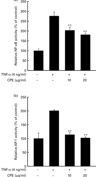
Cocoa polyphenol extract suppresses TNF-a-induced transactivation of NF-kB and activator protein-1 in promotion-sensitive JB6 mouse epidermal cells
The eukaryotic transcription factors NF-kB and AP-1 are key regulators of VEGF expression(16). We next examined whether CPE attenuates TNF-a-induced transactivation of NF-kB and AP-1 using JB6 Pþcell lines stably transfected with an NF-kB and AP-1 luciferase reporter plasmid. Exposure to TNF-a (4 ng/ml) for 4 h increased NF-kB luciferase activity by about 2·5-fold compared with the untreated control, and CPE dose-dependently inhibited these effects (Fig. 2(a)). TNF-a also increased AP-1 luciferase activity by about 2-fold compared with the untreated control, which was attenuated by CPE treatment (Fig. 2(b)).
British
Journal
of
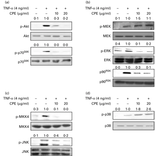
Effects of cocoa polyphenol extract on TNF-a-induced phosphorylation of protein kinase B and mitogen-activated protein kinase
We next examined the PI3K – Akt and mitogen-activated protein kinase pathways, which relate to TNF-a-induced NF-kB and AP-1 transactivation. We investigated the phosphorylation level of the kinase which is involved in these pathways by Western blot analysis. CPE suppressed TNF-a-induced phos-phorylation of Akt, p70S6K, ERK, p90 kDa ribosomal S6 kinase (p90RSK), mitogen-activated protein kinase kinase 4
(MKK4) and c-Jun N-terminal kinase (JNK). Phosphorylation of MEK was not changed by CPE treatment and phosphorylation of p38 was increased slightly by CPE treatment (Fig. 3).
Cocoa polyphenol extract suppresses phosphoinositide 3-kinase activity and phosphoinositide 3-kinase is required for TNF-a-induced vascular endothelial growth factor expression To elucidate whether CPE blocks the phosphorylation of Akt and its downstream kinase, p70S6K, to inhibit PI3K activity, a well-known upstream kinase of Akt, we further examined 350 300 250 200 150 100 50 0 VE GF expression (% of control)
Cell viability (% of control)
– – – + + 5 10 20 + + TNF-α (4 ng/ml) CPE (µg/ml) – – – + + 5 10 20 + + 40 + TNF-α (4 ng/ml) CPE (µg/ml) * ** ** 160 140 120 100 80 60 40 20 0 ** (a) (b)
Fig. 1. Effects of cocoa polyphenol extract (CPE) on TNF-a-induced vascular endothelial growth factor (VEGF) up-regulation in promotion-sensitive (Pþ) JB6 mouse epidermal cells. (a) Inhibition of TNF-a-induced VEGF up-regulation in JB6 Pþcells by CPE. JB6 Pþcells were treated with CPE at the indicated concentrations 1 h before treatment with TNF-a at 4 ng/ml for 18 h. The conditioned medium was then collected and analysed by ELISA as described in Materials and methods. (b) Effect of CPE on JB6 Pþcell viability. JB6 Pþ
cells were treated with CPE at the indicated concentrations 1 h before treatment with TNF-a at 4 ng/ml for 18 h. Cell viability was measured by using the 3-(4,5-dimethylthiazol-2-yl)-2,5-diphenyltetrazolium bromide (MTT) assay as described in the Materials and methods. Values are means, with standard deviations represented by vertical bars. Mean value was significantly different from that of the TNF-a-only-treated cells: * P, 0·05, ** P, 0·01. 350 300 250 200 150 100 50 0 R elati ve NF -κ B acti vity (% of control) R elati ve AP -1 acti vity (% of control) – – – + 10 + 20 + TNF-α (4 ng/ml) CPE (µg/ml) – – – + 10 20 + + TNF-α (4 ng/ml) CPE (µg/ml) ** ** 250 200 150 100 50 0 ** ** (a) (b)
Fig. 2. Effects of cocoa polyphenol extract (CPE) on TNF-a-induced transac-tivation of NF-kB and activator protein-1 (AP-1) in promotion-sensitive (Pþ
) JB6 mouse epidermal cells. (a) Inhibition of TNF-a-induced NF-kB activity by CPE. (b) Inhibition of TNF-a-induced AP-1 activity by CPE. The JB6 Pþ cells, which were stably transfected with an AP-1 or NF-kB luciferase repor-ter plasmid, were pretreated with CPE for 1 h at the indicated concentrations followed by exposure to TNF-a at 4 ng/ml for 4 h. The relative NF-kB and AP-1 activities were measured by the luciferase assay as described in Materials and methods. Values are means from three independent exper-iments, with standard deviations represented by vertical bars. ** Mean value was significantly different from that of the TNF-a-only-treated cells (P, 0·01).
British
Journal
of
the effect of CPE on PI3K activity. CPE at 10 mg/ml (Fig. 4(a)) completely inhibited PI3K activity. To further confirm the mechanism of the inhibitory effect of CPE against PI3K activity, we investigated whether CPE directly binds with PI3K. PI3K was found in the CPE – Sepharose 4B beads (Fig. 4(b); lane 3), but not in the Sepharose 4B beads only (Fig. 4(b); lane 2).
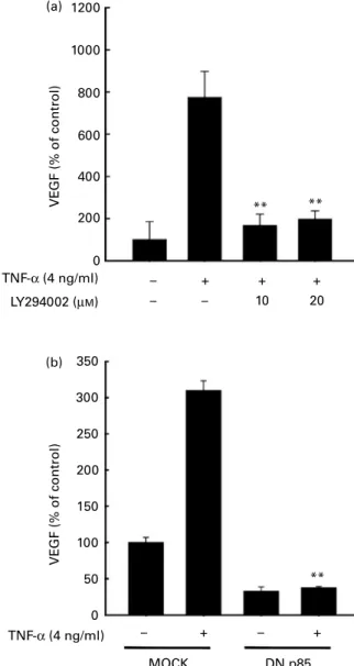
Phosphoinositide 3-kinase is required for TNF-a-induced vascular endothelial growth factor expression
We next examined the involvement of the PI3K signalling pathway in the TNF-a-induced up-regulation of VEGF in JB6 Pþcells. LY294002, a PI3K inhibitor, effectively inhib-ited TNF-a-induced VEGF up-regulation in JB6 Pþ cells (Fig. 5(a)). To confirm the role of PI3K in TNF-a-induced up-regulation of VEGF in JB6 Pþ cells, we used dominant negative p85 (DN-p85) which is the regulatory domain of
PI3K(17). Compared with mock vector transfected cells, TNF-a-induced up-regulation of VEGF markedly decreased in the DN-p85-expressing cells (Fig. 5(b)).
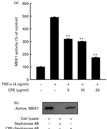
Cocoa polyphenol extract inhibits TNF-a-induced mitogen-activated protein kinase kinase-1 activity and directly binds mitogen-activated protein kinase kinase-1
We previously showed that CPE suppressed 12-O-tetradeca-noylphorbol-13-acetate (TPA)-induced cell transformation and MMP-2 expression though direct inhibition of MEK activity(18,19). Similarly here, CPE inhibited TNF-a-induced phosphorylation of ERK but not MEK. CPE dose-dependently suppressed TNF-a-induced MEK1 activity in JB6 Pþ cells (Fig. 6(a)). To further confirm whether CPE directly interacts with MEK1, we used a CPE pull-down assay. MEK1 in the cell lysate of TNF-a-treated JB6 Pþ cells bound to the CPE – Sepharose 4B beads (Fig. 6(b); lane 3), but not to the TNF-α (4 ng/ml) CPE (µg/ml) TNF-α (4 ng/ml) CPE (µg/ml) – – – + 10 + 20 + – – – + 10 + 20 + TNF-α (4 ng/ml) CPE (µg/ml) TNF-α (4 ng/ml) CPE (µg/ml) – – – + 10 + 20 + – – – + 10 + 20 + p-Akt p-p70S6K p70S6K p90RSK p90RSK p-p38 p38 Akt p-MEK p-ERK p-MKK4 p-JNK JNK MKK4 ERK MEK 0·1 1·0 0·3 0·2 0·1 1·0 1·5 1·1 0·4 1·0 0·1 0·2 0·0 1·0 0·3 0·1 0·0 1·0 0·0 0·0 0·3 1·0 0·1 0·0 0·1 1·0 0·4 0·2 0·0 1·0 1·8 2·6 (a) (b) (c) (d)
Fig. 3. Effects of cocoa polyphenol extract (CPE) on the TNF-a-induced phosphoinositide 3-kinase (PI3K) – protein kinase B (Akt) and mitogen-activated protein kinase (MAPK) pathway. (a) Inhibition of TNF-a-induced phosphorylation (p-) of Akt (Akt) and p70 kDa ribosomal protein S6 kinase (p70S6K) by CPE. Promotion-sensitive (Pþ) JB6 mouse epidermal cells were pretreated with CPE (10 or 20 mg/ml) for 1 h, then stimulated with TNF-a at 4 ng/ml and harvested 2 h later. The levels of phosphorylated and total Akt and p70S6Kproteins were determined by Western blot analysis as described in Materials and methods. (b) Inhibition of TNF-a-induced phosphorylation of extracellular signal-regulated kinase (ERK) and p90 kDa ribosomal S6 kinase (p90RSK) by CPE. Phosphorylation of mitogen-activated protein kinase kinase (MEK) was not inhibited by CPE. (c) Inhibition of TNF-a-induced phosphorylation of mitogen-mitogen-activated protein kinase kinase 4 (MKK4) and c-Jun N-terminal kinase (JNK) by CPE. (d) CPE did not inhibit the TNF-a-induced phosphorylation of p38. JB6 Pþ
cells were pretreated with CPE (10 or 20 mg/ml) for 1 h, then stimulated with TNF-a at 4 ng/ml and harvested 15 min later. The levels of phosphorylated and total MEK, ERK and p90RSKproteins were determined by Western blot analysis as described in Materials and methods. Quantification of phosphoproteins was normalised to total proteins using Image J software (National Institutes of Health, Bethesda, MD, USA).
British
Journal
of
Sepharose 4B beads alone (Fig. 6(b); lane 2), indicating that CPE could directly bind to MEK1 and inhibit MEK1 activity.
Discussion
TNF-a is a key regulator of inflammation and plays vital roles in inflammatory diseases such as cancer, rheumatoid arthritis, Crohn’s disease, diabetes and psoriasis. TNF-a induces the anchorage-independent growth of mouse epidermal JB6 Pþ cells(20). Knockout mice (TNF-a 2 /2 ) are more resistant to TPA-induced skin tumour promotion than wild-type mice(21,22). Neutralising TNF-a with an antibody also inhibits the development of skin tumours(23). These results suggest that targeting TNF-a signalling can be an important strategy for delaying the promotion of skin tumours.
Cocoa contains large amount of flavonoids. It comprises mainly catechins, flavonol glycosides and procyanidins. Our previous study using the identical CPE shows that the CPE had 413 mg epicatechin equivalent flavonoids per g. This demonstrates that 40 % of CPE is comprised of flavonoids(13). We suggested that flavonoids can act as PI3K and MEK inhibitors(15,24,25). Therefore, inhibitory effects of CPE on PI3K and MEK activity could be due to flavonoids contained in CPE.
When individuals consume dark chocolate, the substances epicatechin, catechin, procyanidin B2 or procyanidin B2 gallate are detected in their plasma(26). In a recent clinical study, 45 g dark chocolate containing up to 70 % cocoa was fed to human volunteers. Plasma epicatechin concentration of the volunteers was raised up to 155 ng/ml(27). In this study only epicatechin concentration was measured. However, chocolate is comprised of many other flavonoids(28). The sum of flavonoid concentration would be much higher than that of epicatechin only. The CPE used in the present study was effective, starting from 5 mg/ml. Therefore, the concentration
(5 to 20 mg/ml) of CPE which we used in the present study seems to be a reliable dose.
In the present study, we found that CPE (5 to 20 mg/ml) dose-dependently inhibited TNF-a-induced VEGF expre-ssion in JB6 Pþ cells. Cocoa polyphenols exhibit free PI3K, active (100 ng) PI3K, active Sepharose 4B CPE–Sepharose 4B PI3K CPE (µM) – – – + – – – – + + + + + 10 + 20 + (b) (a)
Fig. 4. Direct inhibition of phosphoinositide 3-kinase (PI3K) by cocoa polyphe-nol extract (CPE). (a) CPE strongly suppresses PI3K activity. A quantity of 100 ng of an active PI3K protein was preincubated with CPE for 10 min at 308C, and then further incubated with phosphatidylinositol substrate and [g-32P]ATP for an additional 10 min at 308C. The resulting32P-labelled phos-phatidylinositol-3-phosphate (PIP3) was measured as described in Materials and methods. Quantification of PIP3 was performed using Image J software (National Institutes of Health, Bethesda, MD, USA). (b) CPE specifically binds to PI3K. PI3K – CPE binding was confirmed by immunoblotting using an anti-body against PI3K. Lane 1, p110 protein standard served as an input control; lane 2, as a negative control, Sepharose 4B was used to pull down p110; lane 3, p110 was pulled down using CPE – Sepharose 4B affinity beads.
1200 1000 800 600 400 200 0 VE GF (% of control) VE GF (% of control) TNF-α (4 ng/ml) TNF-α (4 ng/ml) LY294002 (µM) 350 300 250 200 150 100 50 0 – + – + MOCK DN p85 ** – – – + 10 20 + + ** ** (a) (b)
Fig. 5. Activation of phosphoinositide 3-kinase (PI3K) is required for TNF-a-induced vascular endothelial growth factor (VEGF) expression. Promotion-sensitive (Pþ) JB6 mouse epidermal cells were seeded into ninety-six-well plates, cultured to 70 – 80 % confluence with 5 % fetal bovine serum – Eagle’s minimum essential medium (FBS – MEM), and then starved by replacing the medium with 0·1 % FBS – MEM for 24 h. The cells were then treated with the PI3K inhibitor, LY294002 (a), for 1 h before treatment with TNF-a at 4 ng/ml for 18 h. The conditioned medium was collected and analysed for VEGF expression using ELISA as described in the Materials and methods. Values are means, with standard deviations represented by vertical bars. ** Mean value was significantly different from that of the TNF-a-only-treated cells (P, 0·01). (b) Cell lines stably expressing empty vector (MOCK) or a domi-nant-negative mutant of p85 (DN-p85) were seeded into ninety-six-well plates, cultured to 70 – 80 % confluence with 5 % FBS – MEM, and then starved by replacing the medium with 0·1 % FBS – MEM for 24 h. The cells were then treated with TNF-a at 4 ng/ml for 18 h. The conditioned medium was collected and analysed for VEGF expression using ELISA as described in the Materials and methods. Values are means, with standard deviations represented by vertical bars. ** Mean value was significantly different from that of the TNF-treated MOCK cells (P, 0·01).
British
Journal
of
radical-scavenging activity, as determined by both 2,20 -azino-bis(3-ethylbenzothiazoline-6-sulfonic acid) (ABTS) and 2,2-diphenyl-1-picrylhydrazyl (DPPH) radical-scavenging assays and anti-inflammatory effects in mouse skin(12). Cocoa polyphenols also suppressed the growth of Caco-2 human colon cancer cells through a blockade of the cell cycle at the G2/M phase(29). Further, cocoa polyphenols suppressed lung and pancreatic tumorigenesis in a male rat multi-organ carcinogenesis model(29,30), and TPA-induced tumour promotion in a mouse two-stage skin tumorigenesis model initiated with 7,12-dimethylbenz[a ]anthrathene(31). Thus, the present results support the chemopreventive poten-tial of cocoa polyphenols.
NF-kB and AP-1 are important transcription factors in tumour promotion and are activated by many cytokines and chemicals, including TNF-a(32), leading to VEGF transcrip-tion(33). VEGF mRNA expression in MDA-MB-231 breast cancer cells is associated with NF-kB activation(34). AP-1 regulated by ERK or Akt also mediates VEGF expression(35,36). In the present study the CPE suppressed TNF-a-induced NF-kB and AP-1 transactivation to block VEGF transcription.
The antioxidant effects of CPE may partially contribute to these effects, but it may also target other signalling
molecules(37). Some small molecules in food such as flavonoids also inhibit signalling molecules directly(16,38 – 40). Targeting PI3K or mitogen-activated protein kinase (ERK, JNK and p38) pathways is a good strategy for cancer preven-tion. PI3K phosphorylates the 3 position of the hydroxyl group of the inositol ring of phosphatidylinositol and controls cellular processes such as cell proliferation and apoptosis. It is activated in many tumours and small molecule inhibitors are being developed(41). In the present study the CPE strongly suppressed PI3K activity and bound PI3K directly. These results explain the inhibition of pAkt and p70S6K, which are downstream kinases of PI3K.
Our previous studies demonstrated that CPE inhibited TPA-induced neoplasmic transformation in mouse epidermal cells and suppressed thrombin-induced MMP-2 activation through direct inhibition of MEK1 activity(18,19). A pharmacological inhibitor of MEK suppressed TNF-a-induced VEGF expression in JB6 Pþ cells(42). CPE attenuated TNF-a-induced MEK1 activity, and subsequently phosphorylation of ERK, without affecting MEK phosphorylation. Therefore, inhibition of MEK1 contributes to inhibition of TNF-a-induced VEGF expression by CPE.
Our previous study showed that the MKK4 – JNK pathway is required for TNF-a-induced VEGF expression using specific inhibitors(42). In the present study the CPE inhibited TNF-a-induced phosphorylation of MKK4 and JNK. However, MEK1 and PI3K, which are revealed as targets of the CPE in the present study, cannot explain the phenomenon of inhibition on the MKK4 – JNK pathway by the CPE. Increase of p38 phosphoryl-ation is also hard to explain with MEK1 and PI3K inhibition. Further study must reveal new target proteins of CPE which control the TNF-a-induced MKK4 – JNK and p38 pathway.
Multi-target kinase inhibitors have significant side effects. However, they are in the spotlight since the success of Gleevec targeting Bcr-Abl and KIT kinase due to its powerful effect and a variety of applications(43). Food materials are usually regarded as safe because of their long-term use. There-fore, multi-targeting may be better than specific targeting for food materials. In summary, a CPE suppressed TNF-a-induced VEGF expression via targeting PI3K and MEK1 to inhibit phosphorylation of ERK and Akt in mouse epidermal cells. Taken together, these results indicated that CPE might have chemopreventive potential against pro-inflammatory cytokine-mediated skin cancer and inflammation.
Acknowledgements
The present study was supported by grants from the Basic Research Program (no. R01-2009-000-11957-0), Priority Research Centers Program (2009-0093824) and the WCU Program (R31-10056), through the National Research Foundation of Korea, funded by the Ministry of Education, Science, and Technology, Republic of Korea. K. W. L., C. Y. L. and H. J. L. designed the study and prepared the paper. J. E. K., J. E. S., S. K. J. and N. J. K. contributed to the successful execution of the experimental work. We declare that we have no conflict of interest.
References
1. Balkwill F (2002) Tumor necrosis factor or tumor promoting factor? Cytokine Growth Factor Rev 13, 135 – 141.
MEK1 acti vity (% of control) 600 500 400 300 200 100 0 Active, MEK1 Cell lysate Sepharose 4B CPE–Sepharose 4B TNF-α (4 ng/ml) CPE (µg/ml) – – – + – – – – + + + + + 10 5 + 20 + + ** ** ** (a) (b)
Fig. 6. Cocoa polyphenol extract (CPE) suppresses TNF-a-induced mitogen-activated protein kinase kinase-1 (MEK1) activity and directly binds MEK1. (a) CPE inhibits TNF-a-induced MEK1 activity. Promotion-sensitive (Pþ
) JB6 mouse epidermal cells were pretreated with CPE (5, 10 or 20 mg/ml) for 1 h and then stimulated with TNF-a at 4 ng/ml for 30 min. Cells were harvested, and immunoprecipitation and MEK1 activity assays were performed as described in Materials and methods. Values are means, with standard deviations rep-resented by vertical bars. ** Mean value was significantly different from that of the TNF-a-only-treated cells (P,0·01). (b) CPE specifically binds with MEK1. The MEK1– CPE binding was confirmed by immunoblotting using an antibody against MEK1. Lane 1, MEK4 protein standard served as an input control; lane 2, as a negative control, Sepharose 4B was used to pull down MEK1; lane 3, MEK1 was pulled down using CPE–Sepharose 4B affinity beads.
British
Journal
of
2. Locksley RM, Killeen N & Lenardo MJ (2001) The TNF and TNF receptor superfamilies: integrating mammalian biology. Cell 104, 487 – 501.
3. MacEwan DJ (2002) TNF ligands and receptors – a matter of life and death. Br J Pharmacol 135, 855 – 875.
4. Ferrara N, Gerber HP & LeCouter J (2003) The biology of VEGF and its receptors. Nat Med 9, 669 – 676.
5. Larcher F, Murillas R, Bolontrade M, et al. (1998) VEGF/VPF overexpression in skin of transgenic mice induces angiogenesis, vascular hyperpermeability and accelerated tumor development. Oncogene 17, 303 – 311.
6. Borchers AT, Keen CL, Hannum SM, et al. (2000) Cocoa and chocolate: composition, bioavailability, and health implications. J Med Food 3, 77 – 105.
7. Corti R, Flammer AJ, Hollenberg NK, et al. (2009) Cocoa and cardiovascular health. Circulation 119, 1433 – 1441.
8. Serafini M, Bugianesi R, Maiani G, et al. (2003) Plasma anti-oxidants from chocolate. Nature 424, 1013.
9. McShea A, Ramiro-Puig E, Munro SB, et al. (2008) Clinical benefit and preservation of flavonols in dark chocolate manufac-turing. Nutr Rev 66, 630 – 641.
10. Lee KW, Kim YJ, Lee HJ, et al. (2003) Cocoa has more phenolic phytochemicals and a higher antioxidant capacity than teas and red wine. J Agric Food Chem 51, 7292 – 7295. 11. Lee KW, Hwang ES, Joo KN, et al. (2005) Extraction and
chromatographic separation of anticarcinogenic fractions from cacao bean husk. BioFactors 23, 141 – 150.
12. Lee KW, Kundu JK, Sue OK, et al. (2006) Cocoa polyphenols inhi-bit phorbol ester-induced superoxide anion formation in cultured HL-60 cells and expression of cyclooxygenase-2 and activation of NF-kB and MAPKs in mouse skin in vivo. J Nutr 136, 1150–1155. 13. Lee KW, Kang NJ, Oak MH, et al. (2008) Cocoa procyanidins inhibit expression and activation of MMP-2 in vascular smooth muscle cells by direct inhibition of MEK and MT1-MMP activi-ties. Cardiovasc Res 79, 34 – 41.
14. Kang NJ, Lee KW, Lee DE, et al. (2008) Cocoa procyanidins suppress transformation by inhibiting mitogen-activated protein kinase kinase. J Biol Chem 283, 20664 – 20673.
15. Kwon JY, Lee KW, Kim JE, et al. (2009) Delphinidin sup-presses ultraviolet B-induced cyclooxygenases-2 expression through inhibition of MAPKK4 and PI-3 kinase. Carcinogenesis 30, 1932 – 1940.
16. Lee KW, Kang NJ, Rogozin EA, et al. (2007) Myricetin is a novel natural inhibitor of neoplastic cell transformation and MEK1. Carcinogenesis 28, 1918 – 1927.
17. Nomura M, Kaji A, He Z, et al. (2001) Inhibitory mechanisms of tea polyphenols on the ultraviolet B-activated phosphatidylinositol 3-kinase-dependent pathway. J Biol Chem 276, 46624–46631. 18. Lee KW, Kang NJ, Oak M-H, et al. (2008) Cocoa procyanidins
inhibit expression and activation of MMP-2 in vascular smooth muscle cells by direct inhibition of MEK and MT1-MMP activities. Cardiovasc Res 79, 34 – 41.
19. Kang NJ, Lee KW, Lee DE, et al. (2008) Cocoa procyanidins suppress transformation by inhibiting mitogen-activated protein kinase kinase. J Biol Chem 283, 20664 – 20673.
20. Yan Y, Li J, Ouyang W, et al. (2006) NFAT3 is specifically required for TNF-a-induced cyclooxygenase-2 (COX-2) expression and transformation of Cl41 cells. J Cell Sci 119, 2985–2994. 21. Arnott CH, Scott KA, Moore RJ, et al. (2004) Expression of
both TNF-a receptor subtypes is essential for optimal skin tumour development. Oncogene 23, 1902 – 1910.
22. Moore RJ, Owens DM, Stamp G, et al. (1999) Mice deficient in tumor necrosis factor-a are resistant to skin carcinogenesis. Nat Med 5, 828 – 831.
23. Scott KA, Moore RJ, Arnott CH, et al. (2003) An anti-tumor necrosis factor-a antibody inhibits the development of experi-mental skin tumors. Mol Cancer Ther 2, 445 – 451.
24. Kim JE, Kwon JY, Seo SK, et al. (2010) Cyanidin suppresses ultraviolet B-induced COX-2 expression in epidermal cells by targeting MKK4, MEK1, and Raf-1. Biochem Pharmacol 79, 1473 – 1482.
25. Choi KH, Kim J-E, Song NR, et al. (2010) Phosphoinositide-3-kinase is a novel target of piceatannol for inhibiting PDGF-BB-induced proliferation and migration in human aortic smooth muscle cells. Cardiovasc Res 85, 836 – 844.
26. Taubert D, Roesen R, Lehmann C, et al. (2007) Effects of low habitual cocoa intake on blood pressure and bioactive nitric oxide: a randomized controlled trial. JAMA 298, 49 – 60. 27. Spadafranca A, Martinez Conesa C, Sirini S, et al. (2010)
Effect of dark chocolate on plasma epicatechin levels, DNA resistance to oxidative stress and total antioxidant activity in healthy subjects. Br J Nutr 103, 1008 – 1014.
28. Schroeter H, Heiss C, Balzer J, et al. (2006) (2 )-Epicatechin mediates beneficial effects of flavanol-rich cocoa on vascular function in humans. Proc Natl Acad Sci U S A 103, 1024 – 1029. 29. Yamagishi M, Natsume M, Osakabe N, et al. (2002) Effects of
cacao liquor proanthocyanidins on PhIP-induced mutagenesis in vitro, and in vivo mammary and pancreatic tumorigenesis in female Sprague – Dawley rats. Cancer Lett 185, 123 – 130. 30. Yamagishi M, Natsume M, Osakabe N, et al. (2003)
Chemo-prevention of lung carcinogenesis by cacao liquor proantho-cyanidins in a male rat multi-organ carcinogenesis model. Cancer Lett 191, 49 – 57.
31. Osakabe N, Yamagishi M, Natsume M, et al. (2000) Anti-oxidative polyphenolic substances in cacao liquor. ACS Symp Ser 754, 88 – 101.
32. Szlosarek P, Charles KA & Balkwill FR (2006) Tumour necro-sis factor-a as a tumour promoter. Eur J Cancer 42, 745 – 750. 33. Jos´ko J & Mazurek M (2004) Transcription factors having impact on vascular endothelial growth factor (VEGF) gene expression in angiogenesis. Med Sci Monit 10, RA89 – RA98. 34. Shibata A, Nagaya T, Imai T, et al. (2002) Inhibition of
NF-kB activity decreases the VEGF mRNA expression in MDA-MB-231 breast cancer cells. Breast Cancer Res Treat 73, 237 – 243.
35. Lee CC, Chen SC, Tsai SC, et al. (2006) Hyperbaric oxygen induces VEGF expression through ERK, JNK and c-Jun/AP-1 activation in human umbilical vein endothelial cells. J Biomed Sci 13, 143 – 156.
36. Ouyang W, Li J, Shi X, et al. (2005) Essential role of PI-3K, ERKs and calcium signal pathways in nickel-induced VEGF expression. Mol Cell Biochem 279, 35 – 43.
37. Sebolt-Leopold JS & English JM (2006) Mechanisms of drug inhibition of signalling molecules. Nature 441, 457 – 462. 38. Walker EH, Pacold ME, Perisic O, et al. (2000) Structural
deter-minants of phosphoinositide 3-kinase inhibition by wortmannin, LY294002, quercetin, myricetin, and staurosporine. Mol Cell 6, 909 – 919.
39. Cortes JR, Perez GM, Rivas MD, et al. (2007) Kaempferol inhibits IL-4-induced STAT6 activation by specifically targeting JAK3. J Immunol 179, 3881 – 3887.
40. Kang NJ, Lee KW, Rogozin EA, et al. (2007) Equol, a metabolite of the soybean isoflavone daidzein, inhibits neoplastic cell trans-formation by targeting the MEK/ERK/p90RSK/activator protein-1 pathway. J Biol Chem 282, 32856– 32866.
41. Raynaud FI, Eccles S, Clarke PA, et al. (2007) Pharmacologic characterization of a potent inhibitor of class I phosphatidylino-sitide 3-kinases. Cancer Res 67, 5840 – 5850.
42. Kim JE, Kwon JY, Lee DE, et al. (2009) MKK4 is a novel target for the inhibition of tumor necrosis factor-a-induced vascular endothelial growth factor expression by myricetin. Biochem Pharmacol 77, 412 – 421.
43. Vogt PK & Kang S (2006) Kinase inhibitors: vice becomes virtue. Cancer Cell 9, 327 – 328.