ORIGINAL ARTICLE
Immunohistochemical analysis of estrogen
receptors in the urethra of sexually intact, ovariectomized,
and estrogen-substituted ovariectomized sheep
Heinz R. Augsburger&Constanze Führer
Received: 26 June 2013 / Accepted: 4 November 2013 / Published online: 7 December 2013 # The International Urogynecological Association 2013
Abstract
Introduction and hypothesis Urinary incontinence is preva-lent in postmenopausal women and spayed dogs and is asso-ciated with decreased estrogen plasma concentrations. The objective of the study was to investigate the expression of estrogen receptors (ER) in the urethra of sexually intact, ovariectomized, and estrogen-substituted ovariectomized ewes.
Methods Paraffin cross-sections from each urethral quarter were immunohistochemically analyzed. The reactivity of ER was semiquantitatively assessed employing an immunoreac-tive score (IRS).
Results In contrast to ERβ, ERα was identified in all urethral compartments; the highest IRS was detected in the epithelium of the distal urethra. The immunoreactivity and distribution of ERα did not differ among groups. Highly significant differ-ences in ERα concentrations were observed between consec-utive urethral quarters in each group.
Conclusions Neither ovariectomy nor ovariectomy and estro-gen substitution seem to have a significant effect on overall urethral ERα concentration. The results demonstrate that the precise location of the investigated urethral part is crucial to the reliable evaluation or possible comparison of ERα concentrations.
Keywords Estrogen receptor . Immunohistochemistry . Ovariectomy . Sheep . Urethra . Urinary incontinence
Abbreviations
ANOVA Factorial analysis of variance CEE Conjugated equine estrogen ER Estrogen receptor
ERT Estrogen replacement therapy IRS Immunoreactive score NOC Non-operated control
OVE Estrogen-substituted ovariectomized OVX Ovariectomized
UI Urinary incontinence
U1/4 Urethral quarter 1 (connecting with urinary bladder)
U4/4 Urethral quarter 4 (connecting with vaginal vestibule)
WHI Women’s Health Initiative
Introduction
Urinary incontinence is a common lower urinary tract disorder in both women and female dogs. In women, it is prevalent in the postmenopausal period, whereas in dogs it is the most common undesirable effect of spaying [1–3]. The pathophys-iological mechanism of this lower urinary tract dysfunction is not completely understood. Menopause in women and spay-ing in dogs are accompanied by decreased ovarian steroid hormone plasma concentrations and associated with reduced urethral closure pressures and impaired closure function of the urethra [4–7]. Estrogen deficiency is believed to play an important role in the development of urinary incontinence in both species, as estrogen is essential for physiological main-tenance and the integrity of the female urogenital tract. The specific effects of estrogens are classically mediated via their interaction with corresponding nuclear receptors [8,9]. Apart from other organs and tissues, estrogen receptors have been
H. R. Augsburger
:
C. FührerInstitute of Veterinary Anatomy, Vetsuisse Faculty, University of Zurich, Zurich, Switzerland
H. R. Augsburger (*)
Institute of Veterinary Anatomy, University of Zürich, Winterthurerstrasse 260, 8057 Zürich, Switzerland e-mail: augsbhr@vetanat.uzh.ch
shown to be present in the female urethra of humans [9–11], dogs [12], and laboratory animals [13–15]. The urethra is responsible for urinary continence depending on several ure-thral tissue components, such as smooth and striated muscu-lature, the vascular plexus, connective tissue, as well as periurethral structures [16,17].
A suitable experimental animal model for urinary inconti-nence research has been lacking to date [18]. An appropriate animal model for the investigation of urinary incontinence could reduce the pitfalls associated with studying the disease in humans. The sheep seems to be a promising animal for this purpose. Ovariectomized sheep, for example, have been wide-ly used as a model for postmenopausal osteoporosis research [19]. Ewes are seasonal polyestrous and their estrous cycle extends over a period of about 17 days. The rather unspectac-ular estrus lasts around 24 h and gestation averages 147 days.
This study was undertaken to identify estrogen receptors (ER) in the ovine urethra and to provide insights into the effects of ovariectomy and estrogen substitution on their ex-pression in the different urethral tissue compartments, using immunohistochemistry and semiquantitative analysis. Moreover, the potential of the sheep as an animal model for certain aspects of urinary incontinence was evaluated.
Materials and methods Animals
Eighteen aged Merino ewes (5 years old) were obtained from a single source and selected for uniformity of size, conforma-tion, body condiconforma-tion, and absence of pregnancy. Their average body weight was 58.7 kg. Twelve randomly selected animals were ovariectomized under general anesthesia via a small midline laparotomy incision, while the remaining 6 served as non-operated controls. Furthermore, half of the operated sheep received estradiol implants as described by Adams et al. [20]. The implants were placed subcutaneously in the hairless region of the chest wall below the axilla through a small incision. They released a total of 22.5 μg estradiol-17β (Sigma, St. Louis, MO, USA) per 24 h. The treatment groups (n =6) were therefore: non-operated control (NOC), ovariec-tomized (OVX), and estrogen-substituted ovariecovariec-tomized (OVE). All animals were maintained under identical environ-mental conditions for 6 months, at which time they were slaughtered.
Venous blood samples were collected at 1, 3, and 6 months post-operatively. Plasma estradiol-17ß concentrations were measured using the method described by Webb et al. [21], involving an affinity chromatography extraction procedure followed by double-antibody radioimmunoassay. All animal procedures were approved by the Murdoch University Animal Ethics Committee (R1031/04).
Tissue processing
After slaughter of the sheep, the bladder and urethra including the caudal part of the vagina and the vaginal vestibule were removed from the pelvic cavity. Subsequently, each urethra was excised at the level of the internal and external urethral orifices and mounted on a wooden spatula. The urethrae were then immersion-fixed with Histochoice® tissue fixative (Amresco Inc., Solon, OH, USA) for 48 h and split into four parts of equal length (urethral quarters 1–4), whereby quarter 1 (U1/4) was adjacent to the bladder (proximal) and U4/4 connected with the vaginal vestibule (distal). The fixed specimens were routinely embedded in paraffin and cut into serial cross-sections of 5μm in thickness. For immunohistochemical analysis, three sections from the middle of each urethral quarter were mounted on adhesive SuperFrost Plus® glass-slides (Menzel-Gläser, Braunschweig, Germany) and heat dried at 60 °C for 30 min.
Immunohistochemistry
Immunostaining of ER was performed using the streptavidin– biotin complex peroxidase technique (Dako, Glostrup, Denmark). The sections were deparaffinized and rehydrated in graded ethanol. For retrieval of receptor epitopes, sections were subjected to citrate-based microwave processing (3× 5 min, 103 °C, 600 W) in target retrieval solution pH 9 (Dako). Endogenous peroxidase activity was blocked with 3 % hydrogen peroxide in distilled water for 10 min. Following washing in Tris-buffered saline (TBS, 5 min), the sections were treated with an avidin/biotin blocking kit (Vector Laboratories, Burlingame, CA, USA) to inhibit non-specific staining due to endogenous avidin and biotin. After a further wash in TBS, they were covered with 1 % normal goat serum (Kirkegaard & Perry, Gaithersburg, MD, USA) for 20 min and serum-free protein block (Dako) for 10 min to eliminate nonspecific protein binding. Following removal of excess serum, the primary antibodies, monoclonal mouse human ERα (1ID5, Dako; 1:50) and monoclonal mouse anti-human ERβ1 (PPG5/10, Dako; 1:50), were applied and allowed to incubate for 20 h at 4 °C in a humidity chamber. The sections were then rinsed with TBS and incubated with a biotinylated rabbit anti-mouse secondary antibody (Dako) for 30 min at room temperature before being washed twice in TBS, followed by treatment with a peroxidase conjugated streptavidin label (Dako) for 30 min and a further wash in TBS. The streptavidin–biotin peroxidase reaction was devel-oped with diaminobenzidine (DAB, Dako) as chromogen. The sections were then rinsed with distilled water, counter-stained with Mayer’s haemalaun, dehydrated and mounted in Pertex® (Medite, Nunningen, Switzerland).
Negative controls were obtained by replacing the primary antibody with non-immune mouse-IgG (Dako). Positive con-trols were performed employing uterine tissue.
The immunohistochemical expression and distribution of investigated ER were evaluated by the authors individually at a magnification of ×400 by use of an image analysis system (AnalySIS, Soft Imaging System, Münster, Germany), com-prising a light microscope fitted with a digital camera (Colorview 12, Soft Imaging System). The staining intensity of 500 nuclei was assessed in randomly selected microscopic fields of the urethral compartments epithelium, propria, and smooth musculature using an immunoreactive score (IRS) described by Schäubli et al. [22]. Immunoreactivity was scored as negative (0), weak (1), intermediate (2), or strong (3), correlating with the absence of brown, light brown, and
dark brown staining respectively. Results were expressed as a percentage of positively stained nuclei of the total counted per urethral compartment.
Statistical analysis
Mean ± standard deviation values were computed for all immunoreactivity and hormone data. Data were assessed for normal distribution and subjected to a one-way factorial anal-ysis of variance (ANOVA) by use of StatView® software (5.1; SAS Institute, Cary, NC, USA) to evaluate differences in estradiol and estrogen receptor concentrations among groups
Fig. 1 Representative immunohistochemical reactivity of estrogen receptors-α (brown staining) in the nuclei (arrows) of the epithelium, propria, and smooth musculature in urethral quarters 2–4 (U2/4–U4/4) of the three groups NOC, OVE, and OVX. Bars, 100μm. Smooth musculature in a U2/4 and b U3/ 4 of NOC: no staining of striated musculature (asterisks). Epithelium in c U4/4 of OVE and d OVX: overall, no difference between groups. Epithelium in e U2/4 and f U3/4 of OVE: significant difference between consecutive urethral quarters. Propria in g U2/4 and h U3/4 of OVX: significant difference between consecutive urethral quarters
as well as differences in immunoreactivity between urethral quarters within groups. Significant differences were assessed using the Bonferroni–Dunn procedure as post hoc test. In order to demonstrate a possible relationship between estradiol concentrations at the time of slaughter and the immunohisto-chemical expression of urethral estrogen receptors, the corre-lation coefficients between variables and the corresponding regression analysis with 95 % confidence intervals were calculated.
Results
Distinct immunohistochemical expression of ERα was con-sistently detected in the epithelium, propria, and smooth mus-culature compartments throughout the urethra of all ewes. In contrast, ERβ could not be identified. Immunolocalization of ERα was confined to the nuclei revealing differential staining intensities (Fig. 1). The striated muscle fibers showed no evidence of positive immunolabelling (Fig.1b).
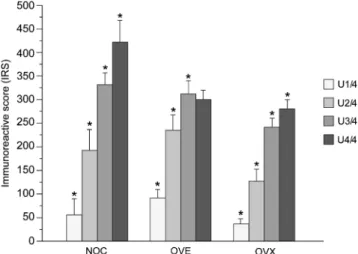
Overall, there were no significant differences in the IRS of the whole urethra among groups (Fig.1c, d). However, dis-tinct differences of statistical relevance were observed in the scores between adjacent urethral quarters within all groups. Apart from the smooth musculature, significant increases (p < 0.01/0.001) in ERα concentrations in the epithelium and proprial connective tissue were ascertained between consecu-tive urethral quarters from the cranial to the caudal end of the urethra in each group (Figs. 1e–h, 2, 3 and Table 1). Furthermore, differences in the IRS of the three urethral compartments analyzed were evident among groups. The IRS of the epithelium was significantly increased (p <0.002)
in the OVX relative to the NOC and OVE sheep. The opposite was true for the smooth musculature: the ERα concentration in the smooth musculature proved to be significantly lower (p <0.001) in the OVX than in the NOC and OVE animals.
Although not always statistically meaningful, mean plasma estradiol concentrations were lower in the OVX group than in groups NOC and OVE (Table2). No correlation was found between estradiol plasma concentrations at the time of slaugh-ter and the corresponding IRS.
Discussion
In contrast to numerous immunohistochemical studies that only rely on small biopsies, this investigation was conducted on full urethral cross-sections throughout the entire organ giving a comprehensive insight into the immunostaining and distribution pattern of ER in the different urethral tissue compartments of OVX, OVE, and NOC ewes. The results of the present study revealed the immunohistochemical expression of ERα in the nuclei of the urethral epithelium, the proprial connective tissue, and the smooth musculature. These findings show the female ovine urethra to be a target organ for the action of estrogen. Overall, no significant differences in the IRS pertaining to the whole urethra were observed among groups. This finding concurs with studies carried out by Blakeman et al. [10], who assessed the incidence and distribution of ER throughout the human female lower urinary tract and compared receptor expres-sion in women of various estrogen statuses. They consis-tently detected ER in the squamous epithelium of the lower urinary tract of all women and found no significant variation in histological score irrespective of estrogen status. This observation was interpreted as the female urinary tract being potentially receptive to the actions of estrogen at all times.
Fig. 3 Mean ERα IRS values (± SD) of the propria compartment in urethral quarters 1–4 of the three groups NOC, OVE, and OVX. Asterisk significant differences between consecutive urethral quarters (p <0.01) in groups NOC and OVE. Asterisk1, 2 Significant differences between
urethral quarters (p <0.01) in group OVX
Fig. 2 Mean ERα IRS-values (± SD) of the epithelium compartment in urethral quarters 1–4 of the three groups non-operated controls (NOC), estrogen-substituted ovariectomized (OVE ), and ovariectomized (OVX). Asterisk significant differences between consecutive urethral quarters (p <0.01)
However, in our study significant differences in the immu-nohistochemical expression of ERα in the epithelial and smooth muscle compartments could be established among groups. The observation of the significantly decreased urethral smooth muscle IRS in OVX compared with NOC and OVE groups is particularly noteworthy since the smooth muscula-ture consisting of essentially circularly oriented fibers is regarded as the most important contributory factor to urinary continence [16,23]. Estrogen has been shown to increase the sensitivity of the urethral smooth musculature toα-adrenergic stimulation [23]. Decreased plasma estrogen levels as a sequel to ovariectomy and the consequent diminished adrenergic sensitivity of the smooth musculature may result in atrophy and/or loss of muscle fibers and this, in turn, could affect the urethral closure mechanism and increase the risk of urinary incontinence development. The role of estrogen replacement therapy (ERT) in postmenopausal and postspaying urinary incontinence (UI) has long been controversial [24–27]. However, according to a more recent publication by investi-gators of the Womens Health Initiative (WHI) [28] ERT has
no beneficial effects with regard to UI. The results from this large, double-blind, placebo-controlled, randomized clinical trial, conducted in multiple centers with healthy postmeno-pausal women, indicate that postmenopostmeno-pausal hormone therapy does not prevent any type of UI. On the contrary, both conju-gated equine estrogen (CEE) alone and CEE plus medroxyprogesterone acetate increased the risk of new onset UI among continent women and worsened the characteristics of UI among symptomatic women. DuBeau [29], who evalu-ated this WHI study concludes that both the scientific rigor of the WHI trial and the issues it raises should prompt continuing investigation of the basic science of estrogen in normal and aging lower urogenital tract and pelvic floor function, and lead to further treatment trials (particularly with topical estrogens) that address the methodological issues of unblinding, UI char-acterization, patient targeting, and comprehensive outcomes assessment.
The reduced concentration of ERα in the smooth muscu-lature of the OVX could be the consequence of lower plasma estrogen levels in this group relative to the other two. The decreased smooth muscle IRS, however, could also be attrib-uted to a reduction in the urethral smooth muscle mass of OVX. In a previous study dealing with the stereological analysis of urethral tissue components in sexually intact and spayed female dogs, we demonstrated decreased smooth mus-cle volumes in the urethra of ovariohysterectomized dogs compared with the intact animals [16].
We failed in our efforts to detect ERβ in all urethral tissue compartments of the groups investigated. In contrast, Tincello et al. [9], who studied ER isoforms in the lower urinary tract of pre- and postmenopausal women, detected ERβ immuno-staining in the urethra and the transitional epithelium of the bladder. Furthermore, they found that hormonal status had no effect on the ERβ staining pattern. The deficiency of ERβ immunoreactivity in our study does not necessarily imply the
Table 2 Plasma estradiol concentrations (mean ± standard deviation) of non-operated control (NOC ), estrogen-substituted ovariectomized (OVE), and ovariectomized (OVX) sheep (n =6) at 1, 3, and 6 months post-operatively (mpo)
Group Plasma estradiol concentrations (pg/ml)
1 mpo 3 mpo 6 mpo
NOC 1.04±0.69a 1.36±0.79 1.36±0.98
OVE 2.01±0.94a, b 2.42±1.37c 1.14±0.38
OVX 0.59±0.11b 0.68±0.26c 0.64±0.28
a, b
Significant differences between group OVE and groups NOC/OVX (p <0.001)
c
Significant difference between groups OVE and OVX (p <0.05) Table 1 Immunoreactive scores
(mean ± standard deviation) of estrogen receptorα in the urethral compartments epithelium, propria, and smooth musculature of urethral quarters 1–4 (U1/4– U4/4) of NOC, OVE, OVX sheep (n =6)
a
Significant differences between consecutive urethral quarters in corresponding compartments within groups (p <0.01)
Group Urethral quarter Urethral compartment
Epithelium Propria Smooth musculature
NOC U1/4 53.3±83.9a 143.7±39.4a 317.6±44.3a U2/4 192.5±106.4a 174.7±41.8a 222.7±19.2a U3/4 329.0±59.6a 236.7±30.7a 183.1±28.3a U4/4 424.6±95.5a 307.7±30.9a 192.7±22.3 OVE U1/4 90.1±43.7a 157.1±31.1a 190.0±23.3a U2/4 233.9±85.7a 196.6±21.4a 221.5±34.8a U3/4 308.6±66.8a 248.4±35.7a 254.4±34.4a U4/4 299.4±52.3 297.0±30.0a 306.3±40.4a OVX U1/4 33.1±27.1a 136.9±27.4a 303.5±22.4a U2/2 123.3±74.0a 189.0±50.9a 284.4±24.8a U3/4 239.9±54.2a 269.1±34.3a 266.3±27.6a U4/4 280.1±46.3a 296.0±36.6a 245.3±39.4
absence of ERβ. The negative staining might either be due to the lack of affinity and/or specificity of the antibody used or may be attributable to loss of antigenicity upon tissue fixation and/or processing. Among other confounding factors, these are the main general limitations of staining fixed antigens by immunohistochemistry.
The sheep has considerable potential as an animal model for certain aspects of human urinary incontinence research because of similar urethral and bladder morphology as well as similar hormone cycles and profiles. In addition, the sheep is docile, easy to handle, relatively inexpensive, and better accepted in society as an experimental animal than companion animals such as dogs and cats [30].
In conclusion, this study revealed that the nuclear ERα concentration in ovine urethral tissues does not seem to cor-relate with estrogen status. Furthermore, the findings ascertain that the precise location of the urethral part investigated is crucial to the reliable evaluation or possible comparison of ERα concentrations. This considerable potential of the sheep as an animal model for human urinary incontinence research warrants further evaluation.
Acknowledgements The authors thank Prof. Martin Cake (College of Veterinary Medicine, Murdoc University, Perth, Australia) for the collec-tion and processing of blood samples and materials.
Conflicts of interest None.
References
1. Townsend MK, Curhan GC, Resnick NM, Grodstein F (2009) Postmenopausal hormone therapy and incident urinary incontinence in middle-aged women. Am J Obstet Gynecol 200:86.e1–86.e5 2. Nygaard IE, Heit M (2004) Stress urinary incontinence. Obstet
Gynecol 104:607–620
3. Holt PE (1990) Urinary incontinence in dogs and cats. Vet Rec 127: 347–350
4. Janssens LA, Peeters S (1997) Comparisons between stress inconti-nence in women and sphincter mechanism incompetence in the female dog. Vet Rec 141:620–625
5. Reichler IM, Pfeiffer E, Piché CA, Jöchle W, Roos M, Hubler M, Arnold S (2004) Changes in plasma gonadotropin concentrations and urethral closure pressure in the bitch during the 12 months following ovariectomy. Theriogenology 62:1391–1402
6. Rymer J, Morris EP (2000) Extracts from“clinical evidence”: men-opausal symptoms. BMJ 321:1516–1519
7. Hilton P, Stanton SL (1983) Urethral pressure measurement by microtransducer. The results in symptom-free women and in those with stress urinary incontinence. Br J Obstet Gynaecol 90:919–933 8. Walters MR, Nemere I (2004) Receptors for steroid hormones:
membrane-associated and nuclear forms. Cell Mol Life Sci 61: 2309–2321
9. Tincello DG, Taylor AH, Spurling SM, Bell SC (2009) Receptor isoforms that mediate estrogen and progestagen action in the female lower urinary tract. J Urol 181:1474–1482
10. Blakeman PJ, Hilton P, Bulmer JN (2000) Oestrogen and progester-one receptor expression in the female lower urinary tract, with reference to estrogen status. BJU Int 86:32–38
11. Iosif CS, Batra S, Ek A (1981) Estrogen receptors in the human female lower urinary tract. Am J Obstet Gynecol 141:817– 820
12. Schulze H, Barrack ER (1987) Immunocytochemical localization of estrogen receptors in normal male and female canine urinary tract and prostate. Endocrinology 121:1773–1783
13. Savolainen S, Santti R, Streng T, Gustafsson JA, Härkönen P, Mäkelä S (2005) Sex specific expression of progesterone receptor in mouse lower urinary tract. Mol Cell Endocrinol 230:17–21
14. Rosenzweig BA, Bolina PS, Birch L, Moran C, Marcovici I, Prins GS (1995) Location and concentration of estrogen, progesterone, and androgen receptors in the bladder and urethra of the rabbit. Neurourol Urodyn 14:87–96
15. Batra SC, Iosif CS (1983) Female urethra: a target for estrogen action. J Urol 129:418–420
16. Augsburger HR, Cruz-Orive LM (1995) Stereological analysis of the urethra in sexually intact and spayed female dogs. Acta Anat 154: 135–142
17. Augsburger HR, Oswald M (2007) Immunohistochemical analysis of collagen types I, III, IV andα-actin in the urethra of sexually intact and ovariectomized beagles. Int Urogynecol J 18:1071–1075 18. Seidlova-Wuttke D, Schultens A, Wuttke W (2004) Urodynamic
effects of estradiol (E2) in ovariectomized (ovx) rats. Endocrine 23: 25–32
19. Turner AS (2002) The sheep as a model for osteoporosis in humans. Vet J 163:232–239
20. Adams E, Turner AS, Wark JD (1995) The potential of sheep for the study of osteopenia: current status and comparison with other animal models. Bone 16:277S–284S
21. Webb R, Baxter G, MacBride D, Nordblum GD, Shaw MP (1985) The measurement of testosterone and oestradiol-17ß using iodinated tracers and incorporating an affinity chromatography extraction pro-cedure. J Steroid Biochem 23:1043–1051
22. Schäubli M, Ritter N, Hässig M, Zerbe H, Bleul U, Boos A (2008) Progesterone receptors, oestrogen receptorα and glucocorticoid re-ceptors in the bovine intercaruncular uterine wall around parturition. Anim Reprod Sci 103:215–227
23. Callahan SM, Creed KE (1985) The effects of oestrogens on sponta-neous activity and responses to phenylephrine of the mammalian urethra. J Physiol 358:35–46
24. Quinn SD, Domoney C (2009) The effects of hormones on urinary incontinence in postmenopausal women. Climacteric 12:106–113
25. Reichler IM, Hubler M, Jöchle W, Trigg TE, Piché CA, Arnold S (2003) The effect of GnRH analogs on urinary inconti-nence after ablation of the ovaries in dogs. Theriogenology 60: 1207–1216
26. Robinson D, Cardozo LD (2003) The role of estrogens in female urinary tract dysfunction. Urology 62(Suppl 4A):45–51
27. Hextal A (2000) Estrogen and lower urinary tract function. Maturitas 36:83–92
28. Hendrix SL, Cochrane BB, Nygaard IE, Handa VL, Barnabei VM, Iglesia C, Aragaki A, Naughton MJ, Wallace RB, McNeeley SG (2005) Effects of estrogen with and without progestin on urinary incontinence. JAMA 293:935–948
29. DuBeau CE (2005) Estrogen treatment for urinary incontinence. Never, now, or in future? JAMA 293:998–1001
30. Newman E, Turner AS, Wark JD (1995) The potential of sheep for the study of osteopenia: current status and comparison with other animal models. Bone 16:277S–284S