J Orofac Orthop 2011; 72:125-132 DOI 10.1007/s00056-011-0010-y
The influence of dental loupes on the quality of adhesive
removal in orthodontic debonding
Einfluss von Lupenbrillen auf die Qualität der
Adhäsiv-Restentfernung bei orthodontischem Debonding
Abstract
Objective. Bracket bonding has been a major advance in ortho-dontic treatment. However, the method of debonding can lead to di-verse problems such as enamel fractures, enamel loss and enamel scratching. In this clinical investigation we aimed to evaluate the in-fluence of wearing dental loupes on enamel damage during the debonding procedure.
Material and Methods. 22 consecutive patients were randomly as-signed in a split-mouth study to evaluate adhesive removal with and without the use of dental loupes (2.5×, LED headlight). Tooth replicas in epoxy resin were made from silicone impressions. Electron micro-scopic images (50× magnification) of 394 buccal enamel surfaces were evaluated according to an enamel damage index (EDI), line an-gle grooves (LAG) and composite residues (CR) on anterior teeth, premolars and molars.
Results. The EDI revealed highly significant advantages for debonding with dental loupes, with which the EDI was significantly higher for molars, while still less than without dental loupes. We de-tected no differences between the tooth groups without dental loupes. We found significantly fewer LAG on anterior teeth debonded with dental loupes. CR were fewer in the dental loupes group, and we noted no significant differences between the tooth groups.
Conclusions. Dental loupes affect the quality of the debonding procedure, resulting in less enamel damage and composite residue, as well as fewer LAG compared to the control group. We therefore strongly recommend the use of dental loupes in orthodontic debond-ing procedures.
Keywords
Debonding · Enamel · Surface · Roughness · Loupe · Magnification
Zusammenfassung
Hintergrund und Ziel. Adhäsivtechniken im Bracketbonding brachten einen großen Fortschritt in der kieferorthopädischen Thera-pie. Die Techniken zur Adhäsiv-Restentfernung nach Debonding kön-nen allerdings zu verschiedekön-nen Problemen führen, wie Schmelzfrak-turen, Schmelzverlust und Riefenbildung. Ziel dieser klinischen Untersuchung war die Evaluation des Einflusses der Benutzung von dentalen Lupenbrillen auf Schäden am Schmelz während der Adhä-siv-Restentfernung.
Material und Methodik. In einem randomisierten Split-Mouth-Versuch wurden 22 konsekutive Patienten bezüglich Adhäsiv-Rest-entfernung mit oder ohne dentaler Lupenbrille (2,5x, LED-Stirnlicht) untersucht. Nach erfolgter Adhäsiv-Restentfernung wurden über Sili-konabformungen Zahnreplika auf Epoxidharzbasis hergestellt. Elekt-ronenmikroskopische Aufnahmen von 394 Bukkalflächen wurden bei 50-facher Vergrößerung nach folgenden Kriterien beurteilt: Schmelz-schadenindex (EDI), „Line-Angle“-Rillen (LAG) und Kompositreste (CR). Dies wurde für die Frontzähne, Prämolaren und Molaren ge-trennt durchgeführt.
Ergebnisse. Der EDI zeigte einen hochsignifikanten Vorteil für das Debonding mit Lupenbrille. Mit Lupenbrille war der EDI an Molaren zwar signifikant höher als in den vorderen Zahnregionen, jedoch im-mer noch tiefer als ohne Lupenbrille. Ohne Lupenbrille konnten keine Unterschiede zwischen den verschiedenen Zahngruppen festgestellt werden. Bei Debonding mit Lupenbrille traten zudem signifikant we-niger „Line-Angle“-Rillen an Frontzähnen auf. Kompositreste fanden sich unter Verwendung einer Lupenbrille seltener, und es bestanden keine signifikanten Unterschiede zwischen den Zahngruppen. Schlussfolgerungen. Die Verwendung dentaler Lupenbrillen be-einflusst die Qualität der Adhäsiv-Restentfernung und hinterlässt we-niger Schmelzschäden, „Line-Angle“-Rillen und Kompositreste. Die Verwendung von dentalen Lupenbrillen kann für die Adhäsiv-Rest-entfernung klar empfohlen werden.
Schlüsselwörter
Debonding · Schmelz · Oberfläche · Rauheit · Lupe · Vergrößerung
Denis F. Baumann
1*, Lorenz Brauchli
1*, Hubertus van Waes
21 Clinic of Orthodontics and Pedodontics, Dental School, University of Basel, Switzerland
2 Pediatric Dentistry Program, University of Zurich, Switzerland
Received: November 15, 2010 ; accepted: December 29, 2010 * D.F. Baumann and L. Brauchli contributed equally to this research.
Introduction
Dental loupes are widely used in dentistry. Advantages in work er-gonomics [4, 8, 9, 10, 18, 20] and precision [8, 17] have been re-ported. Dental loupes are even mandatory in endodontic specialty training in the US [10]; students should master their use as early as possible within the curriculum [18]. Dental loupes also offer eye protection. The majority of ocular traumata suffered by ortho-dontists occur while debonding and trimming acrylic [27].
However, studies on the efficiency of dental loupes in orthodon-tics treatment have not been reported in the literature. Whereas many aspects of orthodontic treatment rely on a good view of one or both dental arches at a time, some, such as bracket placement or adhesive removal after debonding, might profit from enhanced visual control by magnifying loupes.
The bonding of brackets was undoubtedly a major contributor to advances in orthodontic technology in the last century. However, new challenges arose in bracket removal and adhesive clean-up. Various adhesive-removal techniques have been investigated to as-sess the amount of enamel loss [1, 2, 13, 31], and values between 7 µm and 170 µm reported, with most values ranging between 20 µm and 50 µm. Compared to enamel thicknesses of 1500–2000 µm [30], this loss does not seem problematic. However, the outer enam-el layer is harder and more densenam-ely mineralized [22]. This surface layer has a thickness of approximately 20 µm [22] and is removed or reduced during adhesive removal. It therefore seems prudent to re-inforce the enamel by applying fluoride after debonding.
In addition to enamel loss, composite residues and surface dam-age represent two further potential problems. Several studies have reported composite residues [12, 23, 33], and it seems difficult to ensure removal of all composite in a daily clinical environment [3, 5]. Surface damage has been considered in combination with vari-ous adhesive removal protocols using burs, discs, cups, slurry of dif-ferent materials and sequences, as well as air-powder abrasive de-vices [11], lasers [29], ultrasonic techniques [16] and intraoral sandblasting [15]. Most authors agree that any remaining adhesive is best removed with a tungsten-carbide bur [7, 12, 23, 32, 33]. Discs [24, 33], polishing slurry [1, 33], rubber prophy points and fiber-reinforced resin burs [24] have been successfully employed for sub-sequent polishing.
In contrast to an earlier investigation [19] reporting that enamel defects after adhesive removal were reduced by normal wear, no such changes were observed in a recent study using scanning elec-tron microscopy (SEM) [26]. Brown and Way [2] observed less loss of enamel in clinical debonding than in vitro, since the superior vi-sion (due to illumination and better drying) in the in-vitro set-up lead to more thorough (and probably more destructive) cleaning.
It was the aim of this study to investigate the effect of wearing dental loupes on iatrogenic damage to the enamel in the form of surface defects, line angle grooves and the presence of composite residues.
Material and methods
After obtaining approval of our study protocol from the Ethics Committee of the Public Health Administration Canton of Zurich (StV No. 07/05), 22 consecutive patients (13 females, nine males;
Einleitung
Dentale Lupenbrillen werden in der Zahnmedizin weitverbreitet eingesetzt. Vorteile bezüglich Arbeitsergonomie [4, 8, 9, 10, 18, 20] und Präzision [8, 17] wurden diskutiert. Dentale Lupenbrillen sind im Endodontie-Spezialisierungsprogramm in den USA obli-gatorisch [10], und die Studenten werden von Beginn an zur Ver-wendung dentaler Lupenbrillen angehalten [18]. Lupenbrillen schützen zudem die Augen. Die Mehrheit der Augenverletzungen bei Kieferorthopäden erfolgt während Debonding und Beschlei-fen von KunststofBeschlei-fen [27].
Studien zur Effizienz der Verwendung von Lupenbrillen in der orthodontischen Behandlung liegen nicht vor. Während in der Kie-ferorthopädie viele Arbeiten von einer guten gleichzeitigen Über-sicht über beide Zahnbögen abhängen, könnten Lupenbrillen für Arbeiten wie Bracketplatzierung oder Kunststoffentfernung nach Debonding vorteilhaft sein.
Die Bracket-Klebetechniken stellten in der kieferorthopädischen Behandlung im letzten Jahrhundert zweifelsfrei einen großen Durchbruch dar; allerdings entstanden neue Probleme bei der Ent-fernung der Brackets und des Restadhäsivs. Es wurden unterschied-liche Methoden zur Adhäsiv-Restentfernung untersucht, wobei für den Schmelzverlust Werte zwischen 7 und 170 µm bei mittleren Werten von 20–50 µm gefunden wurden [1, 2, 13, 31]. Im Vergleich zur Schmelzdicke von 1500–2000 µm [30] scheint dies unproble-matisch. Allerdings weist die äußere Schmelzschicht eine bessere Mineralisierungsstruktur auf [22]. Diese oberflächliche Schicht hat eine Dicke von ca. 20 µm und wird während der Adhäsiv-Restent-fernung reduziert, oder eliminiert [22]. Deshalb scheint es sinnvoll, den Schmelz nach Debonding mit Fluoriden zu behandeln.
Neben dem Schmelzverlust stellen Kompositreste und Schmelz-schäden zwei weitere potenzielle Problemfelder dar. Kompositreste nach Debonding wurden in verschiedenen Studien [12, 23, 33] do-kumentiert. Es scheint in der täglichen klinischen Arbeit schwierig zu sein, sämtliche Kompositreste zuverlässig zu entfernen [3, 5]. Bezüglich Schmelzschäden wurden in verschiedenen Untersu-chungen unterschiedliche Adhäsiv-Restentfernungsmethoden mit Bohrern, Disks, Näpfchen, unterschiedlichen Pasten sowie Air-Ab-rasion [11], Laser [29], Ultraschall [16] und intraoralem Sandstrah-len [15] untersucht. Die meisten Autoren kamen überein, dass die Adhäsiv-Restentfernung am besten mit Wolfram-Karbidbohrern vorgenommen werden sollte [7, 12, 23, 32, 33]. Für die anschließen-de Politur wuranschließen-den Disks [24, 33], Polierpasten [1, 33], Gummispit-zen oder faserverstärkte Kunststoffbohrer [24] empfohlen.
Die Resultate einer früheren Studie [19], die zum Schluss kam, dass sich Schmelzdefekte nach Adhäsiv-Restentfernung mit der Zeit durch normale Abnutzung verringern, konnten in einer neu-eren REM-Untersuchung nicht bestätigt werden [26]. Braun u. Way [2] stellten zudem fest, dass bei alltäglichem Debonding in der Kli-nik weniger Schmelzverluste gefunden wurden als im In-vitro-Ver-such, weil dort die verbesserten Sichtverhältnisse, aufgrund besse-rer Beleuchtung und Darstellung durch Trocknung der Zähne, eine gründlichere (aber wahrscheinlich auch destruktivere) Adhäsiv-Restentfernung ermöglichen.
Ziel der vorliegenden Studie war, die Auswirkung des Tragens von dentalen Lupenbrillen auf iatrogene Schäden am Schmelz in
mean age 16.9 ± 3.0 years; maximum 27.6 years, minimum 14.0 years) resulting in a total of 394 teeth were included in this study. Teeth with rebonded brackets (n=26) were excluded from our study cohort, as were teeth with buccal caries, demineralization or fillings. We used a split-mouth design in which one quadrant of each jaw was randomly assigned to either the loupes or control group, with the contralateral quadrant assigned vice versa. All pa-tients were debonded by the same orthodontist (mean treatment time 2.2 ± 0.8 years; maximum 3.6 years, minimum 0.6 years).
The dental loupes used were a SwissLoupe TP-710 (SandyGren-del®, Aarburg, Switzerland) with a 2.5× magnification, a working distance of 350 mm, and an additional LED headlight (SandyGren-del®, Aarburg, Switzerland).
Total time for debonding, adhesive removal and polishing was restricted to 45 min. A dental assistant worked together with the operator. The brackets (SPEED System™; Strite Industries, Cam-bridge, Ontario, Canada) were carefully removed from incisors, cuspids, premolars and molars with sharp debonding pliers (Hu-Friedy 678–104, Chicago, IL, USA). A tungsten-carbide bur (H 379AGK FG; Komet−Brasseler GmbH, Lemgo, Germany) was used for initial adhesive removal at 40,000 revolutions per minute and air cooling. A new bur was used for each patient. An ultrasound instru-ment and sodium-bicarbonate air-powder abrasive device (Flow® S2 with the Piezon® scaling instrument type A and the Air-Flow® Powder Classic S2; EMS, Nyon, Switzerland) with 70% power and at a 50% liquid setting were used to clean the cervical and inter-proximal enamel. We used silicone polishers (ABBA® Universal Polisher pre-polisher black; KerrHawe SA, Bioggio, Switzerland) with water cooling and slurry (Hawe Cleanic with fluoride; KerrHawe SA, Bioggio, Switzerland) with a screw-type cup (hard rubber white 1802/30; KerrHawe SA, Bioggio, Switzerland) to polish.
Dental impressions were taken twice with an A-Silicone (Bisico® S1, Bisico® S4; Bielefelder Dentalsilicone, Bielefeld, Germany) in a two-step application. Epoxy resin models were made (Stycast® 1266; Emerson and Cuming, Westerlo, Belgium). The tooth replicas were sprayed with gold (SCD 030; Balzers Union, Balzers, Liechtenstein) for 80 s at 0.08 mbar and 40 mA, resulting in a gold layer of ap-proximately 200 Å. Electron microscopic images were taken (TES-CAN VEGA TS 5136 XM, Cranberry, PA, USA) at a magnification of 50×, and 20 kV.
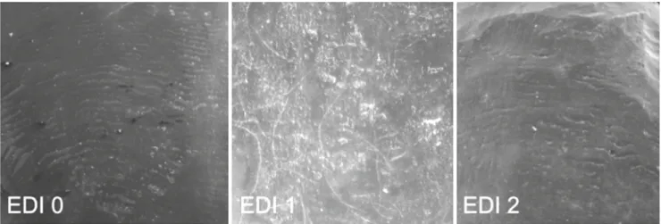
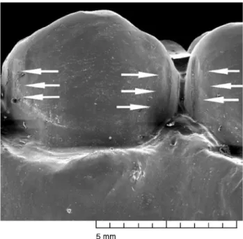
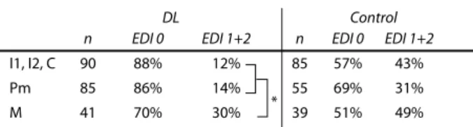
The blinded images were evaluated twice by one examiner at a time interval of 2 weeks. In the case of divergent results from the two assessments, a third evaluation was made using the original epoxy replicas and an optical binocular at a 50× magnification (M7; Wild AG, Heerbrugg, Switzerland). The third evaluation deter-mined the classification. Evaluation included a modified enamel damage index [14] (EDI): 0: smooth surface, perikymata may be visible; 1: acceptable surface, fine, scattered scratches; 2: surface with coarse scratches or gouges (Figure 1). Line angle grooves (LAG) were noted including their location on the mesial or distal interproximal side (Figure 2). The presence of any composite resi-dues (CR) was also recorded (Figure 3).
For the statistical analysis, EDI scores 1 and 2 (damage to the enamel surface) were combined and compared to the EDI 0 scores
Form von Oberflächendefekten und „Line-Angle“-Rillen sowie auf Kompositreste hin zu untersuchen.
Material und Methodik
Nach Zulassung der Studie durch die Ethikkomission der Ge-sundheitsdirektion Kanton Zürich (StV Nr. 07/05) wurden 22 konsekutive Patienten (13 Frauen, 9 Männer; Alter 16,9 ± 3 Jahre; Spannweite 14,0−27,6 Jahre) untersucht und 394 Zähne ausgewer-tet. Zähne, die während der Behandlung neu beklebt werden mussten (n = 26), wurden aus der Studie ausgeschlossen. Eben-falls ausgeschlossen wurden Zähne mit bukkaler Karies, Demine-ralisationen oder Füllungen. Es wurde eine Split-Mouth-Studie durchgeführt, bei welcher ein Quadrant pro Kiefer nach dem Zu-fallsprinzip der Gruppe „Verwendung einer dentalen Lupenbrille“ respektive der Kontrollgruppe zugeteilt wurde. Der kontralaterale Quadrant wurde der jeweils anderen Gruppe zugeteilt. Das De-bonding wurde bei allen Patienten von demselben Behandler durchgeführt (mittlere orthodontische Behandlungszeit 2,2 ± 0,8 Jahre; Spannweite 0,6−3,6 Jahre).
Bei der verwendeten Lupenbrille handelte es sich um eine Swiss-Loupe GT (SandyGrendel®, Aarburg, Schweiz) mit einer 2,5-fachen Vergrößerung, einer Arbeitsdistanz von 350 mm und einem zusätz-lichen LED3-Stirnlicht (SandyGrendel®, Aarburg, Schweiz).
Die festgelegte maximale Arbeitszeit für Debonding, Adhäsiv-Restentfernung und Politur betrug 45 Minuten. Der Behandler wurde von einer Dentalassistentin unterstützt. Die Brackets (SPEED System™; Strite Industries, Cambridge, Ontario, Kanada) wurden vorsichtig mittels einer scharfen Debondingzange (Hu-Friedy 678–104; Chicago, IL, USA) von Inzisiven, Eckzähnen, Prä-molaren und Molaren entfernt. Für die initiale Adhäsiv-Restent-fernung wurde ein Wolfram-Karbidbohrer (H 379AGK FG; Ko-met−Brasseler GmbH, Lemgo, Deutschland) bei 40.000 Umdre-hungen pro Minute unter Luftkühlung verwendet. Für jeden Pa-tienten wurde ein neuer Bohrer benutzt. Ein Ultraschallinstrument und ein Pulverstrahlgerät (Air-Flow® S2 mit Piezon® Scaling Instru-ment Type A und Air-Flow® Powder Classic S2; EMS, Nyon, Schweiz) wurden für die Reinigung im zervikalen und approxima-len Bereich verwendet. Für die Politur wurden Silikonpolierer (AB-BA® Universal Polisher pre-polisher black; KerrHaweSA, Bioggio, Schweiz) mit Wasserkühlung und Polierpaste (Hawe Cleanic mit Fluoriden; KerrHawe SA, Bioggio, Schweiz) im Näpfchen (hard rubber white 1802/30; KerrHawe SA, Bioggio, Schweiz) benutzt.
Die Abformung der Zähne erfolgte mittels A-Silikon (Bisico® S1, Bisico® S4; Bielefelder Dentalsilicone, Bielefeld, Deutschland) in einer zweiphasigen, zweizeitigen Anwendung. Zahnreplika wurden mittels Epoxidharz (Stycast® 1266; Emerson and Cuming, Westerlo, Belgien) hergestellt. Die Modelle wurden 80 Sekunden bei 0,08 mbar und 40 mA mit Gold beschichtet (SCD 030; Balzers Union, Balzers, Liechtenstein), so wurde eine Schichtdicke von ca. 200 Å erreicht. Die REM-Aufnahmen wurden bei einer 50-fachen Ver-größerung bei 20 kV aufgenommen (TESCAN VEGA TS 5136 XM; Cranberry, PA, USA).
Die REM-Bilder wurden von einem Untersucher im Abstand von zwei Wochen zweimal bewertet. Bei unterschiedlichen Resulta-ten wurde die Bewertung ein drittes Mal unter Anwendung eines
(no damage) since only seven teeth in the control group and four in the loupes group had an EDI score of 1. Due to the paucity of LAG, mesial and distal LAG were also combined. χ2 and Fisher´s exact tests were calculated using SPSS® version 16.0 (SPSS, Chicago, IL, USA). The levels of significance were set at p≤0.05 (*), 0.01 (**) and 0.001 (***).
Results
Our results are summarized in Table 1-5, illustrating EDI, LAG and CR.
The EDI scores 0, 1 and 2 scores (p<0.0001) differed significantly in their distribution between the dental loupes and control groups (Table 1). We noted no significant differences (p=0.08) in enamel damage in the control group, but the molars in the dental loupes group revealed significantly more enamel damage (p=0.012) (Table 2).
LAG revealed similar results when comparing the control to the dental loupes group (p=0.13) (Table 3). We observed significantly fewer LAG in anterior teeth in the dental loupes group than in the control group's anterior teeth (p=0.03). There were also fewer LAG
optischen Binokulars mit 50-facher Vergrößerung (M7; Wild AG, Heerbrugg, Schweiz) durchgeführt. Diese Bewertung war die ent-scheidende für die definitive Einteilung. Die Auswertung umfasste einen Schmelzschadenindex [14] (EDI): 0: Glatte Oberfläche, Peri-kymatien zum Teil sichtbar; 1: akzeptable Oberfläche, feine Kratzer; 2: Oberfläche mit groben Kratzern oder Rillen (Abbildung 1). Zu-dem wurden „Line-Angle“-Rillen inklusive deren mesialer und/ oder distaler Lokalisation (Abbildung 2) sowie Kompositreste no-tiert (Abbildung 3).
Da nur 7 Zähne in der Kontrollgruppe und 4 in der Lupenbrillen-gruppe einen EDI von 1 zeigten, wurden für die statistische Analyse die EDI-Werte 1 und 2 (Schmelzoberflächenschäden) kombiniert und mit EDI-Werten 0 (schadlos) verglichen. Aufgrund sehr weni-ger Angle“-Rillen wurden die mesialen und distalen „Line-Angle“-Rillen ebenfalls kombiniert. Die statistische Auswertung erfolgte über χ2- und Fisher's-exact-Tests mittels SPSS® (SPSS 16.0, SPSS, Chicago, IL, USA). Die Signifikanzlevel wurden auf p ≤ 0,05 (*), 0,01 (**) und 0,001 (***) festgesetzt.
Ergebnisse
Die Ergebnisse werden in Tabelle 1-5 für Schmelzschäden, „Line-Angle“-Rillen und Kompositreste dargestellt.
Die Verteilung der Schmelzschäden war für die EDI-Werte 0 so-wie 1 und 2 (p <0,0001) signifikant unterschiedlich im Vergleich der Lupenbrillen- mit der Kontrollgruppe (Tabelle 1). Im Hinblick auf die unterschiedlichen Zahngruppen (Tabelle 2) konnten für die Kontrollgruppe keine statistisch signifikanten Unterschiede (p = 0,08) gefunden werden, allerdings ergaben sich für die Molaren in der Lupenbrillengruppe signifikant mehr Schäden (p = 0,012).
Für „Line-Angle“-Rillen wurden ähnliche Werte für Kontroll- und Lupenbrillengruppe gefunden (p=0,13) (Tabelle 3). Auf Front-zähnen der Lupenbrillengruppe traten signifikant seltener „Line-Angle“-Rillen auf als auf Frontzähnen der Kontrollgruppe (p = 0,03). Zudem traten „Line-Angle“-Rillen auf Frontzähnen der Lu-penbrillengruppe signifikant seltener auf als auf Prämolaren (p = 0,007) und Molaren (p = 0,004) der Lupenbrillengruppe (Tabelle 4).
Figure 1. Enamel damage indices (EDI): EDI 0 no damage, EDI 1 scratches, EDI 2 serious damage, at 50× magnification Abbildung 1. Schmelzschadenindex: EDI 0 (schadlos), EDI 1 (Kratzer), EDI 2 (größere Schäden) bei 50-facher Vergrößerung
Table 1. Enamel damage index (EDI) on teeth debonded with dental loupes (DL) and without dental loupes (Control). Highly significant differ-ences between the DL and the control group were found for EDI 0 and EDI 1+2
Tabelle 1. Schmelz-Schaden-Index (EDI) an Zähnen nach Debonding mit Lupenbrille (DL) und ohne Lupenbrille (Control). Für die EDI-Werte 0 und 1+2 wurden zwischen der Kontroll- und der DL-Gruppe hoch signifikante Unter-schiede festgestellt
Debonding method EDI 0 EDI 1+2
DL (n=189) Control (n=179) p ≤ 0.05 (*), 0.01 (**) and 0.001 (***). 165 114 24 65 *** ***
in premolars (p=0.007) and molars (p=0.004) in the dental loupes group than in the control group's respective teeth (Table 4).
Composite residues were significantly more frequent (p=0.003) in the control group (Table 5). The distribution among the teeth did not differ significantly in the control group (p=0.13) or dental loupes group (p=0.74).
Discussion
Enamel can be damaged from the bracket-bonding procedure to adhesive removal. Research has focused on the materials used for adhesive removal since the first study in 1971 [21]. According to many studies cited in our introduction, tungsten-carbide burs are recommended for adhesive removal rather than diamond burs or grinding stones. However, to our knowledge, other aspects poten-tially affecting adhesive removal—such as differences between or-thodontists, or optimizing visual perception by dental loupes— have rarely been investigated. We identified just one study [26] ad-dressing inter-orthodontist differences. Amazingly, but not sur-prisingly, the same orthodontist scored the best and worst results, indicating that paying close attention could be the most important factor in quality control.
The control group's EDI 1 and 2 scores differed by more than 100% compared to the EDI scores of the dental loupes group, indi-cating greater damage to the enamel. As meticulousness might be a major factor in the quality of adhesive removal, we cannot exclude the possibility of an unconscious bias—namely, that an orthodon-tist may unconsciously take greater care when working with dental loupes. However, a blinded study protocol is not possible.
Kompositreste wurden in der Kontrollgruppe signifikant häufi-ger (p = 0,003) gefunden als in der Lupenbrillengruppe (Tabelle 5). Die Verteilung auf die Zahngruppen war weder für die Kontroll-gruppe (p = 0.13) noch für die LupenbrillenKontroll-gruppe (p = 0,74) signi-fikant unterschiedlich.
Diskussion
Schmelzschäden können über den gesamten Prozess vom Bra-cketbonding bis zur Adhäsiv-Restentfernung entstehen. Seit der ersten Untersuchung im Jahre 1971 [21] fokussierte dabei die For-schung auf die Materialien, die zur Adhäsiv-Restentfernung ver-wendet wurden. Gemäß den in der Einleitung zitierten Studien kann geschlossen werden, dass Wolfram-Karbidbohrer für die Adhäsiv-Restentfernung anstelle von Diamantbohrern oder Kar-borundsteinchen verwendet werden sollten. Hingegen wurden andere Einflussbereiche für die Adhäsiv-Restentfernung, wie Unterschiede zwischen verschiedenen Behandlern oder Verbesse-rung der visuellen Wahrnehmung durch Lupenbrillen, unseres Wissens kaum untersucht. Es wurde eine Studie [26] zu Unter-schieden zwischen verUnter-schiedenen Behandlern gefunden. Erstaun-licherweise, aber nicht überraschenderweise, wurden die besten und schlechtesten Resultate bei ein und demselben Behandler festgestellt, was darauf hinweist, dass die Sorgfalt der wichtigste Faktor für die Behandlungsqualität darstellt.
In der Kontrollgruppe unterschieden sich die EDI-Werte um mehr als 100% von den EDI-Werten der Lupenbrillengruppe, was auf deutlich mehr Schmelzschäden hinweist. Da, wie oben erwähnt, die Sorgfalt einen wesentlichen Einfluss auf die Qualität der Adhä-siv-Restentfernung hat, kann eine unbewusste, unterschiedliche Sorgfalt des Behandlers bei Adhäsiv-Restentfernung mit und ohne Lupenbrille nicht ausgeschlossen werden. Ein Studienprotokoll mit diesbezüglicher Verblindung ist nicht möglich.
Figure 2. Line angle grooves (LAG) were noted including their location at the mesial or distal interproximal surface
Abbildung 2. „Line-Angle“-Rillen (LAG) wurden zusammen mit deren distaler oder mesialer Lokalisation festgehalten
Figure 3. Composite residues (CR) on a lower right molar
The origin of LAG is not clear. It can be compared to the ripples formed on hard surfaces by milling machines, also known as scoring [6]. LAG may also be due to a prolonged procedure or irregular pressure of the bur at the line angles when the direction of the bur's movement is changed. In contrast to the aforementioned defects reflected in the EDI, we detected no significant difference in the quality of adhesive removal in terms of LAG between the two study groups except for the anterior teeth. The anterior teeth were also those least affected in this study. This may be due to better vision and a tooth morphology facilitating adhesive removal. The broad and relatively straight anterior teeth are probably less prone to LAG. The low frequency of LAG and equal distribution between the control and dental loupes groups reveals that the orthodontist performed extremely careful debonding in both groups. If the main factor for the differences in EDI is the orthodontist's meticulousness, this should also be revealed in how the LAG is distributed. However, incorrect bur positions leading to pronounced LAG might have been visible without magnification, whereas smaller errors leading only to scratches were detected and corrected under magnification. Compared to an earlier investigation [26], our results with loupes were favorable with no cervical grooves and many fewer LAG. According to the preference of the orthodontist the H 379AGK FG tungsten-carbide bur with a slightly tapered shape, rounded tip and eight twisted blades specially developed for orthodontic debonding was used. This bur was proven to be less aggressive in morphological evaluations of teeth after adhesive removal [25]. Its hybrid shape— between a classical straight finishing bur for buccal adhesive removal and an egg-shaped lingual finishing bur, as well as the rounded, blunt tip—may have effectively inhibited cervical groove formation. The small bracket bases of the bracket system used in this investigation may also have contributed to fewer LAG, as there was probably need to remove adhesive from interproximal areas susceptible to LAG. Ultrasonic and air-powder devices not usually recommended for adhesive removal due to increased chair time [28] were used as a supplementary tool for interdental and cervical cleaning. This may have lessened the tendency to remove staining with the carbide bur rather than composite in those areas, and thus
Die Ursache der Angle“-Rillen ist nicht geklärt. „Line-Angle“-Rillen könnten mit Riefenbildung auf harten Oberflächen durch Schleifmaschinen verglichen werden, die unter dem Begriff Scoring [6] bekannt sind. Es könnte sich auch um eine verlängerte Einwirkungszeit oder ungleichmäßigen Druck des Bohrers im Umkehrpunkt der Finierbewegung handeln. Im Gegensatz zu den oben genannten Schmelzschäden mittels EDI-Werten konnten, außer für die Frontzähne, zwischen der Kontrollgruppe und der Lupenbrillengruppe keine Unterschiede festgestellt werden. Die Frontzähne waren dabei am wenigsten von „Line-Angle“-Rillenbildungen betroffen. Dies könnte auf eine bessere Sicht und eine günstigere Zahnmorphologie der Front für die Adhäsiv-Restentfernung hindeuten. Die breiten und relativ geraden Zähne sind wahrscheinlich weniger anfällig für das Auftreten von Rillen. Der insgesamt geringe Anteil an „Line-Angle“-Rillen und die Tatsache, dass eine ähnliche Verteilung in Kontroll- und Lupenbrillengruppe vorlag, weist auf ein insgesamt vorsichtiges Vorgehen des Behandlers in beiden Gruppen hin. Wenn die Sorgfalt des Behandlers hauptursächlich für die unterschiedlichen EDI-Werte verantwortlich wäre, müsste sich dies auch in der Verteilung der „Line-Angle“-Rillen niederschlagen. Jedoch scheint es gut möglich, dass größere Fehlstellungen des Bohrers, welche in „Line-Angle“-Rillen resultieren könnten, ohne optische Vergrößerungshilfe gut gesehen werden, während kleinere Fehler, die zu Verkratzungen der Schmelzoberflächen führen, nur mittels Lupenbrille bemerkt und frühzeitig korrigiert werden. Im Vergleich zu einer früheren Untersuchung [26] erwiesen sich die Resultate der vorliegenden Studie in Bezug auf Schmelzschäden günstig. Es wurden keine zervikalen Rillen gefunden und „Line-Angle“-Rillen traten deutlich vermindert auf. Gemäß Präferenz des Behandlers wurde der H 379AGK FG Wolfram-Karbidbohrer mit leicht konischer Form, einer gerundeten Spitze und acht verdrillten Schneiden verwendet, welcher speziell für das kieferorthopädische Debonding entwickelt wurde und sich in morphologischen Untersuchungen von Zähnen nach Adhäsiv-Restentfernung als weniger aggressiv dargestellt hatte [25]. Die Form des Finierers, welcher einer Hybridform zwischen klassisch geradem Finierer für Bukkalflächen und eiförmigem Finierer für Lingualflächen entspricht, und die runde, stumpfe Spitze könnten die Bildung von
DL
n EDI 0 EDI 1+2 n EDI 0 EDI 1+2
Control I1, I2, C Pm M 90 85 41 85 55 39 88% 86% 70% 57% 69% 51% 12% 14% 30% 43% 31% 49% p ≤ 0.05 (*), 0.01 (**) and 0.001 (***). *
Table 2. Distribution of the enamel damage index (EDI) on different tooth groups for the dental loupes (DL) and the control group: anterior teeth (I inci-sors, C canines), premolars (Pm) and molars (M). No differences were found in the control group, but significantly more damage was found for molars in the DL group compared to premolars and anterior teeth
Tabelle 2. Verteilung der EDI-Werte auf den unterschiedlichen Zahngrup-pen nach Debonding mit LuZahngrup-penbrille (DL) und ohne (Control): Frontzähne (Inzisivi (I) und Eckzähne (C)), Prämolaren (Pm) and Molaren (M). In der Kon-trollgruppe wurden keine signifikanten Unterschiede festgestellt. In der DL-Gruppe fanden sich signifikant mehr Schäden an Molaren
p ≤ 0.05 (*), 0.01 (**) and 0.001 (***).
mes & dis LAG
DL (n=378) Control (n=358)
5% 8% 368 teeth = 736 mesial & distal surfaces
Table 3. Line angle grooves (LAG) on mesial (mes) and distal (dis) surfaces debonded with dental loupes (DL) and without dental loupes (Control). No significant differences were found
Tabelle 3. „Line-Angle”-Rillen (LAG) an mesialen (mes) und distalen (dis) Zahnflächen nach Debonding mit Lupenbrille (DL) und ohne (Control). Es wurden keine signifikanten Unterschiede gefunden
have contributed to the lack of cervical grooves and lower frequency of LAG. Debonding via air cooling rather than water cooling for better visual control may also have improved the quality of adhesive removal.
We observed only minor differences among the three arch seg-ments. The fact that the anterior segment in EDI, LAG and CR achieved better results, and the lower scores among all the tooth groups in the dental loupes cohort, where consistent with improved quality by enhanced visual control. This concurs with results from an earlier study in which more defects were detected on the poste-rior than anteposte-rior teeth [26].
Careful adhesive removal and efficient methods of restoring the enamel surface as much as possible to its pretreatment state is es-sential. Our results demonstrate that working with magnification and better illumination results in less severe enamel damage and fewer composite remnants on the enamel surfaces.
Conclusions
1. Enamel damage on tooth surfaces during adhesive removal was considerably reduced by the wearing of dental loupes.
2. Composite residues were significantly reduced when dental loupes had been worn during adhesive removal.
3. We recommend that dental loupes be worn routinely when re-moving the adhesive after bracket debonding.
Acknowledgement
We thank Nils Stucki, Ogar Friedberg, Beatrice Sener and Felix Schmutz for their valuable help and advice, Hans-Ulrich Luder and Margrit Amstad for their assistance with the SEM, and Judith Ball for reviewing the manuscript.
Conflict of interest
The corresponding author states that there is no conflict of inter-est.
zervikalen Rillen vermeiden geholfen haben. Zudem könnten die schmalen Bracketbasen dazu beigetragen haben, dass weniger „Line-Angle“-Rillen entstanden, da weniger Adhäsiv im approximalen Bereich entfernt werden musste. Die Verwendung von Ultraschall und Pulverstrahlgeräten, welche aufgrund der geringeren Effizienz üblicherweise nicht für die Adhäsiv-Restentfernung empfohlen werden [28], wurden begleitend für die Reinigung der interdentalen und zervikalen Bereiche eingesetzt. Dies könnte die Tendenz, Verfärbungen in diesen Bereichen mit dem Finierer zu entfernen, vermindert und dadurch das Auftreten von LAG und zervikalen Rillen reduziert haben. Auch die Adhäsiv-Restentfernung unter Luftkühlung anstelle von Wasserkühlung, welche mit einer besseren visuellen Kontrolle einhergeht, könnte zu den besseren Resultaten beigetragen haben.
Innerhalb der drei untersuchten Zahnsegmente konnten nur kleine Unterschiede festgestellt werden. Die besseren Resultate für das Frontsegment bezüglich Schmelzschäden, „Line-Angle“-Rillen und Kompositresten und die reduzierten Werte für alle Zähne in der Lupenbrillengruppe bestätigen den Einfluss begünstigter Sicht-verhältnisse auf die Qualität der Adhäsiv-Restentfernung. Dies steht in Übereinstimmung mit einer früheren Studie, bei der in posterioren Bereichen mehr Defekte als in anterioren Bereichen ge-funden wurden [26].
Sorgfältige Adhäsiv-Restentfernung und effiziente Methoden zur Wiederherstellung einer möglichst idealen Schmelzoberfläche sollten in der kieferorthopädischen Behandlung Standard sein. Die Resultate zeigten, dass die Arbeit mit Lupenbrille und verbesserter Beleuchtung zu weniger Schmelzschäden und weniger Komposit-resten auf Schmelzoberflächen führte.
Schlussfolgerungen
1. Schmelzschäden auf Zahnoberflächen während der Adhäsiv-Restentfernung wurden durch die Verwendung von Lupenbril-len deutlich reduziert.
Table 4. Distribution of mesial (mes) and distal (dis) line angle grooves (LAG) to the different teeth: anterior teeth (I incisors, C canines), premolars (Pm) and molars (M). Significantly less LAG were found in the dental loupes group (DL) for anterior teeth in comparison to all other groups
Tabelle 4. Verteilung von mesialen (mes) und distalen (dis) „Line-Angle“-Rillen (LAG) auf unterschiedlichen Zahngruppen: Frontzähne (Inzisivi (I) und Eckzähne (C)), Prämolaren (Pm) and Molaren (M). Auf den Frontzähnen der Lupenbrillengruppe (DL) wurden im Vergleich zu allen anderen Gruppen signifikant weniger LAG gefunden
DL (n=189) Control (n=179)
mes + dis mes + dis
LAG n n LAG I1, I2, C Pm M 90 58 41 85 55 39 2% 16% 17% 11% 18% 21% p ≤ 0.05 (*), 0.01 (**) and 0.001 (***). * ** I1, I2, C Pm M (n=189) (n=90) (n=58) (n=41) (n=179) (n=85) (n=55) (n=39) 9% 7% 10% 36% 10% 21% 19% 15% p ≤ 0.05 (*), 0.01 (**) and 0.001 (***). total CR DL CR Control ** ** *
Table 5. Composite residues (CR) after debonding with dental loupes (DL) and without dental loupes (Control). A significant difference was found for the comparison of the DL and the control group and between most tooth groups
Tabelle 5. Kompositreste (CR) nach Debonding mit Lupenbrille (DL) und ohne Lupenbrille (Control). Es fanden sich signifikante Unterschiede zwischen der DL- und Kontrollgruppe sowie zwischen den meisten Zahngru-ppen
2. Kompositreste wurden durch die Verwendung einer Lupen-brille deutlich reduziert.
3. Dentale Lupenbrillen werden für den routinemäßigen Einsatz bei Adhäsiv-Restentfernung empfohlen.
Danksagung
Wir bedanken uns bei Nils Stucki, Ogar Friedberg, Beatrice Sener und Felix Schmutz für ihre Unterstützung, Hans-Ulrich Luder und Margrit Amstad für die Hilfe bei den REM-Aufnahmen und Judith Ball für das Korrekturlesen.
Interessenkonflikt
Der korrespondierende Autor gibt an, dass kein Interessenkonflikt besteht.
Correspondence address Dr. Dr. Lorenz Brauchli
Department of Orthodontics and Pedodontics University of Basel Hebelstrasse 3 4056 Basel Switzerland Phone: (+41/61) 267 26-46, Fax: -57 e-mail: Lorenz.Brauchli@unibas.ch References
1. Al Shamsi AH, Cunningham JL, Laamey PJ, Lynch E (2007) Three-dimensional measurement of residual adhesive and enamel loss on teeth after debonding of orthodontic brackets: An in-vitro study. Am J Orthod Dentofacial Orthop 131:301.e9–301.e15
2. Brown CR, Way DC (1978) Enamel loss during orthodontic bonding and subse-quent loss during removal of filled and unfilled adhesives. Am J Orthod 74:663– 671
3. Caspersen I (1977) Residual acrylic adhesive after removal of plastic orthodontic brackets: a scanning electron microscopic study. Am J Orthod 71:637–650 4. Christensen GJ (2003) Magnification in dentistry: useful tool or another
gim-mick? J Am Dent Assoc 134:1647–1650
5. Diedrich P (1981) Enamel alterations from bracket bonding and debonding: a study with the scanning electron microscope. Am J Orthod 79:500–522 6. DIN Standard 6580 (n d) Terminology of chip removing; movements and
geo-metry of the chip removing process: DIN Group, German Institute for Standard-ization, Berlin, Germany
7. Eliades T, Gioka C, Eliades G, Makou M (2004) Enamel surface roughness follo-wing debonding using two resin grinding methods. Eur J Orthod 26:333–338 8. Forgie AH, Pine CM, Pitts NB (2001) Restoration removal with and without the
aid of magnification. J Oral Rehabil 28:309–313
9. Friedman MJ, Mora AF, Schmidt R (1999) Microscope-assisted precision dentis-try. Compend Contin Educ Dent 20:723–728, 730–731, 735–736, quiz737 10. Friedman MJ (2004) Magnification in a restorative dental practice: from loupes
to microscopes. Compend Contin Educ Dent 25:48, 50, 53–55
11. Hannemann M, Diedrich P (1986) Der Einsatz des Prophy-Jet®-Gerätes zur Schmelzpolitur nach der Bracketentfernung. Fortschr Kieferorthop 47:317–326 12. Hong YH, Lew KK (1995) Quantitative and qualitative assessment of enamel sur-face following five composite removal methods after bracket debonding. Eur J Orthod 17:121–128
13. Hosein I, Sherriff M, Ireland AJ (2004) Enamel loss during bonding, debonding, and cleanup with use of a self-etching primer. Am J Orthod Dentofacial Orthop 126:717–724
14. Howell S, Weekes WT (1990) An electron microscopic evaluation of the enamel surface subsequent to various debonding procedures. Aust Dent J 35:245–252 15. Kim SS, Park WK, Son WS et al (2007) Enamel surface evaluation after removal of
orthodontic composite remnants by intraoral sandblasting: a 3-dimensional surface profilometry study. Am J Orthod Dentofacial Orthop 132:71–76 16. Krell KV, Courey JM, Bishara SE (1993) Orthodontic bracket removal using
con-ventional and ultrasonic debonding techniques, enamel loss, and time require-ments. Am J Orthod Dentofacial Orthop 103:258–266
17. Lussi A, Kronenberg O, Megert B (2003) The effect of magnification on the iatro-genic damage to adjacent tooth surfaces during class II preparation. J Dent 31:291–296
18. Maillet JP, Millar AM, Burke JM et al (2008) Effect of magnification loupes on dental hygiene student posture. J Dent Educ 72:33–44
19. Mannerberg F (1968) Appearance of tooth surface of teeth showing dental fluo-rosis as observed by shadowed replicas. Odontol Revy 19:271–291
20. Millar BJ (1998) Focus on loupes. Br Dent J 185:504–508
21. Newman GV, Facq JM (1971) The effects of adhesive systems on tooth surfaces. Am J Orthod 59:67–76
22. Øgaard B (2001) Oral microbiological changes, long term enamel alterations due to decalcification and caries prophylactic aspects. In: Brantley WA, Eliades T (eds) Orthodontic materials: scientific and clinical aspects. Thieme, Stuttgart, S 124–39
23. Oliver RG, Griffiths J (1992) Different techniques of residual composite removal following debonding – time taken and surface enamel appearance. Br J Orthod 19:131–137
24. Özer T, Başaran G, Kama JD (2010) Surface roughness of the restored enamel after orthodontic treatment. Am J Orthod Dentofacial Orthop 137:368–374 25. Radlanski RJ (2001) A new carbide finishing bur for bracket debonding. J Orofac
Orthop 62:296–304
26. Schuler FS, Van Waes H (2003) SEM-evaluation of enamel surfaces after removal of fixed orthodontic appliances. Am J Dent 16:390–394
27. Sims AP, Roberts-Harry TJ, Roberts-Harry DP (1993) The incidence and preven-tion of ocular injuries in orthodontic practice. Br J Orthod 20:339–343 28. Staribratova-Reister K, Jost-Brinkmann PG (2004) Bonding and debonding
cha-racteristics of APC-plus clarity brackets compared to APC clarity brackets. World J Orthod 5:312–316
29. Strobl K, Bahns TL, Willham L et al (1992) Laser-aided debonding of orthodontic ceramic brackets. Am J Orthod Dentofacial Orthop 101:152–158
30. Thompson RE, Way DC (1981) Enamel loss due to prophylaxis and multiple bon-ding/debonding of orthodontic attachments. Am J Orthod 79:282–295 31. Van Waes H, Matter T, Krejci I (1997) Three-dimensional measurement of enamel
loss caused by bonding and debonding of orthodontic brackets. Am J Orthod Dentofacial Orthop 112:666–669
32. Zachrisson BU, Arthun J (1979) Enamel surface appearance after various debon-ding techniques. Am J Orthod 75:121–127
33. Zarrinnia K, Eid NM, Kehoe MJ (1995) The effect of different debonding techni-ques on the enamel surface: an in vitro qualitative study. Am J Orthod Dentofa-cial Orthop 108:284–293