1
SUPPLEMENTARY MATERIAL
A new procyanidin B from Campylospermum zenkeri (Ochnaceae) and antiplasmodial activity of two derivatives of (±)-serotobenine.
Norbert Mbabi Nyemeck IIa,c, Dominique Serge Ngono Bikoboa*, Auguste Abouem A Zintchema,b, Eva-Maria Schäferd, Christian Bochete, Dieudonné Emmanuel Pegnyemba and Ulrich Koertc.
a
Department of Organic Chemistry, Faculty of Science, University of Yaoundé I, P.O Box 812, Yaoundé, Cameroon
b
Department of Chemistry, Higher Training College, University of Yaoundé I, P.O Box 47, Yaoundé, Cameroon
c
Philipps-Universität Marburg, Faculty of Chemistry, Hans-Meerwein-Strasse, D-35032 Marburg, Germany
d
Philipps-Universität Marburg, Institut für pharmazeutische Chemie, Raum B 407, Marbacher Weg 6, 35032 Marburg, Germany
e
Department Chemie, Universität Fribourg, CH du Musée 9, 1700 Fribourg, Switzerland
2
Abstract
Phytochemical investigation of the stem bark of Campylospermum zenkeri led to the isolation of five known compounds: (Z,Z)-9,12-octadecadienoic acid (1), serotobenine (2), agathisflavone (3), lophirone A (4) and lophirone F (5) together with a new derivative of procyanidin B3, a catechin dimer named zenkerinol (6). Serotobenine (2) is structurally related to decursivine which shows moderate activity against D6 and W2 strains of
Plasmodium falciparum. For a better understanding of structure-activity relationships three
new semi-synthetic derivatives of serotobenine (2) have been prepared. These are: serotobenine monopropionate (2a), serotobenine monopivalate (2b), and serotobenine cyclohexyl ether (2c) respectively. Two of them (2a) and (2b), were evaluated for their antiplasmodial activity against Plasmodium falciparum 3D7 strain in a parasite lactate-dehydrogenase (pLDH) assay. Compound 2b was more active than compound 2a based on their IC50 values (36.6 and 123 PM respectively).
Keywords: Campylospermum zenkeri; Ochnaceae; zenkerinol, flavonoids, serotobenine, hemisynthesis, antiplasmodial activity
Results and discussion (section)
Description of semisynthetic compounds
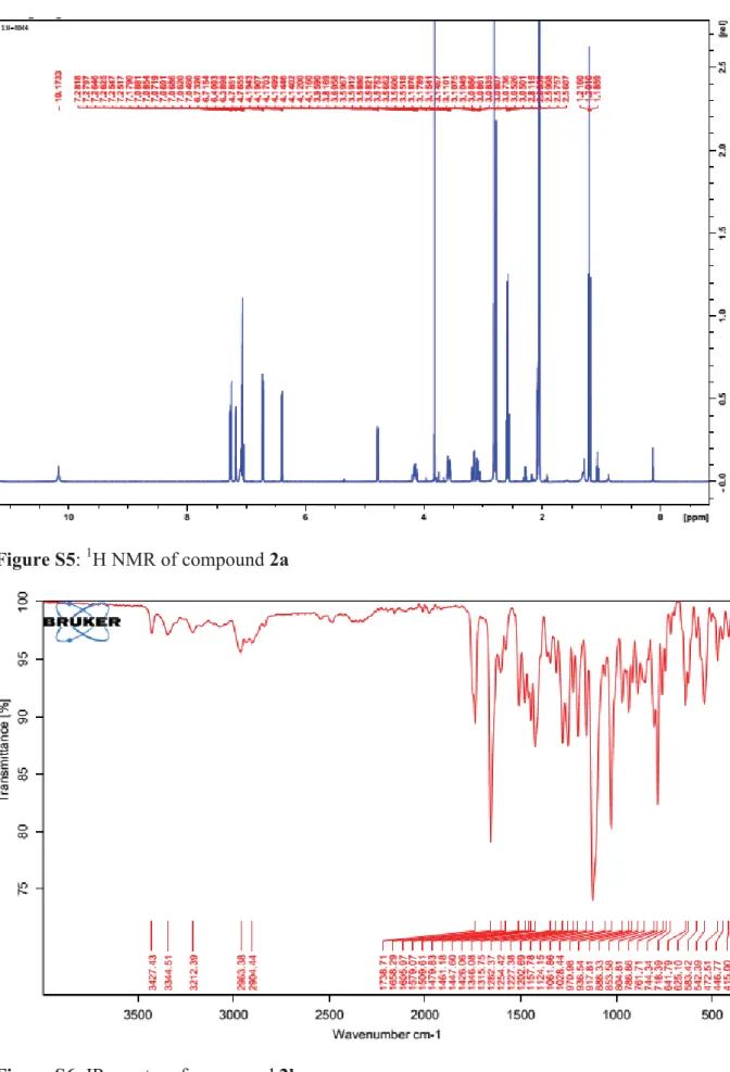
Compound 2a was obtained as a white solid (m.p. 250-252°C), soluble in acetone. The HR-ESI-MS exhibited quasi-molecular ion peak [M-H]- at m/z 405.4307 suggesting a molecular formula of C23H22N2O5 (calcd. 405.4303). In the IR spectum, absorption bands at 1755, 1486, 1425, 1187 cm-1 were respectively attributed to C=O (ester), C-H (methyl group), C-H (methylene group), and C-O (ester) elongations. The large band at 3374 cm-1 usually observed for an O-H (phenol group) elongation in serotobenine IR spectra was replaced by ester elongation described above. Preliminary inspection of 1H-NMR spectrum of compound 2a showed a triplet at δH 1.20 and quadruplet at δH 2.58 ppm strongly deshielded, suggesting its attachment to a carbonyl group. The HMBC spectrum confirmed this connection between H-2′′ (δH 2.58) and C-1′′ (δC 172.6) as well as C-3′′ (δC 9.4), indicating the attachment of propanoyl moiety to the free hydroxyl group of serotobenine (2) (Fig.2). The 1H-NMR spectrum in DMSO (Table S1) also showed an ABX type of aromatic proton at δH 6.72, 7.25 and 7.27 ppm. Thatspectrum showed also in the indole moiety, an AB type of aromatic proton at δH 7.07 and 7.09 ppm. The proton of the pyrrole group was observed at δH 7.18 ppm. Apart from the signals of the propanoyl moiety, these other ones are in agreement with
3 those reported for serotobenine (Sato et al., 1985). The 13C-NMR spectrum (Table S1) showed carbon atoms with resonance of amide and ester groups at δC 171.4 and 172.6 ppm respectively, resonances of the indole moiety at δC 124.9, methoxyl group at δC 56.3, three methylene groups at δC 27.6, 30.7 and 41.5 and, and a methyl group at δ 9.4 ppm belonging to the propanoyl moiety. The correlations observed in the HMBC spectrum and 1H-NMR as well as 13C-NMR confirmed the proposed structure of 2a (Fig. S1 and S2) identified as a new derivative of serotobenine (2).
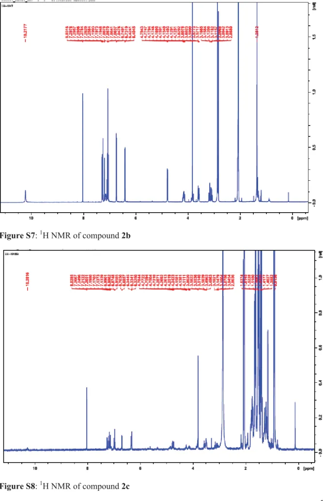
Compound 2b was obtained as a white solid (m.p. 269-272°C) soluble in acetone. The HR-ESI-MS exhibited quasi-molecular ion peak [M-H]- at m/z 433.4845 suggesting a molecular formula of C25H26N2O5 (calcd. 433.4833). Its IR spectum displayed absorption bands at 1738 (C=O, ester), 1157 (C-O, ester) and 1346 (C-H, tert-butyl group) cm-1. Compared to compound 2, the 1H-NMR spectrum of compound 2b showed a signal at δH 1.35 (s) attributable to a tert-butyl unit; the latter was confirmed by a chemical shift at δC 27.3 ppm from the 13C-NMR spectrum which also showed peaks of one quaternary carbon at δC 39.4 and of an ester group at δC 176.4 (Table S1). Selected HMBC correlations between H-3′′ (δH 1.35) and C-2′′ (δC 39.4), H-3′′ and C-1′′ (δC 176.4), and the methoxyl proton (δH, 3.82) and C-3’ (δC 153.5) suggest the occurrence of a tert-butylmethanoate group in serotobenine skeleton indicating the proposed structure of 2b (Fig. S1).
Compound 2c was obtained as a white solid, soluble in acetone. The HR-ESI-MS exhibited pseudo-molecular ion peak [M+Na]+ at m/z 455.4987 suggesting a molecular formula of C26H28N2O4 (calcd. 455.5001). As compound 2, compound 2c showed additional cyclohexyl protons signals between δH 1.39 and 3.50 from 1H-NMR spectrum, and between δC 24.0 – 69.2 from the 13C-NMR giving emphasis to the occurrence of a cyclohexyl unit together with an ether group (Table S1). The correlations observed in the HMBC spectrum (Fig. S1) between H-1′′ (δH 3.50 ppm) respectively with C-4′ (δC 134.0) and C-2′′ (δC 28.2) confirmed the proposed structure of 2c recognised as another new derivative of serotobenine (2) (Fig. S1).
4 Experimental section (Annex)
Hemisynthesis reactions
Esterification of serotobenine with propionic anhydride
rac-Serotobenine 2 (5 mg, 14.29 μmol) was dissolved in pyridine (0.83 mL) and propionic
anhydride (0.75 mL) at 0°C (Koizumi et al., 2008). The solution was allowed to warm to 22°C overnight. Then, an aqueous hydrochloric acid (1M, 5 mL) was added, layers were separated and the aqueous layer was extracted with tert-butylmethyl ether (2x10 mL). The combined extracts were washed with sodium hydrogen carbonate (1M, 10 mL) and brine (10 mL). The organic layer was dried with magnesium sulfate, filtered and the solvent was removed at evaporator under reduced pressure. The residue was purified by CC SiO2 (CHCl3/MeOH :15:1), giving a white solid compound named serotobenine monopropionate 2a (5.5 mg, 95%) (scheme 1)
Esterification of serotobenine with pivalic anhydride
rac-Serotobenine 2 (5 mg, 14.29 μmol) was dissolved in pyridine (0.83 mL) and pivalic
anhydride (1.00 mL) at 0°C. Thereafter, 4-diethylaminopyridine (0.17 mg, 1.429 μmol) was added as a catalyst. The solution was allowed to stirr at room temperature (22°C) overnight. Then, an aqueous hydrochloric acid (1M, 5 mL) was added, layers were separated and the aqueous layer was extracted with tert-butylmethyl ether (2x10 mL). The combined extracts were washed with sodium hydrogen carbonate (1M, 10 mL) and brine (10 mL). The organic layer was dried with magnesium sulfate, filtered and the solvent was removed under reduced pressure. The residue was purified by CC SiO2 (CHCl3/MeOH: 15:1) to afford a white solid compound named serotobenine monopivalate 2b (5.8 mg, 93%) (Scheme 1).
Etherification of serotobenine with cyclohenanol via a Mitsunobu reaction
rac-Serotobenine 2 (10 mg, 28.56 μmol) was added to a solution of cyclohexanol (5.72 mg,
57.12 μmol), tributylphosphine (9.5 μL, 57.12 μmol) and in anhydrous THF under N2 atmosphere at 0°C. The resulting solution was treated with DIAD (11.2 μL, 57.12 μmol ) and the reaction mixture was continuously stirred at room temperature up to completion of the reaction. The solvent was evaporated and the residue dissolved in tert-butylmethyl ether (2x3
5 mL). The combined extracts were washed with sodium hydrogen carbonate (1M, 2 mL) and brine (3 mL). The organic layer was dried with magnesium sulfate, filtered and the solvent was removed under reduced pressure. The residue was purified by CC SiO2 (CHCl3/MeOH: 50:1) to afford a white solid compound, serotobenine cyclohexyl ether (2.5 mg, 20 %). (Scheme 1)
6 Table S1: 1H and 13C-NMR spectroscopic data of compounds 2, 2a, 2b and 2c (500, and 125 MHz in acetone d6) δ in ppm.
2 2a 2b 2c N° δC δH ; m HMBC (C→H) δC δH ; m ; J HMBC (C→H) δC δH, m HMBC (C→H) δC δH, m HMBC (C→H) 1 10.17 10,17 10,21 10,28 2 125.6 7.17 ; s 124,9 7,18 ; s 124,9 7,19 ; s 125,6 7,17 ; s 3 112.2 112,3 112,2 112,2 3a 125,9 123,7 123,8 4 114.4 H-C(8′) 115,0 H-C(8′) 114,7 H-C(8′’) 115,0 5 148.8 152,4 152,2 151,9 6 111.2 7.27 ; d 123,6 7,07 ; d ; 7.0 123,4 7,08 ; d 119,6 7,00 ; d 7 104.5 6.70 ; d 119,1 7,09 ; d ; 7.0 118,9 7,10 ; d 117,6 7,13 ; d 7a 134.4 144,8 140,6 147,8 8a 30,7 3,04-3,09; m 30,7 3,05-3,10; m ; 30,5 3,12-3,09 ; m 29,0 3,10-3,05 ; m 8b 30,7 3,14-3,19; m 30,7 3,15-3,18 ; m 30,5 3,16-3,15 ; m 29,0 3,18-314 ; m 9a 41,5 3,55-3.59 ; m 41,5 3,58 ; m 41,3 3,60-3,57 ; m 41,4 3,51-3,49 ; m 9b 41,5 4.10-4.18; m 41,5 4,17 ; m 41,3 4,17-4,12 ; m 41,4 4,16-4,11 ; m 10 8.17 ; 1H 8.01 ; 1H 8,03 ; 1H 8,02, 1H
7 1′ 147.2 H-C(5′) 141,9 H-C(8′) 141,6 H-C(6′) ; H-C(2′) 136,2 H-C(8′) 2′ 110.7 7.12 ; d 111,3 7,25 ; d 111,1 7,26 ; d 111,7 7,25 ; d 3′ 153.7 153,6 MeOH 153,5 153,7 MeOH 4′ 133.9 H-C(5′) 134,1 H-C(5′) 133,9 H-C(5′) 134,0 5′ 120.1 6.85d 105,4 6,72 ; d ; 9.3 105,3 6,74, d 105,4 6,69, d 6′ 115.1 6.95 ; dd 125,6 7,27 ; dd ; 9.3 125,7 7,29 ; dd 125,8 7,26 ; dd 7′ 85.4 6.32 ; d H-C(8′) ; H-C(2′) 85,0 6,40 ; d ; 10.5 H-C(6′) 84,9 6,41 ; d H-C(8′) ; H-C(6′) 85,3 6,34 ; d 8′ 55.2 4.73 ; d 55,4 4,78 ; d ; 10.5 55,2 4,78 ; d 55,2 4,78 ; d 9′ 171.5 H-C(8′) ; H-C(7′) 171,4 H-C(8′) 171,3 171,5 H-C(8′) 1′′ 172,6 H-C(2′′) 176,4 H-C(3′′) 69,2 3,50 ; m 2′′ 27,6 2,58 ; q H-C(3′′) 39,4 H-C(3′′) 28,2 1,67-1,61 ; m H-C(4′′) 3′′ 9,4 1,20 ; t H-C(2′′) 27,3 1,35 ; s 23,8 1,50-1,54 ; m H-C(4′′) 4′′ 24,0 1,45-1,39 ; m O-CH3 56.2 3.83 ; s 56,3 3,82 ; s 56,1 3,83 ; s 56,2 3,80, s
8 Table S2: 1H and 13C-NMR spectroscopic data of compound 6 (500 and 125 MHz in DMSO) δ in ppm Position δC δH (-OH) ; m ; J (Hz) HMBC (C→H) 1 / / 2 140 .0 / H-C(2′) 3 133.1 / H-C(4) 4 48.8 3.18 ; s OH-C(3) 4a 99.9 / H-C(3); H-C(6); H-C(8) 5 156.5* / H-C(6) 6 95.4 5.88 ; d ; 2.3 / 7 156.5* (8.90) OH H-C(6); HO-C(7) 8 94.4 5.71 ; d ; 2.3 H-C(6) 8a 156.0* / H-C(8); OH-C(7) 1′ 120.2 / H-C(2′) 2′ 115.2 6.89 ; d ; 1.7 H-C(6′) ; OH-C(3′) 3′ 144.8 (8.80) OH H-C(2′) ; OH-C(3′) 4′ 144.7 (8.74) OH H-C(5′) ; OH-C(4′) 5′ 115.2 6.65 ; d ; 7.2 H-C(6′) ;OH-C(4′) 6′ 118.2 6.66 ; d ; 7.2; 1.7 H-C(2′) ;H-C(5′) 1′′ / / / 2′′ 78.3 4.65 ; d ; 9.5 H-C(2′′′) 3′′ 65.1 3.91 ; m ; 9.5; 4.2 H-C(4β′′) ;H-C(4α′′) 4′′ 28.5 2.46 ; dd ; 16.7 ; 4.2 ; 2.66 ; dd ; 16.7 ; 5.9 H-C(2′′) 4a′′ 99.7 / H-C(4β′′) ;H-C(4α′′) 5′′ 156.8 (9.06) OH H-C(4β′′) ;H-C(4α′′); OH-C(5′′) 6′′ 98.8 6.18 ; s OH-C(5′′) 7′′ 156.0* / H-C(6′′) ; OH-C(4) 8′′ 102.4 / H-C(4) ; H-C(6′′) 8a′′ 156.5* / / 1′′′ 130.9 / H-C(2′′); H-C(5′′′)
9 2′′′ 115.2 6.89 ; d ; 1.7 H-C(2′′) ; H-C(6′′′) 3′′′ 144.8 (8.80) OH H-C(2′′′) ; OH-C(3′′′) 4′′′ 144.7 (8.74) OH H-C(5′′′) ; OH-C(3′′′) ; OH-C(4′′′) 5′′′ 115.2 6.65 ; d ; 7.2 H-C(6′′′) ; OH-C(4′′′) 6′′′ 118.2 6.66 ; d ; 7.2; 1.7 H-C(2′′′) ; H-C(5′′′)
* Signals can be interchanged.
Table S3. Inhibition-values of 2a and 2b against P. falciparum. Inhibition is shown in %. SD is the standard deviation. IC50 values of compounds 2a and 2b are respectively 123.2 PM and 36.6 PM. Substance Conc [μM] 2a Mean SD 2b Mean SD 100 45.4 13.03 85.8 11.74 10 0.0 11.12 4.7 7.544 1 4.3 6.783 4.4 9.432 0.1 6.1 3.759 3.6 8.462 0.01 6.8 2.012 3.0 7.66 2a 2b 2c
10 N N O O O H O H O H H H H H H 1'' 2'' 3'' 2 3 8a 9a b 9' 8' 7' 1' 2' 3' 4' 5' 6' b 5 6 7 7a 4 3a N N O O O H O H O H H H H H H 1'' 2'' 3'' 4' 3' 2 3 8a 9a b b 3a 4 5 6 7 7a 9' 8' 7' 1' 6' 5' 2' N N O O O H O H H H H H H 4' 3' 2 3 8a 9a b b 3a 4 5 6 7 7a 9' 8' 7' 1' 6' 5' 2' H H H 1'' 2'' 3'' 4'' 2'' 3'' 2a 2b 2c Figure S2: Selected HMBC correlations of compounds 2a, 2b and 2c.
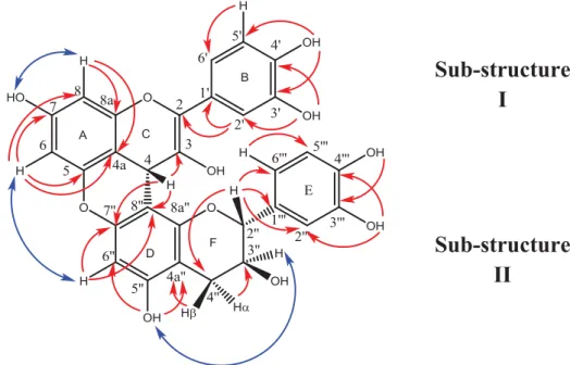
Figure S3: Selected HMBC (
→
) and NOESY (↔
) correlations of compound 6Figure S4: IR spectra of compound 2a
Sub-structure
I
Sub-structure
II
11 Figure S5: 1H NMR of compound 2a
12 Figure S7: 1H NMR of compound 2b
13 Figure S9: 13C NMR spectrum of compound 2c
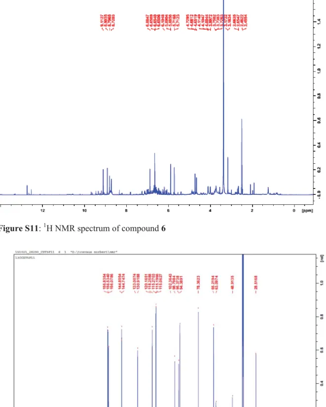
14 Figure S11: 1H NMR spectrum of compound 6
15 Figure S13: HSQC spectrum of compound 6
16 Figure S15: HR-ESI-MS of compound 6
17 Figure S16: LC-MS of compound 6
18 Figure S17: NOESY spectrum of compound 6
Reference
Koizumi Y, Kobayashi H, Wakimoto T, Furata T, Fukuyama T, Kan T. 2008. Total synthesis of (-)-serotobenine. J Am Chem Soc. 130(50): 16854-16855.