R E S E A R C H A R T I C L E
Open Access
Regulation of calretinin in malignant
mesothelioma is mediated by septin 7
binding to the
CALB2 promoter
Walter Blum, László Pecze, Janine Wörthmüller Rodriguez, Martine Steinauer and Beat Schwaller
*Abstract
Background: The calcium-binding protein calretinin (gene name: CALB2) is currently considered as the most sensitive and specific marker for the diagnosis of malignant mesothelioma (MM). MM is a very aggressive tumor strongly linked to asbestos exposure and with no existing cure so far. The mechanisms of calretinin regulation, as well as its distinct function in MM are still poorly understood.
Methods: We searched for transcription factors binding to the CALB2 promoter and modulating calretinin expression. For this, DNA-binding assays followed by peptide shotgun-mass spectroscopy analyses were used. CALB2 promoter activity was assessed by dual-luciferase reporter assays. Furthermore, we analyzed the effects of CALB2 promoter-binding proteins by lentiviral-mediated overexpression or down-regulation of identified proteins in MM cells. The modulation of expression of such proteins by butyrate was determined by subsequent Western blot analysis. Immunohistochemical analysis of embryonic mouse lung tissue served to verify the simultaneous co-expression of calretinin and proteins interacting with the CALB2 promoter during early development. Finally, direct interactions of calretinin with target proteins were evidenced by co-immunoprecipitation experiments.
Results: Septin 7 was identified as a butyrate-dependent transcription factor binding to a CALB2 promoter region containing butyrate-responsive elements (BRE) resulting in decreased calretinin expression. Accordingly, septin 7 overexpression decreased calretinin expression levels in MM cells. The regulation was found to operate bi-directionally, i.e. calretinin overexpression also decreased septin 7 levels. During murine embryonic development calretinin and septin 7 were found to be co-expressed in embryonic mesenchyme and undifferentiated mesothelial cells. In MM cells, calretinin and septin 7 colocalized during cytokinesis in distinct regions of the cleavage furrow and in the midbody region of mitotic cells. Co-immunoprecipitation experiments revealed this co-localization to be the result of a direct interaction between calretinin and septin 7.
Conclusions: Our results demonstrate septin 7 not only serving as a“cytoskeletal” protein, but also as a transcription factor repressing calretinin expression. The negative regulation of calretinin by septin 7 and vice versa sheds new light on mechanisms possibly implicated in MM formation and identifies these proteins as transcriptional regulators and putative targets for MM therapy.
Keywords: Calretinin, Septin 7, Malignant mesothelioma, Transcriptional regulation, Butyrate
* Correspondence:beat.schwaller@unifr.ch
Anatomy, Department of Medicine, University of Fribourg, Route Albert-Gockel 1, CH-1700 Fribourg, Switzerland
© The Author(s). 2018 Open Access This article is distributed under the terms of the Creative Commons Attribution 4.0 International License (http://creativecommons.org/licenses/by/4.0/), which permits unrestricted use, distribution, and reproduction in any medium, provided you give appropriate credit to the original author(s) and the source, provide a link to the Creative Commons license, and indicate if changes were made. The Creative Commons Public Domain Dedication waiver (http://creativecommons.org/publicdomain/zero/1.0/) applies to the data made available in this article, unless otherwise stated.
Background
The Ca2+-binding protein calretinin (CR) serves as an undisputed marker for the diagnosis of human malignant mesothelioma (MM), in particular of the epithelioid type and the epithelioid parts of the mixed type [1,2]; weak CR expression is also found in sarcomatoid MM, possibly indicating a less important and/or different role of CR in this MM cell type [3]. CR is also expressed in human reactive mesothelial cells [4,5], considered as the first step in the transition from healthy flat mesothelial cells covering the pleural cavities to one of the most aggressive and currently therapy-resistant tumor type. Down-regulation of CR by CALB2 shRNA in human MM cell lines profoundly decreases cell growth and viability in vitro: lentivirus-mediated delivery of shCALB2 causes MM cells, in particular the ones with an epithelioid morphology, to enter apoptosis within 72 h post-infection [3]. Under these conditions, the intrinsic caspase 9-dependent pathway is activated. Although the immortal-ized mesothelial cells LP9/TERT1 show strong CR expression (3), shRNA-mediated CR down-regulation dif-ferently affects these non-transformed cells: it inhibits cell proliferation as the result of a G1block. Neither is the
via-bility impaired nor any type of cell death pathway activated. CR is a fast Ca2+ buffer protein [6, 7] modifying the shape of intracellular Ca2+transients [8]; overexpression of CR reduces the mitochondrial Ca2+uptake in primary mesothelial cells [9]. Very little is known about the regulation of CR expression in the various tissues, even in the subpopulation of neurons, where CR is expressed under physiological conditions. It is assumed that CR expression is regulated in a rather similar way in humans and in mice, mostly based on the strong conservation of the proximal promoter regions of the human CALB2 and mouse Calb2 genes [10]. An AP2-like element in proxim-ity of the TATA box confers neuron-specific expression of a luciferase reporter gene (luc+) in cultured neurons [11] (for additional details, see Fig.1in [12]), necessitating the binding of a nuclear protein present in cerebellar granule cells. Of note, this“AP2-like” element in the promoter re-gion has no effect on the transcriptional activity in either MM cells or CR-expressing human colon cancer cells. Down-regulation of β-catenin by its negative regulator Axin2 significantly reduces CR expression in cultured rat thalamic neurons indicating that β-catenin is a positive regulator of the Calb2 gene [13]. A more detailed CALB2 promoter analysis revealed the sequence embracing the− 161/+ 80 bp region to sustain transcriptional activity in MM cells. Cis-regulatory elements within this promoter region including binding sites for NRF-1 and E2F2 are important for CR expression; e.g. siRNA-mediated down-regulation of NRF-1 causes a decrease in CR expression levels indicating that NRF-1 acts as a positive regulator of CR expression (14). Moreover, the strong correlation
between CALB2 mRNA and CR protein expression levels in MM cells is indicative of a control at the transcriptional level [14]. In colon cancer cells, two butyrate-responsive elements (BRE) embracing the TATA box of the CALB2 gene function as butyrate-sensitive repressors of CR ex-pression, while the same sequence has no effect in cells of mesothelial origin, e.g. Met-5A cells [15]. Butyrate (Bt) is the product resulting from intestinal fermentation of diet-ary fibers by bacteria and Bt concentrations in the range of 5–30 mM are present in the chyme/feces of the gut [16]. Bt acts as a modulator of histone acetylation that re-sults in the inhibition of the cell cycle (G1arrest) and leads
to enterocyte (and derived cancer cells) differentiation [17]. Bt exposure of CR-expressing WiDr colon cancer cells results in CR down-regulation [18]. Moreover, gut microbiota might have an influence on respiratory infec-tions [19] also via short chain fatty acids (SFCA) including Bt. Bt is not only produced by the gut microbiota, but also by anaerobic bacteria in the hypoxic environment of cystic fibrosis (CF) airways. Bt concentrations were found to be elevated in the sputum samples of CF patients reaching values of approximately 2 mM [20] and it is conceivable that in the diseased lung of MM patients, Bt may also be increased in the pleural cavity.
In the present study, short-term exposure of MM-derived cell lines to low millimolar Bt concentrations revealed a significant increase in CR expression levels. Thus, we set out to investigate in more detail the promoter region of the CALB2 gene that acts as a Bt-responsive enhancer in MM cells. To address the ques-tion on mechanisms implicated in this regulaques-tion, we searched for proteins binding to this particular promoter region that might be functionally implicated in CR regu-lation in MM cells.
Methods Cell culture
The human mesothelioma cell lines MSTO-211H, H28, H226, the immortalized mesothelial cell line Met-5A and the colorectal adenocarcinoma HT-29 were obtained from the American Type Cell Collection (ATCC, Rock-ville, MD). The human mesothelioma cell lines ZL5, ZL34, ZL55, SPC111 and SPC212 were obtained from the University Hospital of Zurich (Switzerland) de-scribed by Schmitter et al. [21]. Cells were maintained in RPMI1640 (Gibco, Basel, Switzerland) supplemented with 10% fetal bovine serum (FBS, Gibco) and 100 U/ml penicillin and 100μg/ml of streptomycin (1% PS, Gibco). LP9/TERT-1 cells were obtained from the laboratory of Dr. James Rheinwald (Dana Farber Cancer Research Institute, Boston, MA) and were maintained in a medium consisting of 1:1 M199 and MCDB10 medium supplemented with 15% newborn calf serum, 5 ng/mL epidermal growth factor, 0.4 μg/mL hydrocortisone,
2 mM glutamine, 100 U/ml penicillin and 100 μg/ml streptomycin (1% PS) (Gibco, Switzerland).
Generation of reporter plasmids including deletion variants of the humanCALB2 promoter containing BRE7–13
Genomic DNA was isolated from MSTO-211H and ZL55 MM cells using standard methods and purified DNA (0.5μg) was used as template to amplify a 1.3 kb stretch of the human CALB2 promoter embracing the putative Bt-responsive elements (BRE) 7–13; the se-quence and details of primers are shown in Fig. 2 and Table1. The amplicon of 1292 bp containing a SacI and a BglII site at the 5′- and 3′-end, respectively, was cloned into the plasmid pGL3-Promotor (pGL3-P; Pro-mega; # U47298; Wallisellen, Switzerland) linearized with the same restriction enzymes. pGL3-P contains a multiple cloning site, followed by a minimal SV40 pro-moter; the luciferase cassette (luc) in the plasmid is followed by a SV40 late poly (A) signal. This plasmid was then used to generate shorter PCR fragments containing a various number of BREs (see Fig. 2 and Table 1), the fragments were cloned into pGEM-TEasy, sequenced (Microsynth AG, Balgach, Switzerland) and excised by SacI and BglII. The fragments were then inserted into pGL3-P for transfection experiments; DNA (300–500 μg) was isolated using the Wizard Plus Midi-or Maxiprep kit (Promega, DübendMidi-orf, Switzerland).
Luciferase assay
In order to investigate CALB2 promoter activity the dual luciferase assay was used. The constructs were co-transfected with Renilla-Luciferase in 12-well plates (50,000 cells/well) with Mirus lipofection reagent (Mirus, WI, USA). After 48 h (± Bt treatment; 1 mM) the cells were lysed and promoter activity was measured with the dual-luciferase reporter assay (Promega, Dübendorf, Switzerland) on a Turner Designs TD-20/20 Lumin-ometer (Sunnyvale, CA, USA) according to the manufac-turer’s protocol. The details on the normalization of signals and the way to calculate fold changes compared to untreated controls have been described before [15].
DNA binding assay
The fragment containing BRE9–13 (706 bp) serving as capture DNA was synthesized by PCR using 5′-biotinyl-ated primers. The μMACS™ FactorFinder kit (Mylteni Biotech, Bergisch Gladbach, Germany) was used to iso-late proteins interacting with this stretch of the CALB2 promoter. The cleared cell lysate (15,000 x g, 5 min) from 107MSTO-211H cells (100μl) was incubated with 1.5μg of biotinylated BRE9–13 DNA. Putative transcription factor complexes were isolated under non-denaturing (na-tive) conditions according to the manufacturer’s protocol.
Western blot analysis and silver staining
Cell pellets were collected and washed 3 times with CMF-PBS. Cytosolic fractions were isolated as described before [22] and the concentration of proteins was deter-mined using the Bradford method (Bio-Rad, Hercules, CA). Samples were loaded and separated by SDS-PAGE (10% PAA) and subsequently transferred onto nitrocellu-lose membranes (Bio-Rad) by a semidry system (Witec, Litau, Switzerland). Equal protein loading was controlled by transient Ponceau S staining (Sigma) of the mem-branes. The primary antibodies against calretinin (CR7699/4; Swant, Marly, Switzerland) and septin 7 (rabbit polyclonal anti-septin 7; Bethyl Laboratories Inc., Montgomery, TX, USA or Millipore Corp. #ABT354, Te-mecula, CA, USA) were used at a dilution of 1:5000 overnight at 4 °C; anti-GAPDH was from Sigma (ref. G9545) and used at a working dilution of 1:10,000. Rabbit secondary antibody directly linked to horseradish peroxidase (Sigma-Aldrich) was diluted 1:10,000 and membranes were incubated for 2 h at room temperature. The detection was performed with the chemilumines-cent reagent Luminata Classico or Forte (EMD Millipore Corporation, Billerica, MA, USA) and data were col-lected on an imaging system from Cell Biosciences (Santa Clara, CA, USA).
For normalization and quantification of Western blots, densitometric analysis of the Ponceau S-stained mem-branes was performed using the GeneTools software (Syngene, Cambridge, UK). The integral of the signals of all transferred proteins was previously shown to repsent a more reliable way of normalization, since this re-duces the bias towards a specific protein (e.g. GAPDH, α-actin) often used for normalization. Levels of reference (housekeeping) proteins were shown to vary between tis-sues or different cell lines and might also be affected by the experimental manipulations [23,24]. GeneTools soft-ware was also used for the quantification of the specific Western blots signals for CR, septin 7 and GAPDH. In the results section, most often only selected representa-tive regions of the Ponceau S-stained membranes marked as loading control (L.C.) are shown, in particular when Western blot signals were analyzed qualitatively.
Septin 7 cDNA cloning into pLVTHM backbone
A lentiviral system was used to overexpress septin 7. Briefly, the GFP cassette in pLVTHM (Addgene plasmid #12247) was replaced with the human septin 7 (SEPT7) cDNA using the SpeI and PmeI restriction sites. SEPT7 cDNA was obtained from ZL55 cell total RNA. RNA was extracted using the Qiagen RNAeasy kit following the manufacturer’s instructions; cDNA was synthetized from 500 ng of total RNA using the QuantiTect Reverse Transcription kit (Qiagen). Septin 7 was amplified with the following primers: FW_hSEPT7 5′-AGT CGT TTA
AAC ATG TCG GTC AGT GCG AGA TCC-3′ and RV_hSEPT7 5′-AGT CAC TAG TTT AAA AGA TCT TCC CTT TCT T-3′ containing the SpeI and PmeI restriction sites, respectively and the amplicon was inserted into the vector pLVTHM. The correct sequence of the plasmid was verified by colony PCR and subse-quent sequencing of the insert and the novel plasmid was called pLV-hSEPT7.
Lentiviral constructs
Lentivirus particles were produced as described before [3, 25]. Briefly, HEK293T cells were co-transfected by the CaPO4-method with 3 μg of the envelope plasmid
pMD2.G-VSVG (Addgene plasmid #12259), 8 μg of the packaging plasmid psPAX2 (Addgene plasmid # 12260) and either 10 μg of the transfer plasmid pLV-CALB2 as described in [25] or the plasmid containing the shRNA for CALB2 as reported before [3]. Five validated clones of pLKO.1-shRNA SEPT7 plasmids were obtained from Sigma-Aldrich labeled 1–4 and 63 (according to the last digit of The RNAi Consortium (TRC) number). pLV-hSEPT7 was used to overexpress septin 7.
Immunohistochemistry on sections of mouse embryos from E10.5
Mouse embryos were collected at embryonic day 10.5 (E10.5) from C57Bl/6J mice and fixed by immersion in 4% PFA for 72 h, dehydrated and embedded in paraffin. Sections (3 μm) were de-paraffinized and treated with Tris/EDTA (1 mM/0.1 mM, pH 9) for the antigen re-trieval by heating the sections in a boiling water bath for 20 min. Hydrogen peroxide (0.3%; 20 min incubation) served to quench endogenous peroxidases and the tissue was permeabilized with 0.1% PBS Tween20 (5 min incu-bation) followed by blocking with PBS containing 2% BSA and 1% horse serum (20 min incubation). Sections were incubated with primary antibodies (anti-CR 7699/ 4, Swant, Switzerland, anti-septin 7, Bethyl Laboratories, USA) 1:500 overnight at 4 °C. Sections were incubated with secondary antibodies (1:200 dilution) at room temperature for 2 h. After DAB (Sigma-Aldrich) staining, sections were subjected to (weak) hematoxylin counterstaining. Slides
were scanned using a whole-slide imaging system from Ha-mamatsu (Nanozoomer, 2.0-HT).
Immunofluorescence
Cells were seeded on 12-mm glass coverslips and fixed for 15 min with 4% paraformaldehyde. Non-specific binding sites were blocked by incubation with TBS con-taining donkey serum (10%) for 1 h and coverslips were then incubated overnight at 4 °C with the following anti-bodies diluted in TBS 1X: goat polyclonal anti-CR (1: 500; cat# CG1, Swant, Marly, Switzerland) and rabbit polyclonal anti-septin 7 (1:500; Bethyl Laboratories, USA). After washing, coverslips were incubated with secondary antibodies for 3 h at room temperature with the following secondary antibodies: Alexa Fluor 488-conjugated donkey anti-rabbit IgG (1:100, Jackson Immunoresearch Laboratories, West Grove, PA, USA) and Cy5-conjugated donkey anti-goat IgG (1:100; Jack-son). Nuclear DNA was stained using DAPI (5 μg/ml; Molecular Probes, Eugene, OR) and coverslips were mounted with Hydromount solution (National Diagnos-tics, Atlanta, GA). Images were acquired using a Leica fluorescent microscope DM6000B (Wetzlar, Germany) equipped with a Hamamatsu camera C4742–95 (Bridge-water, NJ). For the cells treated with Bt, cells were seeded onto 12-mm glass coverslips pre-coated with Matrigel (Corning, NY, USA) and treated with 1 mM Bt for 48 h.
Co-immunoprecipitation (co-IP)
MM cells at a confluence of 70–90% were washed with ice-cold PBS and lysed with 1 ml ice-cold Triton X-100 lysis buffer (150 mM NaCl, 1% Triton X-100, 50 mM Tris-HCl, pH 8.0) supplemented with protease and phosphatase inhibitors. Cell lysates were incubated on ice for 30 min and then centrifuged (10,000×g, 10 min at 4 °C). To pull down CR, 2–4 μg of a rabbit polyclonal anti-CR antiserum (CR7699/4; Swant) was added to the cleared lysate and incubated for 10 min at 4 °C.μMACS protein A MicroBeads suspension (100μl; Miltenyi Bio-tec, Auburn, CA, USA) was added to the lysate and in-cubated at 4 °C for 30 min. Samples were loaded on
Table 1 All 5′ primers contain a SacI and all 3′ primers a BglII site for cloning into pGl3-P vector
Name BRE 5′ primer (5′-3′) 3′ primer (5′-3′) size (bp)
7–13 GATGAGCTCATGCGAAAGTGAAGGTGGTCCCCAG TGAAGATCTAGCAAGGAGGAACTGGAGATGTCA 1278 9–13 GAGCTCTCGAACCTCTCTTTCCTATCTC AGATCTAGCAAGGAGGAACTGGAGATGTCA 695 10–13 GAGCTCTGGAGCTGAGCCTGGAGAGAAG AGATCTAGCAAGGAGGAACTGGAGATGTCA 530 12–13 GAGCTCTAGGCCATCAGACAGCTCTGAG AGATCTAGCAAGGAGGAACTGGAGATGTCA 127 9 GAGCTCTCGAACCTCTCTTTCCTATCTC AGATCTCTTCTCTCCAGGCTCAGCTCCA 189 9–10 GAGCTCTCGAACCTCTCTTTCCTATCTC AGATCTCCCTCATCTCTCCTTGCAGCT 253 9–11 GAGCTCTCGAACCTCTCTTTCCTATCTC AGATCTAGGCCCTCAAATTCCTCAGAG 348
MACS separation columns (Miltenyi Biotec) and sub-jected to magnetic immunoprecipitation. The columns were washed 3 times with a wash buffer (150 mM NaCl, 1% NP-40, 0.5% sodium deoxycholate, 0.1% SDS, 50 mM Tris-HCl, pH 8.0). Bound protein complexes were eluted in 50 μl of pre-warmed SDS gel loading buffer 1X (50 mM Tris-HCl, pH 6.8, 50 mM DTT, 1% SDS, 0. 005% bromophenol blue, 10% glycerol) and subjected to Western Blotting for septin 7 as described above. A mouse monoclonal anti-Paxillin antibody (1:2000; BD Bioscience) was used as negative control.
MTT assay
To assess the putative toxic effects of Bt in MM cells, the mitochondrial activity of living cells was determined by the 3-(4,5-dimethylthiazol-2-yl)- 2,5-diphenyltetrazo-lium bromide (MTT) quantitative colorimetric assay. MM cells (1500–3000 depending on the cell line) were seeded in 96-well plates. From a stock solution (1 M NaBt in PBS), different amounts were added to yield Bt concentrations from 0.33 to 5 mM. After 3 days of incu-bation with Bt, MM cells were subjected to the MTT assay by adding fresh medium containing 0.5 mg/ml MTT and incubating the plates for 90 min at 37 °C. The supernatant was discarded and DMSO (200 μl) was added to dissolve the formazan crystals. The absorbance at 570 nm was measured using a microplate reader (In-finite Pro200; Tecan Austria GmbH, Groedig, Austria). For each experiment the absorbance value of untreated (control) cells was defined as 1.0 (100%).
Statistical analysis
The data are presented as mean ± standard deviation of multiple experiments (n≥ 3). The statistics were per-formed using StatPlus (AnalystSoft) applying a one-way ANOVA followed by a post-hoc Tukey HSD test. Differ-ences were considered as statistically significant if a p-value was < 0.05.
Results
Butyrate induces CR expression in MM cells and also in immortalized mesothelial cells
We analyzed the effect of Bt on CR expression levels in cells derived from all MM histotypes. Bt concentrations ranging from 0.5–2 mM applied for short periods (48 and 72 h) were tolerated to various degrees; higher Bt concentrations (generally≥2 mM) and prolonged expos-ure (> 6 days) resulted in massive death of most of the investigated MM cell lines (data not shown). Immortal-ized mesothelial cells (Met-5A) and ZL34 cells derived from a sarcomatoid MM showed massive cell death already at 1–2 mM when treated for 6 days. In the few surviving Met-5A cells CR expression levels were insig-nificantly affected as reported before [15]. Here we
investigated the effect of Bt on CR expression levels at 72 h, a time point only mildly affecting cell viability. A quantitative Bt concentration – cell proliferation/viabil-ity curve was determined for MSTO-211H, ZL55 and ZL5 cells, the lines used in most of the further experi-ments (Additional file 1: Figure S1). The weakest effect of Bt on the MTT signal intensity (cell proliferation/via-bility) was observed in ZL5 cells, intermediate in MSTO-211H cells and the strongest Bt-dependent effect was seen in ZL55 cells. The shape of the curves is not reminiscent of a typical sigmoidal toxicity curve, but ra-ther for a growth-inhibiting effect of Bt, as had been ob-served before in Bt-treated colon cancer cells and immortalized LP9/TERT1 mesothelial cells (G1 arrest)
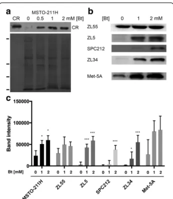
[3, 26]. In support, only few floating (dead) cells were observed at the investigated concentrations and the se-lected time point (72 h). Bt treatment of MSTO-211H (biphasic) cells increased CR protein levels in a Bt concentration-dependent way (Fig. 1a). An increase in CR was also seen Met-5A cells, as well as in the epitheli-oid MM cell lines ZL5 and ZL55, the biphasic cell line SPC212 and the sarcomatoid cell line ZL34 (Fig. 1b). Generally, cells derived from epithelioid and biphasic MM are characterized by higher basal CR expression levels than cells derived from sarcomatoid MM, in par-ticular ZL34 and SPC111 cells, the latter with very low basal CR expression levels [3,14]. Bt treatment of all cell lines led to a concentration-dependent increase in CR levels; semi-quantitative evaluation of the CR signals of the cell lines shown in Fig.1bare depicted in Fig.1c. Of note, CR expression levels were determined in cells that remained attached to the cell culture dishes, i.e. viable cells and not the floating and/or dead cells in the culture medium. Since MM cell lines of the epithelioid and bi-phasic type were previously shown to be strongly af-fected by CR down-regulation, much more than MM cells derived from sarcomatoid tumors [3], further ex-periments were carried out with ZL5 and ZL55 (epitheli-oid) and MSTO-211H (biphasic) cells.
A region in theCALB2 gene promoter containing 7 putative BRE elements acts as a butyrate-responsive enhancer in MSTO-211H and ZL55 MM cells
Analysis of the human CALB2 promoter region re-vealed additional putative BRE elements named BRE7– 13 (Fig.2b) upstream of the previously identified region that contains 2 functional BRE (BRE5–6), which act as transcriptional repressors in colon cancer cells [15]. The newly identified region has a length of 1278 bp and is approximately 3.3 kb upstream of the CALB2 translation start site (ATG) (Fig.2a). This 1.3 kb region was amplified by PCR using genomic DNA from ZL55 and Met-5A cells and primers containing specific re-striction sites (forward primer: SacI site; reverse primer
BglII site) compatible with the reporter luciferase plas-mid pGl3-P. Sequencing revealed the inserts amplified from the 2 cell lines to contain alterations in the nu-cleotide sequence when compared to the sequence an-notated in PubMed (CALB2; gene ID: 794). The three changes (1 point mutation (A/T), 2 deletions of either 1 or 2 nucleotides) were present in the sequences of all clones derived from both cell lines, ZL55 and Met-5A, excluding this being PCR artifacts (Additional file 1: Figure S2). The others were specific for each cell line, consisting of point mutations, 2 in the insert derived from ZL55 cells and 3 in the insert derived from Met-5A cells (Additional file 1: Figure S2). None of these changes directly involved the sequences of the putative BREs. Besides the luc + reporter plasmid containing all 7 putative BREs (BRE7–13), truncated versions were generated by PCR and these included plasmids contain-ing BRE9–13, 10–13 12–13, 9, 9–10 and 9–11 (Fig.2c). In MSTO-211H cells, luciferase activity was increased by 41 ± 16% (mean ± SEM) by the full-length fragment
after Bt treatment for 48 h; however, strongest activa-tion (200 ± 24% of control) was observed with the frag-ment containing BRE9–13, while elimination of BRE9 (fragment BRE10–13), resulted in a luciferase activity close to basal levels (109 ± 9%). In a shorter promoter fragment that included BRE9 (BRE9–11), activity was partially increased (159 ± 14%), but not to values as seen with the BRE9–13 plasmid. Thus, the essential enhancer/activator part appeared to be contained in the region embracing BRE9–11, but the region containing BRE12 and 13 further enhanced the luciferase activity, indicative that the various BREs contributed to the ef-fects in a rather complex manner. Similar results were obtained in ZL55 cells, although the maximal effect in the presence of the BRE9–13 promoter fragment was of smaller magnitude (123 ± 18%). Thus, the effects were nearly identical qualitatively (Fig. 2c, d), yet the effect was approximately 4-times smaller in ZL55 cells. It is noteworthy that the effect on the luciferase activity was rather well correlated with the fold increase in CR ex-pression levels after Bt treatment (Fig.1). The increase in CR expression was + 290% in MSTO-211H cells and + 60% in ZL55 cells, i.e. an approximately 5-fold differ-ence. It appears that in ZL55 cells, where CR levels are rather high in control conditions, the relative Bt-mediated increase in CR expression is smaller than in MSTO-211H cells characterized by a lower “basal” CR expression level [14]. We also tested the BRE7–13 and
BRE9–13 promoter fragments containing the luc + re-porter in the colon cancer cell line Caco-2 in order to see, whether the enhancer function also persisted in colonocytes. A small, yet insignificant increase of + 19 ± 16% was observed in the presence of the BRE9–13 fragment and similar results were also observed with HT-29 colon cancer cells (data not shown). This hints that the BRE9–13-containing promoter region in the CALB2 gene acts as a Bt-activated enhancer, with a strong preference for MM cells.
Identification of proteins binding to CALB2 promoter fragments BRE5–6 and BRE9–13 in a Bt-dependent manner
In a previous study we had demonstrated that both BRE5&6 contribute to the CR-repressing effect in colon cancer cells [15]. Based on the rather complex pattern of BRE-containing fragments on enhancing luciferase activ-ity (Fig.2c) involving up to 5 BREs [9–13], we addressed the question, which proteins might bind to the BRE9– 13-embracing region in a Bt-dependent manner. For this, cell lysates from Bt-treated and control MSTO-211H cells were incubated with the biotinylated BRE9– 13 DNA fragment. Several proteins bound to the DNA and were released in the presence of elution buffer (Fig.3). A side-by-side comparison of silver-stained polyacrylamide
Fig. 1 Upregulation of CR in Bt-exposed immortalized mesothelial (Met-5A) and MM cells a Upper panel: CR Western blot of cytosolic proteins from MSTO-211H cells treated with Bt for 72 h; left lane (CR): human recombinant CR (40 ng) as control. Lower panel: Ponceau Red-stained membrane to check for even loading. Ticks on the left mark the position of marker proteins (in kDa, from top to bottom: 75, 35, 28, 10). The signal for CR is slightly above the 28-kDa marker protein. b Western blots for CR of Bt-treated (72 h) cells: one mesothelial cell line (Met-5A) and 4 MM cell lines: ZL55, ZL5, SPC212, ZL34. Equal loading was confirmed by Ponceau Red staining as shown in a (not shown). c Semi-quantitative CR Western blot results (Mean ± S.D.) from 5 independent representative experiments. *** p < 0.001
gels revealed few proteins bands differentially bound to the BRE9–13 DNA fragment in the 2 conditions (± 1 mM Bt for 72 h). In the Mr range of 35–50 kDa several strongly
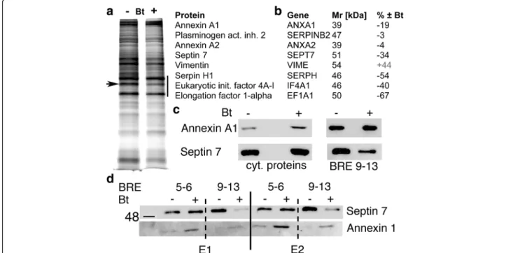
stained bands were excised, digested by trypsin and ana-lyzed by peptide shotgun mass spectroscopy (MS). Highly expressed proteins binding to the CALB2 promoter region included proteins implicated in transcription/translation such as eukaryotic initiation factors 4A-I and–III (IF4A1, EIF4A3) and elongation factors alpha (EF1A1) and 1-delta (EF1D) (Fig. 3a, b). Differentially expressed proteins (± Bt) consisted of annexin A1 (ANXA1; gene ID: 301; Mr
38.7 kDa;− 19%). Even lower amounts of septin 7 (SEPT7; gene ID: 989; Mr50.7 kDa;− 34%), a member of the family
of GTP-binding proteins and serpin H1 (SERPH; − 54%), also known as heat shock protein 47 (HSP47), reported to function as a chaperone for collagen [27], were present in the extracts of Bt-treated cells. Other proteins including annexin A2 (ANXA2; gene ID: 302; Mr38.6 kDa) and
plas-minogen activator inhibitor 2 (SERPINB2; gene ID: 5055; Mr47 kDa) were not differentially bound (Fig.3b). Of
inter-est, the mesenchymal marker vimentin was also present in the complex bound to BRE9–13 DNA and binding was in-creased in the Bt-treated MSTO-211H samples (+ 44%).
Since the decrease in BRE9–13 DNA-bound proteins in-cluding annexin A1 and septin 7 could be the result of a de-creased binding or of a Bt-induced decrease in protein expression, levels of annexin A1 and septin 7 were deter-mined by Western blot analysis of either cytosolic proteins or proteins eluted from the BRE9–13 DNA fragment in control and Bt-treated MSTO-211H cells. Expression levels of annexin A1 were increased in Bt-treated cells, while the amount of annexin A1 bound to BRE9–13 DNA was nearly unaffected by Bt. On the other hand, cytosolic septin 7 levels were slightly decreased by the Bt treatment in these conditions, while the amount of septin 7 bound to BRE9– 13 DNA was clearly lower after Bt treatment (Fig. 3c). To further demonstrate the specificity of the effect, similar ex-periments were carried out with the previously described DNA fragment containing BRE5–6, shown to act as a Bt-responsive repressor element in colon cancer cells [15]. In parallel, we compared binding of annexin A1 to both BRE-containing fragments, BRE5–6 and BRE9–13 using cyto-solic extracts from control or Bt-treated MSTO-211H cells (Fig. 3d). Elution of bound proteins was carried out in 2 conditions: weakly bound proteins eluted in fraction E1 and strongly bound proteins in fraction E2. Since no striking
Fig. 2 Sequence of the human CALB2 promoter region containing putative Bt-responsive elements BRE7–13 a The region of 1278 bp contains 7 putative BREs (cyan). Primer sequences to amplify the 1278-bp fragment are marked in yellow including restriction sites for SacI (Table1) and BglII (red). The start site of translation is marked in red (bold). Primers used to amplify truncated versions are boxed in gray (details are shown in Table1). b Sequence alignment of BRE5 - BRE13 with the BRE consensus sequence (15) shown at the bottom. Nucleotide sequences fully complying with the consensus (100% identity) are marked in blue, medium-conserved sequences are marked in green and nucleotides not conferring to the consensus sequence are marked in black; percentages of identity with the consensus sequence are given in the right lane. c, d Luc + reporter assay in MSTO-211H (upper) and ZL55 MM (lower) cells with CALB2 promoter fragments containing variable numbers of BREs. Luc + activities were normalized to the signal obtained with the control plasmid pGl3-P. As reference for the statistical analysis, BRE5–6 [15] was used. The number of independent experi-ments ranged from n = 4 (e.g. BRE10–13) to n = 11 (e.g. BRE7–13). Values represent mean ± S.D. *p < 0.05 vs. BRE5–6
qualitative differences were observed with respect to pro-teins eluted in fractions E1 and E2, the results from elution conditions E1 and E2 were combined. Similar as shown in Fig. 3c, the signal for septin 7 eluted from immobilized fragment BRE9–13 was clearly weaker using extracts from Bt-treated cells. Yet no significant differences were ob-served when using DNA fragment BRE5–6 as bait. An inverse situation was observed for annexin A1: increased binding to fragment BRE5–6 using extracts of Bt-treated cells and no striking changes in the binding to fragment BRE9–13 (Fig. 3d). In summary, annexin A1 preferentially bound to the DNA fragment BRE5–6 in a Bt-dependent manner, while septin 7 showed a Bt-sensitive decrease in binding to BRE9–13. Since BRE9–13 acted as a Bt-sensitive activator of CALB2 promoter activity and septin 7 bound to BRE9–13 was found to be decreased in extracts from Bt-treated cells, we reasoned that septin 7 might be a negative transcriptional regulator of CR expression by its binding within a protein complex that binds to the CALB2 pro-moter in MSTO-211H cells.
Calretinin and septin 7 act as negative transcriptional regulators of each other in MM cells and are inversely regulated by Bt
First, we investigated CR and septin 7 expression levels in immortalized human mesothelial cells and in human
MM cell lines (Fig.4a). Plotting CR vs. septin 7 levels of all tested cell lines revealed a clear clustering: epithelioid MM (ZL55, ZL5, JL1 and H226) and biphasic MM cells with a major part of epithelioid cells (MSTO-211H) formed the group of high CR-expressing cells (Fig. 4b). Somewhat lower CR levels were found in immortalized mesothelial cells (MeT-5A and LP9/TERT1), followed by the biphasic MM lines with mostly sarcomatoid cells (SPC111, SPC212 and H28) and sarcomatoid cells (ZL34). A clear inverse correlation was also detected in Bt-treated MM cells, where CR and septin 7 levels were determined after 72 h of Bt (1 & 2 mM) treatment. In MeT-5A, ZL55, ZL5, SPC212, ZL34 and MSTO-211H cells CR levels increased (as also shown in Fig. 1), while at the same time septin 7 was clearly down-regulated; representative examples of ZL5 and MSTO-211H cells are shown in Fig.4c1 and c2. Since these results merely indicated inverse regulation between CR and septin 7 expression by Bt in all investigated MM lines, the next experiments were aimed to alter either CR or septin 7 expression levels to determine the effect on the expres-sion levels of the other protein.
Thus, to directly investigate whether septin 7 acts as a negative transcriptional regulator of CR expression, sep-tin 7 levels were increased by lentiviral infection of MSTO-211H cells with LV-SEPT7. The increase in
Fig. 3 Proteins differentially bound to the BRE9–13 DNA fragment a Silver-stained gel of proteins eluted from the BRE9–13 DNA-containing column from control MSTO-211H cells (−) or from Bt-treated (1 mM) cells (+) for 72 h; the region between 35 and 50 kDa (black bar) showed the strongest signals. A band was clearly weaker in extracts from Bt-treated cells (arrow). b Differentially expressed proteins listed (from top to bottom) according to their abundance in the samples analyzed by peptide shotgun MS. c Western blot for annexin A1 (top) and septin 7 (bottom) of either total cytosolic proteins (left) or of the proteins bound to the BRE9–13 DNA (right). d Binding assay of MSTO-211H soluble whole cell extracts with either a DNA fragment containing BRE5–6 or BRE9–13 identified as the septin 7-binding region of the CALB2 promoter. Eluate E1 was obtained after low stringency conditions, E2 under high stringency conditions
Fig. 4 Analysis of septin 7 and CR levels in different cell lines of mesothelial origin and after experimental manipulations. a Relative expression levels of CR and septin 7 in various cell lines of mesothelial origin. b Semi-quantification of CR and septin 7: brown dots represent immortalized mesothelial cells, red dots epithelioid MM, orange dots bi-phasic and yellow dots sarcomatoid MM cell lines. c Effect of Bt administration on CR and septin 7 levels in MSTO-211H (C1) and ZL5 cells (C2). d Extracts of MSTO-211H cells infected with LV-septin7 or LV-GFP; note the decrease in CR in cells overexpressing septin 7. e MSTO-211H cells treated with Bt (+) or not (−) and subjected to down-regulation of septin 7 by shSEPT7; shGFP served as a negative control. f Up-regulation of CR by LV-CALB2 in SPC212 and HT-29 cells; note the decrease in septin 7 levels. The complete Ponceau-stained membranes corresponding to the Western blots shown in a and d are shown in Additional file1: Figure S3
septin 7 levels led to a down-regulation of CR expression (Fig. 4d), consistent with the previous results that lower amounts of septin 7 bound to BRE9–13 after Bt treatment are correlated with increased CR expression (Fig.3c). As a negative control, a lentivirus expressing GFP had no effect on CR expression levels. The opposite, i.e. down-regulation of septin 7 by SEPT7 shRNA strongly inhibited cell proliferation, decreased viability and many dying non-adherent cells were observed; the surviving cells had mostly a spindle-shaped morphology typical for the low CR-expressing subpopulation with sarcomatoid morph-ology of MSTO-211H cells (not shown). While the Bt-induced increase in CR was also seen in shSEPT7-treated cells, septin 7 down-regulation did not noticeably affect CR levels in the surviving MSTO-211H cell population when compared to levels in cells infected with GFP shRNA (Fig.4e). Since the Bt-induced increase in CR was associated with a decrease in septin 7 as evidenced in con-trol and GFP shRNA treated MSTO-211H cells (Fig.4e) indicative of a negative feedback regulation, we investi-gated the direct effects of CR overexpression on septin 7 levels. Lentivirus-mediated upregulation of CR using previously developed tools [25] caused a clear decrease in septin 7, irrespective, whether MSTO-211H cells were treated with Bt or not (Fig.4e). This indicates that CR is a Bt-independent, negative regulator of septin 7. The inverse experiment with shCALB2 was not conclusive, since shCALB2-mediated down-regulation of CR resulted in massive cell death as reported before [3], thus not allow-ing to investigatallow-ing septin 7 levels.
The effect of CR overexpression on septin 7 levels was investigated in SPC212 cells, as well as in the CR-expressing colon carcinoma cell line HT-29, previously shown to negatively modulate CR expression in a Bt-dependent way [15]. In both cell lines, CR overexpres-sion led to a clear decrease in septin 7 levels (Fig. 4f). This supports the hypothesis that CR functions as a negative regulator for septin 7 in two different cell types (mesothelial cells, colonocytes). In summary, treatment with Bt increases CR levels in all histotypes of MM cells and decreases septin 7 levels (data shown for MSTO-211H and ZL5 cells). Finally, overexpression of either one decreases expression levels of the other. These results are indicative of a finely tuned balance between expression levels of the 2 proteins.
Calretinin and septin 7 are co-expressed during mouse embryonic development and septin 7 levels are higher in primary mesothelial cells from mice without a functional Calb2 gene
Based on previous findings that CR is expressed in spe-cific regions within the mesenchyme of murine embryos and in precursor mesothelial cells in the developing lung at embryonic days 14.5 and 16.5 [25], we investigated
the expression of CR and septin 7 on serial sections de-rived from mouse embryos at E10.5. Expression patterns for CR and septin 7 showed an almost complete overlap in the mesenchyme of E10.5 mice (Fig.5a,b); yet the in-tensity of staining for one or the other protein varied no-ticeably. Since transient CR expression is observed in the mesenchyme and developing mesothelial cells [25], we wondered whether CR’s absence during this period in CR−/− mice would affect the expression of septin 7 in the terminally differentiated mesothelium. For this, pri-mary mesothelial cells (prMC) were isolated from either WT or CR−/− mice and kept in cell culture in vitro for 10–15 days. Western blots revealed clearly higher levels of septin 7 in mesothelial cells from CR−/− mice com-pared to WT animals (Fig. 5c). Thus, the transient ex-pression of CR in developing mesothelial cells had a long-lasting effect on septin 7 expression; it appeared that CR acted as a long-lasting repressor of septin 7 synthesis. A similar analysis was carried out in primary mesothelial cells immortalized with SV40. The expres-sion of SV40 Tag/tag was previously shown to substan-tially increase the proliferation rate of primary mesothelial cells, both from WT and CR−/− mice [25]. Interestingly, in SV40-immortalized cells, septin 7 ex-pression levels were strongly increased independent of the genotype (WT vs. CR−/−; Fig.5c, right panel).
Binding of septin 7 to calretinin and co-localization of the two proteins in distinct regions of the cleavage furrow and in the midbody region during cytokinesis
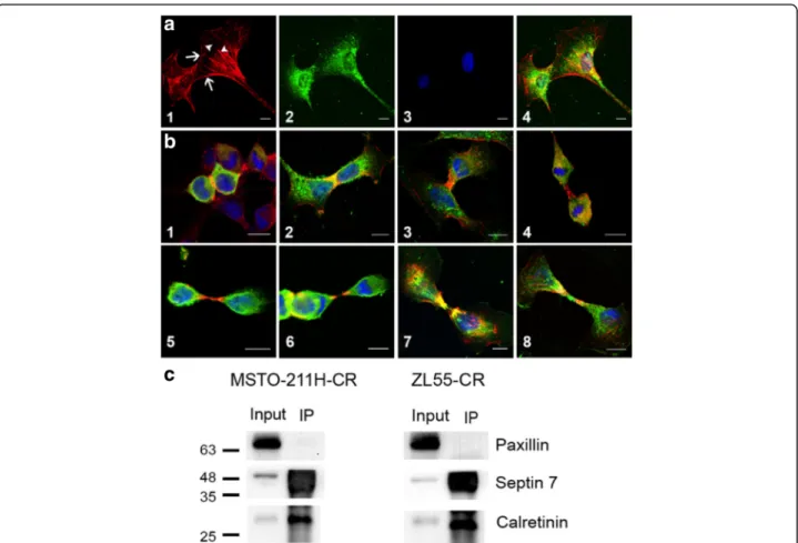
Based on the co-expression of CR and septin 7 in cells from embryonic mesenchymal tissue, we investigated in more detail, the intracellular localization of the two pro-teins in human MM cells. Since CR immunostaining was rather weak in most untreated MM cell lines, im-munofluorescence staining against CR was mostly carried out in cells overexpressing CR or in Bt-treated cells characterized by elevated CR expression levels. Qualitatively, CR intracellular localization was similar in all three conditions: control, CR-overexpressing, Bt-treated cells (data not shown). Septin 7 was either con-fined to the cell cortex, a zone implicated in the dynamic interplay between plasma membrane proteins and the cytoskeleton and to filamentous structures, likely the so-called “septin cytoskeleton” consisting of septin hetero-polymers (Fig. 6a1). In the same cell (e.g. Bt-treated MSTO-211H), the intracellular distribution of CR was rather homogenous (Fig.6a2), yet with a stronger stain-ing of perinuclear intermediate filaments and sometimes microtubules as reported before in WiDr colon cancer cells [28]. In WiDr cells a direct association of CR with these cytoskeletal structures has been reported previously, also based on co-immunoprecipitation experiments. The merged images including DAPI staining (Fig.6a4) showed
some colocalization (yellow color) that was further investi-gated in the different MM cell lines. During telophase sep-tin 7 was strongly localized to the zone of the cleavage furrow (Fig.6b1). Also CR staining was stronger in the re-gion of cell cleavage sometimes resulting in a yellow zone indicating that the proteins were concentrated in this re-gion (Fig.6b2). During the early phase of cytokinesis the center of the midbody showed strong “septin 7 only” staining (red) (Fig.6b3–6), followed first by a yellow zone
(septin 7 and CR) and finally by a green one (CR only). In some rare cases the center of the midbody was stained for CR only (Fig.6b8). Based on the co-localization of these proteins in some regions, we determined whether these two proteins directly interact. Co-immunoprecipitation revealed that in the lysate from MSTO-211H-CR and ZL55-CR cells incubated with anti-CR antibody, septin 7 was pulled down (Fig. 6c). These results suggested that CR and septin 7 have the predisposition to interact dir-ectly, but that other putative binding partners of either CR or septin 7 or different conformations of either protein prevented this interaction to take place throughout the cell. Their close spatial apposition (at times even co-localization indicative of direct interaction) and the pre-cise time points during cytokinesis indicates that the 2 proteins are likely implicated in the same biological
process, namely in cytokinesis, yet have on occasion dis-tinct localizations and thus probably non-identical, but conceivably complementary functions (see discussion).
Discussion
Although CR is currently used as a positive marker for the unequivocal identification of human MM of the epi-thelioid and mixed type [1, 2], still little is known I) about whether CR is implicated in the etiology of MM, II) on the regulation of CR in cells of mesothelial origin and III) about the function(s) of CR in MM cells. Details on the regulation [14] and possible function [3] of CR in MM are only slowly emerging. We had previously reported that Bt acts as repressor of CR expression in CR-positive colon cancer cells via BREs present in the CALB2promoter [15]. The same elements BRE5–6 were
found to be essentially non-functional in the immortal-ized mesothelial cell line Met-5A.
We had hypothesized that injury of the intestinal wall or damage of the lung tissue by e.g. asbestos fibers might lead to the colonization of the tunica serosa of the peri-toneal or pleural cavity by facultative anaerobic bacteria (e.g. Streptococcus pneumoniae, Staphylococcus aureus, Chlamydia pneumoniae in airways or gut microbiota) producing Bt resulting in concentrations in the millimolar
Fig. 5 Co-expression of calretinin and septin 7 Immunohistochemical analysis of E10.5 lung tissue stained for CR a1 and a2 and septin 7 b1 and b2. Scale bars: 250μm for a1 and b1, and 50 μm for a2 and b2. c Left: Septin 7 Western blot of extracts from cultured primary mesothelial cells from C57Bl/6J (WT) and CR−/− mice; note the lower levels of septin 7 in WT cells. Right: Septin 7 Western blot from SV40-immortalzed mesothelial cells derived from WT and CR−/− mice. As loading control (l.c.) an endogenous biotinylated protein (Mr≈ 75 kDa) was used.
range [19]. Thus, we tested the effect of Bt on CR expres-sion levels in immortalized mesothelial cells and several human MM cell lines of all histotypes, the former serving as a model for“reactive” mesothelial cells. The observed strong, rapid and Bt-concentration dependent increase in CR expression depended on the presence of a positive regulatory element in the CALB2 promoter containing additional BREs. Unexpectedly, septin 7, generally consid-ered as a structural protein was found to be part of this regulatory transcriptional complex. Septin 7 belongs to a family of RAS-like GTP-binding and membrane-interacting proteins; currently 13 septin genes subdivided into four dis-tinct groups are known in mammals and septin family members are characterized by a highly conserved domain structure [29]. The different septins are involved in various cellular processes mostly implicated in cell morphology dy-namics including cytoskeleton organization, cytokinesis and
membrane dynamics (e.g. curvature), for a review see [29]. Besides septins’ well-characterized preference for protein-protein interactions among own family members, forming higher-order structures such as filaments, bundles, scaffold-ing structures or rscaffold-ings, many other septin-interactscaffold-ing pro-teins have been identified forming the septin interactome (see Table1in [30]). In many cases these proteins are asso-ciated with the actin and/or microtubule cytoskeleton or with phospholipid membranes. Interestingly, the cdk1-dependent phosphorylation of septin 9 regulates the associ-ation with the proline isomerase (Pin1) implicated in the disjunction of daughter cells. The observed strong staining of septin 7 at the site of daughter cell separation indicates that septin 7 might have a similar function in MM cell cytokinesis. In support, septin 7-deficient fibroblasts show defects in the machinery implicated in cytokinesis including stabilization of microtubules and stalled midbody abscission
Fig. 6 Intracellular localization of calretinin and septin 7 in MM cells a Septin 7 localization (red; a1) and CR (green; a2) in MSTO-211H cells treated with 1 mM Bt. Cortical regions (arrows) and cytoskeletal-like structures (filaments, bundles; arrowheads) are strongly stained for septin 7. a3 Nuclear DAPI staining. a4) Merged images a1–3). Scale bars: 10 μm. b Localization of septin 7 (red) and CR (green) at different stages of cytokinesis starting from early telophase b1 until close to cell separation b8] in MM cell lines. Yellow indicates colocalization of the 2 proteins. DAPI-stained nuclei are shown in blue. Scale bars in all images: 10μm. 1) MSTO-211H-CR; 2) MSTO-211H treated with 1 mM Bt; 3) MSTO-211H-CR; 4) MSTO-211H treated with 1 mM Bt; 5) MSTO-211H-CR; 6) SPC111-CR; 7) ZL55-CR; 8) SPC212-CR. c Co-IP with lysates from MSTO-211H-CR and ZL55-CR cells. Proteins binding to CR were co-immunoprecipitated with a CR antibody. Membranes with PAGE-separated proteins were probed for CR and septin 7 (lane IP). Input samples containing all proteins (prior to immunoprecipitation) were probed for the same proteins. Paxillin was used as negative control
[31]. The latter defect was shown to also lead to multinucleation.
Knowledge on the role of septins in gene regulation is sparse. Septin 9 is capable of interacting with the hypoxia-inducible factor 1 alpha HIF-1α acting as a posi-tive regulator in the hypoxic pathway leading to in-creased proliferation, soft agar clonal survival and tumor growth [32]. In our study we observed that binding of septin 7 to the transcriptional complex driving CALB2 expression was decreased after Bt treatment. Since the Bt-dependent decrease in septin 7 binding to the protein complex bound to CALB2 BRE7–13 leading to increased CR levels might be the result of different processes, we selectively manipulated septin 7 expression in MM cell lines. Of note, Bt treatment had no effect on septin 7 ex-pression levels and moreover down-regulation of septin 7 levels by shSEPT7 had no effect on CR expression. On the other hand overexpression of septin 7 led to a strong decrease in CR expression levels indicating that septin 7 acts as a negative regulator (repressor) of CR expression. The mechanism was operating bi-directionally, i.e. CR overexpression resulted in a decrease in septin 7 levels. In line, a comparison of primary mesothelial cells (prMC) from WT and CR−/− mice revealed higher septin 7 levels in CR−/− prMC, albeit the fact that in these cells isolated from young adult mice, CR expression levels are below the detection limit of Western blot analysis [25].
In summary, these results indicate that both, CR and septin 7 act as negative transcriptional regulators for each other. As CR expression levels in tumor specimen are used to diagnose the clinical outcome, i.e. lower CR levels are a poor prognostic factor [33], also deregulated septin expression has been linked to tumor develop-ment/growth [34]. Increased levels of septin 2, 8, 9, 11 and decreased levels of septin 4 and 10 have been con-sistently reported in various tumor types and are as-sumed to act as oncogenes and tumor suppressor genes, respectively [34]. Evidently in order to exert an antagon-istic regulatory function, CR and septin 7 need to be co-expressed in situ, i.e. within the same cells. This was clearly evident during normal mouse embryonic develop-ment, where the two proteins colocalize in mesenchymal cells during lung development. While our experiments are in clear support of an inverse regulation, the colocalization and/or close apposition of each other might hint towards an implication in the same process, i.e. in cytokinesis. Immuno-EM images of WiDr colon cancer cells stained with an anti-CR antiserum had previously revealed CR staining during early telophase at the midbody, while at later stages the midbody zone was completely negative (see Fig.2f-iin [35]). Essentially identical findings on CR localization during cytokinesis were observed in various MM cell lines. Also septin 7 staining was particularly strong first at the cleavage furrow and then in distinct
parts of the midbody. Of note, strong co-localization was not observed during the entire separation of the daughter cells; at distinct time points and precise midbody localization one or the other protein was more prevalent resulting in a clear separation of the green and red fluores-cence. Yet occasionally, the strong yellow color indicated the 2 proteins to be colocalized at the same regions and co-IP experiments confirmed a direct interaction between CR and septin 7.
Septins have been also shown to associate to the mitotic spindle, being required for cytokinesis [31]; the same had been reported for CR-expressing WiDr cells [35, 36].
Conclusions
In this report we have identified septin 7, an essential cellular component implicated in the final steps of cell division, as a strong Bt-dependent gene regulatory pro-tein binding to the promoter of CALB2. Moreover, septin 7 was negatively regulated by CR forming a feed-back loop. Our study adds knowledge on the molecular mechanism involved in up-regulation of CR caused by Bt, a step that might also be implicated in mesothelio-magenesis. Since CR has been proposed to serve as a pu-tative target for MM therapy due to its essential role in MM cell lines [3], indirectly targeting CR through septin 7 and/or directly targeting septin 7 might represent yet another strategy for the development of a therapy to treat the currently incurable MM.
Additional file
Additional file 1:Figure S1. MTT assay of different MM cell lines exposed to butyrate (Bt). Relative MTT signals for A) MSTO-211H, B) ZL55 and C) ZL5 MM cells exposed to various Bt concentrations ranging from 0.33 to 5 mM. Results are from 3 independent experiments (each sample in triplicate). The value of untreated cells in each experiment was defined as 100%. Results represent mean±SEM. Figure S2. Point mutations in the PubMed database sequence (CALB2; gene ID: 794) of the CALB2 promoter region containing BRE7-13 in comparison to human Met-5A and ZL55 cells are boxed in green (Met-5A) or yellow (ZL55). Insertions or deletions found in the sequence of all analyzed cell lines are boxed in cyan. None of the mutations concern the 7 BRE listed in Fig.2. Figure S3. Top panel: Ponceau-Red stained membrane used for the Western blot shown in Fig.4a. Sizes of marker proteins range from 17 kDa (faintly stained lowest band mark-ing gel front) to 100 kDa (most upper band). Middle panel: Ponceau-Red stained membrane used for the Western blot shown in Fig.4d. The size of marker proteins ranges from 20 kDa to 135 kDa. Lower panel: Ponceau-Red stained membrane used for the Western blot shown in Fig.5c. The size of marker proteins ranges from 20 kDa to 135 kDa. (DOCX 1083 kb)
Abbreviations
BRE:Butyrate-responsive elements; Bt: Butyrate; CMF: Calcium- and magnesium-free; CR: Calretinin; E10.5: Embryonic day 10.5; HRE: Hypoxia-responsive elements HRE; MM: Malignant mesothelioma; prMC: Primary mesothelial cells
Acknowledgements
The authors wish to thank Valérie Salicio, Simone Eichenberger, Nadège Fahrni, Marlène Sanchez and Anne Oberson, University of Fribourg, for their excellent technical assistance.
Funding
The project was supported by the Swiss National Science Foundation (SNF grant no. 130680 and 139226 to B.S.). The funding body was not involved in the design of the study, the collection, analysis, and interpretation of data and in writing of the manuscript.
Availability of data and materials
All data generated or analyzed during this study are included in this published article [and its supplementary information files]. Authors’ contributions
BS conceived the study, MS, WB, JWR, and BS performed experiments; BS, WB, JWR, LP and BS analyzed data and BS, WB, JWR and LP wrote the paper. All authors have read and approved the final version of the manuscript. Ethics approval
All experiments requiring mouse tissue (embryos) were performed with permission of the local animal care committee (Canton of Fribourg, Switzerland) and according to the present Swiss law and the European Communities Council Directive of 24 November 1986 (86/609/EEC). Competing interests
The authors declare that they have no competing interests.
Publisher’s Note
Springer Nature remains neutral with regard to jurisdictional claims in published maps and institutional affiliations.
Received: 3 October 2017 Accepted: 17 April 2018
References
1. Gotzos V, Vogt P, Celio MR. The calcium binding protein calretinin is a selective marker for malignant pleural mesotheliomas of the epithelial type. Pathol Res Pract. 1996;192:137–47.
2. Doglioni C, Tos APD, Laurino L, Iuzzolino P, Chiarelli C, Celio MR, Viale G. Calretinin: a novel immunocytochemical marker for mesothelioma. Am J Surgical Pathol. 1996;20:1037–46.
3. Blum W, Schwaller B. Calretinin is essential for mesothelioma cell growth/ survival in vitro: a potential new target for malignant mesothelioma therapy? In J Cancer. 2013;133:2077–88.
4. Barberis MC, Faleri M, Veronese S, Casadio C, Viale G. Calretinin. A selective marker of normal and neoplastic mesothelial cells in serous effusions. Acta Cytol. 1997;41:1757–61.
5. Kitazume H, Kitamura K, Mukai K, Inayama Y, Kawano N, Nakamura N, Sano J, Mitsui K, Yoshida S, Nakatani Y. Cytologic differential diagnosis among reactive mesothelial cells, malignant mesothelioma, and adenocarcinoma: utility of combined E-cadherin and calretinin immunostaining. Cancer. 2000; 90:55–60.
6. Faas GC, Schwaller B, Vergara JL, Mody I. Resolving the fast kinetics of cooperative binding: Ca2+ buffering by calretinin. PLoS Biol. 2007;5:e311.
https://doi.org/10.1371/journal.pbio.0050311.
7. Schwaller B. The use of transgenic mouse models to reveal the functions of Ca2+buffer proteins in excitable cells. Biochim Biophys Acta. 2012;1820:
1294–303.
8. Pecze L, Blum W, Schwaller B. Mechanism of capsaicin receptor TRPV1-mediated toxicity in pain-sensing neurons focusing on the effects of Na+/
Ca2+fluxes and the Ca2+-binding protein calretinin. Biochim Biophys Acta.
2013;1833:1680–91.
9. Pecze L, Schwaller B. Characterization and modeling of Ca2+oscillations in
mouse primary mesothelial cells. Biochim Biophys Acta. 2015;1853:632–45. 10. Strauss KI, Kuznicki J, Winsky L, Kawagoe JI, Hammer M, Jacobowitz DM. The
mouse calretinin gene promoter region: structural and functional components. Brain Res Mol Brain Res. 1997;49:175–87.
11. Billing-Marczak K, Buzanska L, Winsky L, Nowotny M, Rudka T, Isaacs K, Belin MF, Kuznicki J. AP2-like cis element is required for calretinin gene promoter
activity in cells of neuronal phenotype differentiated from multipotent human cell line DEV. Biochim Biophys Acta. 2002;1577:412–20. 12. Schwaller B. Calretinin: from a“simple” Ca2+buffer to a multifunctional
protein implicated in many biological processes. Front Neuroanat. 2014;8:3. 13. Wisniewska MB, Nagalski A, Dabrowski M, Misztal K, Kuznicki J. Novel
beta-catenin target genes identified in thalamic neurons encode modulators of neuronal excitability. BMC Genomics. 2012;13:635.
14. Kresoja-Rakic J, Kapaklikaya E, Ziltener G, Dalcher D, Santoro R, Christensen BC, Johnson KC, Schwaller B, Weder W, Stahel RA, Felley-Bosco E. Identification of cis- and trans-acting elements regulating calretinin expression in mesothelioma cells. Oncotarget. 2016;7:21272–86. 15. Haner K, Henzi T, Pfefferli M, Kunzli E, Salicio V, Schwaller B. A bipartite
butyrate-responsive element in the human calretinin (CALB2) promoter acts as a repressor in colon carcinoma cells but not in mesothelioma cells. J Cell Biochem. 2010;109:519–31.
16. Cummings JH. Short chain fatty acids in the human colon. Gut. 1981;22:763–79. 17. Schwab M, Reynders V, Ulrich S, Zahn N, Stein J, Schroder O. PPARgamma is
a key target of butyrate-induced caspase-3 activation in the colorectal cancer cell line Caco-2. Apoptosis. 2006;11:1801–11.
18. Marilley D, Vonlanthen S, Gioria A, Schwaller B. Calretinin and calretinin-22k increase resistance towards sodium butyrate-induced differentiation in CaCo-2 colon adenocarcinoma cells. Exp Cell Res. 2001;268:93–103. 19. Shima K, Coopmeiners J, Graspeuntner S, Dalhoff K, Rupp J. Impact of
micro-environmental changes on respiratory tract infections with intracellular bacteria. FEBS Lett. 2016;590:3887–904.
20. Ghorbani P, Santhakumar P, Hu Q, Djiadeu P, Wolever TM, Palaniyar N, Grasemann H. Short-chain fatty acids affect cystic fibrosis airway inflammation and bacterial growth. Eur Respir J. 2015;46:1033–45. 21. Schmitter D, Lauber B, Fagg B, Stahel RA. Hematopoietic growth factors
secreted by seven human pleural mesothelioma cell lines: interleukin-6 production as a common feature. Int J Cancer. 1992;51:296–301. 22. Henzi T, Blum WV, Pfefferli M, Kawecki TJ, Salicio V, Schwaller B.
SV40-induced expression of calretinin protects mesothelial cells from asbestos cytotoxicity and may be a key factor contributing to mesothelioma pathogenesis. Am J Pathol. 2009;174:2324–36.
23. Aldridge GM, Podrebarac DM, Greenough WT, Weiler IJ. The use of total protein stains as loading controls: an alternative to high-abundance single-protein controls in semi-quantitative immunoblotting. J Neurosci Methods. 2008;172:250–4.
24. Calvo AC, Moreno-Igoa M, Manzano R, Ordovas L, Yague G, Olivan S, Munoz MJ, Zaragoza P, Osta R. Determination of protein and RNA expression levels of common housekeeping genes in a mouse model of neurodegeneration. Proteomics. 2008;8(20):4338–43.
25. Blum W, Pecze L, Felley-Bosco E, Schwaller B. Overexpression or absence of calretinin in mouse primary mesothelial cells inversely affects proliferation and cell migration. Respir Res. 2015;16:153.
26. Barnard JA, Warwick G. Butyrate rapidly induces growth inhibition and differentiation in HT-29 cells. Cell Growth Differ. 1993;4:495–501. 27. Widmer C, Gebauer JM, Brunstein E, Rosenbaum S, Zaucke F, Drogemuller
C, Leeb T, Baumann U. Molecular basis for the action of the collagen-specific chaperone Hsp47/SERPINH1 and its structure-collagen-specific client recognition. Proc Natl Acad Sci U S A. 2012;109:13243–7.
28. Marilley D, Schwaller B. Association between the calcium-binding protein calretinin and cytoskeletal components in the human colon
adenocarcinoma cell line WiDr. Exp Cell Res. 2000;259:12–22.
29. Mostowy S, Cossart P. Septins: the fourth component of the cytoskeleton. Nat Rev Mol Cell Biol. 2012;13:183–94.
30. Neubauer K, Zieger B. The mammalian Septin Interactome. Front Cell Dev Biol. 2017;5:3.
31. Menon MB, Sawada A, Chaturvedi A, Mishra P, Schuster-Gossler K, Galla M, Schambach A, Gossler A, Forster R, Heuser M, Kotlyarov A, Kinoshita M, Gaestel M. Genetic deletion of SEPT7 reveals a cell type-specific role of septins in microtubule destabilization for the completion of cytokinesis. PLoS Genet. 2014;10:e1004558.
32. Amir S, Wang R, Matzkin H, Simons JW, Mabjeesh NJ. MSF-A interacts with hypoxia-inducible factor-1alpha and augments hypoxia-inducible factor transcriptional activation to affect tumorigenicity and angiogenesis. Cancer Res. 2006;66:856–66.
33. Kao SC, Klebe S, Henderson DW, Reid G, Chatfield M, Armstrong NJ, Yan TD, Vardy J, Clarke S, van Zandwijk N, McCaughan B. Low calretinin expression and high neutrophil-to-lymphocyte ratio are poor prognostic factors in
patients with malignant mesothelioma undergoing extrapleural pneumonectomy. J Thorac Oncol. 2011;6:1923–9.
34. Liu M, Shen S, Chen F, Yu W, Yu L. Linking the septin expression with carcinogenesis. Mol Biol Rep. 2010;37:3601–8.
35. Gotzos V, Schwaller B, Hetzel N, Bustos-Castillo M, Celio MR. Expression of the calcium binding protein calretinin in WiDr cells and its correlation to their cell cycle. Exp Cell Res. 1992;202:292–302.
36. Gander JC, Gotzos V, Fellay B, Schwaller B. Inhibition of the proliferative cycle and apoptotic events in WiDr cells after down-regulation of the calcium-binding protein calretinin using antisense oligodeoxynucleotides. Exp Cell Res. 1996;225:399–410.