Continuous intravascular blood gas monitoring: development,
current techniques, and clinical use of a commercial device
M. Ganter and A. Zollinger*
Institute of Anaesthesiology and Intensive Care Medicine, Triemli City Hospital Zurich,
Birmensdorferstrasse 497, CH-8063 ZuÈrich, Switzerland
*Corresponding author. E-mail: [email protected]
This review focuses on the development, current techniques, and clinical use of continuous intravascular blood gas monitoring (CIBM) devices in anaesthesia and intensive care. The oper-ating principles, range of application, performance, limitations, costs, and impact on patient treatment and outcome, are discussed. Studies of early and currently available CIBM devices were analysed. At present, the Paratrend 7+â(PT7+â) for adults and NeotrendÔ (NTÔ) for newborns are the only commercially available CIBM systems. The PT7+âcontains three optical sensors to measure PO2, PCO2 and pH, as well as a thermocouple to measure temperature. The NTÔ is a modi®cation of the PT7+âto continuously monitor PO
2, PCO2, pH and tempera-ture in newborns. Under laboratory conditions, good performance over a wide range of blood gas values was observed with the Paratrend 7â(PT7â). Performance in the clinical setting was not as satisfactory, especially for PO2values. However, the performance and accuracy of CIBM devices appear to be suf®cient for clinical use and they are being used clinically in selected patient groups. Several factors affecting the performance of CIBM are considered.
Br J Anaesth 2003: 91; 397±407
Keywords: anaesthesia; blood, gas analysis; equipment, monitors; intensive care; monitoring, continuous intravascular blood gas
Arterial blood gas analyses are essential to monitor gas exchange in critically ill patients and during anaesthesia for major surgery. Usually, arterial blood samples are drawn intermittently and analysed in a central laboratory or by a point-of-care blood gas analyser. Several problems associ-ated with intermittent sampling have been described: indications for sampling are vague; analyses are often performed only after adverse events; sample transportation and analyses may be problematic; a therapeutic response can only be made after a delay; and there is a risk of blood loss and infection.5 15 26 39 60
Pulse oximetry, capnometry and transcutaneous blood gas measurement have been used for many years to assess gas exchange continuously and non-invasively. However, these non-invasive methods cannot fully replace arterial PO2 (PaO2), arterial PCO2(PaCO2), and arterial pH (pHa) analyses
in the clinical setting because of their signi®cant limita-tions.16 54 83Oxygen saturation assessed by pulse oximetry
(SpO2), or intravascular arterial oxygen saturation (SaO2)
sensors cannot detect a high PaO2, and de®nition of a safe
lower saturation limit is dif®cult.61 70 72 Moreover, pulse oximetry may display erroneous readings in the presence of dyshaemoglobinaemia, dyes (e.g. methylene blue, indigo carmine, indocyanine green), ambient light, low peripheral perfusion, motion artefacts, and other technical prob-lems.4 79Measuring end-tidal CO
2concentration by capno-metry only provides an accurate estimation of PaCO2 in
intubated patients with normal pulmonary function, a normal ventilation±perfusion ratio of the lungs, and normal haemodynamics. However, critically ill patients do have varying pulmonary and cardiovascular derangements, lead-ing to unreliable end-tidal CO2 values. Transcutaneous monitoring of PO2 and PCO2 is known to be accurate in small children, but many factors affect this technology in adults, including patient characteristics (variation in skin thickness, oedema, tissue hypoperfusion, administration of
REVIEW ARTICLES
vasoconstrictor agents), or technical problems (trapped air bubbles, improper placement, damaged membranes, inappropriate calibration, and frequent recalibration).9 55
During the past decade, marked advances in continuous intravascular blood gas monitoring (CIBM) have been achieved by miniaturization of the sensors measuring PO2, PCO2 and pH. CIBM appears to be desirable at least in selected patient groups, provided the technique proves to be reliable and cost-effective.
Methods
The literature on CIBM in anaesthesia and intensive care was retrieved using `Medline' searches (PubMed, National Library of Medicine). The following terms alone and in combination were used: continuous, arterial, intra-arterial, intravascular, blood gas, monitoring, measurement, device, sensor, paratrend, and neotrend. Laboratory and clinical evaluation studies, review articles and studies on risk± bene®t, costs and outcome of CIBM were selected. The reference lists of retrieved articles were further studied to complete the search on the topic of this review. Furthermore, manufacturers' instructions were obtained to describe the technology of the currently available commer-cial CIBM devices.
Statistics
Inconsistencies in the statistical analysis of comparisons between different measurement methods have been ad-dressed by Mantha and colleagues.41When assessing new technology, the use of adequate statistical methods and standard nomenclature are essential to draw valid conclu-sions.
To evaluate a new blood gas measuring device, for example a CIBM device, it should be compared with an established one, such as a laboratory blood gas analyser. Agreement between the two measurement techniques is best described by Bland and Altman analysis.6±8 The mean difference between values obtained from the new and the established measurement technique is the estimated bias. Measures of dispersion of this difference represent the random error inherent in either or both devices. It is termed precision in some studies. However, precision is a measure
of repeatability. In the past, the term precision was often incorrectly de®ned and was used in the wrong context. It was therefore suggested that this term should be avoided in measurement comparison studies.41Instead, upper limit of agreement (ULA) as bias +2 SD and lower limit of
agreement (LLA) as bias ±2SDshould be used. Bias 6 2 SD/limits of agreement are only estimates of the values that
apply to all the population measured. To know how precise the estimates are, one should also report the con®dence intervals (CI). Unfortunately, the correct statistical method has only been applied in a few studies on CIBM.
Development of CIBM
Technology and history
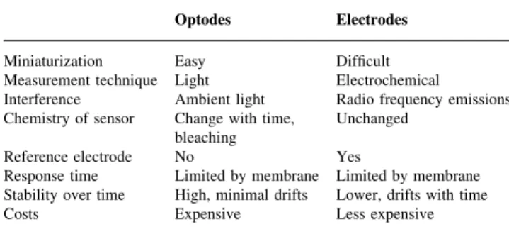
The principles, technology and history of CIBM have been described in detail in previous reviews.39 57 67 78 80 PO
2, PCO2 and pH can be measured by electrochemical and photochemical/optical sensors (Table 1). If both technolo-gies are combined, it is called a hybrid probe.
Electrochemical sensors
Electrochemical sensors have been used for intravascular PO2 measurement. The principle is a modi®ed Clark electrode.10 A small polarizing potential is maintained between the platinum cathode and the silver anode. The electrodes are immersed in an electrolyte solution sur-rounded by an oxygen-permeable membrane. Oxygen diffuses into the chamber, and is reduced at the platinum cathode, producing a current proportional to PO2.
Photochemical/optical sensors: optodes
Sample chambers containing dyes are illuminated with light of a speci®c wavelength via optical ®bres. The illuminating light will be variably transmitted, re¯ected, absorbed and re-emitted depending on the concentration of oxygen, carbon dioxide and hydrogen ions. The photochemical changes of the illuminating light will be used to calculate PO2, PCO2 and pH values.
Early multiparameter CIBM devices
Extensive research was done before commercialization of CIBM technology. Single parameter CIBM devices to measure PO2 electrochemically were developed within a short time of Clark introducing his electrode in 1956.3 10 Within a decade, Lubbers and Opitz published work on a CIBM probe with optodes to measure PO2and PCO2.36 50 Some years later in 1986, the same group described a multiparameter CIBM probe to assess PO2, PCO2and pH optically, and to measure temperature by a thermocouple.21 These devices preceded the early multiparameter CIBM devices (Table 2).
Table 1 Comparison of optical and electrochemical sensor technology Optodes Electrodes Miniaturization Easy Dif®cult Measurement technique Light Electrochemical
Interference Ambient light Radio frequency emissions Chemistry of sensor Change with time,
bleaching Unchanged Reference electrode No Yes
Response time Limited by membrane Limited by membrane Stability over time High, minimal drifts Lower, drifts with time Costs Expensive Less expensive
Table 2 Evaluation studies of early multiparameter CIBM devices. Setting: OR=operating room; ICU=intensive care unit; GS=general surgery; CS=cardiac su rgery; CVS=cardiovascular surgery; NS=neurosurgery; OLV=one lung ventilation; ORTHO=orthopaedic surgery. Insertion (site of measurement): RA=radial artery; FA=femoral artery; BA=brachial artery ; PA=dorsalis pedis artery; SV=superior vena cava. Application: mean in place duration in h. Recalibration: adjusting the original calibration curve using in vitro measured laboratory blood gas determinations Setting Insertion (site) Subjects (n) Samples (n) Application (h) Recalibration P O2 P CO 2 pH Range (kPa) [mean (min/max)] Bias 2 SD Bias 2 SD Range (kPa) [mean (min/max)] Bias 2 SD
Range mean (min/max)]
Bias 2 SD CDI Ô 1000 Blood Gas Monitoring System (CDI-3M Healthcare, Tustin, CA, USA) a Miller et al. ,1987 44 In vitro b ± 4 40 ± ± ± (2.67/13.33) ± ± ± ± ± (1.33/8.00) ± ± ± (7.05/7.65) ± ± Animal, dogs c FA 1 118 4.0 ± ± (±/±) ± ± ± ± ± (±/±) ± ± ± (±/±) ± ± Shapiro et al. ,1989 64 Animal, dogs FA 6 663 6.0 No 22.15 (3.60/41.73) ±2.27 12.35 ± ± 4.53 (2.40/12.27) 0.14 1.01 7.34 (7.05/7.57) ±0.02 0.06 ICU/OR RA 12 79 12.6 Yes 12.91 (4.53/32.93) ±0.16 2.49 ± ± 5.71 (3.20/8.93) 0.06 0.79 7.43 (7.30/7.62) 0.00 0.04 Mahutte et al. ,1990 40 Volunteers RA 4 48 ± Yes ± (6.13/84.40) ±0.39 6.77 ±5.0 23.6 ± (2.93/9.33) 0.10 0.65 ± (7.20/7.59) 0.00 0.04 Barker et al. ,1991 2 OR: GS, NS, ORTHO RA 14 87 4.2 No ± (±/±) ±1.20 6.21 ±6.0 20.0 ± (±/±) ±0.51 1.25 ± (±/±) ±0.03 0.08 Optex Biosentry âSystem (Optex Biomedical, The Woodlands, TX, USA) Smith et al. ,1992 66 OR: GS, NS RA 3 13 2.9 Yes ± (±/±) ±1.98 10.46 ± ± ± (±/±) 0.41 0.53 ± (±/±) ±0.02 0.08 Zimmerman et al. ,1993 84 ICU RA 5 104 55.4 Yes ± (5.87/34.00) ±0.79 3.52 ± ± ± (4.40/9.87) 0.23 1.62 ± (7.35/7.52) ±0.02 0.07 PB 3300 System (Puritan Bennett Corp., FoxS Division, Carlsbad, CA, USA) Lumsden et al. ,1994 38 In vitro ± 8 ± ± ± ± (2.67/33.33) 0.78 0.71 ± ± ± (1.33/13.33) 0.26 0.38 ± (6.80/7.70) 0.00 0.01 Haller et al. ,1994 23 ICU RA, BA, PA 13 487 72.0 No ± (4.00/69.60) ±0.32 1.73 ±2.8 13.2 ± (2.50/11.00) ±0.39 1.04 ± (7.23/7.55) ±0.04 0.06 Larson et al. ,1994 34 OR/ICU: NS, CS RA 29 552 6.0/46.0 No 15.06 (4.27/70.40) ± ± 1.0 30.0 4.93 (3.20/7.20) 0.17 0.88 7.39 (7.23/7.57) 0.01 0.08 Paolillo et al ,1994 52 OR/ICU: CS, CVS RA 27 283 25.0 ± ± (8.80/55.33) ±0.36 3.24 ± ± ± (3.20/8.00) 0.12 0.71 ± (7.14/7.63) ±0.02 0.06 Uchida et al. ,1994 71 OR: OLV, CVS RA 17 196 46.0 No ± (8.00/73.33) 0.12 7.97 ± ± ± (4.00/7.20) ±0.37 0.56 ± (7.28/7.53) 0.00 0.06 ICU 151 No ± 1.13 3.92 ± ± ± 0.52 1.01 0.01 0.07 Kilger et al ,1995 31 ICU RA, BA, PA 10 320 205.0 No ± (6.13/57.73) ±0.57 3.17 ± ± ± (3.33/10.53) ±0.37 1.20 ± (7.25/7.55) ±0.03 0.08 Pappert et al. ,1995 53 ICU RA 10 596 281.0 No ± (6.67/79.33) 0.25 2.11 1.9 11.5 ± (3.47/13.60) 0.08 0.67 ± (7.24/7.67) 0.01 0.04 Kurahashi et al. ,1996 32 OR/ICU: CS RA 46 319 87.0 No ± (±/±) 0.60 4.56 ± ± ± (±/±) 0.60 1.65 ± (±/±) 0.01 0.07 Oropello et al. ,1996 51 Animal, pigs FA 7 98 4.0 No ± (6.40/15.73) ±0.77 2.58 ± ± ± (2.93/5.73) ±0.55 0.78 ± (7.21/7.50) 0.04 0.09 SVC 6 86 4.0 No ± (2.53/13.20) ±1.05 2.28 ± ± ± (3.47/6.93) ±0.49 0.66 ± (7.17/7.48) 0.02 0.06 Roupie et al. ,1996 56 ICU RA 15 260 120.0 No ± (2.40/43.47) 0.21 2.99 ± ± ± (3.60/14.67) ±0.04 0.93 ± (6.84/7.57) 0.01 0.07 aThi s system was previo usly described by G ehrich and co lleagues. 21 bNo Bland± Altman analy ses were done, but linear regression analy sis (r , SEE ,stand ard error of estim ate in kPa): P O2 (0.99, 0.48 ), P CO 2 (0.99, 0.25 ), pH (0.98, 0.03). cN o Bla nd±Alt man ana lyses we re done, but linear reg ression ana lysis (r , SEE ,standa rd error of es timate in kPa): P O2 (0.93, 4.40 ), P CO 2 (0.96, ±) ,p H (0 .99, ±).
Table 3 Evaluation studies of more recent and currently available multiparameter CIBM devices. Probes (CIBM): NT Ô =Neotrend Ô , PT7 â=Paratrend7 â, PT7+ â=Paratrend 7+ â. Setting: OR=operating room, ICU=intensive care unit, CVS=cardiovascular surgery, NS=neurosurgery, OLV=one lung ventilation, thoracic part of surgery, abdominal part of sur gery, CPB=cardiopulmonary bypass, pre-, on-, post-bypass, ORTHO=orthopaedic surgery, LAP=laparoscopic surgery, CS=cardiac surgery. Insertion site: RA=radial artery, FA=femoral artery, CA=carotid art ery, UA=umbilical artery, JV=jugular vein, ChA=chorionic artery, PV=peripheral vein. Application: mean in place duration in hours. Recalibration: adjusting the original calibration curve using in vitro measured laboratory blood gas determinations Probe (CIBM) Setting Insertion (site) Subjects (n) Samples (n) Application (h) Recalibration P O2 P CO 2 pH Range (kPa) [mean (min/max)] Bias 2 SD Bias 2 SD Range (kPa) [mean (min/max)] Bias 2 SD
Range [mean (min/max)]
Bias 2 SD Studies on animals Clutton-Brock et al. , 1994 11 PT7 â Juvenile pigs CA 10 292 8.0 No ± (3.33/66.67) ±0.65 2.32 ±3.8 11.6 ± (2.67/10.67) 0.09 0.83 ± (6.80/7.40) ±0.03 0.08 Devlieger et al. , 2000 17 NT Ô Adult rabbits CA/JV 6 147 ± Yes 11.16 (2.02/22.53) ±0.56 2.91 ± ± 7.95 (3.40/16.27) 0.21 2.19 7.30 (6.98/7.50) ±0.02 0.06 NT Ô Fetal lambs ChA 4 20 2.1 Yes 4.39 (1.60/6.91) ±0.52 1.15 ± ± 7.75 (6.59/8.85) ±0.10 0.98 7.21 (7.14/7.30) 0.00 0.04 Studies on adult patients Clutton-Brock et al. , 1992 12 PT7 â ICU RA 25 461 47.0 ± ± (±/±) 0.29 7.36 ± ± ± (±/±) 0.18 1.40 ± (±/±) ±0.01 0.12 Venkatesh et al. , 1994 75 PT7 â ICU FA 10 71 14.0 Yes ± (12.40/26.90) 0.80 5.40 5.1 28.6 ± (3.30/6.80) 0.22 3.30 ± (7.21/7.53) 0.01 0.14 Venkatesh et al. , 1994 77 PT7 â ICU RA 13 158 42.9 Yes ± (8.00/59.60) 0.38 6.84 4.7 54.7 ± (3.50/6.90) 0.22 1.32 ± (7.31/7.61) 0.01 0.12 Liem et al. , 1995 35 PT7 â OR: LAP RA 27 27 ± ± ± (±/±) ±±± ± ± (±/±) ±0.31 0.55 ± (±/±) 0.00 0.05 Venkatesh et al. , 1995 76 PT7 â OR: CPB, pre RA 20 72 ± ± ± (17.33/61.87) ±0.93 9.07 ±3.0 28.0 ± (3.20/5.47) 0.20 0.53 ± (7.36/7.57) 0.02 0.10 PT7 â OR: CPB, on 157 ± (18.27/68.00) 0.40 12.00 0.5 28.0 ± (2.80/5.47) 0.07 0.53 ± (7.28/7.53) 0.01 0.12 PT7 â OR: CPB, post 174 ± (8.13/40.00) 0.53 7.20 14.0 34.0 ± (2.80/6.67) 0.20 1.07 ± (7.10/7.55) 0.02 0.12 Abraham et al. , 1996 1 PT7 â ICU RA 19 341 69.9 Yes ± (±/±) ± ± ±1.2 25.1 ± (±/±) 0.17 0.66 ± (±/±) 0.01 0.05 Nunomiya et al. , 1996 49 PT7 â ICU RA 9 62 72.0 Yes ± (6.03/72.32) ±0.22 5.33 ± ± ± (3.84/9.13) 0.07 0.54 ± (7.19/7.60) 0.00 0.04 Venkatesh et al. , 1996 79 PT7 â OR: ORTHO RA 10 30 1.4 ± ± (±/±) 0.16 5.20 0.4 28.0 ± (±/±) 0.07 0.48 ± (±/±) 0.02 0.06 Myles et al. , 1997 48 PT7 â OR: CPB RA 20 140 ± ± ± (±/±) ±1.14 11.52 ± ± ± (±/±) ±0.15 0.80 ± (±/±) 0.02 0.07 Zollinger et al. , 1997 85 PT7 â OR: OLV RA 23 138 3.7 No 24.00 (6.10/61.10) 0.38 9.71 ± ± 5.70 (4.10/9.50) 0.31 0.78 7.39 (7.19/7.50) ±0.02 0.07 Ishikawa et al. , 1998 29 PT7 â OR: OLV, thorac RA 12 84 ± Yes ± (6.27/59.87) ±0.13 10.67 0.8 43.2 ± (3.31/7.65) 0.12 0.83 ± (7.30/7.49) 0.00 0.04 PT7 â OR: OLV, abdom ± (9.60/34.00) 0.00 5.60 ±0.2 23.6 ± (4.04/6.21) 0.08 0.83 ± (7.34/7.47) ±0.01 0.08 Zaugg et al. , 1998 83 PT7 â OR: OLV RA 30 76 1.2 No 29.93 (4.93/83.33) 0.24 11.03 ± ± 5.33 (3.60/7.47) 0.01 0.55 7.40 (7.24/7.51) 0.01 0.04 Myles et al. , 1999 47 PT7 â OR: OLV RA 11 55 7.1 No ± (±/±) ±2.93 14.40 ± ± ± (±/±) ±0.21 1.57 ± (±/±) 0.01 0.10 Endoh et al. , 2001 18 PT7 â OR: CVS JV 18 101 ± Yes ± (3.50/16.00) ±0.16 1.20 ± ± ± (3.70/9.60) 0.00 0.92 ± (7.12/7.59) 0.01 0.07
The CDIÔ 1000 Blood Gas Monitoring System (CDI-3M Healthcare, Tustin, CA, USA; Table 2) consisted of ¯uorescent PO2, PCO2and pH sensors and a thermocouple. In vitro and in vivo animal studies by Miller and colleagues,44 and Shapiro and colleagues64showed excel-lent results. High fragility and poor results in clinical studies, especially for PO2, attributable to adherence to blood vessel walls (wall effect), rendered the commercial production and wider use of the system impossible.2 39 40
Smith, King and Schlain,66 and Zimmerman and Dellinger84 studied another CIBM device, the Optex Biosentryâ System (Optex Biomedical, The Woodlands, TX, USA; Table 2). Absorbance sensors were used for pH and PCO2, and a ¯uorescent sensor was used for PO2 assessment. Flexible optical ®bres allowed the indicator dye chamber to be located at the side of the probe, rather than at the tip, thus decreasing the wall effect problem. No further investigations or progress in marketing were made partly because of high costs and also because of poor accuracy of the probe in some studies.39
The PB 3300 (Puritan Bennett Corp., FoxS Division, Carlsbad, CA, USA; Table 2) was a different CIBM device which consisted entirely of ¯uorescent sensors. It was evaluated in laboratory, animal, and clinical studies (Table 2). Some improvements were made with this device by enabling circumferential sensing of gases which was associated with a reduced wall effect. The system has been withdrawn for economical reasons as a result of high costs and poor performance in some clinical investigations.39 56
Current state of continuous intravascular
blood gas monitoring
Paratrend 7+
âAt present, the Paratrend 7+â(PT7+â; Diametrics Medical Inc., High Wycombe, UK; distributed by Philips Medical Systems), and NeotrendÔ (NTÔ) are the only commer-cially available multiparameter CIBM systems (Table 3). Clutton-Brock, Hendry and Fink12described the technology of the Paratrend 7â(PT7â) in 1992. Originally, the PT7â was a hybrid probe incorporating four different sensors: a miniaturized Clark electrode to measure PO2; optodes to determine PCO2and pH (absorbance sensors, phenol red in bicarbonate solution); and a thermocouple (copper, con-stantan) to measure temperature and allow temperature correction of the blood gas values. The sensors were housed in a heparin-coated microporous polyethylene tube that was permeable to the analytes to be measured. In 1999, the manufacturer modi®ed the design of the PT7âand intro-duced a new probe, the PT7+â. The Clark electrode was replaced by an optical PO2-sensor (¯uorescent quenching sensor, ruthenium dye in silicone matrix). The other sensors and the main characteristics of the probe remained un-changed. According to the manufacturer, the new PO2
Ta ble 3 Co ntinued Probe (CIBM) Setting Insertion (site) Subjects (n) Samples (n) Application (h) Recalibration P O2 P CO 2 pH Range (kPa) [mean (min/max)] Bias 2 SD Bias 2 SD Range (kPa) [mean (min/max)] Bias 2 SD
Range [mean (min/max)]
Bias 2 SD Menzel et al. , 2001 43 PT7+ â OR: NS FA 20 124 4.0 ± 36.82 (10.00/50.00) 0.20 4.08 ± ± 5.08 (±/±) 0.25 0.46 7.45 (±/±) ±0.02 0.04 Studies on paediatric patients Weiss et al. , 1996 81 PT7 â ICU FA 5 150 127.0 ± ± (5.20/28.53) ± ± 1.9 34.2 ± (3.60/14.00) ±0.10 1.25 ± (7.12/7.58) 0.01 0.05 Hatherill et al. , 1997 27 PT7 â OR/ICU: CS FA 10 100 27.0 No 5.30 (2.50/8.20) 0.04 0.87 ± ± 4.76 (2.71/7.09) ±0.44 0.74 7.39 (7.14/7.59) 0.02 0.06 Tobias et al. , 1998 69 PT7 â ICU PV 4 17 99.0 ± ± (±/±) ± ± ± ± 5.07 (3.87/6.80) 0.40 0.37 7.38 (7.16/7.50) 0.04 0.04 Morgan et al. , 1999 46 NT Ô ICU UA 27 753 120.7 Yes ± (±/±) ±0.19 1.98 ± ± ± (±/±) 0.26 1.04 ± (±/±) 0.00 0.04 Weiss et al. , 1999 82 PT7 â ICU RA/FA 24 414 101.0 Yes ± (±/±) 0.16 6.40 ± ± ± (±/±) ±0.24 1.68 ± (±/±) 0.01 0.06 Tobias et al. , 2000 68 PT7 â ICU PV 23 100 115.2 ± ± (±/±) ±±± ± ± (3.33/10.40) ±0.28 0.72 ± (7.24/7.54) 0.03 0.06 Coule et al. , 2001 14 PT7 â ICU RA/FA 50 1445 108.0 Yes ± (4.53/64.00) 0.10 6.60 ± ± ± (1.81/15.23) ±0.05 1.28 ± (6.99/7.66) 0.00 0.08
sensor measures PO2more precisely, with a faster response time and less artefacts. Unfortunately, most evaluation studies published thus far have been of the PT7â; only one study by Menzel and colleagues43 has evaluated the new PT7+âprobe (Table 3).
NeotrendÔ
NeotrendÔ (NTÔ; Diametrics Medical Inc., High Wycombe, UK; distributed by Philips Medical Systems), a modi®cation of the PT7+â, was designed for CIBM in the umbilical artery of newborn infants. As in the PT7+â, PO
2, PCO2 and pH are measured by ®breoptic sensors and the temperature is determined by a thermocouple (Table 3).
Current clinical use
Site of measurement
The most common site for CIBM measurement is the radial artery in adults and the femoral artery in children, particu-larly if they are <5 yr old. There are some disadvantages of the radial approach: it is more susceptible to motion and positional artefacts, vasospasm, and changes in peripheral blood ¯ow. Nevertheless, this approach is chosen routinely in adults and older children, because of easy access, the double blood vessel supply of the hand, and low compli-cation rates.75 82 The umbilical artery is used for probe insertion in neonates.46 The incidence of catheter-related complications with the NTÔ is low and does not differ from that observed with a standard umbilical artery catheter.13 Other sites of measurement are peripheral veins in chil-dren,68 69the jugular venous bulb in adults,18 and carotid arteries, chorionic arteries and jugular veins in animal studies.11 17
Evaluation studies of current CIBM devices, and
ranges of application
All the evaluation studies published with currently available commercial CIBM devices are listed in Table 3. CIBM has
been applied in various clinical settings in the operating room and the intensive care unit, as well as in experimental studies (Table 4). Different study designs and different statistical analyses render comparison of these studies dif®cult. Moreover, there are methodological limitations. For example, several studies corrected a perceived bias during the study period by adjusting the original calibration curve using in vitro laboratory blood gas determinations (`recalibration'). This may be necessary after prolonged monitoring in the clinical setting, according to the recom-mendations of the manufacturer. However, the issue of `recalibration' was not mentioned in all of the studies, and `recalibration' was performed at different time points. Similarly, the temperature at which PO2, PCO2 and pH were measured and compared was not mentioned in all the studies. Most authors did not address the issue of tempera-ture correction (i.e. the use of alpha-stat or pH-stat). Furthermore, the number of measurements per patient varied in all the studies. Some performed a large number, others only a few measurements with a single probe or patient.
An animal study with the PT7âshowed good perform-ance (Table 3) over a wide range of blood gas values under laboratory conditions.11Another study on animals with an NTÔ probe presented the feasibility and accuracy of ®breoptic multiparameter sensing in fetal monitoring. The NTÔ probe showed good performance at low PO2levels (PO2<6.7 kPa; bias (2SD): ±0.2 (1.5) kPa). However, as a result of movement and interference by the endoscopic light, readings for all variables were only available for about 50% of the operating time. The authors concluded that extensive modi®cation of the sensor design would be necessary before the sensor could be used routinely in this area.17
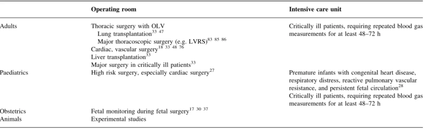
There are several clinical evaluation studies from the operating theatre and the intensive care unit. Most studies in the operating room are in adults undergoing one lung ventilation for major thoracic surgery (thoracoscopic sur-gery, lung transplantation),47 83 85or major thoracoabdom-inal surgery (oesophagectomy).29Studies in cardiac and/or vascular surgery,18 27 48 76 major orthopaedic surgery,79
Table 4 Applications of CIBM. OLV=one lung ventilation, LVRS=lung volume reduction surgery
Operating room Intensive care unit Adults Thoracic surgery with OLV
Lung transplantation33 47
Major thoracoscopic surgery (e.g. LVRS)83 85 86
Critically ill patients, requiring repeated blood gas measurements for at least 48±72 h
Cardiac, vascular surgery18 33 48 76
Liver transplantation33
Major surgery in critically ill patients33
Paediatrics High risk surgery, especially cardiac surgery27 Premature infants with congenital heart disease,
respiratory distress, reactive pulmonary vascular resistance, and persistent fetal circulation28
Critically ill patients, requiring repeated blood gas measurements for at least 48±72 h
Obstetrics Fetal monitoring during fetal surgery17 30 37
laparoscopic surgery,35and neurosurgery43have also been done. Blood gas values were measured over wide ranges (PO2 3.5±83.3 kPa, PCO2 1.8±15.2 kPa, pH 6.99±7.66). Overall performance in PO2 measurement was relatively poor (bias ±2.9 to 0.8 kPa, 2 SD of bias 0.5±14.4 kPa).
However, looking at the clinically important lower range of PO2, much better results were obtained. Zaugg and colleagues83found a bias (2SD) of ±0.5 (2.2) kPa for PO
2 <13.3 kPa in patients undergoing thoracoscopic surgery; Nunomiya and colleagues49 showed a bias (2 SD) of ±0.3 (1.7) kPa for PO2 <13.3 kPa; Coule and colleagues14 measured a bias (2SD) of 0.24 (2.18) kPa for PO2<8.0 kPa in children in the intensive care unit (in this article the term precision was used and assumed to be 1 SDof the bias);
Weiss and colleagues82 obtained a bias (2SD) of 0.1 (2.5) kPa for PO2<9.3 kPa in children in the intensive care unit; and Hatherill and colleagues27presented a bias (2SD) of 0.0 (0.8) kPa for PO2values ranging from 2.5 to 8.2 kPa (mean 5.3 kPa) in children with cyanotic heart disease. These results are comparable with the animal study by Devlieger and colleagues.17 Performance of PCO
2and pH measure-ment was acceptable over the whole ranges.
Studies in the intensive care unit were done in adults,1 12 49 75 77 as well as in children and neo-nates.14 27 46 68 69 81 82Despite poor reporting, the ranges of measured blood gas values were smaller compared with the operating theatre studies, despite the probes being used over a much longer time period. The reported mean longest duration of use was 127 h (5.3 days),81 and the single longest duration of use was 429 h (17.8 days).46 Furthermore, the number of measurements per patient or probe, respectively, was much greater in the intensive care unit (17 measurements per patient or probe in the intensive care unit vs six in the operating room), and `recalibrations' were done more frequently in the intensive care unit.
Two studies reported on the PT7âsensors inserted in a peripheral vein in paediatric patients.68 69Only the values for i.v. PCO2 and pH were compared with the respective arterial values.
Reliability, accuracy and consistency
In the technical speci®cation sheet of the PT7+â and the NTÔ, the manufacturer presents promising in vitro data with blood gases and temperature measured over wide ranges (PO2 2.6±66.6 kPa, PCO2 1.3±10.6 kPa, pH 6.80± 7.80, temperature 10±42°C) with a good performance. Using gas-tonometered solutions, 95% con®dence limits were: 65% or 60.4 kPa (whichever is greater) of the actual values for a PO2<16 kPa, and 610% for a PO2>16 kPa; 60.4 kPa for PCO2; 60.03 for pH; and 60.3°C for temperature. At 37°C, it took <15 s for the sensor to start responding to a change in blood gases, and the 90% response time for the sensor was 180 s or less. Drifts of the sensors were <0.5% h±1for PO
2and PCO2, and <0.005 pH units h±1.1
Comparison of blood gas variables from continuous intravascular sensors with those from laboratory blood gas analysers is a controversial issue. Blood gas variables vary substantially within short periods of time even in stable patients in the intensive care unit. The accuracy of laboratory blood gas analysers can be quanti®ed in the laboratory where the values to be measured are known. However, this is not the case in clinical studies. Thus, the clinical performance of an optode-based intravascular probe must be judged in comparison with an electrode-based blood gas analyser, for which clinical performance cannot be speci®cally quanti®ed.62Moreover, resulting values for bias (2SD) of a CIBM device not only re¯ect the accuracy of
the intravascular device, but also depend on the accuracy of the laboratory blood gas analyser used as a reference. Laboratory blood gas analysers (even between individual analysers of the same manufacturer), also have inconsist-encies.24 25The bias of intravascular sensors could either be reduced or increased if the blood gas analysers also showed high levels of bias. Poor repeatability [bias (2 SD)] of
variables obtained by the blood gas analyser would, in contrast, inherently result in poorer repeatability for the variables from the intravascular sensors.11Blood gas values measured by a laboratory blood gas analyser may also be affected by pre-analytic sample errors. Therefore, even with an ideal laboratory blood gas analyser, the measured blood gas values would never completely re¯ect the real blood gas values in vivo. Since intermittently drawn blood samples analysed by laboratory blood gas analysers are the clinical standard of care, all authors used this procedure as a reference method to assess the accuracy of CIBM.
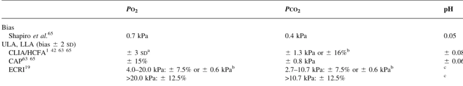
No of®cial recommendations concerning performance of continuous intravascular blood gas devices exist. However, several guidelines for laboratory blood gas analysers have been published by the Clinical Laboratory Improvement Amendment/Health Care Finance Administration (CLIA/ HCFA), the College of American Pathologists (CAP), and the Emergency Care Research Institute (ECRI) (Table 5). If they are applied to the published evaluation studies of PT7â and NTÔ (Table 3), most of the bias values lie within these recommended ranges. Concerning repeatability, the situ-ation is less clear as a result of poor reporting of the distribution of the measured PO2, PCO2and pH values. PO2 measurements met the recommendations least, although a much better performance was obtained in the clinically important lower PO2range as discussed above. In contrast, values for PCO2 and pH were more acceptable and comparable to the given recommendations.
As temperature in the radial artery re¯ects an intermedi-ate or even peripheral temperature, it is not surprising that values for temperature with the PT7â were lower (bias ± 0.5°C) than those recorded rectally.85
On the screen of CIBM devices, additional values such as oxygen saturation, bicarbonate concentration, and base excess are provided, which are continuously calculated based on algorithms and normograms. These variables
agreed poorly with those calculated by laboratory blood gas analysers in a number of studies.18 27 85 This may re¯ect differences in the built in algorithms.73 Interestingly, the poor agreement between two different blood gas analysers for bicarbonate and base excess was similar.22 Such variations between laboratory blood gas analysers render estimation of the accuracy of these calculated values impossible.
Response times were analysed and found to correlate well with the values in the manufacturer's ®gures for the PT7â. Mean 90% in vitro response time was 70 s for PO2, 143 s for PCO2 and 78 s for pH.77 Response times in vivo were comparable, as shown in clinical studies. The PT7âwas also evaluated during cemented total hip replacement. As a result of the instantaneous effect of acetabular and femoral cementation, mean PaO2values dropped signi®cantly within
90 s (mean onset time) following the application.79 Furthermore, response times from four sensors were inves-tigated after using them in patients undergoing cardiopul-monary bypass. The mean 90% post-in vivo response time was 100 s for PO2, 122 s for PCO2, and 123 s for pH, respectively. Additionally, these sensors were exposed to known concentrations of en¯urane, iso¯urane and ha-lothane. There was no interference with the pH and PCO2 sensors by any of the anaesthetic gases tested, but halothane interfered with PO2 measurement.76 Other investigations exposed PT7âsensors to the ¯uorescent drug propofol as well as to bone cement (methyl methacrylate), and sterile polymer powder in vitro. No interference of these sub-stances with blood gas measurement was found.77 79 Halothane interference with PO2 measurement in the PT7âprobe was attributable to the electrochemical reduc-tion of halothane by the Clark electrode used on the PT7â probe. None of the anaesthetic agents have any effect on the optical measurement of PO2, and so the PT7+âprobe should be free from this interference.
Sensor drifts in vivo were reported in an early animal study. Mean variation of the bias per hour was acceptable at ±0.59% for PO2, 0.42% for PCO2 and ±0.002 pH units, respectively.11 These results were con®rmed in humans undergoing cardiopulmonary bypass (i.e. mean variation of
the bias per hour was ±0.43% for PO2, 0.62% for PCO2and 0.001 pH units).76
Limitations and complications
Reliable intravascular blood gas measurement depends on a number of mechanical, electrical and physicochemical properties of the CIBM probe as well as the conditions of the vessel into which the probe is inserted. Blood ¯ow in the vessel containing the probe is most critical. CIBM becomes unreliable if blood ¯ow decreases or stops, for example during in¯ation of a sphygmomanometer cuff, because of vasospasm, or during cardiopulmonary resuscitation. PO2 was shown to be the most ¯ow-dependent variable. Insertion of the probe into a femoral artery yields more reliable results compared with those from the radial artery during low ¯ow states.74 75If the sensor becomes attached to the wall of the vessel, thus measuring a combined blood and tissue PO2, the oxygen value may be falsely low. Hybrid probes (PT7â) are less susceptible, as the PO2electrode has a larger surface area. Arterial catheters are ¯ushed in vivo with a continuous solution. If the sensing probe is not inserted for an adequate distance over the tip of the arterial catheter, it may measure the blood gas variables in the ¯ush solution, resulting in errors known as the `¯ush effect'.78 Interference from electrocautery and ambient or endoscopic light may be another source of erroneous blood gas values.17 82Damping of the arterial pressure tracing or dif®culty in withdrawing blood from the arterial catheter was reported in one study, and occurred after an average of 35.4 h of measurement in only four patients (15%).1 However, even without an indwelling CIBM probe, the incidence of damping or dif®culty in withdrawing blood from radial artery catheters was reported to be 17±26%.58Without any adverse events, clot formation (incidence 8±30%) was observed in three studies.29 68 82Poor robustness of the ®breoptic cables led to bending and kinking in up to 20% of patients, resulting in probe malfunctioning and a short user life.1 27 68 82Finally, before measurement, a 30 min warm-up time is mandatory for calibration. Hence, the device is not immediately available. The unit is also quite bulky.
Table 5 Quality recommendations for laboratory blood gas analysers. ULA=upper limit of agreement, LLA=lower limit of agreement, CLIA/HCFA=Clinical Laboratory Improvement Amendment/Health Care Finance Administration, CAP=College of American Pathologists; ECRI=Emergency Care Research Institute
PO2 PCO2 pH
Bias
Shapiro et al.65 0.7 kPa 0.4 kPa 0.05
ULA, LLA (bias 6 2SD)
CLIA/HCFA1 42 63 65 6 3SDa 6 1.3 kPa or 6 16%b 6 0.08
CAP63 65 6 15% 6 0.8 kPa 6 0.06
ECRI19 4.0±20.0 kPa: 6 7.5% or 6 0.6 kPab 2.7±10.7 kPa: 6 7.5% or 6 0.6 kPab c
>20.0 kPa: 6 12.5% >10.7 kPa: 6 12.5% c aSDof a pro®ciency blood gas analyser used as reference;bwhichever is greater;cthere is no reference method for pH measurement for laboratory
Costs
Costs are important, and new monitoring devices are warranted only if cost effectiveness may be shown. Either reduction in overall cost or an improvement in patient outcome is required. Some cost±bene®t analyses on CIBM have been done, but different results have been obtained. Some authors recommended the use of CIBM and presented cost-savings,20 82 others argued against this technology because of high costs and low bene®ts (i.e. the monitoring system far exceeding the clinical needs).28 This may be attributable to a differing basis for the analyses. Costs assumed for one conventional, intermittent laboratory blood gas analysis varied between 2.50 and 80.00 euros per sample. Some investigations assessed only the direct costs of CIBM, in other words, approximately 20 000 euros for the satellite monitor including a patient data module, 15 000 euros for the calibration unit, and 500 euros for each one way sensor. Others also took indirect costs into account, for example administrative costs, laboratory support, and specialized personnel. Therefore, no concluding data are currently available on the cost±bene®t ratio of CIBM.
Outcome
The impact of undetected changes in arterial blood gases on patient outcome has not yet been investigated. Experimental studies reported myocardial ischaemia with a potential risk of myocardial damage even when the period of hypoxia was short, if coronary blood ¯ow was limited.59Pa
O2<8.0 kPa,
as often observed during thoracic surgery with one lung ventilation, is thought to be the threshold that should be detected. It is associated with an increased risk of perioperative myocardial ischaemia in susceptible individ-uals.45 83
Although CIBM appears to be advantageous, there are no prospective, randomized, double-blind studies of its impact on morbidity and mortality, length of intensive care unit and hospital stay, or myocardial and cerebral ischaemia. Future outcome studies should focus on well-de®ned groups of selected patients who might bene®t from CIBM (e.g. critically ill patients with potentially rapid and unexpected changes in blood gas values). Nevertheless, it may be dif®cult, if not impossible, to show a positive impact of CIBM on patient outcome.
Conclusions
Development of CIBM devices has resulted in the com-mercially available PT7+âprobe for adults and NTÔ probe for newborns. Changes in blood gas values may be immediately and reliably detected in the clinical setting without signi®cant side-effects or complications. Indications for CIBM, in terms of evidence-based medicine, are still lacking since the cost±bene®t ratio and the impact on patient outcome are unknown. Nevertheless, CIBM is
being used by clinicians in selected patient groups in the operating theatre and the intensive care unit. It is useful during surgery where blood gas values can change rapidly and unexpectedly, for example during thoracic surgery with one lung ventilation, cardiovascular surgery and organ transplantation. CIBM is useful in critically ill patients needing a considerable number of blood gas determinations over a long period, for example premature infants with severe cardiopulmonary disease, and patients with acute respiratory distress syndrome, sepsis, and severe trauma.
References
1 Abraham E, Gallagher TJ, Fink S. Clinical evaluation of a multiparameter intra-arterial blood-gas sensor. Intensive Care Med 1996; 22: 507±13
2 Barker SJ, Hyatt J. Continuous measurement of intra-arterial
pHa, PaCO2, and PaO2in the operating room. Anesth Analg 1991;
73: 43±8
3 Barker SJ, Tremper KK. Intra-arterial oxygen tension monitoring. Int Anesthesiol Clin 1987; 25: 199±208
4 Benson JP, Venkatesh B, Patla V. Misleading information from pulse oximetry and the usefulness of continuous blood gas monitoring in a post cardiac surgery patient. Intensive Care Med 1995; 21: 437±9
5 Biswas CK, Ramos JM, Agroyannis B, Kerr DN. Blood gas analysis: effect of air bubbles in syringe and delay in estimation. Br Med J (Clin Res Ed) 1982; 284: 923±7
6 Bland JM, Altman DG. Comparing methods of measurement: why plotting difference against standard method is misleading. Lancet 1995; 346: 1085±7
7 Bland JM, Altman DG. Comparing two methods of clinical measurement: a personal history. Int J Epidemiol 1995; 24 [Suppl. 1]: S7±14
8 Bland JM, Altman DG. Statistical methods for assessing agreement between two methods of clinical measurement. Lancet 1986; 1: 307±10
9 Cassady G. Transcutaneous monitoring in the newborn infant. J Pediatr 1983; 103: 837±48
10 Clark LC. Monitor and control of blood and tissue oxygen tensions. Trans Am Soc Artif Intern Organs 1956; 2: 41±8 11 Clutton-Brock TH, Fink S, Markle D, Luthra AJ, Hendry SP. The
evaluation of a new intravascular blood gas monitoring system in the pig. J Clin Monit 1994; 10: 387±91
12 Clutton-Brock TH, Hendry SP, Fink S. Preliminary clinical evaluation of the Paratrend 7 intravascular blood gas monitoring system. Intensive Care Med 1992; 18: S154
13 Cohen RS, Ramachandran P, Kim EH, Glasscock GF. Retrospective analysis of risks associated with an umbilical artery catheter system for continuous monitoring of arterial oxygen tension. J Perinatol 1995; 15: 195±8
14 Coule LW, Truemper EJ, Steinhart CM, Lutin WA. Accuracy and utility of a continuous intra-arterial blood gas monitoring system in pediatric patients. Crit Care Med 2001; 29: 420±6
15 Dennis RC, Ng R, Yeston NS, Statland B. Effect of sample dilutions on arterial blood gas determinations. Crit Care Med 1985; 13: 1067±8
16 Desiderio DP, Wong G, Shah NK, Liu J, Loughlin CJ, Bedford RF. A clinical evaluation of pulse oximetry during thoracic surgery. J Cardiothorac Anesth 1990; 4: 30±4
17 Devlieger R, Gratacos E, Wu J, et al. Continuous monitoring of
in animal models reproducing in utero conditions. Fetal Diagn Ther 2000; 15: 127±31
18 Endoh H, Honda T, Oohashi S, Nagata Y, Shibue C, Shimoji K. Continuous intra-jugular venous blood-gas monitoring with the Paratrend 7 during hypothermic cardiopulmonary bypass. Br J Anaesth 2001; 87: 223±8
19 ECRI (Emergency Care Research Institute). Blood gas/pH analyzers. Health Devices 1995; 24: 208±43
20 Franklin ML, Peruzzi WT, Moen SG, Shapiro BA. Evaluation of an on-demand, ex vivo bedside blood gas monitor on pulmonary artery blood gas determinations. Anesth Analg 1996; 83: 500±4 21 Gehrich JL, Lubbers DW, Opitz N, et al. Optical ¯uorescence
and its application to an intravascular blood gas monitoring system. IEEE Trans Biomed Eng 1986; 33: 117±32
22 Graystone SJ. Continuous intra-arterial blood gas monitoring. Br J Anaesth 1997; 79: 815±16; author reply: 816±17
23 Haller M, Kilger E, Briegel J, Forst H, Peter K. Continuous intra-arterial blood gas and pH monitoring in critically ill patients with severe respiratory failure: a prospective, criterion standard study. Crit Care Med 1994; 22: 580±7
24 Hansen JE, Casaburi R, Crapo RO, Jensen RL. Assessing precision and accuracy in blood gas pro®ciency testing. Am Rev Respir Dis 1990; 141: 1190±3
25 Hansen JE, Jensen RL, Casaburi R, Crapo RO. Comparison of blood gas analyzer biases in measuring tonometered blood and a ¯uorocarbon-containing, pro®ciency-testing material. Am Rev Respir Dis 1989; 140: 403±9
26 Harsten A, Berg B, Inerot S, Muth L. Importance of correct handling of samples for the results of blood gas analysis. Acta Anaesthesiol Scand 1988; 32: 365±8
27 Hatherill M, Tibby SM, Durward A, Rajah V, Murdoch IA. Continuous intra-arterial blood-gas monitoring in infants and children with cyanotic heart disease. Br J Anaesth 1997; 79: 665±7 28 Hoffer JL, Nor¯eet EA. Con: is continuous intra-arterial blood gas and pH monitoring justi®able? J Clin Monit 1996; 12: 183±9 29 Ishikawa S, Makita K, Nakazawa K, Amaha K. Continuous
intra-arterial blood gas monitoring during oesophagectomy. Can J Anaesth 1998; 45: 273±6
30 Jauniaux E, Kiserud T, Ozturk O, West D, Hanson MA. Amniotic gas values and acid-base status during acute maternal hyperoxemia and hypoxemia in the early fetal sheep. Am J Obstet Gynecol 2000; 182: 661±5
31 Kilger E, Briegel J, Schelling G, et al. Long-term evaluation of a continuous intra-arterial blood gas monitoring system in patients with severe respiratory failure. Infusionsther Transfusionsmed 1995; 22: 98±104
32 Kurahashi K, Hirose Y, Yamada H, Toyoshima M, Usuda Y. Intra-arterial blood gas monitoring system: more accurate values can be obtained. J Clin Monit 1996; 12: 141±7
33 Larson CP. Continuous arterial blood gas monitoring: a technology in transition. Intensive Care Med 1996; 22: 1141±3 34 Larson CP, Vender J, Seiver A. Multisite evaluation of a
continuous intraarterial blood gas monitoring system. Anesthesiology 1994; 81: 543±52
35 Liem MS, Kallewaard JW, de Smet AM, van Vroonhoven TJ. Does hypercarbia develop faster during laparoscopic herniorrhaphy than during laparoscopic cholecystectomy? Assessment with continuous blood gas monitoring. Anesth Analg 1995; 81: 1243±9
36 Lubbers DW, Opitz N. [The PCO2-/PO2-optode: a new probe for
measurement of PCO2 or PO2 in ¯uids and gases (author¢s
translation)]. Z Naturforsch [C] 1975; 30: 532±3
37 Luks FI, Johnson BD, Papadakis K, Traore M, Piasecki GJ. Predictive value of monitoring parameters in fetal surgery. J Pediatr Surg 1998; 33: 1297±301
38 Lumsden T, Marshall WR, Divers GA, Riccitelli SD. The PB3300 intraarterial blood gas monitoring system. J Clin Monit 1994; 10: 59±66
39 Mahutte CK. On-line arterial blood gas analysis with optodes: current status. Clin Biochem 1998; 31: 119±30
40 Mahutte CK, Sassoon CS, Muro JR, et al. Progress in the development of a ¯uorescent intravascular blood gas system in man. J Clin Monit 1990; 6: 147±57
41 Mantha S, Roizen MF, Fleisher LA, Thisted R, Foss J. Comparing methods of clinical measurement: reporting standards for Bland and Altman analysis. Anesth Analg 2000; 90: 593±602
42 Medicare MaCp. Regulations implementing the Clinical Laboratory Improvement Amendments of 1988 (CLIA)Ð HCFA. Final rule with comment period. Fed Regist 1992; 57: 7002±186
43 Menzel M, Henze D, Soukup J, et al. Experiences with continuous intra-arterial blood gas monitoring. Minerva Anestesiol 2001; 67: 325±31
44 Miller WW, Yafuso M, Yan CF, Hui HK, Arick S. Performance of an in vivo, continuous blood-gas monitor with disposable probe. Clin Chem 1987; 33: 1538±42
45 Moller JT, Johannessen NW, Espersen K, et al. Randomized evaluation of pulse oximetry in 20 802 patients: II. Perioperative events and postoperative complications. Anesthesiology 1993; 78: 445±53
46 Morgan C, Newell SJ, Ducker DA, et al. Continuous neonatal blood gas monitoring using a multiparameter intra-arterial sensor. Arch Dis Child Fetal Neonatal Ed 1999; 80: F93±8 47 Myles PS, Buckland MR, Weeks AM, Bujor M, Moloney J.
Continuous arterial blood gas monitoring during bilateral sequential lung transplantation. J Cardiothorac Vasc Anesth 1999; 13: 253±7
48 Myles PS, Story DA, Higgs MA, Buckland MR. Continuous measurement of arterial and end-tidal carbon dioxide during
cardiac surgery: Pa-E¢CO2gradient. Anaesth Intens Care 1997; 25:
459±63
49 Nunomiya S, Toshihide T, Masaru T, Naohiro M, Kazuei O, Tatsuya K. Clinical evaluation of a new continuous intraarterial blood gas monitoring system in the intensive care setting. J Anesth 1996; 10: 163±9
50 Opitz N, Lubbers DW. Theory and development of ¯uorescence-based optochemical oxygen sensors: oxygen optodes. Int Anesthesiol Clin 1987; 25: 177±97
51 Oropello JM, Manasia A, Hannon E, Leibowitz A, Benjamin E. Continuous ®beroptic arterial and venous blood gas monitoring in hemorrhagic shock. Chest 1996; 109: 1049±55
52 Paolillo G, Tosoni A, Mariani MA, Venturino M. Continuous intra-arterial blood gas monitoring. A clinical experience. Minerva Anestesiol 1994; 60: 355±9
53 Pappert D, Rossaint R, Lewandowski K, Kuhlen R, Gerlach H, Falke KJ. Preliminary evaluation of a new continuous intra-arterial blood gas monitoring device. Acta Anaesthesiol Scand Suppl 1995; 107: 67±70
54 Reich DL, Timcenko A, Bodian CA, et al. Predictors of pulse oximetry data failure. Anesthesiology 1996; 84: 859±64
55 Rithalia SV. Developments in transcutaneous blood gas monitoring: a review. J Med Eng Technol 1991; 15: 143±53 56 Roupie EE, Brochard L, Lemaire FJ. Clinical evaluation of a
continuous intra-arterial blood gas system in critically ill patients. Intensive Care Med 1996; 22: 1162±8
57 Royston BD. Continuous monitoring of arterial blood gases. Int Anesthesiol Clin 1993; 31: 1±22
58 Russell JA, Joel M, Hudson RJ, Mangano DT, Schlobohm RM. Prospective evaluation of radial and femoral artery
catheterization sites in critically ill adults. Crit Care Med 1983; 11: 936±9
59 Scharf SM, Graver LM, Balaban K. Cardiovascular effects of periodic occlusions of the upper airways in dogs. Am Rev Respir Dis 1992; 146: 321±9
60 Scott PV, Horton JN, Mapleson WW. Leakage of oxygen from blood and water samples stored in plastic and glass syringes. Br Med J 1971; 3: 512±16
61 Seguin P, Le Rouzo A, Tanguy M, Guillou YM, Feuillu A, Malledant Y. Evidence for the need of bedside accuracy of pulse oximetry in an intensive care unit. Crit Care Med 2000; 28: 703±6 62 Shapiro BA. Clinical and economic performance criteria for intraarterial and extraarterial blood gas monitors, with comparison with in vitro testing. Am J Clin Pathol 1995; 104: S100±6
63 Shapiro BA. Evaluation of blood gas monitors: performance criteria, clinical impact, and cost/bene®t. Crit Care Med 1994; 22: 546±8
64 Shapiro BA, Cane RD, Chomka CM, Bandala LE, Peruzzi WT. Preliminary evaluation of an intra-arterial blood gas system in dogs and humans. Crit Care Med 1989; 17: 455±60
65 Shapiro BA, Mahutte CK, Cane RD, Gilmour IJ. Clinical performance of a blood gas monitor: a prospective, multicenter trial. Crit Care Med 1993; 21: 487±94
66 Smith BE, King PH, Schlain L. Clinical evaluation±continuous real-time intra-arterial blood gas monitoring during anesthesia and surgery by ®ber optic sensor. Int J Clin Monit Comput 1992; 9: 45± 52
67 Sza¯arski NL. Emerging technology in critical care: continuous intra-arterial blood gas monitoring. Am J Crit Care 1996; 5: 55±65 68 Tobias JD, Connors D, Strauser L, Johnson T. Continuous pH
and PCO2monitoring during respiratory failure in children with
the Paratrend 7 inserted into the peripheral venous system. J Pediatr 2000; 136: 623±7
69 Tobias JD, Meyer DJ, Helikson MA. Monitoring of pH and PCO2
in children using the Paratrend 7 in a peripheral vein. Can J Anaesth 1998; 45: 81±3
70 Tremper KK, Barker SJ. Pulse oximetry. Anesthesiology 1989; 70: 98±108
71 Uchida T, Makita K, Tsunoda Y, Toyooka H, Amaha K. Clinical assessment of a continuous intraarterial blood gas monitoring system. Can J Anaesth 1994; 41: 64±70
72 Van de Louw A, Cracco C, Cerf C, et al. Accuracy of pulse oximetry in the intensive care unit. Intensive Care Med 2001; 27: 1606±13
73 Venkatesh B. Continuous intra-arterial blood-gas monitoring. Br J Anaesth 1997; 79: 815
74 Venkatesh B, Clutton-Brock TH, Hendry SP. Continuous intra-arterial blood gas monitoring during cardiopulmonary resuscitation. Resuscitation 1995; 29: 135±8
75 Venkatesh B, Clutton-Brock TH, Hendry SP. Continuous measurement of blood gases using a combined electrochemical and spectrophotometric sensor. J Med Eng Technol 1994; 18: 165±8
76 Venkatesh B, Clutton-Brock TH, Hendry SP. Evaluation of the Paratrend 7 intravascular blood gas monitor during cardiac surgery: comparison with the C4000 in-line blood gas monitor during cardiopulmonary bypass. J Cardiothorac Vasc Anesth 1995; 9: 412±19
77 Venkatesh B, Clutton Brock TH, Hendry SP. A multiparameter sensor for continuous intra-arterial blood gas monitoring: a prospective evaluation. Crit Care Med 1994; 22: 588±94 78 Venkatesh B, Hendry SP. Continuous intra-arterial blood gas
monitoring. Intensive Care Med 1996; 22: 818±28
79 Venkatesh B, Pigott DW, Fernandez A, Hendry SP. Continuous measurement of arterial blood gas status during total hip replacement: a prospective study. Anaesth Intens Care 1996; 24: 334±41
80 Wahr JA, Tremper KK. Continuous intravascular blood gas monitoring. J Cardiothorac Vasc Anesth 1994; 8: 342±53 81 Weiss IK, Fink S, Edmunds S, Harrison R, Donnelly K.
Continuous arterial gas monitoring: initial experience with the Paratrend 7 in children. Intensive Care Med 1996; 22: 1414±17 82 Weiss IK, Fink S, Harrison R, Feldman JD, Brill JE. Clinical use of
continuous arterial blood gas monitoring in the pediatric intensive care unit. Pediatrics 1999; 103: 440±5
83 Zaugg M, Lucchinetti E, Zalunardo MP, et al. Substantial changes in arterial blood gases during thoracoscopic surgery can be missed by conventional intermittent laboratory blood gas analyses. Anesth Analg 1998; 87: 647±53
84 Zimmerman JL, Dellinger RP. Initial evaluation of a new intra-arterial blood gas system in humans. Crit Care Med 1993; 21: 495±500
85 Zollinger A, Spahn DR, Singer T, et al. Accuracy and clinical performance of a continuous intra-arterial blood-gas monitoring system during thoracoscopic surgery. Br J Anaesth 1997; 79: 47± 52
86 Zollinger A, Zaugg M, Weder W, et al. Video-assisted thoracoscopic volume reduction surgery in patients with
diffuse pulmonary emphysema: gas exchange and