Université de Montréal
Effect of Spatial Learning on the Protein Tyrosine Phosphatase STEP
Par
Christina McAnulty
Département de pharmacologie et physiologie de l’Université de Montréal Faculté de médicine
Mémoire présenté en vue de l’obtention
du grade de Maîtrise en pharmacologie, option neuropharmacologie
Avril 2020
Université de Montréal
Département de pharmacologie et physiologie de l’Université de Montréal Faculté de médicine
Ce mémoire intitulé
Effect of Spatial Learning on the Protein Tyrosine Phosphatase STEP Présenté par
Christina McAnulty
A été évalué(e) par un jury composé des personnes suivantes René Cardinal Président Jonathan Brouillette Directeur Pierre-Paul Rompré Membre du jury
Résumé
La protéine Striatal-Enriched Protein Tyrosine Phosphatase (STEP) joue un rôle important dans la régulation de la force synaptique, notamment par sa capacité à s'opposer au renforcement synaptique et à encourager la dépression à long terme. Des niveaux anormaux de STEP peuvent altérer l'apprentissage et la mémoire et ont été impliqués dans une variété de troubles neuropsychiatriques tels que la maladie d'Alzheimer. Bien qu'il existe de nombreux substrats et régulateurs connus de STEP, la gamme complète des molécules capables d'intéragir avec STEP reste à découvrir. Dans cette étude, nous avons utilisé deux méthodes complémentaires afin de trouver de nouveaux intéracteurs de STEP: l'identification par proximité à la biotine (BioID) et la purification par affinité couplée à la spectrométrie de masse (AP-MS). Nous avons ensuite utilisé le protocole de la piscine de Morris chez le rat afin de déterminer l'effet d'un apprentissage spatial sur les niveaux de STEP61, STEP non phosphorylé, le récepteur 1 de la neuromédine U (NMUR1) et la neurologine-1 (NLGN-1) dans l'hippocampe des rats. Nous avons observé qu'un environnement naturel riche en indices distaux radicalement différents les uns des autres était plus propice à l'apprentissage spatial qu'un environnement plus uniforme avec uniquement des images disponibles pour être utilisées comme indices distaux. Nous avons également constaté que la protéine STEP61 totale, la STEP non-phosphorylé et la NMUR1 n'ont pas changé à la suite d'un apprentissage spatial, mais que la NLGN-1 change dans l'un des protocoles utilisés. Enfin, nous n'avons pas été en mesure d'induire des changements dans les niveaux de STEP grâce à l'utilisation de NMDA ou de DHPG pour induire une dépression à long-term dans des cultures hippocampiques dissociées. Des recherches supplémentaires seront nécessaires afin de déterminer la nature des nouvelles interactions découvertes, ainsi que la façon dont celles-ci sont affectées par un apprentissage spatial, et le rôle de la dépression à long terme ou de la potentialisation à long terme dans ces processus.
Abstract
The Striatal-Enriched Protein Tyrosine Phosphatase (STEP) plays an important role in the regulation of synaptic strength, namely through its ability to oppose synaptic strengthening and encourage long term depression. Abnormal levels of STEP can impair normal learning and memory, and have been implicated in a variety of neuropsychiatric disorders such as Alzheimer's Disease. Though there are many known substrates and regulators of STEP, the full range of STEP interactions remains to be discovered. In this study, we used Proximity-dependent Biotin Identification (BioID) and affinity-purified mass spectrometry (AP-MS) in order to identify novel interactors of STEP. We then used the Morris water maze (MWM) protocol in rats to determine the effect of a spatial learning event on STEP61, non-phosphorylated STEP, neuromedin U receptor
1 (NMUR1) and neurologin-1 (NLGN-1) levels in the hippocampus of rats. Throughout our experiments, we determined that a natural environment rich with dramatically different distal cues was more conducive to spatial learning than a more uniform environment with only images available to be used as distal cues. We found also that total STEP61, non-phosphorylated-STEP,
and NMUR1 did not change as a result of a spatial learning event, but that NLGN-1 was increased in one of the protocols used. Finally, we were unable to induce changes in STEP levels through the use of NMDA or DHPG to induce long-term depression (LTD) in dissociated hippocampal cultures. Further research is required in order to determine the nature of the novel interactions discovered, as well as how these are impacted by a spatial learning event, and the role of LTD or long-term potentiation (LTP) in these processes.
Table of Contents
Résumé ... 3
Abstract ... 4
Table of Contents ... 5
List of Figures ... 8
Liste des Acronyms and Abbreviations ... 9
Acknowledgments ... 12
Chapter 1 – Introduction ... 13
1.1 Learning and Memory ... 13
1.1.1 Overview ... 13
1.1.2 Molecular Mechanisms of Memory ... 14
1.1.3 The Hippocampus and Spatial Memory ... 16
1.2 The Striatal-Enriched Protein Tyrosine Phosphatase (STEP) ... 17
1.2.1 Overview ... 17
1.2.2 Structure ... 17
1.2.3 Substrates ... 18
1.2.4 Regulation ... 22
1.2.5 Role in Memory/Psychiatric Disorders ... 24
1.2.6 STEP and Learning ... 25
1.2.7 The Search for Interacting Proteins ... 26
2.2 Objectives ... 29
2.2.1 Objective 1 ... 29
2.2.2 Objective 2 ... 29
2.2.3 Objective 3 ... 30
Chapitre 3 – Materials and Methods ... 31
3.1 Objective 1 ... 31
3.1.1 Proximity-Dependent Biotin Identification (BioID) ... 31
3.1.2 Affinity-purifiation Mass Spectrometry ... 32
3.2 Objective 2 ... 32
3.2.1 Animals ... 32
3.2.2 Experimental Design ... 32
3.2.3 Morris Water Maze ... 33
3.2.4 Protein Extraction and Western Blot ... 35
3.2.5 Statistical Analysis ... 36
3.3 Objective 3 ... 37
3.3.1 Animals ... 37
3.3.2 Neuron Cultures ... 37
3.3.3 Neuron Culture Treatments ... 38
Chapitre 4 – Results ... 40
4.1 Objective 1 ... 40
4.1.1 Interactome Screenings ... 40
4.2 Objective 2 ... 41
4.2.1 Morris Water Maze ... 41
4.2.1.2 Protocol 2 ... 44 4.2.1.3 Protocol 3 ... 49 4.2.1.4 Protocol 4 ... 50 4.3 Objective 3 ... 53 4.3.1 NMDA ... 53 4.3.2 DHPG ... 54 Chapter 5 – Discussion ... 56
5.1 BioID and AP-MS Screening ... 56
5.2 Morris Water Maze ... 58
Overview ... 58
Protocol 1 ... 58
Protocol 2 ... 59
Protocol 3 ... 62
Protocol 4 ... 63
5.3 Hippocampal Neuron Cultures ... 65
5.4 Conclusion ... 66
References ... 67
List of Figures
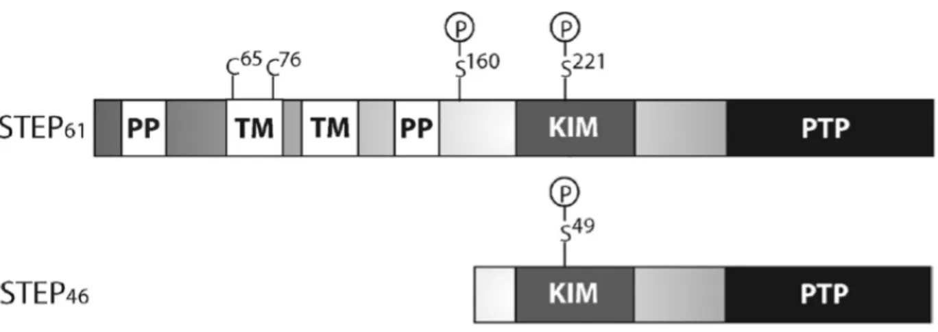
Figure 1 - Structure of STEP61 and STEP46 Isoforms... 18
Figure 2 - STEP61 Regulation of Substrates... 22
Figure 3 - Morris Water Maze Protocol 1... 43 Figure 4 - Effect of MWM Protocol 1 on STEP61, NLGN-1, and non-phospho-STEP protein
... 44 Figure 5 - Morris Water Maze Protocol 2 ... 46 Figure 6 - Effect of MWM Protocol 2 on STEP61, NLGN-1, NMUR1 and non-phospho-STEP
protein ... 47 Figure 7- Morris Water Maze Protocol 3 ... 48 Figure 8 - Effect of MWM Protocol 3 on STEP61, NLGN-1, NMUR1 and non-phospho-STEP
protein ... 49 Figure 9 -Morris Water Maze Protocol 4 ... 52 Figure 10 - Effect of MWM Protocol 4 on STEP61, NLGN-1, NMUR1 and non-phospho-STEP
protein ... 53 Figure 11 - Effect of Previously Frozen 20μM NMDA treatment on Total STEP61 in Primary
Hippocampal Neuron Cultures ... 54 Figure 12 - Effect of 20 μM of Freshly Prepared NMDA treatment on Total STEP61 in
Primary Hippocampal Neuron Cultures ... 54 Figure 13 - Effect of 100μM DHPG treatment on Total STEP61 in Primary Hippocampal
Neuron Cultures ... 55 Supplemental Figures ... 73
Liste des Acronyms and Abbreviations
Αβ: Beta-amyloidAD: Alzheimer's Disease
aMCI: Amnesic mild cognitive impairment
AMPA: α-amino-3-hydroxy-5-methyl-4-isoxazolepropionic acid
AMPAR: α-amino-3-hydroxy-5-methyl-4-isoxazolepropionic acid receptor ANOVA: Analysis of variance
AP-MS: Affinity Purification Mass Spectrometry BioID: Proximity-dependent Biotin Identification
CARD10: Caspase Recruitment Domain-Containing Protein 10 CO-IP: Co-immunoprecipitation
CPE: Cytoplasmic polyadenylation element
CREB: Cytoplasmic polyadenylation element binding protein DHPG: (s)-3,5-Dihydroxyohenylglycine
EPSC: Excitatory postsynaptic current
ERK1/2: Extracellular Signal Regulated Kinases 1/2 FMRP: Fragile X mental retardation protein
GIMAP6: GTPase of Immunity-Associated Protein 6
GluA2: α-amino-3-hydroxy-5-methyl-4-isoxazolepropionic acid receptor subtype A2 GluN2B: N-methyl D aspartate receptor subtype 2B
HBSS: Hank's Balanced Salt Solution HEK293: Human embryonic kidney 293 K+: Potassium Ion
KIM: Kinase Interacting Motif KO: Knock out
LTP: Long-term potentiation
MAPK: Mitogen-activated protein kinase
MEM-HS: Minimum Eagle Essential Medium-Horse Serum METTL26: Methyltransferase-like 26
Mg2+: Magnesium ion
mGluR: Metabotropic glutamate receptor MWM: Morris Water Maze
Na+: Sodium Ion
NLGN-1: Neurologin-1
NMDA: N-methyl-D-aspartate
NMDAR: N-methyl-D-aspartate receptor NMUR1: Neuromedin U receptor 1 PER1: Period Circadian Regulator PP: Polyproline-rich
PP1: Protein phosphatase 1
PTP: Protein tyrosine-phosphatase
PTPN4: Protein Tyrosine Phosphatase Non-Receptor Type 4 SAINT: Significance analysis of interactome
STEP: Striatal-Enriched Tyrosine Phosphatase
Acknowledgments
A special thanks to Benoit Coloumbe and his team from the Montreal Clinical Research Institute for performing the BioID and AP-MS screening, as well as to Maude-Éloïse Piché-Lemieux for her help in data collection and Julien Dufort-Gervais for teaching me the techniques required for my project.
Chapter 1 – Introduction
1.1 Learning and Memory
1.1.1 Overview
In the most general terms, learning and memory can be thought of as the ability of an organism to first obtain information from the environment and the subsequent retention of this information for future recall and use. The neurological basis of memory is complex and is still being explored, though it is well established that synaptic plasticity (the ability for synaptic transmission to change in response to stimuli) plays an important role.
The most well-studied forms of synaptic plasticity thought to be involved in the formation of memories include long-term potentiation (LTP) and long-term depression (LTD) in the hippocampus. First proposed by Donald Hebb in 1949, the Hebbian theory proposed that the firing of a postsynaptic neuron while the presynaptic neuron is active could lead to changes within synapse that would strengthen the connection between the two. Evidence for this process, which we now refer to as LTP, was first provided decades later when it was shown that synapses in the hippocampus could be potentiated (i.e. signal transmission between neurons is increased) for days following repeated activation using high frequency stimulation (Bliss and Gardner-Medwin, 1973; Bliss and Lomo, 1973). LTD was later described in 1982 by Ito and colleagues (Ito and Kano, 1982; Ito et al., 1982). It is thought that the strengthening and weaking of connections in the brain through LTP and LTD, respectively, likely play a role in the coding and storing of information (.i.e. memory) in the brain.
Various forms of LTP and LTD have been documented, with different brain regions exhibiting different forms. However, most of the research has focused on the hippocampus due to
strong evidence for its involvement in memory (Martin et al., 2000; Zola-Morgan and Squire, 1993, Voss et al., 2018). The exact role of the hippocampus in memory is still being explored, though it is known to have a role in episodic and spatial memory (Bird & Burgess, 2008, Voss et al., 2018).
1.1.2 Molecular Mechanisms of Memory
Amongst the various molecular mechanisms underlying LTP and LTD are N-methyl-D-aspartate receptor (NMDAR)-dependent LTP, NMDAR-dependent LTD and mGluR1-dependent LTD.
Perhaps the most well-known form of LTP in the hippocampus, NMDAR-dependent LTP relies on the activation of NMDA as well as the α-amino-3-hydroxy-5-methyl-4-isoxazolepropionic acid receptors (AMPARs) in the postsynaptic neuron. Usually found colocalized on dendritic spines, these receptors function together to induce LTP. LTP is triggered by a burst of high frequency stimulation at the presynaptic neuron which causes it to release glutamate into the synaptic cleft. Following the binding of glutamate to AMPA receptors in the postsynaptic neuron, Na+ and K+ are allowed through the AMPAR ion channels. If there is sufficient cation influx, sufficient depolarization of the membrane occurs to allow for the removal of a magnesium ion that usually blocks the NMDAR channel, which then permits a subsequent influx of cations (Mayer et al., 1984; Nowak et al., 1984). If sufficient calcium enters the postsynaptic neuron through NMDAR channels, LTP is induced (Malenka, 1991; Malenka and Nicoll, 1993). The expression of NMDAR-dependent LTP in the CA1 hippocampal synapses appears to depend on alterations in AMPAR trafficking that lead to the increased insertion of calcium permeable AMPARs in the postsynaptic membrane (Bredt and Nicoll, 2003; Derkach et al, 2007; Malenka and Nicoll, 1999; Malinow and Malenka, 2002; Song and Huganir, 2002).
Interestingly, calcium influx through NMDARs is also involved in NMDAR-dependent LTD. While LTP requires large increases in postsynaptic calcium levels, LTD seems to occur when lower frequency stimulation is applied repetitively, thus still allowing calcium influx through NMDA receptors, though in a much smaller concentration (Cummings et al., 1996). This form of LTD involves, at least in part, removal of AMPA receptors from the postsynaptic membrane (Bredt and Nicoll, 2003; Collindgride et al., 2004; Derkach et al., 2007; Malenka and Bear, 2004; Malinow and Malenka, 2002). Metabotropic glutamate receptor-dependent LTD is less well studied, but also appears to involve the removal of AMPARs from the synapse (Snyder et al., 2001; Wang and Linden, 2000; Xiao et al., 2001).
Most research into LTP and LTD have taken place in vitro, and thus the way in which these processes affect behaviour are still poorly understood. There is, however, evidence to suggest a role for LTP in hippocampal memory. For instance, mice with increased NMDAR function had evidence of increased LTP in a subset of hippocampal synapses as well as improvements in spatial learning performance (Whitlock et al., 2006). In other research the abolition of LTP maintenance using a protein kinase Mzeta inhibitor was shown to cause the loss of a spatial memory in rats (Pastalkova et al., 2006). Though much evidence points to the involvement of LTP in memory, causality has not yet been firmly established (Stuchlik, 2014).
Although the involvement of LTP in memory formation and retention has long been suspected, research also suggests a role for hippocampal LTD. Dong et al., for instance, showed in 2012 that memory enhancements induced by novelty exploration in rats was increased when NMDAR-dependent LTD was facilitated, and blocked when LTD was inhibited, giving evidence that LTD has its own role to play in memory acquisition. Later, they also demonstrated a role for LTD in spatial reversal learning during the Morris water maze (Dong et al., 2013). Thus, the
formation and maintenance of memories through synaptic plasticity appears to be dynamic in nature, requiring coordinated changes within the synapse that involve both long-term depression and long-term potentiation. Importantly, these studies and others have demonstrated that a behavioural experience can induce the formation of LTP and LTD (Dong et al., 2012; 2013; Kemp and Manhan-Vaughan, 2004; Manaha-Vaughan and Braunewell, 1999).
1.1.3 The Hippocampus and Spatial Memory
Memory disturbances due to hippocampal damage have long been known to occur, though the precise nature of its role in various types of memory remains debated. What is clear, however, is that it plays an important role in the formation and maintenance of long-term memories (Baddeley and Warrington, 1970; Cave & Squire, 1992). In addition, the hippocampus is known to be implicated in spatial memory, and damage to the area frequently causes issues with forgetting where an object has been placed or with the ability of an individual to properly navigate (Bird and Burgess, 2008). It comes at no surprise, then, to learn that the hippocampus is one of the first areas of the brain to be affected by Alzheimer's disease (AD) (Frisoni et al., 2010), where the first symptoms individuals display often include a tendency to forget directions and to misplace objects. It is thought that the importance of the hippocampus in spatial memory is, at least in part, due to the presence of "place cells," that is, cells that have been shown to specifically fire when an animal is in a specific location in an environment or "place field." Place cells were first discovered in rodents but were later also discovered in primates as well as in humans (O'Keefe, 1971; Ekstrom AD, 2003; Ono, 1991). Due to the relative ease with which spatial memory can be assessed in laboratory animals, spatial memory tasks are a widely used tool to study hippocampal-dependent memory in rodents.
1.2 The Striatal-Enriched Protein Tyrosine Phosphatase (STEP)
1.2.1 Overview
First discovered nearly 30 years ago, the Striatal-Enriched Protein Tyrosine Phosphatase (STEP) is, as its name suggests, a tyrosine-specific phosphatase that is found in large quantities within the striatum in the brain (Lombroso et al., 1991; 1993). In addition to its high concentration in the striatum, it is also found in the other structures of the brain, excluding the cerebellum. STEP isoforms are not only differentially expressed throughout different tissues (Lombroso et al., 1991; 1993), but through developmental time as well (Raghunathan et al., 1996). STEP can be found in both excitatory and inhibitory neurons (Oyama et al., 1995; Choi et al., 2007). Depending on the brain region under observation, STEP can be found either throughout the neurons (Boulanger et al., 1995), or in some cases only in neurites (Kim et al, 2008), and in some cases, such as under ischemic conditions or optic nerve damage, expression has been seen in astroglia (Hasewaga et al., 2000; Lorber et al., 2004). Abnormal levels of STEP have been linked to various neuropsychiatric disorders and problems with memory and cognition, such as Alzheimer's Disease and Schizophrenia, amongst others.
1.2.2 Structure
The STEP protein is encoded by the ptpn5 gene and alternative splicing leads to four distinct isoforms. Two of these isoforms (Fig. 1) contain an active phosphatase domain which allows STEP to dephosphorylate specific tyrosine residues on its substrates (STEP61 and STEP46), while the other
two are lacking a phosphatase domain (STEP38 and STEP20) (Sharma et al., 1995; Bult et al., 1996,
1997). The functions of STEP38 and STEP20 are not currently known, though it has been proposed
that they may have a regulatory function and compete with active STEP variants for binding sites (Goebel-Goody, 2012). All of the splice variants of STEP contain a kinase interacting motif (KIM)
domain that allows for binding to substrates, and a polyproline-rich (PP) present in some isoforms (STEP61 and STEP38) appears to confer substrate specificity (Nguyen et al., 2002). STEP61
contains an extra sequence that targets it to cell membranes, including the postsynaptic density (Oyama et al., 1995) and the endoplasmic reticulum, while STEP46 lacks this sequence and is
instead found in the cytosol (Lombroso et al., 1993; Bult et al., 2006).
Figure 1 - Structure of STEP61 and STEP46 Isoforms. These main isoforms of STEP both contain
a kinase interacting motif (KIM) domain, as well as a tyrosine phosphatase (PTP) domain. Phosphorylation of Ser221 on STEP
61 or Ser49 on STEP46 inactivates STEP by sterically interfering
with substrate binding. STEP61 contains a transmembrane domain (TM) as well as
popypropoline-rich domains (PP) which are hypothesized to have a role in substrate specificity.
1.2.3 Substrates
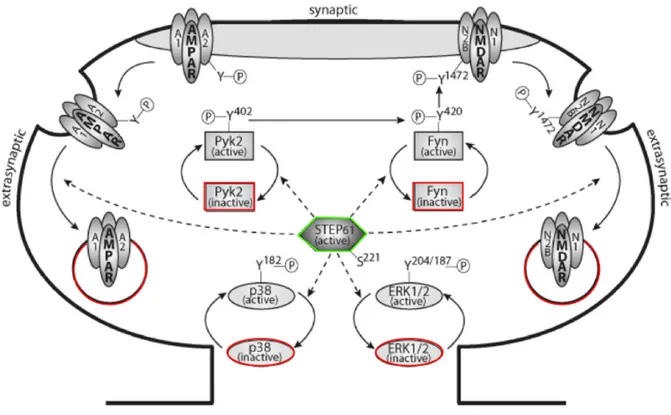
Known STEP substrates include ERK1/2, Pyk2, p38 and Fyn, as well as AMPA and NMDA receptor subunits (Fig. 2).
Extracellular Signal Regulated Kinases 1/2 (ERK1/2)
ERK1/2 is a member of the mitogen-activated protein kinase (MAPK) family that is known to have an important role in the induction and maintenance of synaptic plasticity (Sweatt, 2004),
and is a known substrate of STEP (Munoz et al., 2003; Paul et al., 2003). Following NMDAR stimulation, STEP deactivates ERK1/2 by dephosphorylation of two tyrosine residues, which limits the amount of time that the enzyme stays active (Paul et al., 2003; Valjent et al., 2005), suggesting a place for STEP in the opposition of synaptic strengthening. In addition, ERK1/2 phosphorylation is increased in the hippocampus and several other brain structures in STEP KO mice, further supporting the important of STEP in ERK1/2 regulation (Venkitaramani et al., 2009, 2011).
p38
Another member of the MAPK family, p38, is also a substrate of STEP and opposes the actions of ERK1/2. While ERK1/2 encourages synaptic strengthening and promotes cell survival, p38 is implicated in glutamatergic excitotoxicity and cell death pathways (Hardingham et al., 2002; Semenova et al., 2007; Ivanov et al., 2009; Poddar et al., 2010). Prolonged stimulation of extrasynaptic NMDARs leads to cleavage and inactivation of STEP61. STEP becomes unable to
dephosphorylate Tyr182 of p38, which promotes its activity and leads to neuronal death (Xu et al.,
2009).
GluN2B
NMDARs contain four subunits, two of which are obligatory GluN1 subunits, and two of which are usually either both GluN2 units or a GluN2 and GluN3 subunit (Paoletti et al., 2013, Regan et al., 2015). Different splice variants of the subunits exist, one of which (GluN2B) is regulated by STEP. STEP regulates phosphorylation of the NMDAR subunit GluN2B both directly
(Snyder et al., 2005; Kurup et al., 2010). This is correlated with an increased association of GluN2B with clathrin adapter proteins, which promotes endocytosis of the GluN2B-containing NMDAR (Nakazawa et al., 2006), and in fact, we see an increase in surface GluN2B receptors in STEP KO mice (Zhang et al., 2010; Venkitaramani et al., 2011). In addition to directly regulated GluN2B phosphorylation, STEP also indirectly regulates it by dephosphorylating and inactivating of Src-family kinase Fyn (Nguyen et al., 2002). Active Fyn phosphorylates GluN2B on three tyrosine residues, and STEP inhibits this activity by dephosphorylating a tyrosine residue (Tyr420) on Fyn
(Nguyen et al., 2002). Consistent with the evidence that STEP mediates endocytosis of GluN2B-containing NMDARs is the reduction in excitatory postsynaptic currents (EPSCs) and LTP following the application of STEP to hippocampal slices and in contrast, inhibition of tonic levels of STEP enhances NMDAR EPSCs and prevents LTP (Pelkey et al., 2002). This supports a role for STEP in the tonic opposition of synaptic strengthening, which implies that STEP must be inactivated or removed in order for LTP to occur, and that higher amounts of STEP contribute to LTD (Zhang et al., 2008; Gladding et al., 2009; Chen et al., 2013).
GluA2
AMPARs contain four possible subunits (GluA1-GluA4) that form heterotetramers, most commonly found as GluA1/GluA2 or GluA2/GluA3 within the hippocampus (Park, 2018). In addition to its role in the regulation of NMDAR trafficking, STEP also regulates synaptic plasticity through dephosphorylation of a tyrosine residue on the AMPAR subunit GluA2 following group I mGluR stimulation using (s)-3,5-Dihydroxyohenylglycine (DHPG) (Zhang et al., 2008). This dephosphorylation results in internalization of the AMPA receptors and is reduced by a substrate-trapping STEP mutant (Zhang et al., 2008), suggesting that STEP is a requirement for this process.
In addition, STEP KO mice had higher levels of GluA2 containing AMPA receptors at the surface of neurons (Zhang et al., 2008, Venkitaramani et al., 2011). Recently, Won et al. (2019) demonstrated that the interaction between GluA2 and STEP is direct, and does not take place through an intermediary protein.
Pyk2
Finally, the focal adhesion kinase Pyk2 also appears to be a substrate of STEP (Xu et al., 2010; Venkitaramani et al., 2011). When activated Pyk2 phosphorylates and activates Fyn, which is then available to phosphorylate GluN2B, thereby increasing the surface expression of GluN2B-containing NMDARs (Besshoh et al., 2005; Le et al., 2006). In addition, Pyk2 activates ERK1/2, further promoting LTP (Nicodemo et al., 2010). By dephosphorylating and inactivating Pyk2, STEP again opposes the formation of LTP by reducing the activity of ERK1/2 and Fyn.
Figure 2 - STEP61 Regulation of Substrates. Well known substrates of STEP61 include Pyk2,
Fyn, p38, ERK1/2, as well as AMPA and NMDA receptors. When phosphorylated at Ser221,
STEP61 dephosphorylates regulatory tyrosine residues on Pyk2, Fyn, p38 and ERK 1/2 and
consequently inactivates them. In addition, STEP61-mediated phosphorylation of regulatory
tyrosine residues on AMPA and NMDA receptors lead to their endocytosis, further opposing synaptic strengthening.
1.2.4 Regulation
STEP is regulated through a variety of mechanisms including phosphorylation, ubiquitination, proteolytic cleavage, dimerization and local translation.
Phosphorylation of Ser221 of STEP
61 or Ser49 in STEP46 within the KIM domain reduces
STEP activity by sterically hindering its ability to bind to substrates (Paul et al., 2000; 2003). Protein kinase A (PKA) is one enzyme known to phosphorylate (and deactivate) STEP following DR1-like dopamine receptor stimulation (Paul et al., 2000). In addition to directly phosphorylating
STEP, PKA also activates DARPP-32, which regulates STEP through the inhibition of protein phosphatase 1 (PP1) (Valjent et al., 2005). An opposing process occurs following the activation of NMDARs, which leads to increased PP1 activity via calcineurin, and hence, increased STEP phosphorylation and activity (Paul. et al., 2003; Snyder et al., 2005; Valjent et al., 2005). Interestingly, though NMDAR activation can lead to increased STEP activity through PP1, it has also been shown to lead to rapid degradation of STEP through ubiquitination (Xu et al., 2009). Extrasynaptic NMDAR stimulation has also been shown to reduce STEP activity through a mechanism involving calpain-mediated proteolytic cleavage within the KIM domain (Xu et al., 2009). This cleavage results in a STEP33 fragment which is no longer capable of binding to
substrates. Consequently, the MAPK p38 (which is usually inactivated by STEP) becomes activated and triggers an apoptotic signaling cascade; blocking the proteolytic cleavage of STEP61
protects against glutamate neurotoxicity (Xu et al., 2009). Dimerization of STEP61 occurs through
cysteine residues within the amino terminus in low amounts basally, and in higher amounts upon the induction of oxidative stress, and this dimerization is also associated with a reduction in phosphatase activity (Deb et al., 2011).
Local dendritic translation of STEP61 is thought to play a role in mGluR-dependent LTD
(Zhang et al., 2008), and the presence of cytoplasmic polyadenylation elements (CPEs) within the 3' UTR of STEP suggests that this translation may be regulated by the CPE binding protein (CREB) (Goebel-Goody et al., 2012; Piqué et al., 2008). The fragile X mental retardation protein (FMRP) is also thought to regulate translation of STEP through a G-quartet sequence in the 3' UTR (Darnell et al., 2011). The current hypothesis is that CREB and FMRP bind and repress STEP mRNA, and that the dissociation of these proteins following synaptic stimulation leads to the rapid translation of STEP (Goebel-Goody et al., 2012).
1.2.5 Role in Memory/Psychiatric Disorders
The dysregulation of STEP is linked to a large number of neuropsychiatric disorders. Numerous investigations have found abnormal levels of STEP in neurological conditions such as Alzheimer's disease, schizophrenia, Fragile X, as well as substance abuse disorders, amongst others.
Evidence suggests that at least some of the cognitive decline observed in patients who have Alzheimer's disease (AD) is due to abnormally elevated levels of STEP. This is apparently due to modulation of STEP by beta-amyloid (Αβ) protein that is increased in AD. Αβ increases STEP activity by inhibiting the ubiquitin-proteasome system, which therefore slows down the rate of STEP degradation (Kurup et al., 2010). In addition to this mechanism, Αβ also activates calcineurin and PP1, which dephosphorylate and activate STEP61 (Snyder et al., 2005). The subsequent
increase in NMDAR endocytosis leads to cognitive decline (Kurup et al., 2010; Zhang et al., 2010). Consistent with this hypothesis, several animal models of AD as well as post-mortem samples of prefrontal cortex from AD patients show increased STEP activity (Chin et al., 2005; Kurup et al., 2010; Zhang et al., 2010). Recent research has shown that STEP is also increased in the hippocampus of animal models and humans with amnesic mild cognitive impairments (Castonguay et al., 2018), which are part of the prodromal phase of AD.
STEP also appears to have a role in alcohol-induced memory loss, which prevents the formation of new memories while intoxicated (McIntosh and Chick 2004). STEP is required for ethanol-induced inhibition of NMDAR function and LTP, as well as for ethanol's ability to inhibit fear learning (Hicklin et al., 2011). Treatment of wild type and STEP KO brain slices with ethanol back up these findings by demonstrating that ethanol only leads to reduced phosphorylation of the GluN2B subunit in the wild type (Hicklin et al., 2011).
1.2.6 STEP and Learning
Much of the research on STEP has examined the effect of STEP on memory. There is very little research, however, that examines the effect of learning on STEP. One paper that looked at the effect of fear conditioning on STEP found that fear induction in rats resulted in the de novo translation of both STEP61 and STEP46 in the amygdala within 10 min, and infusion of a STEP
substrate-trapping mutant into the amygdala interfered with fear conditioning memory and LTP induction (Paul et al., 2007). Paul et al. demonstrated that experiences can affect STEP regulation, and that at least in some circumstances, STEP may be required for LTP induction in the lateral amygdala, despite its well-known role in opposing the strengthening of synapses in the hippocampus. The effect of learning a hippocampal-dependent spatial task on STEP has, to the best of our knowledge, not yet been studied. In our study, we determined the effect of training in a spatial task (the Morris water maze) on the level of total STEP61 protein and on the active form of
STEP61 in the rat hippocampus. In addition, we examined the effect of DHPG and NMDA
treatments (meant to induce LTD) on STEP61 in primary hippocampal neuron cultures.
The negative consequences of elevated STEP levels suggest that the reduction of STEP may in fact improve certain aspects of cognition. In fact STEP KO mice have shown improved performances in both hippocampal-dependent (Venkitaramani et al., 2011) and amgydala-dependent learning (Olausson et al., 2012). For instance, though STEP KO mice were able to learn the Morris water maze task (which is hippocampal-dependent) as well as their wild type counterparts, they also performed better than the wild type when it came to a reversal learning task where the platform was moved (Venkitaramani et al., 2011). This suggests that they had greater cognitive flexibility, or perhaps improved extinction learning. In addition, the STEP KO mice
committed fewer reference and working memory errors in the radial-arm water maze, a much more difficult spatial learning task.
More recent evidence shows a role for STEP in age-related cognitive decline in several different species. STEP levels were found to be higher in cognitively impaired aging mice, rats, monkeys, as well as post-mortem amnesic mild cognitive impairment (aMCI) human hippocampi, compared to non-cognitively impaired controls (Castonguay et al., 2018). In addition, the overexpression of STEP in the CA1 region of the hippocampus of young mice was able to induce similar declines in performance during MWM acquisition (Castonguay et al., 2018); subsequent injection of the small molecule STEP inhibitor TC-2153 also restored cognitive performance in mice overexpressing STEP as well as in aged rats. Unfortunately, TC-2153 is difficult to dissolve and has an unstable sulfur ring structure, which consequently make it not ideal for the purpose of drug development and has led to hesitancy to test it in clinical trials. However, it has helped to support STEP as a potentially powerful drug target (Lombroso et al., 2019). To date, however, there are no commercially available drugs that selectively target STEP. Alternatively, it might be possible to affect the pathways that STEP is involved in either through the targeting of STEP regulators or its substrates.
1.2.7 The Search for Interacting Proteins
Though many substrates of STEP have been identified and studied, the full scope of STEPs interactions are unknown. Recently, however, Won et al. (2019) used liquid chromatography tandem mass spectrometry (LC/MS/MS) to identify proteins that co-immunoprecipitated with STEP61 in mouse cortex and hippocampus lysate. The co-immunoprecipitation yielded several
hundred potential interactors, and a major finding of this study was that STEP61 binds directly to
et al., 2002). Importantly, this highlights the possibility that novel interactors of STEP might be found through the use of more modern research techniques and/or equipment.
Proximity-dependent Biotin Identification (BioID) is a recent advance to the interactome discovery toolkit. The process requires the fusion of a promiscuous biotin ligase to the end of a protein of interest. The protein of interest can then be expressed in living cells and, upon addition of biotin, it will biotinylate proteins that pass within 10 μm of the biotin ligase region. The method allows for the detection of proteins that are found in close proximity at any point in time throughout the experiment and does not require direct interaction of the proteins. As such, BioID helps to identify proteins that are localized within the same space as the bait protein as well as potential interactors of the bait protein that may interact only transiently or weakly, and therefore are unlikely to show up when using other screening methods such as co-immunoprecipitation. Affinity-purified mass spectrometry (AP-MS) is a complementary method to screen proteins that relies on the identification of proteins through the analysis of co-precipitated proteins through the chemical analysis of their components identified through mass spectrometry. We used results from both BioID and AP-MS in order to uncover a list of potential STEP-interacting proteins. We assessed the level of one of these proteins (neuromedin-U receptor 1, NMUR1) in the hippocampus of rats following MWM training. NMUR1 is primarily expressed in the peripheral nervous system, though it is also found in the hippocampus (Zhang et al., 2010). In addition, neuromedin U is capable of inhibiting memory impariments in mice and neuronal death in cultured neurons exposed to inflammation (Iwai et al., 2008). Furthermore, neuromedin exposure in neuromedin U neurons of the dorsal root ganglion has been linked to increases in ERK1/2, a known substrate of STEP (Zhang et al., 2012), making it an interesting potential STEP-interacting protein.We also assessed the level of neurologin-1 (NLGN-1), a synaptic anchoring protein recently shown to interact with STEP61
(Won et al., 2019), in addition to STEP61 total protein and active STEP61 (not phosphorylated on
Chapitre 2 – Hypotheses & Objectives
2.1 Hypotheses
Our main hypothesis was that a spatial learning task would increase the level and/or activity level of STEP protein in the hippocampus of rats. We also hypothesized that these changes would be associated with changes in LTD and/or LTP in the hippocampus. Finally, we expected that there are a variety of STEP substrates and/or regulators that have not as of yet been discovered that may play a role in spatial learning, LTD and/or LTP.
2.2 Objectives
2.2.1 Objective 1
Our first objective was to develop a list of possible novel interactors of STEP using proximity-dependent biotin identification (BioID) and affinity purification mass spectromery (AP-MS).
2.2.2 Objective 2
Our second objective was to examine the effect of a spatial learning task (the Morris water maze) on STEP61 total protein and on STEP61 phosphorylation on Ser221 in the hippocampus of rats.
At the same time, we looked at the effect on neuroligin-1 (NLGN-1) and neuromedin U receptor 1 (NMUR1).
2.2.3 Objective 3
Our third objective was to develop an in vitro protocol that could chemically induce LTD in order to study the effect of LTD on STEP and potentially interacting proteins in rat primary hippocampal neuron cultures
Chapitre 3 – Materials and Methods
3.1 Objective 1
Proximity-dependent Biotin Identification (BioID) and affinity-purified mass spectrometry (AP-MS) were used in order to identify potentially novel substrates or regulators of STEP. BioID and AP-MS, as well as all protein expression and cultures required in order to perform the these experiments were carried out by the team of Dr. Benoit Coulombe at the Montreal Clinical Research Institute. Significance analysis of interactome (SAINT) scores were assigned by Dr. Coulombe's team to the proteins identified using BioID and FLAG AP-MS. Our team used the SAINT scores provided to help narrow down which proteins were the most likely to be interacting with STEP.
3.1.1 Proximity-Dependent Biotin Identification (BioID)
BioID was used in order to identify proteins existing in the same cellular environment as STEP61 and STEP46, as previously published (Cloutier et al., 2017). In short, BioID consists of a
technique in which a promiscuous biotin ligase (BirA*) is fused to a protein of interest. The fusion protein can then be expressed in living cells and will biotinylate proteins that are within a 10 μm distance of the protein of interest (or more specifically, within range of the biotin ligase) when biotin is added to the cells. The cells are later lysed and biotinylated proteins are pulled down and identified using mass spectrometry. Thus, potential interacting proteins may be revealed as well as proteins that are found in the near environment, with which protein of interest does not necessarily directly interact. The experiments were repeated 3 times in HEK293 cells. False positive interactions were reduced through the use of the Decontaminator computational approach (Lavallee-Adam et al., 2010).
3.1.2 Affinity-purifiation Mass Spectrometry
Affinity-purification mass spectrometry was used in order to identify potential directly-interacting STEP substrates and/or regulators, according to previously published protocols (Thiffault et al., 2015, Cloutier et al., 2017). In short, STEP46 was fused with a FLAG-tag and
expressed in human embryonic kidney 293 (HEK-293) cells. The FLAG-tagged STEP46 was then
immunoprecipitated and proteins bound to STEP were eluted and identified using mass spectroscopy. The experiment was repeated 3 times. False positive interactions were reduced through the use of the Decontaminator computational approach (Lavallee-Adam et al., 2010).
3.2 Objective 2
3.2.1 Animals
3-month old male Long-Evans rats (N=54) were purchased from Charles River (Kingston, On). Rats were kept on a 12-hour light/12-dark cycle (lights on at 8am) at an ambient temperature between 22°C and 25°C. They had ad libitum access to food and water for the entire duration of the study. Rats were single-housed and allowed to acclimate to the animal facility for one week before undergoing 5 days of handling habituation. All procedures were approved by the appropriate ethics committee (Le comité d'éthique de l'expérimentation du centre de recherche de l'Hôpital du Sacré-Coeur de Montréal).
3.2.2 Experimental Design
In order to determine the effect of a spatial learning task on STEP, we first trained rats on the Morris water maze (MWM) task (N=54). Those rats were separated into two groups, that is, one group that completed a typical probe test (n=22) without the platform at the end of training,
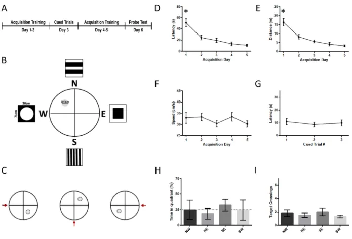
and those who essentially underwent a final training session with the platform still available (n=23), rather than the probe test. THE NUMBER OF ANIMALS PER GROUP VARIED. INSERT HERE. A control group consisted of 9 rats who underwent all of the same handling as the test rats but were never released into the water. One hour following the probe test the rats were euthanized by decapitation, and the brains were flash frozen on dry ice and stored at -80°C until further processing. The protocol for the Morris water maze was changed several times throughout the study (see Fig. 3A, 5A, 7A, 9A), as described below.
3.2.3 Morris Water Maze
The Morris water maze consists of three main components : 1) An acquisition phase (lasting 4 to 5 days), in which rats are trained to use distal cues to find a hidden platform, 2) A series of cued trials (3 over the same day) where the platform is made visible and moved around the pool in order to ensure that the rodents have adequate vision and the motivation to escape onto the platform, and 3) A probe trial (on the final day), in which the platform is removed and learning through the use of distal cues can be inferred through the amount of time the rodent spends in the area where the platform was previously located.
The Morris water maze (MWM) task was carried out in a pool 180 cm in diameter that was filled with a mixture of water and white non-toxic paint in order to avoid visibility of the submerged platform. One hour prior to testing, the rats were brought into the testing room in order to acclimate to the environment. The lights were left on until immediately prior to testing, when a solitary light was left on above the pool only. Any time a rat was removed from the water, it was immediately toweled dry and then remained in a clean cage on top of a heating pad for several min before being returned to its cage, in order to avoid hypothermia. Control rats were brought to the water but were only touched to the surface of the water before being towelled off and brought to the heating pad.
At all times, the room was kept at 25°C, and the water in the pool was kept between 23°C-25°C. Distal cues present around the pool were changed several times throughout the study (Fig. 3B, 5B, 7B, 9B).
During the acquisition phase (4 or 5 days), the platform (10.5 cm or 15 cm) was located in the centre of the North-West quadrant of the pool, 0.5 cm below the surface of the water. The rats were placed gently in the water at semi-random locations around the edge of the pool (Supplemental Figures 4, 6, 8, 10), and were released facing the outer edge of the pool. Rats were given a maximum of 90 sec to find the hidden platform, after which they were guided to through the water towards the platform. After all acquisition trials, rats were left on the platform for 10 sec in order to give them time to observe the surroundings. These trials were repeated 3 times per acquisition day, with a minimum interval of 45 min between each trial.
The cued trials consisted of a series of trials where the platform was made visible by raising it 0.5 cm above the surface of the water and covering the sides with black tape. The cued trials consisted of 3 x 30 sec trials, each trial being at least 45 min apart. The cued trials took place either in the morning on the day before acquisition training or in the afternoon after the third acquisition training day. The platform was moved to a different quadrant for each cued trial (Fig. 3A). Rats that failed to find the platform after 30 sec were led to it.
The probe trial took place on the final day (Day 6 or Day 7). One group of rats underwent a standard probe trial where the platform was removed from the water and the rats were allowed to search for it for 30 sec (probe group). Another group was also given 30 sec to find the platform, but in this group the platform was not removed (platform group). Rats were euthanized one hour following the probe trial.
For all Morris water maze manipulations, the movements of the rats were captured with a camera and analyzed using SMART software (Harvard Apparatus). Using the SMART software, we were able to determine the speed of displacement, latency to target, distance travelled, and the percentage of time spent in different zones of the pool.
3.2.4 Protein Extraction and Western Blot
Rats were euthanized by decapitation 1 hour following the probe test. Their brains were rapidly removed and flash frozen on dry ice and then kept at -80°C. Later, one hemispere was sliced 500 μm thick at -20°C, and the dorsal hippocampus was excised. The protein from these sections was extracted for analysis by Western blot. The hippocampi were kept on ice and were homogenized in 1X Cell Lysis Buffer (Cell Signaling, Cat. No 9803), 0.5% CHAPS (Fisher BioReagents, Cat. No. BP571-1) and a protease and phosphatase inhibitor (Thermo Scientific Cat. No. A32961). The samples were then sonicated at an amplitude of 20% (18 x 1 s on, 1 s off), following which they were rotated for 1 hour at 4°C. Finally, the samples were centrifuged at 14 000 rcf for 20 min, still at 4°C. Following centrifugation the supernatant containing the protein was stored in clean tubes and the pellet was discarded. The proteins were quantified using the Pierce BCA Protein Assay Kit (Thermo Scientific Cat. No. 23225).
Proteins were diluted to a final concentration of 1μg/ μl. LDS sample buffer (Thermo Fisher, Cat. No. NP 007) and Bolt reducing agent (Invitrogen, Cat. No. B0009) were added. This solution was heated to 70°C for 10 min. 20 μl of each sample was loaded onto 4-12% Bis-Tris Plus 15 well gels (Thermo Fisher, Cat. No. NW04125BOX), and an intergel control was loaded onto each gel in order to allow for comparison between samples that were not on the same gel. The gels underwent electrophoresis for 90 min at 100V in MOPS running buffer with 500μl Bolt antioxidant (Invitrogen, BT0005). The proteins were later transferred to PVDF membranes (Immobilon-Psq,
Cat. No. ISEQ00010, 0.2μm pored) in NuPage-Methanol Transfer Buffer with antioxidant for 90 min at 100V. The membranes were then blocked using either Odyssey Blocking Buffer (PBS) (LiCor, Cat. No. 927-40000) or Intercept Blocking Buffer (PBS) (LiCor, Cat. No. 927-70001) for an hour at room temperature, and later allowed to incubate with the primary antibodies anti-STEP(D9H3) (Cell Signaling, Cat. No. 9069), anti-non-phospho-STEP(Ser221) (Cell Signaling, Cat. No. 5659), anti-GAPDH (Santa Cruz, Cat. No. sc-25778), anti-NMUR1 (Abbexa, Cat. No. abx217197), anti-neuroligin-1 (Santa Cruz, sc-365110) overnight at 4°C. Primary antibodies were all diluted to 1:1000 in Odyssey Blocking Buffer (PBS) and 0.2% Tween 20 (Fisher BioReagents, Cat. No. BP337-500). The following day the membranes were rinsed for 4 times in PBS-T 1X (0.1% Tween 20) for 5 min, after which they were kept in the dark and incubated with secondary antibodies IRDye 800CW (LiCor, Cat. No. 32211) or IRDye 680RD (Licor, Cat. No. 926-68070) for an hour at room temperature. The membranes were again rinsed 4x 5 min in PBS-T 1X, after which they were rinsed for 5 min with PBS 1X in order to remove residual Tween. The membranes were allowed to dry and were later scanned using the Odyssey CLx apparatus (Licor, Cat. No. 9140) and Image Studio 3.1 software. Band quantification was performed using ImageJ software.
3.2.5 Statistical Analysis
Latency, distance, and speed data for the acquisition and cued trials was analyzed out using one-way repeated measures analysis of variance (ANOVA) with Bonferroni adjustment for multiple comparisons. In the cases where data was non-spherical, the Greenhouse-Geisser F value correction was applied. In the event of a statistically significant result (p<0.05) pairwise comparisons were performed as appropriate. Latency to the platform target during the probe trial was assessed using the two-tailed independent samples t-test and % time spent in the target
quadrant versus other quadrants as well as platform crossings in the target quadrant vs other quadrants was analyzed using a two-tailed paired-samples t-test. All statistical analyses were carried out using IBM SPSS Statistics software version 25. Western blots were analyzed using one-way ANOVA. All results are reported as group mean +/- s.e.m.
3.3 Objective 3
3.3.1 Animals
Three Female Long Evans rats (Charles River, Kingston, ON) were mated at our facility. Matings were timed so that cultures occurred on embryonic day 18 or 19. All procedures were approved by the appropriate ethics committee (Le comité d'éthique de l'expérimentation du centre de recherche de l'Hôpital du Sacré-Coeur de Montréal).
3.3.2 Neuron Cultures
Primary hippocampal cell cultures were prepared from female Long-Evans rats on E18 or E19. Females were decapitated and the embryos were rapidly removed by caesarean section and stored in a HBSS buffer containing: Hank’s Balanced Salt Solution (HBSS, Sigma Cat. No. H1641), HEPES 1M (Gibco, Cat. No. 15630-080), 1X Amphotericin B, Penicillin and Streptomycin (Gibco, Cat. No. 15240-062), 7.5% NaHCO3 (Gibco, Cat. No. 15630-080). Embryos
were immediately transferred to a biological safety cabinet, and all solutions and procedures were carried out under sterile conditions. Embryonic hippocampi were dissected as rapidly as possible and stored in groups of 6 hippocampi for 1mL HBSS at 37°C. Once all hippocampi had been dissected, 400 μl of 2.5% trypsin (Life Technologies, Cat no. 15090-046) was added to each of the tubes, which were then rotated at 900 rpm for 15 min, still at 37°C. Following trypsinization, the
hippocampi were washed twice with HBSS, following which they were dissociated in 1 mL HBSS with the aid of a 21G and 28G needle. Between 150 μl and 200 μl (depending on the culture) of the resulting solution was used to inoculate each well. The 12-well plates (Costar, Cat. No. 3513) contained 1 mL MEM-HS (MEM (Sigma, Cat. No. M0275), Horse Serum (Sigma, Cat. No. H1138), NaHCO3, D-Glucose (Sigma, Cat. No. BP-250), GlutaMax (Sigma, Cat. No. RNBD9302),
1X Amphotericin B, Penicillin and Streptomycin) and were previously coated with poly-D lysine hydrobromide (Sigma, Cat. No. P6407-5MG). Cultures were kept at 5% oxygen, 37°C for approximately 24 hours, when the MEM-HS was replaced by 2 mL of Neurobasal Medium (Neurobasal Medium (Gibco, Cat. No. 21103-049), B27 supplement (Gibco, Cat. No. 17504-044), Glutamax, 1X Amphotericin B, Penicillin and Streptomycin). Half of the Neurobasal medium was removed and replaced with fresh media on day 7. On day 13, the volume of Neurobasal medium was reduced to 1 ml per well in order to simplify treatments, which took place on day 14.
3.3.3 Neuron Culture Treatments
We used both DHPG treatments and NMDA treatments in order to chemically induce LTD. Picrotoxin (GABAA receptor antagonist; 50μm, Tocris Cat. No. 1128, 0.1% DMSO) and
L-689,560 (NMDA antagonist; 5μM, Tocris, Cat. No. 0742, 0.2% DMSO) were added 20 min before DHPG treatments, after which 100 μM DHPG (Tocris. Cat. No. 0342) was added for 10 min according to previousy published protocols (Gladding et al., 2009). Control wells were treated with DMSO. NMDA treatments (20μM, Tocris, Cat. No. 0114) lasted 3 min (protocol from Lee et al., 1998, Holman et al., 2007), and the control wells were treated with sterile water. All treatments took place on culture day 14. At the end of the treatment, media was removed from the wells and washed with ice cold PBS 1X. 150μl ice cold cell lysis buffer was added to each well. Cells were scraped off the bottom of the wells into the lysis buffer, and then removed and stored on ice in
clean microfuge tubes. The proteins were then extracted using the same protocol as the brain tissue samples (see section 3.2.4), with the exception that the cells were not incubated at 4°C for an hour. The protein was quantified and western blots were performed using STEP(D9H3) and GAPDH antibodies.
Chapitre 4 – Results
4.1 Objective 1
4.1.1 Interactome Screenings
BioID and FLAG AP-MS screening was carried out by Dr Coulombe and his team at the IRCM and the results were provided to us with accompanying SAINT confidence scores. Due to potential difficulties in the purification of membrane proteins, BioID was performed using both the transmembranous STEP61 and cytosolic STEP46 fusion proteins as bait. The STEP46 BioID yielded
a much higher number of results that STEP61. A large number of proteins found during the BioID
and FLAG AP-MS screening (Supplemental Fig. 1-3). In order to help narrow these down to a manageable list of potential interactors, we considered only those proteins that had a SAINT confidence score of 1 (the highest possible score, indicating the highest probability that the interaction is real) for the STEP46 results, and a SAINT score of 0.9 for STEP61. The BioID
screenings resulted in eighty-nine proteins having a SAINT score of 1 using STEP46 as bait
(Supplemental Fig. 1), as compared to fourteen when using STEP61 (Supplemental Fig. 2.).
Lowering the SAINT score cutoff to 0.9 for STEP61 added another thirteen proteins to our list.
Eleven proteins obtained through AP-MS had SAINT scores of 1 (Supplemental Fig. 3). We then narrowed the list of proteins down further, base primarily on the localization of the protein in hippocampal neurons, and the commercial availability of antibodies. Additional criteria considered were having a tyrosine residue known to be regulated by phosphorylation, known expression in the hippocampus and previous evidence supporting a role in learning and memory, though these were not strict criteria. We obtained commercial antibodies for ten of these proteins and retained the most reliable antibody, which targeted Neuromedin U receptor-1 (NMUR1) (Data not shown).
4.2 Objective 2
4.2.1 Morris Water Maze
We performed various analyses in order to ensure that the Morris water maze protocol induced a spatial learning event in the rats. First, we assessed latency to the target, distance travelled to the target, and mean speed of the rats during the acquisition trials. In the event that learning has taken place, we expect to see a decrease in the latency and distance travelled to the target, and sometimes an increase in the mean speed. We analyse the cue latency in order to ensure that the animals have the physical ability to see the platform, swim to it in a timely manner, and climb onto it, as well as the motivation to do so. We expect that the rat should succeed at this task at least once during the three cued trials. Finally, in order to differentiate spatial learning from other types of learning, we determine the percentage of time the rats spend in the target 'platform' quadrant versus the other three quadrants. In the event that the rats have used a platform searching strategy other than the use of distal cues, we expect that the percentage of time spent in the target quadrant will not be significantly higher than in the other three quadrants. Four different protocols were tested throughout the experiments, and these analyzed separately below. For the acquisition and cued trials, data for the probe and platform groups were combined for analysis after we ensured that there were no significant differences for the results between each group (for analysis between groups, see Supplemental Figures 5, 7, 9, 11). The probe group consists of the rats that underwent the typical MWM probe trial with platform removed, while the platform group did not have the platform removed on the day of the probe trial.
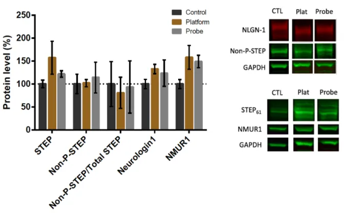
Following the MWM, we performed western blots using protein from the hippocampus and probed for total STEP61, non-phosphorylated-STEP61 and NMUR1 (in the last three protocols), as
well neuroligin-1 (NLGN-1). Outliers that were 2 SD from the mean were identified by SPSS and removed before data analysis. Protein levels are expressed as a ratio of protein over GAPDH.
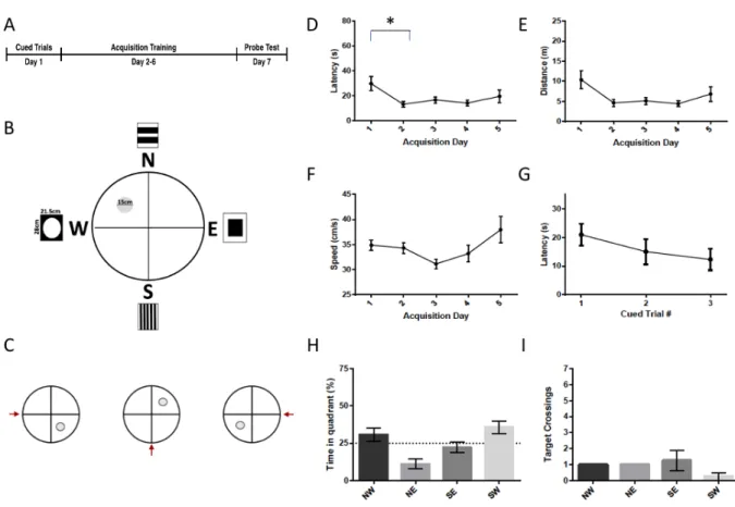
4.2.1.1 Protocol 1
The first MWM protocol (probe n = 4, platform n = 4) (Figure 3) consisted of 3x 30 sec cued trials on the first day of training, followed by five days of acquisition training (3x 90 sec/day), with a 30 sec probe test on the final day (Fig. 3A). We placed the cued trials before the acquisition training in order to ensure that changes to STEP and other proteins would be due to a long term learning effect, rather than a short term learning that would occur had the cued trials been placed at the end of training. The platform was 15 cm in diameter. Curtains were hung on all sides of the pool, from which were hanging four different small (28 x 21.5 cm) rectangular 'distal cues' (Fig. 3B). The starting locations of the rats for each trial are shown in Supplemental Figure 4. There was a reduction in latency to the platform and distance during the acquisition training (F4,28 = 3.838,
p= .022 and F4,28 = 3.3069, p= .032; Fig. 3D & 3E) with the reduction in acquisition latency
between day 1 and day 4 being statistically significant (p=.038), but no increase in swim speed (F4,28 = 2.184, p= .097; Fig. 3F). Latency did not change from one cued trial to another (F2,14= .962,
p= .406; Fig. 3G). During the probe trials, the rats did not spend more time in the NW (target) quadrant as compared to the other three quadrants, nor did they cross the NW platform area more frequently than the equivalent areas in the other three quadrants (p>.05; Fig. 3H & 3I).
There were no significant differences in STEP61 (F2,13 = .171, p=.844) , NLGN-1 (F2,13 =
1.691, p=.222) or non-phospho-STEP (F2,13 = .458, p=.642) total protein levels or in the ratio of
non-phosphorylated to total STEP61 (F2,13 = .148, p =.864) between the probe (n=3), platform (n=4)
(F1,13=5.021, p=.043) lower in the MWM conditions (79.9% +/- 5.3) as compared to controls
(100% +/-7.19) (Supplemental Fig. 12).
Figure 3 - Morris Water Maze Protocol 1. (A) Timeline of the Morris water maze protocol 1. In this protocol, the cued trials took place on day 1, acquisition took place from day 2-6, and the probe test took place on day 7. (B) Setup of the tank during the acquisition phase. Rats must find their way to the 15 cm platform located in the NW quadrant using the 21.5 cm x 28 cm distal cues. During the probe phase, the platform is removed. (C) Set up of the tank during the cued trials. The platform is placed in a different location for each of the 3 trials. The red arrow
indicates the location where the rat was placed into the water. Distal cues are still present, though not shown in the schematic. (D) Latency to the platform during acquisition. There was a
significant reduction in latency to the platform during acquisition training. The reduction was statistically significant on day 4 relative to day 1. (E) Distance travelled to the platform during acquisition training. There was a significant reduction in the distance travelled to the platform. (F) Swim speed was not significantly changed throughout the acquisition training. (G) Latency during the cued trials. Rats were able to find the platform within 30 sec during the cued trials. The latency did not change significantly throughout the cued trials. (H) Percent time spent in the target quadrant during the probe trial. The rats did not spend significantly more time in the target (NW) quadrant than in any of the other quadrants (p>.05) (I) Target crossings during the probe trial. The rats did not spend more time in a zone of 30 cm in diameter located in the center of the
Figure 4 - Effect of MWM Protocol 1 on STEP61, NLGN-1, and non-phospho-STEP protein
There were no significant differences in STEP61, NLGN-1 or non-phospho-STEP between the
probe group, the platform group, or the control group.
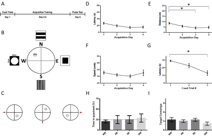
4.2.1.2 Protocol 2
During the second water maze protocol (probe n = 7, platform n = 8) (Figure 5), the smaller signs that served as distal cues surrounding the pool were removed and were replaced by larger signs (56 cm x 71 cm), in an effort to improve their visibilty to increase the likelihood that the rats would see them and use them in order to spatially navigate. These were placed in the same location on the curtains as in the previous protocol. Since the acquisition performance appeared to plateau towards the end of acquisition training, the acquisition period was shortened from five days to four. All other aspects of the test remained the same as in the first protocol. The rats did not start in the South-West until the probe trial, in order to discourage the memorization of a route to the platform. The starting locations of the rats for each trial are shown in Supplemental Figure 6. As in group
1, there was a reduction in latency and distance to the platform during acquisition training (F3,42 =
3.267, p = .03 and F3,42 =3.879, p = .016; Fig. 5D & 5E), and no change in swim speed during the
acquisition training (F3,42 = 1.113, p = .354, Fig. 5F). However, there was a decrease in the latency
during the cued trials (F2,28 = 11.039, p <.001), from a mean of 29 +/- 1s during the fist cue to a
mean of 13 +/- 3 s in the last trial (Fig. 5G). During the probe trial, there was not a significant difference in the percentage of time spent in the target quadrant (NW) versus the other quadrants, nor in the amoun of times the target platform area was crossed versus the analogous area in other quadrants (p>.05; Fig 5H & 5I).
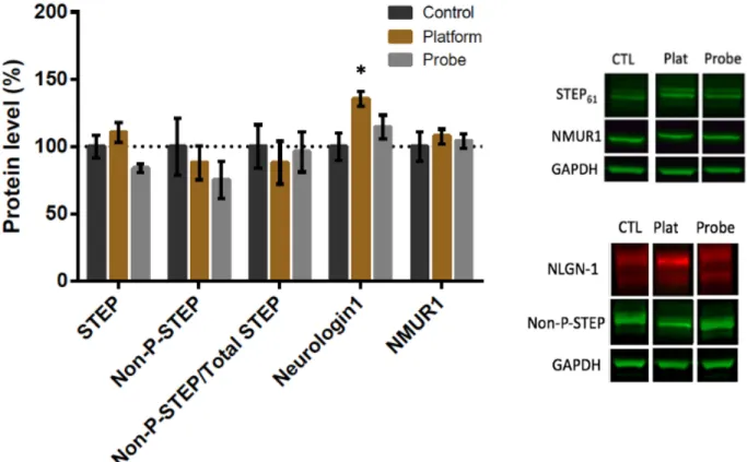
There was a significant difference in NLGN-1 protein expression in the hippocampus (F2,21
= 4.633, p =.022) during the second MWM protocol (Fig. 6). Post hoc analysis showed significantly higher expression of NLGN-1 in the platform group (n=8, 135% +/- 5.4) vs the control group (n=9; 100% +/- 10%, p = .017). There was no significant difference between NLGN-1 expression in the probe group (n=7) vs the platform (p= .229) or control group (p = .476). There were no significant differences between the groups with regards to level of STEP61, NMUR1,
non-phosphorylated-STEP, or the ratio of non-phosphorylated STEP to total STEP61 (F2,20 = 3.049, p =
.070; F2,17 = .264, p = .771; F2,21 =.512, p =.606; F2,20 =.156, p = .857) (Fig. 6). When the MWM
conditions were combined (Supplemental Fig. 13) NLGN-1 expression was also higher in the MWM training group (126% +/- 5.7) vs the control (100% +/- 10.1) (F1,22 = 5.875, p =.024).
Figure 5 - Morris Water Maze Protocol 2 (A) Timeline of the Morris water maze protocol 2. In this protocol, the cued trials took place on day 1, acquisition training took place on day 2-5, and the probe test took place on day 6. (B) Setup of the tank during the acquisition phase. The distal cues surrounding the pool were larger for this protocol (56cm x 71cm). (C) Set up of the tank during the cued trials. The platform is placed in a different location for each of the 3 trials. The red arrow indicates the location where the rat was placed into the water. Distal cues are still present, though not shown in the schematic. (D) Latency to the platform during acquisition. There was a significant reduction in latency during the acquisition phase. (E) Distance travelled to reach the target during acquisition. There was a significant reduction in the distance travelled to reach the target during the acquisition phase. (F) Average swimming speed during the
acquisition phase. The average swimming speed did not change significantly during acquisition training. (G) Latency during the cued trials. There was a significant reduction in latency during the cued trials. (H) Percent time spent in the target quadrant during the probe trial. The rats did not spend significantly more time in the NW quadrant as opposed to the NE, SE, or SW quadrant (p>.05) (I) Target crossings during the probe trial. The rats did not spend more time in a zone of 30cm in diameter located in the center of the NW quadrant than in the equivalent areas in the other three quadrants (p>.05)
Figure 6 - Effect of MWM Protocol 2 on STEP61, NLGN-1, NMUR1 and non-phospho-STEP
protein. NLGN-1 expression differed between groups (F2,21 = 4.633, p =.022), which can be
attributed to a higher level of NLGN-1 in the platform group relative to the control (p=.017). STEP61, NMUR1, non-phosphorylated-STEP and the ratio of non-phosphorylated STEP to total
Figure 7 - Morris Water Maze Protocol 3 (A) Timeline of the Morris water maze protocol 2. In this protocol, the cued trials took place on day 1, acquisition training took place on day 2-5, and the probe test took place on day 6. (B) Setup of the tank during the acquisition phase. Rats must find their way to the 10.5 cm platform located in the NW quadrant using the 21.5cm x 28cm distal cues. During the probe phase, the platform is removed. (C) Set up of the tank during the cued trials. The platform is placed in a different location for each of the 3 trials. The red arrow indicates the location where the rat was placed into the water. Distal cues are still present, though not shown in the schematic. (D) Latency to the platform during acquisition. The reduction in latency was statistically significant. Pairwise interactions showed that latency was lower in the last four acquisition days as compared to the first day. (E) Distance travelled to the platform during acquisition training. Distance to the platform was significantly reduced during acquisition. Pairwise analysis showed significant reductions on days 2-5 as compared to day 1. (F) Swim speed. There was no difference in swimming speed throughout acquisition training. (G) Latency during the cued trials. There was no significant difference in latency to find the platform between different cued trials. (H) Percent time spent in the target quadrant. Rats did not spend
significantly more time in the target (NW) quadrant than in the other quadrants during the probe trial (p>.05) (I) Target crossings during the probe trial. There was no difference in the number of platform crossings in the NW quadrant as compared to the other quadrants (p>.05).
Figure 8 - Effect of MWM Protocol 3 on STEP61, NLGN-1, NMUR1 and non-phospho-STEP
protein. There were no significant differences in the level of STEP61, non-phosphorylated-STEP,
NMUR1, or NLGN-1 between groups.
4.2.1.3 Protocol 3
During the third MWM protocol (N=11) (Fig. 7), the 15 cm platform was exchanged for a 10 cm platform, in order to increase the difficulty of finding the platform, to increase the chances that the rats used distal cues to find the platform, rather than accidentally happening upon it. In addition, the cued trials were moved from the morning of day 1 to the afternoon of the third acquisition day, in order to provide a more accurate learning curve as measured through acquisition latency and distance to the platform. An extra acquisition day was added, so that the total number of acquisition days was one again 5, to ensure that the cued trials on the third day did not negatively impact performance on the probe trial. The probe trial took place on day 6. All other aspects of the