ORIGINAL ARTICLE
Characterization of a novel chitinase from a moderately
halophilic bacterium, Virgibacillus marismortui strain M3-23
Badiâa Essghaier&Abdeljabbar Hedi&Mohamed Bejji&Haϊssam Jijakli&
Abdellatif Boudabous&Najla Sadfi-Zouaoui
Received: 16 September 2010 / Accepted: 13 July 2011 / Published online: 5 August 2011 # Springer-Verlag and the University of Milan 2011
Abstract A new chitinase produced by the moderately halophilic bacterium Virgibacillus marismortui strain M3-23 was identified and characterized. Distinguishable charac-teristics of high activity and stability at different pH, temperatures and salinity of M3-23 chitinase are reported. Analysis of the catalytic domain sequence from the enzyme highlighted its relationship to glycosyl hydrolase family 18. Comparison of the deduced chitinase sequence from strain M3-23 to known chitinases from Bacillus species showed low similarity (82%), suggested its novelty. This is the first report of the characterization of chitinase from the species V. marismortui. The halo- and thermo-tolerant nature of the chitinolytic enzyme allows its potential use in agricultural and industrial applications.
Keywords Chitinase . Halotolerant . Thermotolerant . Virgibacillus marismortui . Catalytic domain
Introduction
Chitin, a homopolymer of N-acetyl-D-glucosamine (GlcNAc)
residues linked byβ-1,4 bonds, is the second most abundant polymer in nature after cellulose. It is widely distributed as a
structural component of the shells of crustaceans, insect exoskeletons and other arthropods, as well as a component of the cell walls of most fungi and some algae (Patil et al. 2000). Chitin is degraded by chitinases, which are produced by a large variety of chitin-degrading organisms including bacteria, fungi, insects and plants. Chitinolytic microorganisms or chitinolytic enzymes have shown immense potential for application in the biological control of plant pathogenic fungi and insects in agricultural or environmental applications (Freeman et al.2004; Jung et al.2005; Chuan2006).
Several bacteria produce enzymes that degrade chitin, including many Bacillus species, e.g., B. circulans (Watanabe et al. 1990), B. licheniformis (Trachuck et al. 1996), B. cereus (Pleban et al.1997), B. subtilis and B. pumilus (Wang et al. 2006; Ahmadian et al. 2007), Streptomyces species (Tsujibo et al. 1993a) and Alteromonas sp. (Tsujibo et al. 1993b)—all these cited species have been found to be potential biocontrol agents. However, our laboratory was the first to take an interest in the study of antifungal chitinase from moderately halophilic bacteria such as Virgibacillus marismortui, Terribacillus halophilus and Planococcus rifi-toensis. These moderately halophilic bacteria were isolated previously from shallow salt lakes in Tunisia and selected as strong antagonists of Botrytis cinerea—the causal agent of grey mold disease on strawberries and tomatoes under the commercial standard conditions applied in Tunisia (Essghaier et al.2009; Sadfi-Zouaoui et al.2008).
It should be noted that, on the one hand, the use of the present enzyme toolbox is limited as supply is insufficient to meet most industrial demands. Therefore, efforts are still being made to find newer sources of enzymes, higher-yielding production techniques and novel applications of these enzymes in unexplored fields. In view of these restrictions, researchers have diverted their attention to the isolation and characterization of enzymes from
extremo-B. Essghaier
:
A. Hedi:
A. Boudabous:
N. Sadfi-Zouaoui (*)Laboratoire Microorganismes et Biomolécules Actives, Faculté des Sciences de Tunis, Université de Tunis El Manar, Campus Universitaire,
2092, Tunis, Tunisia
e-mail: zouaouinajla@yahoo.fr
B. Essghaier
:
M. Bejji:
H. JijakliUnité de Phytopathologie, Faculté Universitaire des Sciences Agronomiques de Gembloux,
philic organisms, including halophiles (Rothschild and Mancinelli2001).
On the other hand, in Tunisia, large yield losses are caused by variations in temperature and high salinity. The presence of high salt concentrations characterizes some Tunisian soils, especially in Sahel regions due to the extensive use of a heavy dressing of manure and irrigation with brackish waters containing high concentrations (3–4 g/l) of NaCl (Messaï et al. 2006). Consequently, many investigations based on biotechnological approaches (Liu and Li 1991; Messaï et al. 2006) have been directed towards regenerating salt-tolerant plants adapted to the conditions of high salinity found in Tunisia. Finding a broad antifungal chitinase able to function under such environmental conditions is relevant for biocontrol purposes.
The present work is a continuation of a project focused on discovering new biological control agents isolated from extreme saline soil from Tunisia that are able to reduce grey mold disease on strawberries and tomatoes. Previous research also aimed to identify the mode of action of the most successful isolates as this information may help optimize their biocontrol efficiency in the field. Under-standing the mechanisms involved in biological control may enable efficacy to be enhanced (Sadfi-Zouaoui et al. 2008; Essghaier et al. 2009). We reported previously the efficiency of the moderately halophilic Virgibacillus maris-mortui strain M3-23 in biological control, and its high production of chitinase. The purpose of this study was to evaluate the effect of three variables (pH, temperature and salt concentration) on the activity and stability of the chitinase produced by strain M3-23. The partially purified chitinase was also characterized biochemically and the gene encoding it was sequenced.
Materials and methods
Screening and taxonomic analysis of strain M3-23
A new strain of moderately halophilic bacterium Virgiba-cillus marismortui strain M3-23 was isolated from a Tunisian shallow salt lake (Essghaier et al. 2009). The morphological, physiological and molecular characteristics of this strain were previously reported and its nucleotide sequence of 16S rDNA has been deposited in the GenBank database under the accession number GQ2825501 as previously described (Essghaier et al.2009).
Media composition and culture conditions for chitinase production
Bacterial growth was carried out on Mc medium as described by Leelasuphakul et al. (2006), with minor modifications. Mc
medium is nutrient broth medium (Difco, Detroit, MI) supplemented with yeast extract, 0.3% (w/v) and 0.5% (w/ v) colloidal chitin prepared according to Rodriguez-Kabana et al. (1983). Cultures were incubated at 37°C for 5 days on a rotary shaker (150 rpm). After centrifugation at 8,000 rpm for 10 min, the cell-free supernatant from culture was used for measurement of chitinase activity.
The effect of salinity on chitinase production was evaluated using a series of media containing various concentrations of salt (0, 5, 10, 15, 20, 25 and 30% NaCl w/v) (Essghaier et al.2010).
Chitinase assay
Chitinase activity was determined according to the method of Gomez Ramirez et al. (2004) as previously detailed (Essghaier et al. 2009, 2010). A 1:1 mixture (v/v) of cell free supernatant and 10% (w/v) colloidal chitin in 0.2 M phosphate buffer pH 7 was incubated for 1 h at 50°C. The reaction was stopped by adding 1 ml 1% NaOH and shaking. The concentration of reaction products was determined by 3,5-dinitrosalicylic acid assay, and the absorbance was measured at 535 nm. The chitinase activity was defined as the amount of enzyme required to produce 1 μmol N-acetylglucosamine (Sigma, St. Louis, MO) per hour per milliliter of supernatant (Roja-Avelizapa et al. 1999). All experiments were carried out in triplicate.
Influence of pH and temperature on chitinase activity and stability
The optimal temperature for enzyme activity was determined by monitoring activity at pH 8 at various temperatures ranging from 40 to 90°C. Heat stability was analyzed by measuring the residual activity after preincubation of the enzyme solution (cell free supernatants) for 30 min at various temperatures in the interval between 40 and 90°C (Smaali et al.2003).
The pH optimum of enzyme was determined by applying substrate solution at different pH values (5–12), and activity was measured at optimum temperature (70°C). pH values were adjusted using the following buffers: 0.2 M phosphate buffer (pH 5.0–6.0), 0.2 M Tris-HCl buffer (pH 7.0–8.0), 0.2 M H3BO3-NaOH buffers (pH 9.0–10.0) and 0.2 M
Na2HPO4-NaOH (pH 12.0). The pH stability was examined
by incubating enzyme solution in the above buffers for 1 h at 4°C before adding the substrate. The remaining activities (%) were subsequently determined (Ellouze et al.2007). Crude enzyme preparation
Bacterial strain M3-23 was grown in 500-ml flasks containing 200 ml Mc medium supplemented with 10 % NaCl (w/v) at 37°C for 5 days with stirring at 150 rpm. The
culture fluid was centrifuged at 8,000 rpm at 4°C for 10 min. The supernatant was subjected to precipitation with ammonium sulphate to 80% saturation at 4°C with constant stirring overnight. The precipitate was collected by centri-fugation at 9,000 rpm for 30 min at 4°C, dissolved in an appropriate volume of 0.2 M phosphate buffer (pH 9), and dialysed extensively against the same buffer. The resultant dialysate was used as chitinase crude extract, and was sterilized by filtration through a 0.2μm pore size filter (Life Sciences, PALL, Ann Arbor, MI, Acrodisc 32 mm syringe filter with 0.2μm Supor membrane) and stored at −20°C until further use for electrophoresis.
Polyacrylamide gel electrophoresis and zymogram analysis Native PAGE was performed at 4°C with a 10% polyacryl-amide gel using a BioRad Mini Protean II apparatus at 100 V (Bio-Rad, Hercules, CA). After electrophoresis, one part of the gel was stained with Coomassie brilliant blue R-250 and the other was incubated for 2 h in a solution of carboxymethylchitin Remazol Brilliant Violet (CM-chitin-RBV) at 2 mg/ml as substrate (Loewe Biochemica, Nordring, Germany) at 37°C. The substrate solution was changed at least twice, and the gel was then rinsed with de-mineralized water. The clear zones corresponding to enzyme activities were visualized by Coomassie brilliant Blue R-250 staining. The molecular mass of the isoforms was estimated using standard markers (BioRad Range Protein Molecular Weight Markers 225 kDa, Promega, Madison, WI).
Identification and characterization of the chitinase gene sequence
Genomic DNA of strain M3-23 was extracted using a Wizard Genomic DNA Purification Kit (Promega) as previously described (Essghaier et al.2009), and stored in TE buffer at −20°C for further study and analysis. The genomic DNA was used as a template to amplify a fragment of the chitinase-encoding gene by polymerase chain reaction (PCR) with a pair of degenerate oligonucleotide primers designed on the basis of chitinase gene sequences from bacterial species deposited in the GenBank database (NCBI): forward primer, KTF1: 5′AGCCAYATTAAT TATGCCTTTGC-3′; reverse primer, KTR2: 5′-GCGCCAT TAAAGTCATAGGTCAT-3′. PCR cycling conditions were 3 min at 94°C for predenaturation step, 34 cycles of 30 s at 95°C, 45 s at 50°C and 30 s at 72°C for amplification. After cycling, the reaction mixture was kept for 10 min at 72°C for extension.
The PCR product obtained (638 bp) was purified with a QIAEX gel elution kit (Qiagen) and cloned using the Gene Jet Kit Cloning (Fermentas, St. Leon-Rot, Germany) and transformed into INVαF′ competent cells (Invitrogen, La
Jolla, CA). Cells carrying the recombinant plasmids were screened and cultured individually in Luria-Bertani (LB) medium supplemented with 50 μg/ml ampicillin. After growth on a rotary shaker at 37°C for 20 h, cells were harvested by centrifugation (8,000 rpm, 10 min). The pellet obtained was retained for the extraction and purification of recombinants plasmids using the Gene jet TM plasmid miniprep Kit (Fermentas); plasmid DNA was eluted in distilled water.
DNA sequencing and analysis were performed on an automated system (GATC Biotech, Germany). DNA and deduced amino acid sequences were compared with those of other chitinase genes in GenBank, using the BLAST (http://www.ncbi.nlm.nig.gov/blast) and CLUSTALW mul-tiple sequence alignment tools.
The nucleotide sequence of the chitinase fragment determined in this study has been deposited with the NCBI GenBank database under the accession number GQ274956.
Results and discussion
Chitinolytic enzymes have been used to control fungal plant pathogens, since chemical fungicides have adverse envi-ronmental effects and are hazardous to human health. Thus, agricultural industries need in novel biofungicide formula-tions (Gohel et al. 2007). Chitinolytic enzymes, which are key players in the lysis of cell walls of higher fungi, are produced by several microorganisms including bacteria (Sadfi-Zouaoui et al.2008; Essghaier et al.2009). Recently, a number of commercial biocontrol products have been developed from rhizobacteria, and many plant disease biocontrol products that contain Bacillus spp. are being used (Schisler et al.2004). Although moderately halophilic bacteria have great biotechnological potential, only a few studies have been carried out concerning the production of extracellular enzymes from these organisms (Ventosa et al. 1998; Sanchez-Porro et al. 2003). Thus, our laboratory is pioneering the study of antifungal enzymes (chitinases, proteases, cellulases, β-1.3-glucanases) from halophilic bacteria such as Halomonas elongata, Terribacillus halophi-lus, Planococcus rifitoensis and Virgibacillus marismortui. Previously, we have demonstrated the ability of some halophilic bacteria to inhibit B. cinerea on strawberries and tomato fruits, and their ability to produce high levels of chitinase inducible by the presence of B. cinerea (Sadfi-Zouaoui et al. 2008; Essghaier et al. 2009). These observations are of great importance for agricultural purposes. Thus, studies to select the best producers of chitinase should be pursued further, and investigations should be directed towards the in-depth characterization of these antifungal enzymes. Because of the need for novel enzyme formulations and the interest in enzymes from halophilic bacteria, it is of
significant interest to focus on isolation and characterization of chitinase from the rarely explored group of halophilic bacteria. For this reason, in the present paper we have selected the moderately halophilic bacterium Virgibacillus marismortui M3-23 in order to characterize its antifungal chitinase. Furthermore, extensive examination of the sparse literature in this field found no detailed information about chitinase produced by this genus.
Chitinase production
Virgibacillus marismortui M3-23 produces an enzyme capable of degrading chitin when grown in nutrient broth medium containing 0.5% colloidal chitin. In examining the effect of salinity on chitinase activity, the results showed that V. marismortui can produce chitinase in the absence of salt as well as in the presence of high salinity (25–30% NaCl w/v), with maximal activity at 5–10% with 10 U/ml and 14.5 U/ml, respectively (Fig. 1). These data confirm the halotolerant nature of the enzyme. Previously described halophilic bacteria such as Salinivibrio costicola 5SM-1 (Aunpad and Panbangred2003), Vibrio harveyi and Alteromonas sp. strain O-7 (Sivitil and Kirchman 1998; Chang et al. 2003), and Planococcus rifitoensis (Essghaier et al.2010) were able to produce chitinase in the absence of salt, but their enzymatic activity decreased at high salinity and was not seen at greater than 15% NaCl. The activity of such enzymes at high salinity has great biotechnological potential, which prompted us to investigate them in more detail. Moreover, for the production of a stable salt-tolerant enzyme from extremophiles, it is important to use a strain such as M3-23 as chitinase producer to control disease in salt-tolerant plants (tomato, potato…) regenerated recently in Tunisia and adapted to high salinity.
However, it is worth mentioning that this study revealed the activity of the enzyme in vitro and further investigation is needed to verify the effectiveness of these antagonists or their enzymes under long-term storage conditions and their ability to persist on plants for a long time.
Chitinase characterization
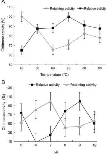
The effect of temperature on the activity and stability of the chitinase is presented in Fig.2a. Maximal chitinase activity was found at 70°C, and the enzyme retained greater than 87.7% of its activity over the range of 50–90°C. Thus, the chitinase was fairly stable over a broad range of temperatures between 40 and 90°C retaining 61% of its activity. Although a large number of chitin hydrolyzing enzymes have been isolated, only a few thermostable chitin-hydrolyzing enzymes are known to be active at above 70°C (Niehaus et al. 1999). The optimum temperature of M3-23 chitinase at
0 2 4 6 8 10 12 14 16 0 5 10 15 20 25 30 Chitinase (U/ml) NaCl (%)
Fig. 1 Effect of salinity on Virgibacillus marismortui M3-23 chitinase production, after 5 days of incubation at 37°C. Chitinase activity is expressed in units per milliliter of cell-free supernatant. Values shown are the average from triplicate experiments. Error bars Standard deviation
Fig. 2 Effect of temperature and pH on the chitinase of V. marismortui M3-23. a Effect of temperature on chitinase activity and stability at pH 8.0. b Effect of pH on enzyme activity and stability at 70°C. The values shown are percentages of the maximum activity of the enzyme, which is taken as 100%. Values shown are the average from triplicate experiments. Error bars Standard deviation
70°C was similar to that of thermostable chitinases from B. stearothermophilus CH-4 (75°C) (Sakai et al. 1994), Streptomyces thermoviolaceus OPC-520 (70–80°C) (Tsujibo et al.1993a), and P. rifitoensis (Essghaier et al.2010).
The effect of pH on activity and stability of the chitinase are illustrated in Fig.2b, which shows that the enzyme has the highest activity at pH 9.0 and was active over a broad pH range between 5 and 12, retaining greater than 52% of its original activity. Strain M3-23 produces an alkaline chitinase; similar observations were reported for Alcali-genes xylosoxydans (pH 8.0) (Vaidya et al.2001), Strepto-myces thermoviolaceus OPC-520 (pH 8.0–10) (Tsujibo et al.1993a) and Planococcus rifitoensis (pH 8.0) (Essghaier et al. 2010). These characteristics of thermo-tolerant and halotolerant alkaline enzyme are of great importance in metabolic production, and for the biocontrol ability against fungi and for protecting plants under biocontrol saline
conditions as well as in future applications of these enzymes in any biotechnological process that depends on high salinity or osmotic pressure for long incubation periods (Margesin and Schinner 2001).
Analysis of the partially purified enzyme from strain M3-23 of V. marismortui by electrophoresis followed by activity staining with a specific substrate revealed one large clear zone on the native PAGE gel corresponding to chitinase activity, but whether a single chitinase or more is produced should be confirmed. Many chitinase-producing strains, such as B. cereus 65 (Chang et al.2003), B. cereus YQ308 (Pleban et al.1997), B. subtilis W-118 (Wang et al. 2006), B. subtilis IMR-NK1 (Chiang et al. 2003), and Aeromonas hydrophila JP101 (Wu et al. 2001), produce a single chitinase in the cell supernatant. However, prelimi-nary results from the electrophoretic methods reported in this study should be confirmed by the total purification of
Fig. 3 a Phylogenetic relationships based on partial amino acid
sequences of family 18 chitinases. b Alignment of partial domain catalytic sequence from V. marismortui M3-23 with those of other Bacillus species. The phylogenetic tree was calculated by the neighbor-joining method implemented in the program CLUSTAL X and drawn using the program Tree View. Dark gray shading Active
site characteristic of 18 family glycoside hydrolases; asterisks 100% conservation. GenBank accession numbers: B. pumilus (ABI15082), B. subtilis (ABG57262.1); B. licheniformis ATCC 14580 (YP_077581.2); B. clausii (YP_174146.1); B. halodurans (NP_241782.1); B. cereus (ZP_04265861.1); B. mycoides (ZP_04167090.1); B. thuringiensis (ZP_04063375.1) and V. marismortui (GQ274956.1)
chitinase by other methods such as chromatography and analytical ultracentrifugation to estimate the molecular mass exactly and to check whether the chitinase has two or several isoforms.
Identification and characterization of chitinase gene sequence
The nucleotide sequence obtained from strain V. marismortui M3-23 was 638 bp, and encodes a partial protein of 213 amino acids. The sequence of the gene and the deduced amino acid sequence were used to query the GenBank database using BLAST. A neighbour-joining tree based on comparison of the sequence from strain M3-23 with other chitinases from Bacillus species is presented in Fig.3a. The results found no significant homology with the nucleotide sequence from strain M3-23. Alignment of the chitinase amino acid sequence from strain M3-23 with other chitinases are presented in Fig. 3b. Analysis of the amino acid sequences revealed that, in this region, the amino acid sequence from residue 125 to 133 is homologous to the active site motif (FDGVDLDWE) of enzymes in glycosyl hydrolase family 18. The deduced chitinase sequence from strain M3-23 showed low similarity to chitinases in GenBank. The highest similarity of 82% was obtained with B. halodurans C-125 (NP-241782) and B. licheniformis ATCC14580 (YP-077581), and less homology (80%) was shown with B. clausii KSM-K16 (YP-174146) and B. pumilus (ABI15082). The M3-23 catalytic domain nucleotide sequence seemed to belong to the chiA gene encoding a modular enzyme composed of a family 18 catalytic domain responsible for chitinase activity. Furthermore, on the basis of published chitinase gene sequences, and the corresponding deduced amino acid sequences, the novelty of chitinase M3-23 was probably confirmed (similarity 82%). Determination of the entire sequence gene and the presence of other domains responsible for chitinase activity should confirm the present results. Even within the same family, members exhibit diversity and variations in enzymatic properties. Perhaps the most remarkable difference among chitinases is the large range in their sizes. The smallest chitinases consist primarily of a catalytic domain, whereas the largest are often organized into multiple domains with distinct functions (Morimoto et al. 1997). Typically, chitinase enzymes are composed of at least three functional domains: a catalytic domain, a chitin-binding domain, and a cadherin-like domain or fibronectin type III-like domain, and other conserved domains of unknown function (Blaak et al.1993). Chitinases also differ in their pH optima and catalytic parameters. Multiple forms of chitinases are produced to provide synergistic degradation of the complex and resilient chitin polymer for complete degradation to N-acetylglucosamine.
Conclusion
This study showed that V. marismortui strain M3-23 produced a novel chitinase in cell supernatants. The described chitinase was tolerant and stable in the presence of extreme conditions (pH, temperature and high salt concentrations) compared to other bacterial chitinases. Therefore, the biotechnological exploitation of this enzyme could be of great importance in agro-industries.
Acknowledgment This work was supported by funds from the
Ministry of Higher Education and Scientific Research (Tunisia).
References
Ahmadian G, Degrassi G, Venturi V, Zeigler DR, Soudi M, Zanguinejad P (2007) Bacillus pumilus SG2 isolated from saline conditions produces and secretes two chitinases. J Appl Micro-biol 103:1081–89
Aunpad R, Panbangred W (2003) Cloning and characterization of the constitutively expressed chitinase c gene from a marine bacterium,
Salinivibrio costicola strain 5SM-1. J Biosci Bioeng 96:529–536
Blaak H, Schnellmann J, Walter S, Henrissat B, Schrempf H (1993) Characteristics of an exochitinase from Streptomyces olivaceovir-idis, its corresponding gene, putative protein domains and
relationship to other chitinases. Eur J Biochem 214:659–669
Chang WT, Chen CS, Wang SL (2003) An antifungal chitinase produced by Bacillus cereus with shrimp and crab shell powder
as a carbon source. Curr Microbiol 47:102–108
Chiang CL, Chang CT, Sung HY (2003) Purification and properties of chitosanase from a mutant of Bacillus subtilis IMR-NK1.
Enzyme Microb Technol 32:260–267
Chuan LD (2006) Review of fungal chitinases. Mycopathology 161:345–60
Ellouze O, Mejri M, Smaali I, Limam F, Marzouki MN (2007) Induction, properties and application of xylmanase activity from Slerotinia Sclerotiorum S2 fungus. J Food Biochem 31:96–107 Essghaier B, Fardeau ML, Cayol JL, Hajlaoui MR, Boudabous A,
Jijakli H, Sadfi-Zouaoui N (2009) Biological control of grey mould in strawberry fruits by halophilic bacteria. J Appl
Microbiol 106:833–46
Essghaier B, Rouaissi M, Boudabous A, Jijakli H, Sadfi-Zouaoui N (2010) Production and partial characterization of chitinase from a halotolerant Planococcus rifitoensis strain M2-26. World J
Microbiol Biotechnol 26:977–984
Freeman S, Minzm O, Kolesnik I, Barbul O, Zveibil A, Maymon M, Nitzani Y, Kirshner B, Rav-David D, Bilu A, Dag A, Shafir S, Elad Y (2004) Trichoderma biocontrol of Colletotrichum acutatum and Botrytis cinerea and survival in strawberry. Eur J
Plant Pathol 110:361–370
Gomez Ramirez M, Rojas Avelizapa LI, Rojas Avelizapa NG, Cruz CR (2004) Colloidal chitin stained with Remazol Brillant Blue R®, a useful substrate to select chitinolytic microorganisms and to evaluate chitinases. J Microbiol Method 56:213–219 Gohel V, Maisuria V, Chhatpar HS (2007) Utilization of various chitinous
sources for production of mycolytic enzymes by Pantoea dispersa in
bench-top-fermenter. Enzyme Microb Technol 40:1608–1614
Jung WJ, Kuk JH, Kim KY, Kim TH, Park RD (2005) Purification and characterization of chitinase from Paenibacillus illinoisensis
Leelasuphakul W, Sivanunsakul P, Phongpaichit S (2006) Purification,
characterization and synergetic activity ofβ-1, 3-glucanase and
antibiotic extract from an antagonistic Bacillus subtilis NRS 89–
24 against rice blast and sheath blight. Enzyme Microb Technol 38:990–997
Liu KB, Li SX (1991) Effect of NaCl on element balance, peroxidase iso-enzyme and protein banding pattern of Lycopersicon leaf cultures and regenerated shoots. Sci Hortic 46:97–107
Margesin R, Schinner S (2001) Potential of halotolerant and moderately halophilic microorganisms for biotechnology.
Extremophiles 5:73–83
Messaï A, Hannachi C, Zid E (2006) In vitro regeneration of NaCl-adapted tomato plants (Lycopersicon esculentum Mill.).
Tropi-cultura 24(4):221–228
Morimoto K, Karita S, Kimura T, Sakka K, Ohmiya K (1997) Cloning, sequencing, and expression of the gene encoding Clostridium paraputrificum chitinase ChiB and analysis of the functions of novel cadherin-like domains and a chitin-binding
domain. J Bacteriol 179:7306–7314
Niehaus F, Bertoldo C, Kahler M, Antranikian G (1999) Extremophiles as a source of novel enzymes for industrial application. Appl Microbiol Biotechnol 51:711–729
Patil RS, Ghormade V, Deshpande MV (2000) Chitinolytic enzymes: an exploration. Enzyme Microb Technol 26:473– 483
Pleban S, Chernin L, Chet I (1997) Chitinolytic activity of endophytic
strain of Bacillus cereus. Lett Appl Microbiol 25:284–288
Rodriguez-Kabana R, Godoy G, Morgan-Jones G, Shelby RA (1983) The determination of soil chitinase activity: conditions for assay
and ecological studies. Plant Soil 75:95–106
Roja Avelizapa LI, Cruz CR, Guerro MI, Rodriguez VR, Ibarra JE (1999) Selection and characterization of a proteo-chitinolytic strain of Bacillus thuringiensis, able to grow in shrimp waste
media. World J Microbiol Biotechnol 15:299–308
Rothschild LJ, Mancinelli RL (2001) Life in extreme environments.
Nature 409:1092–1101
Sadfi-Zouaoui N, Essghaier B, Hajlaoui MR, Fardeau ML, Cayol JL, Ollivier B, Boudabous A (2008) Ability of moderately halophilic bacteria to control grey mould disease on tomato fruits. J Phytopathol 156:42–52
Sakai K, Narihara M, Kasama Y, Wakayama M, Moriguchi M (1994) Purification and characterization of thermostable beta-N-acetylhexosaminidase of Bacillus stearothermophilus CH-4
isolated from chitin-containing compost. Appl Environ
Micro-biol 60:2911–2915
Sanchez-Porro C, Martin S, Mellado E, Ventosa A (2003) A. Diversity of moderately halophilic bacteria producing extracellular hydro-lytic enzymes. J Appl Microbiol 94:295–300
Schisler DA, Slinnger PJ, Behle RW, Jackson MA (2004) Formulation of Bacillus spp. for biological control of plant diseases.
Phytopathology 94:1267–1271
Sivitil AL, Kirchman DL (1998) A chitin-binding domain in a marine bacterial chitinase and other microbial chitinases: implications for
the ecology and evolution of 1,4-β-glycanase. Microbiology
144:1299–1308
Smaali MI, Gargouri M, Limam F, Fattouch S, Maugard T, Legoy MD, Marzouki N (2003) Production, purification and biochemical
characterization of twoβ-glucosidases from Sclerotinia
sclero-tiorum. Appl Biochem Biotechnol 11:29–40
Trachuck LA, Revina LP, Shemyakina TM, Chestukhina GG, Stepanov VM (1996) Chitinases of Bacillus licheniformis
B6839: isolation and properties. Can J Microbiol 42:307–315
Tsujibo H, Minoura K, Miyamoto K, Endo H, Moriwaki M, Inamori Y (1993a) Purification and properties of a thermostable chitinase from Streptomyces thermoviolaceus OPC-520. Appl Environ Microbiol 59:620–622
Tsujibo H, Orikoshi H, Tanno H, Fujimoto K, Miyamoto K, Imada C, Okami Y, Inamori Y (1993b) Cloning, sequence, and expression of a chitinase gene from a marine bacterium, Alteromonas sp.
strain O-7. J Bacteriol 175:176–181
Vaidya RJ, Shah IM, Vyas PR, Chhatpar HS (2001) Production of chitinase and optimization from a novel isolate Alcaligenes xylosoxydans: potential in antifungal biocontrol. World J
Micro-biol Biotechnol 17:691–696
Ventosa A, Nieto JJ, Oren A (1998) Biology of moderately halophilic
aerobic bacteria. Microbiol Mol Biol Rev 62:504–544
Wang SL, Lin TY, Yen YH, Liao HF, Chen YJ (2006) Bioconversion of shellfish chitin wastes for the production of Bacillus subtilis
W-118 chitinase. Carbohydr Res 341:2507–2515
Watanabe T, Oyanagi W, Suzuki K, Tanaka H (1990) Chitinase system Bacillus circulans WL-12 and importance of chitinase A1 in chitin degradation. J Bacteriol 172:4017–4022
Wu ML, Chuang YC, Chen JP, Chen CS, Chang MC (2001) Identification and characterization of the three chitin-binding domains within the multidomain chitinase Chi92 from