ORIGINAL ARTICLE
Mucosal malignant melanomas in head and neck surgery:
a retrospective study of six patients and review
of the literature
Astrid L. D. Kruse&Marc O. Riener&
Klaus W. Graetz&Heinz-Theo Luebbers
Published online: 12 March 2010 # Springer-Verlag 2010
Abstract
Introduction Of all malignant processes of the oral mucosa, 0.5% are malignant melanomas. Because of late diagnosis, pattern of growth, close proximity to the bone (particularly in palatinal localizations), and the correlated infiltration, malignant melanomas have a bad prognosis.
Patients and methods In this retrospective study, six cases of patients with oral mucosal malignant melanoma are evaluated, and a critical review of the literature is presented. The female to male proportion was 1:1 with an average age of 60.2 years; all patients were treated between January 1999 and July 2007. A neck dissection was performed on two patients because of clinically positive lymph nodes; one patient received interleukin 2 therapy, and three patients received postoperative radiotherapy. Two male patients died.
Conclusions We recommend biopsy on every growing lesion, pigmented or nonpigmented, for the required diagnosis and, in cases of malignant melanoma, wide excision as a second step. Neck dissections should be
performed in patients with clinically positive lymph nodes. Concerning interleukin 2 therapy, further studies should be performed in order to evaluate a routine application.
Keywords Malignant melanoma . Oral mucosa . Head and neck oncology
Introduction
Melanoma of the mucosa is very rare, presenting in only 1.3% of all melanomas [1], and it is especially rare in patients below the age of 30 years [2]. The peak age is between 65 and 79 years [3]. No risk factors for intraoral lesions have been clearly identified to date [4]. For mucosal melanomas, distribution on the head and neck occurs in 55.4% of cases, followed by anal/rectal (23.8%), female genital tract (18%), and urinary tract sites (2.8%) [1]. The most frequently affected sites in the oral cavity are the maxillary gingival and the palate [5,6], and they are more commonly affected in Japan and Africa than in Western countries [2, 7, 8]. The development of oral malignant melanomas (OMM) and the function of melanocytes in the oral mucosa have still not been clearly identified. One reason for the rare appearance of OMM might be the low concentration of melanocytes in the oral mucosa. In single tissue sections of the gingiva, the ratio is one melanocyte to 15 keratinocytes, whereas in the skin of the cheek, the ratio is 1:4 and in the trunk 1:14 [5]. Melanocytes migrate much less frequently to endodermally derived mucosa—e.g., nasopharynx, larynx, esophagus, or tracheobronchial tree;
A. L. D. Kruse
:
K. W. Graetz:
H.-T. Luebbers (*) Clinic for Craniomaxillofacial and Oral Surgery, University Hospital Zurich,Frauenklinikstr. 24, 8091 Zürich, Switzerland e-mail: t.luebbers@gmail.com M. O. Riener
Department of Pathology, University Hospital Zurich, Zurich, Switzerland
therefore, mucosal malignant melanomas are rare in these locations [3]. Concerning the difference between oral melanomas arising in the squamous or the respiratory (sinonasal) mucosa, the clinical outcome is slightly, but not significantly, worse in sinonasal melanomas. Prasad et al. [9] studied the difference in clinical behavior of 36 oral versus 59 sinonasal malignant melanoma. The latter demonstrates more aggressive morphologic features, but prognosis remains similar in both groups. It is striking, however, that in the squamous mucosa men are affected 3.5 times more frequently than women [9].
Three criteria for the diagnosis of primary oral melano-ma were proposed by Greene et al. [10]: (1) demonstration of malignant melanoma in the oral mucosa, (2) presence of “junctional activity” in the lesion, (3) and inability to show malignant melanoma at another primary site. But junctional activity is now seen as an unreliable indicator that the lesion is primary because melanoma metastasis can also involve the epithelium junction.
For determining the stage of cutaneous malignant melanomas, Breslow’s depth of penetration and Clark’s level of invasion, which correspond to the regional lymph node status and presence/absence of metastasis, have been used. Furthermore, two different growth patterns have been mentioned: (1) a radial growth phase that tends not to invade the underlying reticular corion but is associated with metastasis and (2) the vertical growth pattern, invasive and growing before metastasis can occur [3]. For mucosal malignant melanomas, Clark’s level of invasion cannot be applied because of missing histological landmarks like papillary and reticular dermis [11]; in addition, oral malignant mucosas have a radial growth phase [2] with early metastasis.
Therefore, the purpose of this study was to determine the initial symptoms, localization, stage, presence of regional or distant metastases, and treatment.
Patients and methods
The records of all patients with mucosal malignant melanoma from January 1999 to July 2007, six patients (three females, three males) treated at the Clinic for Craniomaxillofacial and Oral Surgery, University Hospital Zurich, were systematically reviewed. Criteria for inclusion were histologically proven primary malignant melanoma of the oral cavity. Criteria for exclusion were inadequate information, secondary oral malignant melanoma, and recurrent disease. Data of site of tumor, histopathology, stage, presence of regional or distant metastases, treatment, recurrence, and outcome were analyzed.
All patients had incisional biopsy performed before definitive treatment.
Results
Age, sex, and primary sites
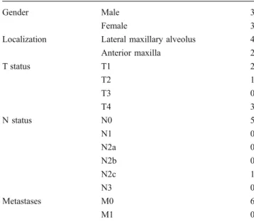
Subjects involved were three female and three male patients with an average age of 60.2 years. All patients revealed an OMM of the maxilla. The initial clinical presentation is listed in Table1.
Initial symptoms, stages, and course of the disease
The primary symptoms were pigmentation in three cases and painless swelling in three cases (Fig.1). No bleeding or pain as a first symptom was reported. In all patients, a biopsy was first performed, and wide excision followed as a second step. Two patients presented primary positive lymph nodes, so a neck dissection was immediately performed.
Histological findings
In two (with clinical T1 status) out of the four patients, the primary incisional biopsy was taken alio loco and a clear determination of infiltration depth was impossible, but in one of the two patients a superficial spreading growth pattern (patient 6 in Table 2) was observed. Concerning the melanoma marker, HMB-45, S-100 protein, and tyrosinase were used. One case was an amelanotic melanoma.
The tumors consisted of highly atypical melano-cytes with partial pigmentation. They showed focal necrosis and infiltration of the gnathic bones (Fig. 2a,
Table 1 Initial clinical presentation
Gender Male 3
Female 3
Localization Lateral maxillary alveolus 4 Anterior maxilla 2 T status T1 2 T2 1 T3 0 T4 3 N status N0 5 N1 0 N2a 0 N2b 0 N2c 1 N3 0 Metastases M0 6 M1 0
b). Three of the cases were composed of spindle cell melanomas.
Treatment results
Two (male) patients out of six died because of the malignant melanoma. After 3 months, one of these two patients had developed lung metastases; he received interleukin 2 therapy after metastasis, but he died. One male patient had a recurrence.
Three of the six patients underwent directly postsurgical radiation therapy. None of the patients received chemother-apy (Table2).
Discussion
Clinically, OMM can be divided into five types: (1) pigmented nodular, (2) nonpigmented nodular, (3) pig-mented macular, (4) pigpig-mented mixed, and (5) nonpig-mented mixed type [12]. Differential diagnosis include drug-, disease-, pregnancy-, or smoking-associated mela-nosis, oral melanotic maculae, Kaposi’s sarcoma, physio-logic pigmentation, melanoacanthoma, and nevus.
Outcome
It is generally accepted that the outcomes for oral malignant melanomas are worse than those for the cutaneous form. About 10% of oral melanomas are amelanotic, and more than 95% of the lesions are anti-S-100 antigen positive, with more specific markers including HMB 45, Melan-A, and antityrosinase [13]. These amelanotic malignant mela-nomas have a worse prognosis than other kinds because of the delays in establishing the correct diagnosis and also because they are thought to be more aggressive than pigmented melanomas [8]. One of the two patients that died had an amelanotic malignant melanoma (patient 2 in Table2) and presented early lung metastases.
Rogers et al. [14] reported that 50% of all patients with oral melanoma had regional lymph node metastases. In our patients, one with OMM revealed directly positive lymph nodes, so a neck dissection was performed. We did no elective neck dissection on N0 tumors. Lens et al. [15] did a meta-analysis of 1,522 cases and showed that there is no
Fig. 1 Painless swelling as a first symptom
Table 2 Initial staging, treatment, and outcome of the presented patients
Patient Sex cTNM Treatment Local recurrence Metastases Survival Follow-up time (months)
1 m T4N0M0 Resection, RT – Skin metastases DOD 12
Recurrence (skin) Involvement of skull base 2 m T3N2c0o Resection, bilat. neck
dissection, RT
Local recurrence (17 months)
Ileum metastases DOD 9 Pulmonary and mesenterial
metastases
3 f T4N0M0 Resection, RT – – Alive,
tu-free
32 4 f T1N0M0 Resection – Metastases lower eyelid Alive,
tu-free
42 5 f T4N0M0 Resection – Pulmonary metastasis Palliative 26 6 m T1N0M0 Resection – Contralateral cervical lymph
node metastasis
Palliative 13 Cheek metastasis
Cervical lymph node metastasis
Pulmonary, mediastinal, liver metastases
significant overall survival benefit for patients undergoing elective neck dissections.
There are different reasons for aggressive behavior in oral malignant melanomas. One reason is, on the one hand, the presence of the vertical more aggressive growth (nodular) with invasion of the underlying submucosa and, on the other hand, the radial growth (superficial spreading) phase that is characterized by early metastasis [5]. In the present study, one patient revealed a superficial spreading growth pattern (patient 6 in Table2) with primary T1 status and early metastases. Furthermore, the diagnosis can be delayed because most of the oral forms are painless in the early stages and do not reveal ulceration like the oral squamous cell carcinomas. In the present study, no patient had ulceration, bleeding, or pain as a first symptom. However, the overall 5-year survival rate is <30% [16–18]. Another important prognostic factor is the early metas-tasis in oral malignant melanomas because, in certain localizations like the palate, the tumor does not need to invade very deeply before reaching the bone, and radiologic evidence of bony erosions has been associated with early hematogenous spread [2]. In our study population, all patients had malignant melanomas in the upper jaw.
In the oral cavity, wide excisions are limited because of the anatomical structures; therefore, surgical results cannot always be compared between cutaneous and oral forms. The average age of patients with OMM in our study population was 60.2 years of age; the age is, in general, higher in comparison to squamous cell carcinomas. The highest incidence in the group is between 40 and 70 years as reported [13,19]. Therefore, the higher age also plays an important role in the outcome.
Bad prognostic factors, in general, correlate with tumor thickness and ulceration. Several studies also showed that male patients had a worse prognosis; the two patients that died and the one patient with a recurrence were men. In general, malignant melanomas are found almost three times more
frequently in men than in women, though in the present retrospective study the male–female distribution was 1:1.
Gingival melanoma has a slightly greater 5-year survival rate than that for palatinal melanoma, with a longer median survival period (46 vs. 22 months). Nodal involvement with oral melanoma reduces median survival substantially from 46 months to only 18 months [5]. In the literature, different survival times are mentioned for OMM. Chang et al. [1] reported a 5-year disease-specific survival for OMM of 31.7%. Meleti et al. [7] reported a 5-year survival of only 15%.
To sum up, the factors that are significant in lower disease-specific survival rates are primary high clinical stage, tumor thickness greater than 5 mm, presence of vascular invasion, absence of melanosis, and development of lymph node or distant metastases.
Excisional versus incisional biopsy
Cytological tests on brushed samples seem not to be reliable [4] and are, like fine-needle aspiration, contra-indicated [3]. Concerning biopsies in malignant melano-mas, dissemination of malignant cells in the tissue, blood, or lymphatic stream is discussed. Bong et al. [20], in a retrospective study of 265 patients having an incisional biopsy of cutaneous melanoma and 496 control cases of excisional biopsy, reported no correlation of malignant spreading; and Lederman et al. [21] reported similar results. One patient had been lasered before because of pigmen-tation. We recommend that any growing lesion, pigmented or nonpigmented, should be biopsied without delay for a diagnosis.
Treatment
Most authors advocate wide local excision of the tumor [4,
22]. Oral malignant melanomas are not very radiosensitive, but postoperative radiotherapy is generally recommended in
Fig. 2 a Melanoma infiltrating the maxillary bone (arrow), H&E, ×100. b Melanoma cells positive for HMB-45 (clone HMB-45, 1:50, DAKO, ×200)
multiple positive nodes, in extranodal spread, and in poor prognostic pathologic features [7,23].
The goal of immunotherapy is an activation of immune-competent cells in the tumor defense when, for example, interferon or interleukin are administered [24]. Verma et al. [25] were able to show in a review that a high dose of interferon is associated with a significant improvement in disease-free survival and a reduction of 2-year mortality.
Concerning interleukin 2 therapy, between 1985 and 2006, Petrella et al. [26] reviewed noncomparative phase II trials of high-dose, single-agent interleukin 2 with a response rate of 10–33% and with a complete response rate from 0% to 15%. The one patient that was treated with interleukin 2 in the present study already had lung metastasis and died. There is still a need for further investigations concerning immunotherapy in this particular group of malignancies.
Conclusion
Every growing lesion, pigmented or nonpigmented, requires a diagnosis; therefore, we recommend biopsy and, because of anatomical limits, wide excision as a second step. In cases of clinically positive lymph nodes, a neck dissection should be done immediately. In patients with negative lymph nodes, an elective neck dissection should not be offered directly. Despite the improvement of surgical techniques and the introduction of new chemotherapeutic agents, prognosis of this malignancy remains poor.
Competing interests The authors declare that they have no competing interests.
References
1. Chang AE, Karnell LH, Menck HR (1998) The National Cancer Data Base report on cutaneous and noncutaneous melanoma: a summary of 84, 836 cases from the past decade. The American College of Surgeons Commission on Cancer and the American Cancer Society. Cancer 83(8):1664–1678
2. Rapini RP et al (1985) Primary malignant melanoma of the oral cavity. A review of 177 cases. Cancer 55(7):1543–1551 3. Femiano F et al (2008) Oral malignant melanoma: a review of the
literature. J Oral Pathol Med 37(7):383–388
4. Garzino-Demo P et al (2004) Oral mucosal melanoma: a series of case reports. J Craniomaxillofac Surg 32(4):251–257
5. Hicks MJ, Flaitz CM (2000) Oral mucosal melanoma: epidemi-ology and pathobiepidemi-ology. Oral Oncol 36(2):152–169
6. Gu GM, Epstein JB, Morton TH Jr (2003) Intraoral melanoma: long-term follow-up and implication for dental clinicians. A case report and literature review. Oral Surg Oral Med Oral Pathol Oral Radiol Endod 96(4):404–413
7. Meleti M et al (2007) Oral malignant melanoma: a review of the literature. Oral Oncol 43(2):116–121
8. Notani K et al (2002) Amelanotic malignant melanomas of the oral mucosa. Br J Oral Maxillofac Surg 40(3):195–200
9. Prasad ML et al (2003) Clinicopathologic differences in malignant melanoma arising in oral squamous and sinonasal respiratory mucosa of the upper aerodigestive tract. Arch Pathol Lab Med 127 (8):997–1002
10. Greene GW et al (1953) Primary malignant melanoma of the oral mucosa. Oral Surg Oral Med Oral Pathol 6(12):1435–1443 11. Prasad ML et al (2004) Primary mucosal melanoma of the head
and neck: a proposal for microstaging localized, stage I (lymph node-negative) tumors. Cancer 100(8):1657–1664
12. Tanaka N et al (2004) Primary malignant melanoma of the oral cavity: assessment of outcome from the clinical records of 35 patients. Int J Oral Maxillofac Surg 33(8):761–765
13. Rapini RP (1997) Oral melanoma: diagnosis and treatment. Semin Cutan Med Surg 16(4):320–322
14. Rogers RS 3rd, Gibson LE (1997) Mucosal, genital, and unusual clinical variants of melanoma. Mayo Clin Proc 72(4):362–366 15. Lens MB, Nathan P, Bataille V (2007) Excision margins for
primary cutaneous melanoma: updated pooled analysis of ran-domized controlled trials. Arch Surg 142(9):885–891, discussion 891–893
16. Thompson LD, Wieneke JA, Miettinen M (2003) Sinonasal tract and nasopharyngeal melanomas: a clinicopathologic study of 115 cases with a proposed staging system. Am J Surg Pathol 27 (5):594–611
17. Yii NW et al (2003) Mucosal malignant melanoma of the head and neck: the Marsden experience over half a century. Clin Oncol (R Coll Radiol) 15(4):199–204
18. Patel SG et al (2002) Primary mucosal malignant melanoma of the head and neck. Head Neck 24(3):247–257
19. Gorsky M, Epstein JB (1998) Melanoma arising from the mucosal surfaces of the head and neck. Oral Surg Oral Med Oral Pathol Oral Radiol Endod 86(6):715–719
20. Bong JL, Herd RM, Hunter JA (2002) Incisional biopsy and melanoma prognosis. J Am Acad Dermatol 46(5):690–694 21. Lederman JS, Sober AJ (1985) Does biopsy type influence
survival in clinical stage I cutaneous melanoma? J Am Acad Dermatol 13(6):983–987
22. Mendenhall WM et al (2005) Head and neck mucosal melanoma. Am J Clin Oncol 28(6):626–630
23. Barker BF et al (1997) Oral mucosal melanomas: the WESTOP Banff workshop proceedings. Western Society of Teachers of Oral Pathology. Oral Surg Oral Med Oral Pathol Oral Radiol Endod 83 (6):672–679
24. Mohr P, Weichenthal M, Hauschild A (2003) Adjuvant therapy in melanoma. Onkologie 26(3):227–233
25. Verma S et al (2006) Systematic review of systemic adjuvant therapy for patients at high risk for recurrent melanoma. Cancer 106(7):1431–1442
26. Petrella T et al (2007) Single-agent interleukin-2 in the treatment of metastatic melanoma: a systematic review. Cancer Treat Rev 33 (5):484–496