Introduction
The use of mobile bearing implant systems such as the LCS (low contact stress) knee (DePuy AG, Cham, Switzer-land) for total knee replacements has proven to be very successful with an excellent long-term survivorship his-tory [3]. After the introduction of the A/P-Glide tibial component, a posterior cruciate retaining design, an in-crease of complaints about persisting anterior knee pain was observed postoperatively, which led to further inves-tigations. In this work we present the early follow-up re-sults for 230 knees. To illustrate the soft-tissue irritation, five cases were studied with a PET 18F-FDG scan in a pi-lot project prior to revision surgery.
The aim of this publication is to communicate the higher-than-usual revision rate, to demonstrate the intraoperative
findings of fat-pad impingement and to describe the typi-cal symptoms.
Methods Subjects
During a four-year period, 230 knee replacements with the LCS A/P-Glide bearing were performed in 218 subjects. The subjects were followed prospectively. Standard follow-up consul-tations were performed 3, 6 and 24 months postoperatively. Ad-ditional consultations were needed in the symptomatic cases. The clinical and radiological data was fed into a database using IDES software. The rate of revision surgery was documented. The decision to undertake revision surgery was made according to the severity of symptoms and the clinical findings. The reason for revision was documented on the clinical chart and the surgery protocol.
Abstract Early follow-up (15.8 months;1–48) of 230 knee replace-ments with an LCS A/P Glide com-ponent indicated an increased occur-rence of anterior knee pain due to a fat-pad impingement, necessitating early revision surgery. Unsatisfactory results were observed in 28 knees (12.2%). Thirteen knees (5.7%) were revised on finding the fat-pad im-pingement, and four knees (1.7%) were scheduled for later revision surgery; the remaining 11 subjects (4.8%) had revision surgery for a dif-ferent reason. Twenty-six subjects (11.3%) complained about milder but typical symptoms of a fat-pad im-pingement, and 22 subjects (9.6%) had unspecific mild symptoms. 151 knees (65.7%) were free of pain and
demonstrated an excellent result. The total revision rate of 10.4% (24 knees) is higher than described for other implant systems. However, the revision needed to treat the fat-pad impingement (5.7%) consisted of mi-nor surgery only, such as exchange of the mobile bearing or reduction of the fat pad by arthroscopy. The fem-oral and tibial components were able to be left untouched. Resection of the Hoffa’s fat pad is recommended when such an implant system is used, and possible impingement should be investigated intraopera-tively before closure.
Keywords Knee · Arthroplasty · Fat-pad impingement · Hoffa’s disease · Revision surgery DOI 10.1007/s00167-004-0492-x
Inès A. Kramers-de Quervain Ivette Engel-Bicik
Wolfgang Miehlke Tomas Drobny Urs Munzinger
Fat-pad impingement
after total knee arthroplasty
with the LCS A/P-Glide system
Received: 15 April 2003 Accepted: 8 December 2003 Published online: 16 March 2004 © Springer-Verlag 2004
I. A. Kramers-de Quervain (✉) ·
W. Miehlke · T. Drobny · U. Munzinger Schulthess Clinic,
Lengghalde 2, 8008 Zürich, Switzerland Tel.: +41-1-3857433,
Fax: +41-1-3857578,
e-mail: [email protected] I. Engel-Bicik
Department of Radiology, Division of Nuclear Medicine, University of Zürich,
Implant design: LCS A/P-Glide
The components include a cobalt chrome tibial tray, an ultra high molecular weight polyethylene bearing and a combination poly-ethylene and cobalt chrome control arm. The design rationale is to allow simultaneous axial rotation and anterior/posterior translation of the polyethylene tibial bearing relative to the fixed metal tray. Medial/lateral motion is constrained in the same manner as the ex-isting rotating-platform knee. The A/P Glide tibial component is kinematically equivalent to the meniscal bearing component, in that they have the same degrees of freedom. However, the design characteristics reduce the potential for bearing dislocation when compared to the existing mobile-bearing design, without compro-mising kinematics, contact stress or range of movement.
Pilot PET-scan study
In a pilot project, five subjects already scheduled for revision sur-gery had access to a PET 18F-FDG scan. The objective of this pi-lot project was to study subjects with soft tissue irritation due to a mechanical problem which will be verified during surgery, in or-der to investigate if this method might be suitable to detect me-chanical irritations. Positron emission tomography (PET) with 18-Fluoro-2-deoxy-D-glucose (FDG), a glucose analogon, is a diag-nostic method based on the imaging of positron emission of Fluor-18. FDG uptake is increased in cells with high glucose consump-tion and accumulates as phosphorylated FDG-6Phosphate. An in-crease in FDG uptake is therefore found in neoplastic cells, mak-ing this method a valuable diagnostic tool in stagmak-ing and therapy monitoring of various tumours [11, 22]. It has been shown that FDG-PET can also be used for imaging of patients with acute in-flammatory or infectious diseases, as activated granulocytes, leuko-cytes and macrophages metabolise FDG to a higher degree [1, 2, 21, 23]. For this study, all patients fasted at least 4 to 6 hours prior to the FDG-PET scan. The patients’ position was supine. The scan-ner employed was a whole-body PET scanscan-ner (GE Advance, GE Medical System, Waukesha, WI, USA). Data acquisition started 30 min after intravenous injection of at least 280 MBq of 18-Flu-oro-2-deoxy-D-glucose (manufactured in-house). Two bed posi-tions were acquired, centred at the knee joint. Transaxial images were reconstructed using filtered backprojection (Hanning filter with a 4 mm cut-off, 128×128 matrix, 2.34×2.34×4.25 mm voxel size). Images were displayed in coronal, sagittal and transversal planes. The contralateral knee was used as individual control.
Results
During the period of observation, follow-up information was available for all except one subject (see Table 1). The average postoperative observation period was 15.8 months (1–48). Two subjects died for reasons unrelated to the sur-gery. Thirteen patients (5.7%) had revision surgery when a fat-pad impingement was found intraoperatively. Revi-sion surgery included the resection of the Hoffa’s fat pad and posterior cruciate ligament, and the exchange of the A/P-Glide bearing for a rotating-platform bearing in 12 cases. In one case with milder symptoms an arthroscopic reduction of the fat pad was performed. The tibial and Table 1 Follow-up results of the 230 knees with the A/P-Glide system
Patient status Time since surgery (months) n %
Pain free, excellent result 16.4 (SD 10.3, range 2–48) 151 65.7%
Symptoms of fat pad impingement
Mild to moderate anterior knee pain, tenderness of the Hoffa’s fat pad, no revision necessary at the time. Satisfactory result
16.1 (SD 9.1, range 5 – 46 26 11.3% Revision surgery recommended, fat-pad impingement anticipated 15.8 (SD 5.68, range 12 – 24 4 1.7% Revision surgery performed, fat-pad impingement confirmed 14.5 (SD 6.7, range 6 – 33 13 5.7% Other symptoms
Mild-to-moderate symptoms not typical for a fat-pad impingement. Satisfactory result
14.8 (SD 8.89, range 5 – 32 22 9.6% Revision surgery performed for different reasons: progressive ligament
insufficiency (n=3), arthrofibrosis (n=2), tibial component loosening (n=2), patella revision (n=1), other (n=3)
12.7 (SD 9.68, range 2.5 – 35 11 4.8%
Died during the observation period 2 0.9%
Lost to follow up 1 0.4%
Fig. 1 An anterior position of the A/P-Glide bearing is seen in flexion, with decreased space for the fat pad between the anterior edge of the A/P-Glide bearing and the patellar ligament or the dis-tal patellar pole. The arrow indicates the prominent anterior border of the polyethylene
femoral components were able to be left untouched in all revised cases. Revision surgery was deferred in four cases (1.7%). A further 26 patients (11.3%) complained about mild or moderate anterior knee pain of the same nature as the revised ones – suggesting a fat-pad impinge-ment – which did not necessitate a surgical revision at that time. Eleven subjects (4.8%) had revision surgery for a different reason (see Table 1) without clinical signs of a fat-pad impingement. In those subjects, no imprints were found in the fat pad. The reason for revision surgery in those subjects was: (a) progressive ligament insuffi-ciency (n=3), (b) arthrofibrosis (n=2), (c) tibial compo-nent loosening (n=2), (d) patella revision (n=1), and (e) other (n=3). Twenty-two subjects (9.6%) had mild or moderate symptoms which were not typical for a fat-pad impingement and which did not necessitate treatment at that time.
Clinical symptoms
The following symptoms were associated with a fat-pad impingement:
1. Anterior knee pain
2. Tenderness of the anterior soft tissue (Hoffa’s fat pad) to palpation
Radiological findings
The radiological finding seen in the cases of a fat-pad im-pingement is demonstrated in Fig. 1: an anterior position of the A/P-Glide bearing is seen in flexion, decreasing the space for the fat pad between the anterior edge of the A/P-Glide bearing and the patellar ligament or the distal patel-lar pole.
PET-18F-FDG findings
The individual results are listed in Table 2. All subjects had increased FDG uptake in the involved knee. No or mini-mal uptake was noted in the contralateral asymptomatic knees, even when these knees were fitted with an arthro-plasty (see Fig. 2).
Intraoperative findings
Macroscopic local necrosis and fibrosis of the fat tissue below the distal pole of the patella were found in all cases where a fat-pad impingement was anticipated. The poly-ethylene of the A/P-Glide bearing gave a clear imprint in the fat tissue (see Fig. 3). Such an imprint was not seen in the cases that were revised for other reasons.
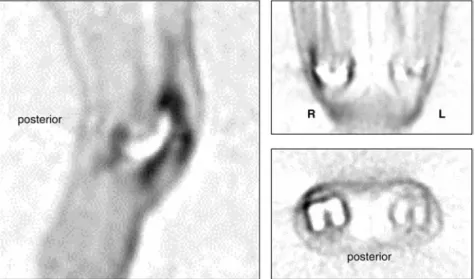
Fig. 2 Sagittal, coronal and axial view of a FDG PET-scan of a patient with both sided knee implants; high FDG up-take at the antero-lateral aspect of the right knee, where intra-operatively a fat-pad impinge-ment was confirmed. The pros-thesis is seen as a defect in FDG accumulation
Table 2 PET-18F-FDG findings
Case HJ Increased FDG uptake was seen at the antero-lateral border of the patella of the symptomatic knee fitted with the A/P-Glide system. An expansion into the cranial recessus was noted. There was no uptake on the contralateral side, where the patient was fitted with an unicondylar arthroplasty
Case OD Increased FDG uptake was seen on the lateral aspect of the knee and in the central intercondylar part Case GA Increased FDG uptake was seen in the anterolateral
and medial region of the symptomatic knee fitted with the A/P-Glide system. Only minimal uptake was noted in the contralateral knee, which was also fitted with a A/P-Glide system, which was, however, pain-free Case HE Increased FDG uptake was seen in the anterolateral
region of the symptomatic knee
Case BA Increased FDG uptake was noted on the medial and lateral side as well as in the suprapatellar and infrapatellar region of the knee fitted with the A/P-Glide system
Postoperative follow-up information
One year after the revision surgery five subjects were pain-free, five stated that their knee had improved and three complained about unchanged symptoms.
Discussion
The A/P-Glide component of the LCS knee-replacement system has a promising design as far as its kinematic abil-ity and stabilabil-ity are concerned. However, unlike the previ-ous LCS designs our early follow-up results with a total of 10.4% revisions demonstrated an increased revision rate. Whilst 4.8% of the cases were revised for commonly-recognised reasons, such as progressive ligament insuffi-ciency, arthrofibrosis and component loosening, an addi-tional 5.7% were revised as a result of finding the fat-pad impingement. This indicates the fat-pad impingement to be a new complication specific to the A/P-Glide design.
The fat pad has been described as an area with a high density of sensory nerve endings [26, 27], which explains the high rate of disabling pain. Dye [7] reported severe pain of the anterior synovium, the fat pad and the joint capsule when these structures were palpated arthroscopi-cally in a self-assessment study. “Hoffa’s disease” has been widely described by several authors [6, 9, 16, 18] as a cause for anterior knee pain, with serious functional implications in athletes and in subjects after anterior cruciate ligament reconstruction and other knee pathologies. After total knee
replacements, other impingement problems have been widely described by several authors, such as: (a) impinge-ment due to hypertrophic fibrous tissue or nodules in the intercondylar notch [4, 12], (b) impinging hypertrophic synovitis [5], (c) intraarticular fibrous plicae and bands [14, 24], (d) soft tissue under the patella consistent with the “clunk” syndrome [5, 15], (e) impinging PCL stump [5], (f) impingement caused by cement extrusion and proximal tibiofibular instability [19], and (g) fabellar im-pingement [8, 25]. Patellotibial imim-pingement has been de-scribed by Grigoris [10] in Kinemax-stabilised total knee replacement and by Patel [20], who reported mild patellar impingement symptoms in 8% of 157 patients with a pos-teriorly-stabilised (Insall-Burstein) knee replacement.
With the A/P-Glide component, the Hoffa’s fat pad was impinged between the anterior edge of the polyethyl-ene bearing and the distal pole of the patella, causing an-terior knee pain. In four knees (1.7%), revision surgery was planned to be performed later; in 13 knees (5.7%) re-vision surgery was performed, confirming the fat-pad im-pingement. Another 26 knees (11.3%) displayed mild-to-moderate symptoms. The high number of symptomatic cases indicates a significant complication resulting from this de-sign. However, revision surgery for this specific problem is comparatively simple, since only the polyethylene com-ponent need to be exchanged.
The other cases revised for different reasons did not demonstrate fat-pad impingement. This complication ap-pears to occur less frequently with the rotating-platform or meniscal bearing designs. Although the rotating plat-form has a similar shape of the polyethylene, an anterior impingement is not likely to occur due to the lack of ante-rior translation of the bearing. Meniscal bearings, how-ever, translate anteriorly, but appear to impinge less due to a lack of polyethylene centrally.
Fat-pad impingement may be anticipated where there is a history of anterior knee pain and tenderness of the an-terior region to palpation. The anan-terior position of the A/P-Glide polyethylene may be seen on radiographs. How-ever, the final diagnosis is only confirmed intraopera-tively where local tissue necrosis and fibrosis is found with an imprint of the bearing in the fat tissue. Up to now there are no imaging tools available to prove fat-pad im-pingement prior to revision. MRI has been described as capable of diagnosing alterations in the Hoffa’s fat pad with high accuracy [9, 13, 17]. This imaging technique, however, is not feasible in the presence of an arthroplasty due to artefacts resulting from the implant. The results of the PET-18F-FDG scan in the five subjects who had ac-cess to this investigation technique in a pilot project sug-gest that this is a possible tool to visualise soft-tissue irri-tation. The location of the increased uptake was in agree-ment with the intraoperative findings of soft-tissue irrita-tion due to the impingement. No uptake was seen in the asymptomatic contralateral knees, even when those knees were also fitted with an arthroplasty. The number of our subjects however is too small to be conclusive about the Fig. 3 Intraoperative findings. Macroscopic local necrosis and
fi-brosis of the fat tissue is present below the distal pole of the patella. The polyethylene of the A/P-Glide bearing gives a clear imprint in the fat tissue
sensitivity and the specificity of this imaging technique. Further investigation of this technique is needed to answer this question. Disadvantages of this technique are its cost and limited availability.
Conclusion
The Hoffa’s fat-pad impingement is a complication in to-tal knee replacement seen in systems with a central mo-bile-bearing component which has the freedom of anterior translation, such as the LCS A/P-Glide bearing. Since the fat pad is highly innervated, the impingement may cause anterior knee pain with functional implications necessitat-ing early revision surgery. Resection of the Hoffa’s fat pad
is recommended when such an implant system is used, and possible impingement should be investigated intra-operatively before closure. Fat-pad impingement may be anticipated in cases complaining about anterior knee pain and demonstrating anterior soft tissue tenderness to palpa-tion. The diagnosis is confirmed intraoperatively during revision surgery. As an imaging technique, PET-18F-FDG may be useful to visualise the soft tissue irritation prior to revision. However, due to the high cost of this technique and its limited availability other imaging techniques such as sonography should be evaluated as well.
Acknowledgements Many thanks to M. Pidermann and his team for entering and retrieving the data to and from the database. No fi-nancial assistance was provided for this work. This work complies with the current law of Switzerland.
1. Bakheet SM, Powe J, Ezzat A, Rostom A (1998) F-18-FDG uptake in tubercu-losis. Clin Nucl Med 23:739–742 2. Bicik I, Bauerfeind P, Breitbach T,
von Schulthess GK, Fried M (1997) Inflammatory bowel disease activity measured by positron-emission tomog-raphy. Lancet 350:262
3. Buechel FF, Pappas MJ (1990) Long-term survivorship analysis of cruciate-sparing versus cruciate-sacrificing knee prostheses using meniscal bearings. Clin Orthop 260:162–169
4. Carro LP, Suarez GG (1999) Inter-condylar notch fibrous nodule after total knee replacement. Arthroscopy 15:103–105
5. Diduch DR, Scuderi GR, Scott WN, Insall JN, Kelly MA (1997) The effi-cacy of arthroscopy following total knee replacement. Arthroscopy 13: 166–171
6. Duri ZA, Aichroth PM, Dowd G (1996) The fat pad. Clinical observa-tions. Am J Knee Surg 9:55–66 7. Dye SF, Vaupel GL, Dye CC (1998)
Conscious neurosensory mapping of the internal structures of the human knee without intraarticular anesthesia. Am J Sports Med 26:773–777 8. Erichsen H (1997) Bilateral fabellar
impingement after knee replacement – a case report. Acta Orthop Scand 68: 403
9. Faletti C, De Stefano N, Giudice G, Larciprete M (1998) Knee impinge-ment syndromes. Eur J Radiol 27: S60–69
10. Grigoris PH, Treacy RB, McMinn DJ (1992) Patellotibial impingement in Kinemax-stabilised total knee replace-ment. J Bone Joint Surg Br 74:472– 473
11. Hawkins RA, Hoh C, Glaspy J, Choi Y, Dahlbom M, Rege S, Messa C, Nietszche E, Hoffman E, Seeger L et al (1992) The role of positron emission tomography in oncology and other whole-body applications. Semin Nucl Med 22:268–284
12. Hirsh DM, Sallis JG (1989) Pain after total knee arthroplasty caused by soft tissue impingement. J Bone Joint Surg Br 71:591–592
13. Jacobson JA, Lenchik L, Ruhoy MK, Schweitzer ME, Resnick D (1997) MR imaging of the infrapatellar fat pad of Hoffa. Radiographics 17:675– 691
14. Jerosch J, Schroder M (1996) Clinical symptoms caused by intra-articular fi-brous plicae after knee replacement. Arthroscopic diagnosis and therapy. Arch Orthop Trauma Surg 115:195– 198
15. Lucas TS, DeLuca PF, Nazarian DG, Bartolozzi AR, Booth RE Jr (1999) Arthroscopic treatment of patellar clunk. Clin Orthop 367:226–229 16. Magi M, Branca A, Bucca C,
Langerame V (1991) Hoffa disease. Ital J Orthop Traumatol 17:211–216 17. Morini G, Chiodi E, Centanni F,
Gattazzo D (1998) Hoffa’s disease of the adipose pad: magnetic resonance versus surgical findings. Radiol Med (Torino) 95:278–285
18. Ogilvie-Harris DJ, Giddens J (1994) Hoffa’s disease; arthroscopic resection of the infrapatellar fat pad. Arthros-copy 10:184–187
19. Otani T, Fujii K, Ozawa M, Kaechi K, Funaki K, Matsuba T, Ueno H (1998) Impingement after total knee arthro-plasty caused by cement extrusion and proximal tibiofibular instability. J Arthroplasty 13:589–591
20. Patel DV, Aichroth PM, Wand JS (1991) Posteriorly-stabilised (Insall-Burstein) total condylar knee arthro-plasty. A follow-up study of 157 knees. Int Orthop 15:211–218
21. Peters AM (1998) The use of nuclear medicine in infections. Br J Radiol 71: 252–261
22. Reske SN, Bares R, Bull U, Guhlmann A, Moser E, Wannenmacher MF (1996) Clinical value of positron emission tomography (PET) in oncologic ques-tions: results of an interdisciplinary consensus conference. Schirmerreschaft der Deutschen Gesellschaft for Nuk-learmedizin. Nuklearmedizin 35:42–52 23. Sugawara Y, Braun DK, Kison PV,
Russo JE, Zasadny KR, Wahl RL (1998) Rapid detection of human in-fections with fluorine-18 fluorodeoxy-glucose and positron emission tomog-raphy: preliminary results. Eur J Nucl Med 25:1238–1243
24. Thorpe CD, Bocell JR, Tullos HS (1990) Intra-articular fibrous bands. Patellar complications after total knee replacement. J Bone Joint Surg Am 72:811–814
25. Wang JW (1995) Fabellar impinge-ment after total knee replaceimpinge-ment – a case report. Chang Keng I Hsueh Tsa Chih 18:185–189
26. Witonski D, Wagrowska-Danielewicz M (1999) Distribution of substance-P nerve fibers in the knee joint in pa-tients with anterior knee pain syn-drome. A preliminary report. Knee Surg Sports Traumatol Arthrosc 7: 177–183
27. Wojtys EM, Beaman DN, Glover RA, Janda D (1990) Innervation of the hu-man knee joint by substance-P fibers. Arthroscopy 6:254–263