;a-I
Acetylene: Dispersed Fluorescence Spectroscopy and Intramolecular
Dynamics
by
Jonathan Paul O'Brien
B.S. Chemistry (1991)
Hope College
Submitted to the Department of Chemistry in Partial Fulfillment of the
Requirements for the Degree of
DOCTOR OF PHILOSOPHY
at the
MASSACHUSETTS INSTITUTE OF TECHNOLOGY
September 1997
© 1997 Massachusetts Institute of Technology.
All rights reserved
Signature of Author ,. ._ Department of Chemistry June 27, 1997 Certified by Robert W. Field Professor of Chemistry Thesis Supervisor Accepted by --Dietmar Seyferth F
f
Chairman, Department Committee on Graduate Students
S
TEPCHM1OL7
SEP
171971
This doctoral thesis has been examined by a Committee of the Department of Chemistry as follows:
Professor Moungi G. Bawendi Chairperson
Professor Robert W. Field Thesis Supervisor
Professor Sylvia T. Ceyer
Acetylene: Dispersed Fluorescence Spectroscopy and Intramolecular Dynamics
by
Jonathan Paul O'Brien
Submitted to the Department of Chemistry on June 27, 1997 in Partial Fulfillment of the Requirements for the Degree of Doctor
of Philosophy in Chemistry
ABSTRACT
In this thesis a study of the Intramolecular Vibrational Redistribution (IVR) on the ground (01+~ ) state of acetylene is presented. For this purpose, several dispersed
fluorescence (DF) spectra were recorded. The transitions originate from different vibronic levels on the first excited singlet A1 Au state but terminate on the same vibrational levels of the
5l07. state.
Each of the DF spectra are composed of several overlapping progressions of the same
AAA. - X11,, initially excited states, the Franck-Condon allowed zero-order bright states (ZOBS). These are the CC stretching (vi') and trans-bending (vi') vibrational modes. Each ZOBS is associated with a distinct fractionation pattern caused by anharmonic coupling with surrounding Franck-Condon dark states ( v" = 0, v" = 03, v' = 0) on the RIlx. state.
Identification of the distinct ZOBS fractionation patterns is accomplished by simultaneously comparing the relative intensities of the DF spectra. A manual pattern identification
procedure is presented first.
Significant changes to the technique of traditional DF spectroscopy are described. These enable high quality frequency (± 4.0 cm') and intensity (± 20%) calibration for spectra recorded over a 20,000 cml' energy range. DF spectra recorded in this way are used in a new
statistical pattern recognition technique.
From the disentangled fractionation patterns, AAu. ---> Rl1 Franck-Condon factors and deperturbed energies for progressions of the ZOBS (nv2,mv4) are be obtained. These
results are useful in defining an effective Hamiltonian model which describes the ZOBS fractionation patterns. Qualitative IVR trends along each ZOBS progression are also investigated. The results presented here indicate that initial excitation in the CC stretch and
trans-bend vibrations is redistributed by strong Bend-Bend interactions prior to weaker
Stretch-Bend or Stretch-Stretch interactions.
Several previously undocumented features were observed in the DF spectra and the process of identifying them is described.
Thesis Supervisor: Robert W. Field Title: Professor of Chemistry
Acknowledgments:
Looking back over the past six years, reminiscing about my experience at MIT, I am immediately drawn to one of the most difficult times; this past year. A little over a year ago, I under went an experimental eye surgery to repair a laser injury that occurred in one of our laboratories. As I recovered, lying face down for almost two months, I honestly wondered if I would ever be able to finish my degree. While I have recovered quite nicely, I would never have made it back without the loving support of my wife, my research group, my research supervisor, and the other physical chemistry graduate students at MIT. For all of their help, I am extremely grateful and will always remember their enormous amount of kindness and generosity.
I am truly grateful to Bob Field. He has been an incredible supervisor over the last six years. He is packed full of enthusiasm and ingenious ideas about spectroscopy and dynamics. He never ceased to amazed me with his ability to derive back-of-the-envelope calculations that always seemed to point in the favor of his argument. Bob is an unique manager! He lets his students have considerable intellectual freedom but is careful to add the necessary
prodding to keep a student on track. I appreciate the freedom he gave me in reorganizing the spectroscopy laboratory and in running the acetylene DF experiments.
I spent a large portion of my first few semesters in the Undergraduate
Chemistry Office. Launa Calendar and Melinda Cerny always made me smile. I am thankful for my interactions with Melinda both as a teacher, professional advisor, and friend. I wish her the best.
During my six years, I have interacted with many undergraduates both as a teaching assistant and through the undergraduate research program. I was very fortunate to meet a very special person, Jennifer Sokol. Beyond her natural gifts, she is one of the most motivated and hard working undergraduates that I have ever encountered. She was incredibly helpful in the laboratory and made substantial contributions to my research. I wish her the best as she starts a new phase in her life. I know that she will succeed at anything she decides to do.
I was fortunate to have worked with many talented Field graduate students during my early years at MIT. Dave Jonas, Jim Lundberg, Bhavani Rajaram, Mike McCarthy, Jonathan Bloch, Nicole Harris, George Adamson, and Chris Gittins were extremely helpful. I also was able to work with many graduate students from other labs. Jody Klassen and I spent a month one summer recording energy transfer data on acetylene. One semester late at night, Manoj Nirmal and I tried desperately to record gain measurements of their CdSe quantum dots. Ken Kuno and I tried to revive the same project a couple of years later. It was always pleasant to interact with David Norris and his wife Beth.
I helped to setup a Resonance Raman experiment with Brian Gilbert. Brian talked me through some difficult times. We also drank and brewed a lot of beer. Steve Drucker and I collaborated on the acetylene triplet work described in Chapter 8 of this thesis. I enjoyed the numerous conversations we had about life, science, and acetylene.
The current Field group members are incredible! I have enjoyed my interactions with Richard Duan, James Janni, Leah Ruslen, Michelle Silva, Ilia Dubinsky, David Moss, and
Sergey Panov. Jason Clevenger was extremely helpful this past year with computer questions and in reorganizing the superlab disaster. I don't think that anyone could have done the job he's had to do for the past 10 months. (I know that I would not have!) I enjoyed our
occasional trip to the real world for dinner. Kevin Cunningham has always been willing to lend an ear to my rantings. His presence has kept me from the edge and in good spirits. It has been fun living with Fred Mikulec for the past year.
After ten years and nearly a million people hours, the acetylene isomerization problem may finally be solved! There are two unique individuals that I have had the pleasure of working with on the dynamics of acetylene:
Stephani Solina was an inspiration that drew me towards the world of dynamics and acetylene. Over the years, her friendship, constant excitement, and encouragement helped me to better formulate who I am. Her ambitious style started numerous theoretical and
experimental collaborations that have become an essential part of the acetylene project at MIT. I wish her the best in her new career and look forward to her continued friendship.
Matt Jacobson and I have developed a strong collaboration on acetylene which I hope continues beyond my years at MIT. The beautiful results presented in this thesis would never have been possible without Matt's hard work and input. I enjoyed our conversations about acetylene, dynamics, and the future of physical chemistry. During the writing phase of this thesis, Matt and Judy surprised me with a dinner and musical. That evening out really helped me to relax.
Steve Empedocles and I frequented the Muddy Charles to share some spirits and conversation. We also have spent some time in the gym playing basketball. Even though we are both atrocious, it was fun! The meals and slide shows courtesy of Steve and Marianne gave me an opportunity to relax and enjoy life a bit.
I had many great times with my good friend Bashir and his wife Rasha. Now that they have returned to Saudia Arabia, I will miss their home cooked meals, movies, and
adventurous drives to where ever Bashir wanted to eat. I wish them both the best of luck with their new move and newest addition to their family, Dana.
My family has been a source of great inspiration to me. For their constant support and understanding I am extremely grateful. My wife, Amelia, continues to be my best friend. I would not have made it without her by my side. She was always understanding when it came to late nights in the lab, the occasional science banter at MIT functions, and the mood swings accompanying my apparent successes and failures.
Table of Contents
Title Page 1 Signature Page 3 Abstract 5 Dedication 7 Acknowledgments 9 Table of Contents 11Chapter 1 Understanding the IVR Processes in Acetylene: Dispersed Fluorescence Spectra, Theoretical Models, and Pattern Recognition
Motivation 14 1.1 Introductions 14 1.2 Background 20 1.3 Previous Work 24 1.4 Current Work 26 1.4.1 Effective Hamiltonian 27
1.4.2 Dispersed Fluorescence Spectra 29
1.5 Future Work 33
1.6 Notation 33
1.7 References 36
Chapter 2 Preliminary Dispersed Fluorescence Spectra
2.1 Introduction 39
2.2 Experimental Setup 40
2.2.1 Spectra Purity of the PUMP Transitions 42 2.3 Effective Hamiltonian and Separation of Polyads 44 2.3.1 The Original Effective Hamiltonian 48
2.3.2 Utilizing the Appropriate Data Set 49
2.4 Polyad Pattern Recognition 52
2.5 Analysis of States Below 10,000 cm' 55
2.5.1 Pure Bending Polyads (N,=0) 57
2.5.2 Polyads with CC Stretch (N,>0) 58
2.5.3 Internal Structure of the Polyads 58
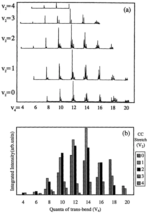
2.5.4 Intramolecular Vibrational Redistribution 63 2.5.5 Acetylene Polyad Franck-Condon Factors 66 2.6 Comparison of the N,=0 to the N,= 1 Polyads 69
2.8 Conclusion 75
2.9 References 77
Chapter 3 Dispersed Fluorescence Spectroscopy Methodology
3.1 Introduction 80
3.2 Experimental Setup 83
3.2.1 Production of Light 84
3.2.2 Molecules 87
3.2.3 Detection 91
3.3 Recording Dispersed Fluorescence Spectra 92
3.4 Frequency Calibration 102
3.4.1 Individual Segment Calibration 102
3.4.2 Baseline Functions 107
3.4.3 Grand Calibration 108
3.5 Concatenation 115
3.6 Relative Intensity Corrections 118
3.7 Conclusion 121
3.8 References 123
Chapter 4 Numerical Pattern Recognition: Polyad Zero-Order Energies and Franck-Condon Factors for 12C 2H2 XIA, - X1~: Emission
4.1 Introduction 124
4.2 Experimental 130
4.3 Pattern Recognition 133
4.3.1 Manual Pattern Recognition 133
4.3.2 XCC Pattern Recognition 135
4.4 Results 145
4.4.1 DF Features 145
4.4.2 Ns=0 Polyad Patterns 148
4.4.3 Ns>0 Polyad Patterns 151
4.4.4 ZOBS Zero-Order Energies 153
4.4.5 Franck-Condon Factors 156
4.5 Extra Emission Features 159
4.6 Discussion 166
4.7 References 168
Chapter 5 Polyad Patterns in the 12,000-16,000 cnm' region of the 11B,; State of Acetylene
5.1 Introduction 170
5.2 Experimental 170
5.3.1 The N.=0 Polyads 5.3.2 The N,=1 Polyads 5.3.3 The Ns>1 Polyads 5.4 Effective Hamiltonian
Chapter 5 Polyad Patterns in the 12,000-16,000 cml region of the X'tl State of Acetylene 5.5 5.6 5.7 5.8 16,000-18,000 cm-' and V 2 =V4 ZOBS Acetylene"-Vinylidene Interactions Discussion References
Chapter 6 Unexpected Emission Features from the XiA,, -+ XKi Dispersed Fluorescence Spectra
6.1 Introduction
6.2 Molecular Emission or Experimental Artifact 6.3 Interpolyad Interactions
6.4 Discussion 6.5 References
Chapter 7 XIA, K- 1i• Dispersed Fluorescence Spectra From VIK3 and
[4Vb] 7.1 7.2 7.3 7.4 Chapter 8 8.1 8.2 8.3 8.4 8.5 8.6 8.7 Introduction Experimental Discussion References
The Effects of Triplet Perturbers on Photophysical Processes in the
XIA, State of Acetylene
Introduction Experiment Results Discussion Preliminary Conclusions Recent Work References 177 180 180 187 189 190 193 195 197 199 211 230 233 235 235 237 244 245 246 247 249 255 256 259
Chapter 1: Understanding the IVR Processes in Acetylene:
Dispersed Fluorescence Spectra, Theoretical
Models, and Pattern Recognition
Motivation
The following summary describes the research conducted for this dissertation and serves as a general introduction to the main body of the thesis. This chapter is organized into five major sections: Introduction, Background on Acetylene, Current Work, Future Work, and Notation.
1.1 Introduction
Chemists have always sought to understand the nature of chemical reactions. All
chemical reactions require the "breaking and making" of single and sometimes multiple chemical bonds. These chemical processes involve complex motion of atoms contained in a molecular
species. Over the last one hundred years, physical chemists have studied a multitude of
molecular species using quantum mechanical, kinetic and thermodynamic theory. The advent of lasers in the 1960's provided revolutionary ways for physical chemists' to study chemical
reactivity.15 Scientists believed that lasers could eventually be used to control chemical reactivity. Initial experiments hoped to control reactivity by using laser light to excite very specific high vibrational levels of a molecule. In principle, this excitation process could activate chemical reactivity by increasing the probability that a selected bond will be broken or would become more susceptible to reactive attack. However, results from those experiments revealed that the excitation did not remain localized in a specific bond. The initial vibrational energy was redistributed throughout the rest of the molecular framework before the molecule could undergo chemical reaction.1'68 These redistribution processes have made most laser control strategies
Initial Excitation
Local Motion
Redistributed Motion
I II -I
I
)I
Figure 1.1: Schematic representation utilizing balls and springs of the redistribution of localized vibrational motion. At early times the initial vibrational motion stays localized. However, the localized motion is soon distributed through out the molecular framework.
activate chemical reactivity within a single chemical bond mostly ineffective. (Recently, Crim and co-workers have demonstrated how lasers can be used to control the chemical selectivity of dissociation processes.') This energy randomization can be thought of in terms of a set of
coupled balls and springs. We know that if we displace one of the balls and release it at t=O, the
excitation of the specific plucked ball and spring will remain localized for some time, but energy eventually will leak to the other balls and springs and they will start to move as well, see Figure
1.1. Physical chemists involved in the study of molecular dynamics have been striving to understand exactly how, when and where does the redistribution of energy take place for real molecules. A comprehensive understanding of these processes may lead to possible control mechanisms for many molecules.
For molecules near their equilibrium, with low internal energy, we have very good descriptions, harmonic oscillator and rigid rotor, of the vibrational and rotational motions.
However, chemical reactions seldom occur near equilibrium. Therefore, we have chosen to focus on highly excited molecular species which contain significant amounts of internal energy.
To develop an effective laser control strategy, we begin by asking where the molecule redistributes vibrational energy. Does the rate and primary path for redistribution of vibrational
motion differ for initial excitation in a pure stretching vibration, a pure bending vibration, or a combination of stretch and bend? Molecular interactions with the environment, such as with solvents, can make it difficult, due to the high density of states and the loss of frequency domain resolution, to interpret the results of experiments designed to answer questions of this nature. Our group's research avoids these problems by conducting low pressure gas phase experiments which are directed at the characterization of the vibrational redistribution processes of an isolated molecule. The characterization of the important molecular reaction pathways of vibrational redistribution necessitates a strong knowledge of the intramolecular vibrational redistribution (IVR). IVR processes for many molecular species have been the subjects of intense study, leading to a general and fundamental understanding of the IVR of many molecular systems.9,'0 The complexity of the IVR changes as the molecular species is altered, i.e. as the size, functional groups, and isotopes are changed. However, we do not know quantitatively how or why IVR changes from molecule to molecule or within a molecule at different vibrational energies. To develop a global standard or model for IVR of a particular molecule, we need to learn about individual IVR processes for a range of initially excited molecular vibrations and to try to understand how the IVR mechanism changes for different initial excitation conditions. The
multi-dimensional potential energy surface of a polyatomic molecule, V 3N-6- i Q , controls
the IVR coupling. In practice, we are limited by the types of initial molecular excitations that we can create in the laboratory, probing only a small region of the full potential energy surface. We hope that this localized view of the molecular dynamics gives us enough insight so that we will
be able to extrapolate the dynamics over a range of different initial excitation conditions. This knowledge will help to develop laser control strategies which utilize IVR and laser excitation as useful ways to control chemical reactivity.
We chose to determine the IVR structure of a molecular species by conducting a series of high- and low- resolution frequency-domain laser experiments. The complementarity of
frequency and time-domain is well established, see References 11 and 12 for examples. Information extracted from a frequency domain spectrum will give the same insight into the molecular dynamics as a time domain experiment. For either a high resolution (long time) or low resolution (short time) frequency domain experiment, one samples the eigenstates of a system. Confusion usually arises when one starts to think about interpreting an eigenstate spectrum in terms of yielding information about dynamics or energy flow. Eigenstates describe stationary probability distributions. They cannot move. A frequency domain experiment measures the molecular eigenstate spectrum, a distribution of energies and intensities, which arises from the coupling of an initial bright state, I (B ), to a series of dark levels, Y I OD)i , through high-order (cubic and quartic) potential energy coupling, VBD, see Figure 1.2. Any eigenstate of a system may be expanded by using a linear superposition of an orthonormal set of basis states,
I)
= C )i(1.1)For our work we expand the vibrational eigenstates using a zero-order basis of harmonic normal mode vibrational "states",
I
4 D)i (dark) andI
DB) (bright).I
'j)
= ajBI (DB) + CjDIl (D)i (1.2)The observed spectral intensities are proportional to the fractional bright state character in the jth eigenstate.
I c
lajBI
2Observed Spectrum
Energy
AUt leffU
1B)
0D
I
Figure 1.2: The observed spectrum of molecular eigenstates is equivalent to diagonalizing the
interactions among the basis states,
I
(,) and I D,). The observed intensity is derived from the fraction of ZOBS,I
B,), in each molecular eigenstate.1.0
0.8
-_ - 0.4-V 0.2-0.0 -1.0 0 V 0.8 0.6 0.4 5 5 20 WMW&P4WWi4 I* i 5 10 15 20 Time (ps)Figure 1.3: The top panel displays a schematic time dependent probability of the ZOBS, I cB(t)). The
lower panel displays the time dependent probability of the ith zero-order dark state, OD,(t))i
We can also express the zero order bright state (ZOBS), at t=O, in terms of the molecular
eigenstates.
(1.4)
(DB(O) = Cil LT
where
cj = (yj
I
B).
(1.5)The ZOBS, which is not an eigenstate of the system, will evolve in time according to (1.6)
I
B(t)) =x
CjI Yj)e-i(Ejt/h) j.~ .
.
.I ,This state will have a time dependent probability distribution
,,12 -i(EJ"- i)/thA/ (1.7)
P(t) =
(o)
,
I
B(It)=
I
ic,(4
,
2
+2
I
I
)
EJ+ c.
c (1.7)
Figure 1.3 displays a schematic survival probability of an initial ZOBS and the time dependent probability of the i-th dark state. The energy splittings and intensity information extracted from the high-resolution, frequency-domain spectrum reveals the nature of the IVR ("dynamics") for
an initial zero-order bright state. Realize that we are not discussing the "dynamics" of individual eigenstates, which are by definition stationary. The frequency distribution of the molecular
eigenstates (and the intensity distribution of transitions terminating on them) provides us with a "picture" of the IVR dynamics for an initially localized (and perfectly known) excitation! High resolution frequency domain experiments provide information about long time because the static eigenstate picture contains information pertaining to the long time evolution dynamics, which is exquisitely sensitive to an enormous number of weak interactions of the initially localized excitation and the dark states. In contrast, a low resolution frequency domain experiment contains only the short or early time dynamics, a few strong interactions between the initially localized excitation and the dark states.
1.2 Background for the study of acetylene IVR
The study of C2H2 is ideal for five reasons. First, the small size--four atoms--and lack of
complicated molecular structure, two carbons and two hydrogens, simplify the IVR processes. More specifically, a tetratomic species with only two heavy atoms lowers the density of states that can participate in the IVR processes at a given energy. Second, in the + state, So, C2H2
exists only as two chemical geometric isomers, acetylene and vinylidene. The C2H2 geometrical
isomer can react via an unimolecular isomerization, the simplest chemical reaction, to produce the other isomer. The transition state to this chemical reaction is predicted by high level quantum chemical calculations to lie near Evib= 16,000 cml above the linear acetylene zero-point level of
the ground state. 13 Third, the study of small unsaturated hydrocarbons such as C2H2 is important
for a wide range of applications, such as in combustion processes, atmospheric chemistry, astrophysics, and synthetic chemistry.
16,000
cm'-0 cm
1-C
[C
H
0-0
- *
e H
Acetylene
soReaction Coordinate
Figure 1.4: The ground electronic "'ZI state (So) of C2H2. The vinylidene (C2v) isomer has a minimum
Fourth, vinylidene is of special interest to scientists because it is a highly reactive species, a carbene, which survives for only a few picoseconds before isomerizing to form acetylene.14
Synthetic chemists have posited that carbenes are important as intermediates in many chemical reactions. Ultimately, we hope to learn more about the properties of vinylidene through the study
of the acetylene <-> vinylidene isomerization. Fifth, acetylene, a permanent gas, is inexpensive and easy to purify and use.
My research has focused on the study of the IVR processes that are expected to be important in the acetylene isomerization to vinylidene. With this knowledge, we may be able to control the production of vinylidene such that we may learn more about its physical properties. We have started to obtain this knowledge by employing laser spectroscopic and theoretical
methods. The focus of my research has been to understand the IVR structure that occurs in the acetylene So spectrum at low vibrational energies. After modeling the low vibrational energy IVR we can begin to extrapolate our model to vibrational energies at and above the energy of the isomerization barrier.
Infrared absorption and Raman experiments of the R15V state are plentiful and have been
used to determine the fundamental vibrational parameters, o , xii, xi5 15-19 The study (i=1 i=1 i j
of highly vibrationally excited eigenstates by absorption methods is made difficult by
increasingly weak transitions. As a rule of thumb, the successive overtone transitions for the CH stretches will have intensities which scale as 20,21
where
3N-5
AV= I IAv .- (1.9)
At high internal energies, the only eigenstates with significant transition moments in an
absorption experiment are the X-H stretching modes. Therefore, the intensity scaling limitations have forced many of the absorption studies to focus on the IVR of X-H stretches, where X=C, N, or O. We are interested in probing the high lying vibrational states of acetylene which will have favorable interaction with vinylidene states. Figure 1.5 displays the normal modes and
fundamental frequencies for linear (D-h), trans-bent (C2h) and vinylidene (C2v) C2H2. While, the
5th overtone of the acetylene CH stretch will occur in the 16,000 cmn ' energy region, a pure
stretching vibrational motion does not contain the large amplitude local CC-H bending needed to facilitate an acetylene <-+vinylidene interaction, see Figure 1.4. To reach Evib=16,000 cm-' via
pure overtone and combination bands of the CC-H bends, the CC-H bending frequencies,
o40
= 620 cm- and coP = 720 cm1, would necessitate the spectroscopy of the20th bending overtone.
These transitions are extremely weak.
By utilizing the first excited singlet (A 'Au) state as an intermediate in a double resonance experiment, Xi'V state spectra can be recorded which contain long progressions in highly
excited Franck-Condon active vibrational modes, trans-bending and moderately excited CC stretching. The Franck-Condon activity of these modes arises from the specific geometrical differences between the first excited singlet state (A 'Au) of acetylene, which has a trans-bent
The AAA, (Kr*' <- i) R1L+ transitions will have favorable Franck-Condon intensity for the trans-bending, AOCCH = 600, and CC stretching, Ar = 0.18
A
vibrations.There are two basic types of double resonance experiments conducted in our laboratory for recording spectra of the highly excited bending vibrational levels in the R ,state. One of these, stimulated emission pumping (SEP) spectroscopy, was invented by our group and has been extensively documented.22-25 Briefly, SEP spectroscopy excites a single rovibrational level in the
first excited state,
A
'Au, of acetylene with a nanosecond laser, called the PUMP. Molecules in the A 'Au state spontaneously fluoresce to R E' .A second nanosecond laser, called theDUMP, stimulates allowed A 1Au - X1gV transitions. This causes a depletion of the total
spontaneous emission. The SEP signal is observed by detecting the difference between the total
amount of spontaneous emission in the presence and absence of the DUMP laser. The other technique, dispersed fluorescence (DF) spectroscopy, utilizes only one laser, which is used to populate a single rovibrational level within the A 'Au state. This serves the same purpose as the
PUMP laser used in the SEP scheme. The total spontaneous emission from molecules in the
•A 'Au state is collected and imaged into a monochromator, dispersed by a grating, and the
" A'Au-+ X I' spectrum is recorded as a function of emission frequency.
1.3 Previous Work
In the late 1980's, SEP experiments conducted by our research group recorded acetylene X1' spectra which sampled vibrational energies slightly below and above the isomerization barrier.26,27 The complexity and incomplete understanding of the IVR
Symmetric
-A 1ACH Stretch
Am
AM
v,= 3372.87
+ Ss+cg
Aa+
CC Stretch
.,•4~ddL,.dh_...~giL.•,
v
2=1
+9
974.32
Ss+
Aa+
Sv 2=1386.90
Ss+g Aa+
antisymmetric CH Stretch
t~~l~A..v
3=3288.68
Sa+ rU As+trans-bend
IOM
A, a%
v
4=612.098
Ig Ss+Sa-Aa+
As-cis-bend
v5
=7
nu g A Aa+k"
Ss-u
Aa-A
26.835
Ss-
Sa+
Aa- As+
D u fl aTAs+
A4
AS+03
= 1165 Ss+ Al As+ 04 =835 Sa-B1Aa-05=3050
B2Sa+
Aa+
06=320
Sa+ B2 Aa+Figure 1.5: The normal modes for linear (D-,h), trans-bent (C2h), and vinylidene (C2v) structures of C2H2.
Note that the frequencies are expressed in terms of vi = + x for linear and trans-bent C2H2 while the
vinylidene frequencies are expressed in terms of co") 's. The double headed dashed arrows indicate the correlation of the different vibrational motion between linear and trans-bent C2H2
-Ag Ss+
Aa+
.3= 3025
Ss+ A As+ 02=1635 +sScAs+
Ss+
~processes made these spectra of acetylene appear to be intrinsically unassignable. Many plausible explanations for the discrepancies observed in the early SEP spectra were made, but none fully accounted for the complexity of the IVR at such high vibrational energies. Since that time, SEP experiments conducted by Jonas et. al. in the 7,000 cm' energy region have provided the most useful information in developing an initial picture of IVR at low vibrational energy.7'28 Analysis of these experiments revealed that on the shortest time scales, vibrational excitation
initially localized in the trans-bend and CC stretch, is redistributed into cis-bending and CH stretching vibrations. These results have prompted our group to design a series of experiments which would extend our knowledge of IVR to highly excited vibrational energies.
1.4 Current Work
Development of a new laser strategy to probe the acetylene <-+vinylidene interactions relies on achieving two research objectives. First, we need to develop a complete understanding of the acetylene spectrum and IVR at Evib below the barrier to isomerization. This approach necessitates a complementary use of both experiment and theory. We begin by recording high quality dispersed fluorescence (DF) spectra of the low energy region, Evib< 16,000 cml1, of the acetylene Ri'V state. The method for recording such high quality DF spectra will be discussed
at length in Chapter 3. By extracting energy positions and intensity distributions from the DF spectra, we can create an effective Hamiltonian model (Heff) that models quantitatively the IVR processes that occur at low vibrational energy. Second, once we are confident about the
dynamics at low Evib, we can extrapolate our effective Hamiltonian model to higher energy regions and look for deviations in the recorded DF spectra from the scaled predictions of the IVR
1.4.1 Effective Hamiltonian
The successful theoretical modeling of spectra at Evib below the barrier to isomerization creates a strong foundation that is useful in predicting and interpreting spectra recorded at higher energies. A theoretical model, an effective Hamiltonian, currently being developed to describe the IVR of acetylene, is based upon a normal mode harmonic oscillator basis set.28 30
, 32,33 This
choice of basis set allows us to conveniently express the resonances which are responsible for coupling vibrational energy from one normal mode into another. The theoretical model is a matrix where all of the diagonal terms contain the oscillator zero-order energies and
anharmonicities.31 TvrGv + Fr (1.10) where Gv=Gv(v, V2, V3, V414, 515 ; J 1)= 5 55 55 55 5 555 X vig, + x ttxltlt, lYttlt"VtVtVt+,--
+
+X
t1no~vi
+
1xO,3viv3 + 1180t,1le~
, E7t.t
vt y"L
Yi tviltlt' +...-By 12i=l i=1 j=i t=4t'=t t=4t'=tt"=t' i=1 t=4t'=t
Fr = Fr(VI, V2, 3,V44,v5 es Je) = BvJ(J + 1)+... and 5 55 B, =B- av
o
+ I (Y ttVtV, t~et
t )--... i=1 t=4t'=tThe off-diagonal matrix elements are expressed in terms of the potential energy,
V
(-
-Q
Q
, anharmonic oscillator coupling terms, such as kQiQjQkQ. 34 36 All of therules. Therefore, knowledge of the low energy DF (and/or absorption) spectrum can be used to determine the basic coupling strengths, Kij,k and Kij,k,1. The low Evib coupling is extrapolated to higher Evib by the harmonic oscillator scaling rule,
3N+5 M 3N+5 AV n (1.11)
i= -=1 i=1
The structure of this matrix is not universal for all chemical species, but is dictated by the unique set of states and state-specific coupling of each molecule. In the case of acetylene, the effective Hamiltonian matrix can be block diagonalized, see Figure 1.6. All of the states within a particular block interact via off-diagonal anharmonic couplings with each other, but do not interact with oscillators belonging to different blocks. Each block of oscillators is called a polyad. Each polyad is associated with a set of approximate polyad quantum numbers
[Ns, N,,e, 1]; where N, is the total number of quanta of stretching vibrational motion, Nres reflects
the vibrational frequency ratios (5o)1: 3co2: 55: o4: os), and I is the total vibrational angular momentum.37,38 Depending upon which states have optical activity, the coupling scaling and
selection rules from the Heff model indicate that each polyad will be represented by a unique intensity and frequency pattern within the acetylene spectrum. These patterns are dependent upon the identity of the initially excited state(s) within the polyad block. In the case of acetylene, the optically bright initial states are called the zero-order bright states (ZOBS). Both the strength of the resonances and the specific set of zero-order energies and anharmonicities control each spectral pattern, which may be as simple as a three line pattern spread over 30 cmn', or as complex as a 200 line pattern spread over 550 cml'. (Some anharmonic resonances cause
stronger mixing than others, and any given zero-order state is assumed to interact only with a certain subset of other zero-order states.)
The effective Hamiltonian model predicts that acetylene spectra of the X1 V state will be
composed of many partially overlapping polyad patterns. The onset of the acetylene<->vinylidene isomerization will introduce new resonances, causing interactions between polyads to turn on abruptly. This will likely cause the polyad patterns to be destroyed or degraded in the energy region of the acetylene <-> vinylidene interaction. A complete picture of the complex IVR involved in the acetylene isomerization could in principle be generated by observing the onset of the polyad pattern breakdown. Developing a clearer understanding of the role that IVR plays in these processes can only occur once the breakdown has been observed. We can begin to look for polyad breakdown once we have recorded and completely understood the low Evib DF spectra.
1.4.2 Dispersed Fluorescence Spectra
The A'An -- Xi' I dispersed fluorescence (DF) spectrum will contain progressions of the Franck-Condon active modes, (these are the zero-order bright states, ZOBS)
(v1,-
vI v , v v o =4+ (0, n,, mr0),lTm o ='m.m - 2,m - 4....-m (1.12) where the values of m and ITotI, the total vibrational angular momentum, are determined by the intermediate vibrational level chosen in the A 'Au state. TheA
'A, +<-* '+ transition followsc-type selection rules (the transition moment lies along the symmetry c-axis) AJ=0,±+, AK=Al--±1. ( K= K' is the vibrational angular momentum of the near prolate top A 'Au state projected onto the symmetry a axis), g<->u, and parity e<+-f for AJ=O, andf<->f or e<--e for AJ=-1.4 5 Because
Seff
[N
s,
Nres
,
1]
-[ N
s, Nres,
i ]
Figure 1.6: The effective Hamiltonian model is a block diagonal matrix. Each block is described by a set
of unique polyad quantum numbers, [N,,Ne.,1], and contains the zero order energies of the bright and dark states on the diagonal and with anharmonic couplings off the main diagonal.
The projection of the quanta in the trans-bending vibration, quantized in units of ýh, can only have
values of 14=v4, V4-2, v4-4, ...- v4 (V4 141).4 39 Any intermediate state with K = odd will
iII
I
terminate on ITotal= even and vice versa. This forces the number of quanta of v4 to be either even
(K# = odd) or odd (K/ = even). Due to both the structure of the effective Hamiltonian and the A' Au--R X1ZV acetylene electric dipole transition selection rules, there is only one ZOBS per polyad, see Chapter 2.
In order to observe individual polyad patterns and their breakdown, high quality spectra recorded over several thousand cml, at and below the energy of isomerization, with excellent frequency and intensity calibration, are necessary. This is accomplished by our optimized implementation of dispersed fluorescence spectroscopy, see Chapter 3. At low vibrational energies, Evib< 7,000 cmn1, the polyad patterns are clear and are easily extracted from the DF spectra. However, the polyad patterns at higher Evib are difficult to identify for two reasons. First, there is an increasing number of ZOBSs in a given energy region. Second, some of the polyads become highly fractionated due to stronger IVR interactions.
Regardless of the spectrum quality, the overlap of adjacent polyad patterns would often appear to corrupt polyad pattern recognition. Within the regions probed by dispersed
fluorescence, there are more than 100 overlapping polyad patterns. Unlike the relative intensity distribution within a polyad, the relative intensity distribution between polyad patterns is dependent upon the specific intermediate level used in the A'Au state. By recording and comparing the intensity patterns from several dispersed fluorescence spectra which utilize different intermediate rovibronic states in S1, the overlapping polyad patterns can in principle be
extracted. In practice, however, once the spectra are recorded, it is difficult to accurately recognize complex overlapping polyad patterns by eye. The lack of a clear criterion for
recognizing patterns causes ambiguities in assigning spectral line positions and intensities, see Chapter 2. This in turn damages our ability to create a reliable understanding of IVR processes.
Z.O.B.S
Tier 1
Tier 2
Tier 3
Bend-Bend
B
Stretch-Bend
A=B=C
Z.O.B.S <-> Tier 1 > Z.O.B.S <-> Tier2 > Z.O.B.S <-> Tier3
Figure 1.7: The IVR in acetylene is organized into tiers of interacting states: bend-bend, stretch-bend, and stretch-stretch. The hierarchical structure of the IVR is due to the strengths of the different anharmonic couplings, see Chapter 2 for more details.We have successfully developed a rigorous and unbiased statistical method (Extended Cross Correlation) for recognizing spectral polyad patterns, see Chapter 4.40,41
Several dispersed fluorescence spectra have been recorded and calibrated. Recently, the
XCC statistical method has successfully identified all polyad patterns with significant
Franck-Condon factors up to 15,000 cmrl, just below the isomerization barrier. This offers strong evidence that our simple model of coupled harmonic oscillators is valid in predicting the IVR.
C
Next, the nature of the IVR can be characterized by examining the intensity and frequency distributions within each polyad. From this type of analysis, the importance of the resonances can be ordered in terms of the strongest to weakest interactions, see Chapter 2. In our experiments, the order from strongest to weakest is as follows: bend-bend interaction (between trans and cis bending vibrations), stretch-bend interactions, and stretch-stretch interactions, see Figure 1.7.32,33 These are the controlling features of the IVR processes in acetylene.
1.5 Future Work
This thesis represents our nearly complete understanding of the IVR processes in
acetylene at Evib below the isomerization barrier. We must extrapolate our understanding of the IVR processes to energy regions near the isomerization barrier where we will begin to look for a breakdown of the polyad patterns. The greater understanding of IVR and the role it plays in the acetylene <-> vinylidene isomerization will be essential for devising an optimal laser control strategy. There are several recommendations in this thesis for future DF experiments. In the acetylene <-> vinylidene isomerization region, there may be a specific breakdown of the polyad structure for polyads with moderate CC stretch and high bending vibrational motion. Polyads with only bending or stretching motions may be less susceptible to isomerization. In any event, we are extremely close to generating a complete understanding of the early-time molecular dynamics of acetylene up to 16,000 cm-1.
1.6 Notation
Figure 1.5 displays the normal mode numbering and symmetry, following the format of Herzberg.3 1 Notice, that due to the reduced symmetry of the A'Au state (C2h point group)
compared that of the X'Z1 state (D**h point group) the vibrational mode number of the trans-bending vibration changes from v4 in the ground state to v3 in S1. Therefore, the
A'AU <--
50
transitions involving the trans-bending mode will be denoted by vV'. The9v4
subscript and superscript describe the initial and final number of quanta of bending, respectively. In the case of A'A, *- Xi 1 transitions involving CC stretch, the vibrational mode numbering is
the same in the So and S, states and will be denoted by 2s0.
We use Hougen and Watson's isomorphic Hamiltonian to express the linear acetylene rotational wavefunctions in a prolate top basis with the constraint that K-1.42 (K is the vibrational angular momentum in the S1 state). The A1'AU state is a near prolate top (the
moments of inertia have the following characteristics: A >> B = C) and K=Ka. 1 is the vibrational angular momentum in the X1 ' state, as discussed in 1.4.2. Throughout this thesis
the compact vibronic notation, VsS KK, introduced by Watson, will be used to describe the
A'Au - XR g transitions.43
The rotational prolate top wavefunctions are described in a signed-k basis set, where k
represents either signed-Ka or 1. This basis is extremely useful in writing matrix elements. However, photons and the molecule have a definite parity (inversion of the space fixed axis system), the signed-k basis set is not an eigen-representation of the parity basis. Therefore we use the Condon and Shortley phase convention as adopted by Hougen and Watson44
HrI
J,+k) = (- 1)J-KI J,-k) (1.13)Value of k Linear Combination Rotational Parity
Ik
=
0
IJ,
k = 0)
1
Ikl
20
o)"'
(I
J,+k) +I
J,-k)) (- 1)JJkl 0
/,j
(I J,+k) - I J,-k)) (- 1)J-k+1The total parity alternates with J. Therefore we employ e andf parity labels, where e parity is defined as even total parity with even J and odd total parity with odd J, and thef parity label is defined as even total parity with odd J and odd total parity with even J. Note that the total parity is determined by the parity of the rotational, vibrational, and electronic wavefunctions. (A simplified way of describing e and
fparity
is (-1)J and -(-1)J, respectively.)In this thesis, the most common 'A <-- -X' transition will be via the Q(1) rotational transition, where a Q branch implies AJ=0 and e<--f. The (1) refers to the initial value of J in the
RlEg state. This excitation process is denoted by AKAJK(J) For example, the transition
1.7 References
1. G.J. Scherer, K.K. Lehmann, and W. Klemperer. J. Chem. Phys. 78, 2817 (1983). 2. F.F. Crim, Ann. Rev. Phys. Chem. 35, 675 (1984) and references cited therein.
3. T.R. Rizzo, C.C. Hayden, and F.F. Crim, J. Chem. Phys. 81, 4501 (1984). 4. D.S. King, Adv. Chem. Phys. 50, 105 (1982).
5. J.J. Scherer, A.L. Cooksey, R. Sheeks, J. Heath, and R.J. Saykally, Chem. Phys. Lett. 172,
4 (1990).
6. S.L. Coy, R. Hernandez, and K.K. Lehmann, Phys. Rev. A, 40, 10 (1989).
7. K. Yamanouchi, N. Ikeda, S. Tsuchiya, D.M. Jonas, J.K. Lundberg, G.W. Adamson, and R.W. Field, J. Chem. Phys. 95, 6330 (1991).
8. Y. Chen, D.M. Jonas, C.E. Hamilton, P.G. Green, J.L. Kinsey, and R.W. Field, Ber.
Bunsenges. Phys. Chem. 92, 329 (1988).
9. Crim, F.F. J. Phys. Chem. 100, 12725 (1996) and references cited therein.
10. Nesbitt, D.J.; Field, R.W. J. Phys. Chem. 100, 12735 (1996) and references cited therein. 11. E.J. Heller, J. Chem. Phys. 68, 3891 (1978).
12. K. Kulander and E.J. Heller, J. Chem. Phys. 69, 2439 (1978).
13. M.M. Gallo, T.P. Hamilton, H.F. Schaefer III., J. Am. Chem. Soc. 112, 3714 (1990) and
references cited therein.
14. K.M. Ervin, J. Ho, and W.C. Lineberger, J. Chem. Phys. 91, 5974 (1989). 15. B.C. Smith and J.S. Winn, J. Chem. Phys. 89, 4638 (1988).
16. B.C. Smith and J.S. Winn, ibid. 94, 4120 (1991). 17. J. Pliva, J. Mol. Spectrosc. 44, 165 (1972).
18. W.J. Lafferty and A.S. Pine, ibid. 141, 223 (1990).
19. H. Finsterholzl, H.W. Schrotter, and G. Strey, J. Raman Spectrosc. 11, 375 (1981). 20. R.G. Bray and M.J. Berry, J. Chem. Phys. 71, 4909 (1979).
21. K.K. Lehmann and A.M. Smith, J. Chem. Phys. 93, 6140 (1990).
22. C. Hamilton, J.L. Kinsey, and R.W. Field, Ann. Rev. Phys. Chem. 37, 493 (1986). 23. C. Kittrell, E. Abramson, J.L. Kinsey, S.A. McDonald, D.E. Reisner, and R.W. Field, J.
Chem. Phys. 75, 2056 (1981).
24. D.E. Reisner, R.W. Field, J.L. Kinsey, and H.L. Dai, J. Chem. Phys. 80, 5968 (1984). 25. C. Kittrell, Stimulated Emission Pumping by Fluorescence Dip: Experimental Methods,
Molecular Dynamics and Spectroscopy by Stimulated Emission Pumping, World Scientific, New Jersey (1995).
26. E. Abramson, R.W. Field, D. Imre, K.K. Innes, and J.L. Kinsey, J. Chem. Phys. 83, 453 (1985).
27. Y. Chen, D.M. Jonas, J.L. Kinsey, R.W. Field, J. Chem. Phys. 91, 3976 (1989). 28. Jonas, D.M.; Solina, S.A.B.; Rajaram, B.; Silbey, R.J.; Field, R.W.; Yamanouchi, K;
Tsuchiya, S.J. J. Chem. Phys. 1993, 99, 7350.
29. M.A. Abbouti-Temsamani and M. Herman, J. Chem. Phys. 102, 6371 (1995). 30. M.A. Abbouti-Temsamani, M. Herman, S.A.B. Solina, J.P. O'Brien, R.W. Field, J.
Chem. Phys. 105, 11357 (1996).
31. G. Herzberg, Molecular Spectra and Molecular Structure, Vol. II Infrared and Raman
Spectroscopy of Polyatomic Molecules, Kreiger, Malabar (1991).
32. Solina, S.A.B.; O'Brien, J.P.; Field, R.W. J. Phys. Chem. 100, 7797 (1996).
33. Solina, S.A.B.; O'Brien, J.P.; Field, R.W. Ber. Bunsen-Ges. Phys. Chem. 99, 555 (1995). 34. W. H. Shaffer and A.H. Nielson, J. Chem. Phys., 9, 847 (1941).
35. G. Strey and I.M. Mills, J. Mol. Spectrosc., 59, 103 (1976).
36. E.B. Wilson, J.C. Decius and P.C. Cross, Molecular Vibrations, McGraw-Hill, New York (1955).
37. M.E. Kellman, J. Chem. Phys. 93, 6630 (1990). 38. M.E. Kellman and G. Chen, ibid. 95, 8671 (1991).
39. C.H. Townes and A.L. Schallow, Microwave Spectroscopy, Dover Publications, Inc.,
New York, NY (1975).
40. M. Jacobson, S.L. Coy and R.W. Field, J. Chem. Phys., accepted. 41. S.L. Coy, M. Jacobson and R.W. Field, J. Chem. Phys., submitted.
42. P.R. Bunker, Molecular Symmetry and Spectroscopy, pp. 330-332, Academic Press, New York (1979).
43. J.K.G. Watson, M. Herman, J.C. van Craen, and R. Colin, J. Mol. Spectrosc. 95, 101 (1982).
44. J.T. Hougan, J.K.G. Watson. Can. J. Phys. 43, 298 (February 1965). 45. C.K. Ingold, and G.W. King, J. Chem. Soc. 00, 2702 (1953).
Chapter 2: Preliminary Dispersed Fluorescence Spectra
This chapter represents a three year collaboration with Stephani Solina. At the end of this chapter, results from a recent collaboration with Professor M. Herman are presented. Parts of this chapter have been published in the Ber. Bunsenges. Phys. Chem., the Journal of Physical
Chemistry and the Journal of Chemical Physics.16,38,39
2.1 Introduction
Describing a system in a zero-order basis (uncoupled harmonic oscillator product states) that accurately depicts the initial state prepared by the laser enables elucidation of the dominant intramolecular vibrational redistribution (IVR) pathways. These pathways are equivalent to the anharmonic resonances described by matrix elements evaluated in the anharmonic oscillator product zero-order basis. Although high-resolution, frequency-domain spectroscopy is often naively considered to determine only "static" information, such as equilibrium molecular structure, spectroscopy can also generate a complete, compact, and intuitive description of complex dynamical processes, such as IVR, by the elucidation of the subset of resonances most important to the dynamics. This compact, intuitive description is an effective Hamiltonian model.
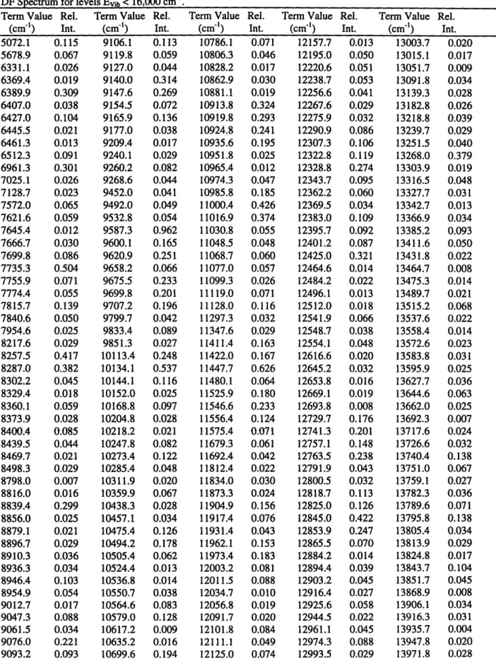
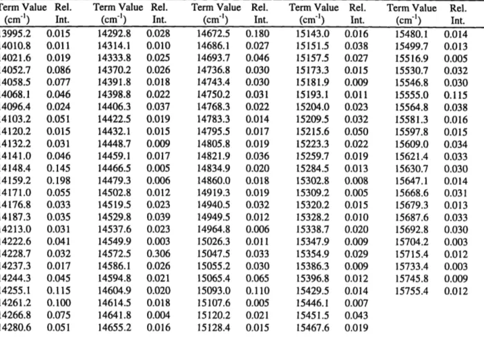
This chapter will focus on the preliminary analysis of the states at Evib< 10,000 cm- 1 in a
new high resolution A -4 R VO0Kb dispersed fluorescence (DF) spectrum of acetylene, Figure 2.1. The improved frequency resolution, when compared to previous DF spectra, and meaningful relative intensities, compared to those in stimulated emission pumping (SEP) spectra, allow this new VoOK 0 DF spectrum to be of great use in the initial stages of developing an effective Hamiltonian model and in elucidating the initial energy-flow pathways responsible for IVR.
1.0 0.8 0.6 0.4 0.2 0.0 *p~w~nn ~ -w~I'wF I . I
w
Origin Band
[~~E.il I.II
4 11, kII i ~ld Iva I * I c' I * I 4,000 6,000 8,000 10,000 12,000 14,000 16,000 18,000 INTERNAL ENERGY ( cm-1)Figure 2.1: High-resolution A Au -, -4X dispersed fluorescence (DF) spectrum, of the ground state, X state, of acetylene-h2 recorded from the zero-point level (Origin band) of the A state.
2.2 Experimental Setup
The high resolution DF spectra that sample the electronic ground state of acetylene-h2,
g, were recorded originating from the zero-point level, VOK, of the first excited singlet
electronic state, A 'Au. The third harmonic of a Lumonics HY-1200 Nd:YAG laser pumped a Lumonics HyperDYE-300 dye laser. The Coumarin 480 output of the dye laser was doubled in a 13-BBO crystal to produce ~3mJ of tunable 240 nm radiation which was used to populate the
V0`K0 JKa,Kc = Ka,Kc rovibronic level.' Matheson Gas Products purified acetylene (99.6% min.),
L
-v ----.. Il1 M" -VWIrý F. .- ... 'dllRNW%used without further purification at a backing pressure of 1.5 atm, was expanded into vacuum through a 0.8mm diameter General Valve Series 9 pulsed nozzle at 900 to the excitation laser beam, producing an effective pressure of acetylene in the beam of 350mTorr. The relative rotational line intensities obtained from a fluorescence excitation (FE) scan were consistent with a rotational temperature of 30K.
The fluorescence was collected in a direction perpendicular to both the laser excitation beam and the jet and focused into a 3/4 meter monochromator (Spex 1700). The monochromator was operated in first and second orders, utilizing a 1200gr/mm grating blazed at 500nm, and the dispersed fluorescence was imaged onto a Princeton Instruments intensified CCD (ICCD) Model
576LDG/RB. Roughly, 6000 laser shots were averaged for each segment. The advantages of using an ICCD are twofold. First, 576 spectral elements are recorded in parallel; i.e. a large
segment of the spectrum is recorded simultaneously. At each monochromator setting the ICCD covers a spectral width of -200
AS
in first order. Second, and most importantly, frequency andintensity calibration can be carried out directly following each DF scan without moving the
grating. This arrangement avoids corruption of the wavelength calibration by the stick/slip
problems associated with many scanning monochromators.
A complete description of the calibration routine is forthcoming, see Chapter 3. Briefly, we used thorium, neon and mercury atomic transitions for calibration, and by comparing
successive slightly overlapping scans along a series, systematic errors could be compensated for, allowing overall high accuracy and precision. The precision was found to be 1.5cmn' 2 - at 7.0 cm-1 resolution by comparing peak centers in spectra recorded on separate days. The accuracy
stimulated emission pumping (SEP) spectra recorded previously with absolute experimental errors of 2 F =0.020 cm>.3
Since the fluorescence intensity was the limiting factor in sensitivity, a series of spectra was recorded with entrance slit widths corresponding to resolutions of 10, 12 and 30 cm-' at 355 nm in first order and 7 cmr' for 355 nm photons in second order (grating position at 710 nm). Features with very low intensity in the higher resolution spectra were easily discernible in the spectra recorded at larger slit widths.
2.2.1 Spectral Purity of the PUMP Transition
Since the PUMP transition in this experiment was the Q(1) rotational line
(JKa,Kc 1
, Jparity = 1,e ) of the origin band, the intermediate level cannot be affected by B-type Coriolis4 interactions (AKa= +1, AK&= +1), and there are also no AKa= +2 asymmetric top interactions for this level. Since the closest C-type Coriolis perturber would be the v3 =1048 cm
1 level with a matrix element on the order of C, J(J -I1)-_ 1 cm'1, an estimate of the maximum contribution of K' = 0 character would be = 0.0001%. The contribution of K, = 0 character in
the intermediate level, which arises from the axis-switching mechanism at J'=1, is on the order of 0.001%.5 Therefore, with its exceedingly pure K'= 1 character, the only allowed rovibronic transitions from the selected single rovibronic intermediate level terminate on X -state rotational
levels with Ji,parity= 10,e or 22,f*
Since we use a normal mode product basis set, the X -state Coriolis coupling is
minimized. In a worst case, the proportion of admixed 1=1 fractional character would be roughly
~VStretchVBend X 10-5.6 The DF spectrum of acetylene has a smaller sensitivity range (100:1) than
the SEP spectrum of acetylene (1000:1).7 Indeed, the fluorescence intensity from the particular
intermediate state we have selected here was at least three orders of magnitude weaker than that from the more Franck-Condon allowed 2V3 or 3V3 intermediate levels excited under comparable conditions. Therefore, we do not anticipate significant contributions in the VooKb DF spectrum from levels with l= 1.
Furthermore, the only rotational structure in our DF spectra will be collapsed into a single feature even at higher resolution. This can be seen from the modified Dunham expression,3
Tvr=Gv + Fr (2.1)
where
Gv=Gv(v1, v2, V3, V414, 55 ; J 1, parity)
5 55 5 5 55 5 5 555
10)0 Vi +XIIxviv + + gotItlt +
X
7Yttt,,V+tIVt,
+ I-IIB2ttXVtlty"
i=1 i=1 j=i t=4t'=t t=4t'=tt"=t' i=1 t=4t'=t
Fr = Fr(Vi,V 2 V3,V4 ,v5 ;Je,Parity )=BJ(J+ 1)+...
and
5 55,
B, = B -
a°v +
(YttVtvt
,+y
,t~•
)+...
i=l t=4t'=t
For the 10,e and 22,f pairs of rotational levels with the same V = (v,,v,v2 3,v4,v5 ) and
14=0 or 15=0, Tvr(V;10,e) - Tvr(V;2 2,f) since
Fr(V; lo,e) -Fr(V;22,f) = B(1)(2) -Bv(2)(3)+... = -4By (2.2)
and
Gv(V;10,e) -Gv(V;22,f) = ...-By(0)2 + By(2)2 = 4By (2.3)
This rotational simplicity, combined with the accurate frequency calibration and meaningful relative intensity information, should make this data set of substantial interest to theoreticians. Although SEP has a much higher resolution and allows simplified rotational
assignments (since it is a double resonance technique), SEP's relative intensities are inaccurate due to saturation effects.7 Hence, theoretical analysis requiring moderate resolution and accurate intensities benefits from the use of high resolution DF spectra.
2.3 The Effective Hamiltonian and Separation of Polyads
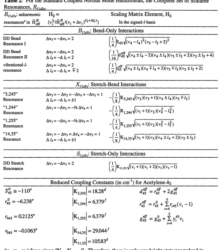
Our effective Hamiltonian, HRff , is expressed in terms of a normal mode product basis set, where the zero-order states, see Table 2.1, are coupled by harmonic oscillator matrix elements associated with the set of all resonanceso, R,c2,H, which have been identified in the
previous IR, Raman and CH- overtone spectra, as well as the SEP study, see Table 2.2. Each
resonance is characterized by a selection rule (which defines a group of strongly coupled zero-order levels) and a scalable harmonic oscillator matrix element. The resonances may be summarized as follows:
RcfH2 =BC2Hm 2 Xc2H2 u Sc2m I (2.4)
where
Bend-Bend Resonances:
Bcm=={DD Bend I, DD Bend II,vib-1-res) (2.5) Bend-Stretch Resonances:
XC2 2= { "3,245", "1,244", "1,255", "14,35"} (2.6)
Stretch-Bend Resonances:
Sc2H2={DD Stretch}. (2.7)
By inspection of the set of all knownlo resonances, Rc2H2, listed in Table 2.2, or by simple linear
algebra 3.11-13, it can be shown that out of the seven zero-order vibrational quantum numbers, there remains a set, Nc2H, of three conserved quantities or polyad numbers,
Nc2 = [Ns, NRes , l], (2.8)
where
Ns = V1 + V 2 + V 3 (2.9)
Nrs = 5vl + 3 v2 + 5 V3 + V 4 + 5 (2.10)
1=14+15 (2.11)
Ns is the total stretch quantum number, I is the total vibrational angular momentum quantum number, and Nres is a "resonance" quantum number arising from the frequency ratios of the normal modes, see Reference 3. The conserved polyad numbers mean that the effective Hamiltonian anharmonic oscillator product basis set for acetylene is effectively block diagonalized into groups of zero-order states, each labeled by [Ns , NRes , 1]. Each group of strongly coupled states is called a "polyad". The set of known resonances, Table 2.2, connects the zero-order states within each polyad but never between polyads. These dynamically
conserved polyad numbers (conserved on the timescale, ie resolution, of the experiment) can be used to label the eigenstates in addition to the rigorously conserved symmetries of acetylene that correlate to J, g/u point group inversion, and +/- parity labels. The three polyad numbers plus the J, g/u, and +/- parity labels provide a unique name for each polyad as well as a recipe for
automatically setting up individual polyad blocks in an effective Hamiltonian matrix.
In contrast to the linear and CC triple-bonded ground state geometry, acetylene's first excited singlet electronic state, A 'Au, has an equilibrium geometry that is trans-bent and has a
nominal CC double bond. This results in Franck-Condon (FC) activity in very high excitation of the trans-bend, v4, and moderate excitation in the CC stretch, v2, for spectroscopic schemes that
Table 2.1: Molecular Constants (in cm-') for Acetylene-h2 04 x34 0 x35 0 X44 0O X45 0 X55 0 g44 0 44 Y4 45 y55 y4 Ys 45 y5 55 Y5 -6.96"a -8.69a 3.082b -2.406b -2.335b 0.759b 6.541b 3.490b 0.0083b 0.0114b -0.0632b 0.0639b 0.0518b
-0.0070"
co o a0 0 X04 o xl20 x12 0 x13 0 x14 0 X15 0 X22 0 x23 0 X24 0 X25 0 X33 a See 3398.74a 1981.71a 3316.09" 609.016" 729.170a -26.57" -12.62a -105.09" -15.58" -10.85a -7.39" -6.10" -12.48a -1.57" -27.41" Ref (3) Y444 y445 Y455 0 Bo0
ai a4o0 745 75544r2
45
744
r55
c SeeNote: the effective Hamiltonian constants listed here somewhat out dated, see Chapter 5.
These two particular FC-active modes, v2 and v4, in combination with the particular set of
active resonances, Rc2H2, lead to a fortuitous coincidence such that there will be only one FC
bright state per polyad. This can be explained as follows. To a good approximation, the
symmetric CH stretch (vl), the anti-symmetric-CH stretch (v3) and the cis-bend(vs) are
FC-inactive, whereas the CC stretch(v2) and the trans-bend(v4) are FC-active. This defines a set of
zero-order bright states, or "chromostates", for SEP or DF spectra as the set of (vI,V2, V3, V4,
vs)'s where vl, v3, and vs5 are held constant and V2 and V4 are variable.14 By inspection of the
definitions of [Ns , NRes , 1], it is impossible to find more than one (V2,V4) pair with the same
b See Ref(18) 0.0062b -0.0379" 0.1576" 0.0141b 1.176608c 0.00686c 0.00621c 0.00560c -0.00129c -0.00215c 0.0000010b -0.0000236b 0.0000167b


![Figure 2.7: Structure of the low-energy extreme of the [low Ns, high NRes] polyads](https://thumb-eu.123doks.com/thumbv2/123doknet/13829244.443157/61.918.92.759.122.653/figure-structure-low-energy-extreme-high-nres-polyads.webp)

![Figure 2.10: Comparison of the observed and theoretical spectra for the pure bend polyad series and the Ns=1 polyad series ( [0,8], [0,10], [0,12] and [1,11], [1,13], [1,15]](https://thumb-eu.123doks.com/thumbv2/123doknet/13829244.443157/63.918.79.756.109.626/figure-comparison-observed-theoretical-spectra-polyad-series-polyad.webp)