Actin Remodeling in Motile Cells
By Eric A. Osborn
B.S.E. Biomedical Engineering
Duke University, 1998
S.M. Mechanical Engineering
Massachusetts Institute of Technology, 2001
Submitted to the Harvard-MIT Division of Health Sciences and
Technoloov in Partial Fulfillment of the Reouirements for the Degoree
of
Doctor of Philosophy in Medical Engineering
at theMassachusetts Institute of Technology
,..~ rx tx ,4~~~~~~June 20U4
~l'qkr'~g A · v ., .m · · 11 i
©/ ZU4 Mlassaclusetts Institute o0 I ecinology. All
rigts
reserved.ARCHIVES
Signature of Author
_
Harvard-MIT Division of Health Sciences and Technology
May,21, 2004Certified. by
C. Forbes ejvey, Jr., Ph.D.
ofessor of MechM
icil Engineering
Massachusetts Institu-f
Technology
Thesis Supervisor
Certified byJohn H. Hartwig, Ph.D.
Professor of Cell Biology
Harvard Medical School
Thesis Supervisor
Accepted byartha L. Gray, Ph.D.
Edward Hood Taplin Professor of Medical and Eectrical Engineering
Co-Director, Harvard-MIT Division of Health Scien es and Technology
LIBRARIES
-
_ -=I
__ __ _ __ - --- I __ ___- --- -- I,-- _ c_Actin Remodeling in Motile Cells
by Eric A. Osborn
Submitted to the Harvard-MIT Division of Health Sciences and
Technology on May 21, 2004 in Partial Fulfillment of the Requirementsfor the Degree of Doctor of Philosophy in Medical Engineering
Abstract
Non-muscle cell shape change and motility depend primarily on the dynamics and
distributions of cytoplasmic actin. In cells, actin cycles between monomeric and
polymeric phases tightly regulated by actin binding proteins that control cellular
architecture and movement. Here, we characterize actin remodeling in shear stress
stimulated endothelial cells and in actin networks reconstituted with purified proteins.Fluid shear stress stimulation induces endothelial cells to elongate and align in the
direction of applied flow. Alignment requires 24 h of exposure to flow, but the cells
respond within minutes to flow by diminishing their movements by 50%. Although
movement slows, actin filament turnover times and the amount of polymerized actin in
cells decreases, increasing actin filament remodeling in individual cells composing a
confluent endothelial monolayer to levels used by disperse, non-confluent cells for rapid
movement. Hours later, motility returns to pre-shear stress levels, but actin remodeling
remains highly dynamic in many cells. We conclude that shear stress initiates a
cytoplasmic actin remodeling response that is used to modify endothelial cell shape
instead of bulk cell translocation.
We determine the steady state dynamics of purified actin filament networks in the
entangled state and after orthogonal cross-linking with filamins using a novel,
non-perturbing fluorescence system. Human filamin A or Dictyosteliun discoidium filamin
slow actin filament turnover by 50% and recruit much of a significant population of
actin oligomers that we measure are present in polymerized purified actin solutions into
the immobile filament fraction. Surprisingly, these observations occur at very low
stoichiometry to actin, approximately requiring only one filamin molecule bound per
actin filament, similar to the amount required for actin filament gelation in vitro.
Networks formed with filamin truncates localize this activity to the actin binding domain
and reveal that dimerization and orthogonal cross-linking are not required for dynamic
stabilization. Re-expression of filamin A with or without the actin binding domain in
human melanoma cells that naturally lack this protein support the findings in purified
actin networks. These results indicate that filamin cross-linking stabilizes filament
dynamics by, slowing filament subunit cycling rates and by either decreasing spontaneous
filament fragmentation or promoting filament annealing.
Thesis Supervisor: C. Forbes Dewey, Jr., Ph.D.
Title: Professor of Mechanical Engineering, MIT
Thesis Supervisor: John H. Hartwig, Ph.D.Acknowledgements
I would like to express my deep gratitude to my advisors, John H. Hartwig and C. Forbes
Dewey, Jr. for their wonderful ideas, advice, and support throughout the process of
shaping this research. I would also like to sincerely thank the members of my Ph.D.
Thesis Committee, Frank Gertler and Doug Lauffenburger, for their guidance and insight.
I am grateful for the continued mentorship and collaboration of James L. McGrath, who has immeasurably helped me focus and guide my thoughts about actin dynamics and cellmovement. Special thanks to everyone in the Hematology Division at Brigham and
Women's Hospital and the Microfluids Laboratory at MIT who contributed their time and
expertise to teach me and help me achieve this goal. Finally, I would like to thank thestudents and faculty of the Harvard-MIT Division of Health Sciences and Technology, a
group with which I am grateful to be associated.
This is dedicated to my parents, Margie and Larry, for all of their faith, support, and love,
and to Teresia, without whom this would not have been possible.Biography
Eric Alan Osborn was born on October 2, 1975 in Battle Creek, MI to Larry and Margie
Osborn. In 1994, Eric graduated from Port Huron Northern High School in Port Huron,
MI and proceeded to attend college at Duke University in Durham, NC where he
graduated in 1998 with a B.S.E. in Biomedical Engineering. In 1999, Eric was awarded aWhitaker Foundation Graduate Fellowship and began graduate studies in the Department
of Mechanical Engineering at MIT and the Medical Engineering/Medical Physics
program at the Harvard-MIT Division of Health Sciences and Technology. His doctoral
research was performed at the Hematology Division of Brigham and Women's Hospital
located in Boston, MA.Table of Contents
Abstract ...
...2
Acknowledgements
... ...
4
Biography ...
5
Table of Contents .
...
...
...6
List of Figures ...
9
List of Tables ...
10
Goals of this Thesis .
... 11
Chapter
1:
Background and Literature Review ... 12Actin monomer ...
13
Polymerization of purified actin ...
...
14
Actin filaments
...
...
...
14
Actin filament turnover ...
15
The actin cytoskeleton ... 16
Regulation of actin remodeling by actin binding proteins ... 17
Gelsolin ...
... ... 18
The Arp2/3 complex ... ... 19
ADF/cofilins ...
20
Profilin ...
21
Ena/VASPs ...
21
Tropomodulins ... 22
Thymosins ...
22
Filamins ...
23
Cell crawling ... 24Fluorescence techniques for measuring actin dynamics ... 25
A mathematical description of actin dynamics ... 26
Potential deleterious effects of photoactivation and photobleaching .
...
27
Endothelial cells ... 28
Endothelial cell actin dynamics in static culture ... 30
References ...
32
Chapter II: Endothelial Cell Actin Cytoskeleton Remodeling During Mechanostiumlation
with Fluid Shear Stress ... 41Abstract ...
42
Introduction ... ... 4.3
Results ...
.... ...
... ... 46
46...
Fluorescent actin analogs function similar to native actin in endothelial cells ... 46Actin fluorescence recovery and decay curves are biphasic ... 48
Endothelial cells respond rapidly to shear stress by net depolymerization of their
cytoskeletons ...
48
Actin remodeling is transiently decoupled from motility during shear stress
exposure ... .0Actin remodeling remains enhanced in many endothelial cells after shear
stress-induced endothelial shape change ... 51Discussion ...
52
Short term response to shear stress (0 - 30 min) .
...
52
Relationship between shear stress-induced actin remodeling and endothelial cell
motil
.
ity
...
55
Actin dynamics during the phase of decreased endothelial cell
movement (1 - 6 h) ...
56...56
Actin remodeling in shear stress accomodated endothelium (~24 h) ... 57
Materials and Methods ... 59
Reagents
...
... .. 59
Cell culture, microinjection, and transfection ...
... 59
Immunofluorescence ...
...
60
SDS-PAGE and immunoblotting ...
60
Shear stress...
61
PA F and FRA P ... 61
Motility ...
62
Online supplemental material ...
...
63
Acknowledgements ...
64
Abbreviations list ...
65
R eferences ... ... 66
Figure legends ...
...
70
Online supplemental material ...
...
72
Figures ...
73
Chapter III.: Filamin Cross-linking Stabilizes Actin Filament Dynamics ... 78
Summary ...
79
Introduction ...
80
Experimental Procedures ...
83...
83
Caged resorufin iodoacetamide-labeled actin (CR-actin) ... 83
Filamin purification ...
...
...
84
F-actin co-sedimentation ...
84
Measurement of filament network gelation ... 85
Critical concentration ... 85
Reconstitution of purified actin filament networks ... 86
Cell culture, motility, microinjection, and transfection ... 86
Reconstituted actin network theory and simulations .
...
87
PAF and FRAP ...
89
Fluorescence measurement of actin assembly ... 91
Measurement of actin filament lengths... 91
Immunofluorescence ...
...
92
Results ...
93
Monomer diffusion and the amount of polymerized actin in reconstituted
networks ...
93
Actin dynamics in reconstituted networks of entangled purified filaments ... 94
Effect of cross-linking on reconstituted network actin dynamics ... 95
Localization of the stabilizing effect of filamins on purified actin dynamics ... 97
Rate constants for filament turnover in entangled and cross-linked filament
netw orks ... ... 98Effect of FLNa on cellular actin dynamics ... 99
DIiscussion
...
...
...
... 103
Significance of the purified protein PAF system for studying reconstituted actin
netw orks... 103Cross-linking stabilizes actin filament turnover ... 104
Filamins may function as a molecular 'clutch' ...
105
Filamins trap actin oligomers in reconstituted actin networks by altering filament
fragmentation and/or annealing... 106The spectin superfamily of cross-linking proteins influence the dynamic stability
of cytoplasmic gels ...
107
Acknowledgements ...
109
R eferences... ... 110F ootnotes... ... 115
Figure legends ... ... 116
Supplemental videos... 119
T ables ... 120 Figures...1... 122Chapter IV: Discussion and Future Directions ... 129
Dissecting the endothelial mechanotransduction cascade ... 131
Endothelial shear stress-sensing mechanisms ... 133
Filamin-A, a shear stress mechanosensor? ...
...
... 134
Filamin-A dynamics in endothelium ... 135
A molecular 'Clutch' or an adhesive 'Trap'? ... 137
Building an actin motor...
...
... 138
List of Figures
Description
Page
Fig. I-i: Actin filament treadmilling ...
16
Fig. 1-2: The actin cytoskeleton ... 17
Fig. 1-3: Schematic design of a PAF/FRAP microscopy system ...
25
Fig. 1-4: Actin dynamics correlate with cell speed ... 30
F'ig. II-1: EGFP-actin localization and distribution in endothelial cells ... 73
Fig. 11-2: Enclothelial cell fluorescent actin dynamics and modified by 12 dyn/cm2fluid
shear stress ...
,...74
Fig. 11-3: Short term shear stress response of endothelial cell actin remodeling . ... 75
Fig. 11-4: Endothelial cell motility during shear stress stimulation ... 76
Fig. 11-5: Actin remodeling response of endothelial cells after accommodation to shear
stress ... ...
77
Fig. II- 1: Simulations of PAF experiments with 30 m and 230 pm wide photoactivated
bands ... 122Fig. 11-2: Actin monomer diffusion from a 230 pm wide photoactivated band ... 123
Fig. 111-3: Steady state critical concentration of CR-actin and the effect of filamin
cross-linking.124
linking
...
124
Fig. 111-4: Characterization of actin dynamics in reconstituted networks of purified,
entangled actin filaments ...
125
Fig. 111-5: Filamin-induced cross-linking stabilizes reconstituted actin network
dynam ics ...
126
Fig. III-6: Actin binding, but not dimerization or cross-linking, is required for filamin-mediated stabilization of purified actin filaments ... 127
Fig. 111-7: FLNa modulates actin dynamics in melanoma cells ... 128
List of Tables
L)escription
Page
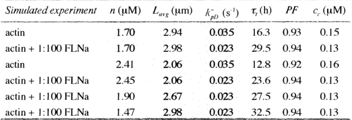
Table III-1: Predictions of purified actin dynamics with a mechanistic model of the actin cycle ... 120
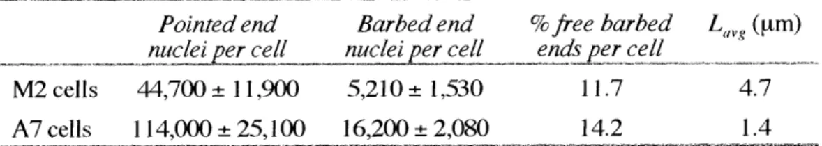
Table 111-2:
Characterization of filament ends and lengths in melanoma cell
Goals of this Thesis
The fundamental goals of this Thesis are to explore the dynamics and regulatory controls
of actin-based processes in an effort to further understand the molecular events governing
non-muscle cell shape change and movement. Within this context, the work presented
here focuses on two specific entities: the shear stress-induced shape change response ofendothelial cells (Chapter II) and the effect of the filamin family of actin filament
cross-linking proteins on the actin cycle (Chapter III). The individual goals of each
investigation are as outlined below.Endothelial cells in confluent monolayers subjected to steady, laminar fluid shear stress
change shape by elongating and aligning in the direction of applied flow, a process driven by dynamic changes in the actin cytoskeleton. Despite this striking morphological changeand its well-described relationship to the cytoskeleton, the dynamics of actin during this
process are unknown. How is the endothelial actin remodeling response modulated in
single cells before, during, and after endothelial cells accommodate their shapes to
the imposed fluid shear stress?
Filamins are known for their ability to create stiff actin networks by cross-linking
neighboring actin filaments. However, even in cross-linked networks, actin filaments
exchange subunits at their ends over time with the unpolymerized actin pool. Filamin
binding interactions with actin filaments, in addition to its cross-linking effect, may alterthe dynamic properties of actin filaments by unknown mechanisms. How does the
filamin family of actin filament cross-linking proteins affect actin filament diffusion,
turnover, length, and the extent of polymerization?
Chapter
I
Background and Literature Review
Non-muscle cells move and change shape by activating cascades of signals and molecular
events that require the integrated function of a large number of independent proteins
[2-71. Despite this complexity, these events all converge to influence the cytoplasmic proteinactin, which is the final substrate in this pathway. Inside the cell, actin coexists in a
dynamic exchange between monomeric and polymeric states [8-1 11. Actin polymers arebound together by accessory proteins to construct a dense three-dimensional network
1121. This actin polymer network, commonly referred to as the actin cytoskeleton,
dictates the underlying mechanical structure of the cell and determines both coarse and
fine aspects of cellular shape [13, 14]. Actin polymers incorporated into the cytoskeleton
are not simply mechanical struts, but exhibit a highly dynamic component in which
monomeric subunits 'turnover' by adding and subtracting from the different polymer
ends 115, 161. External biological, chemical, and mechanical stimuli can significantly
modulate a cell's baseline cytoskeleton organization and dynamics 1121. Understanding
these changes underpins the efforts to decipher the vast array of physiologic and
pathophysiologic events that rely on cell shape and movement 1171.Actin monomer
Actin is a highly conserved, globular 42 kDa protein 118, 191 that is one of the most
abundant proteins contained within the cytoplasm of eukaryotic cells 1201. Actin is of
considerable biological significance due to its involvement in many integral cellular
functions involving cell shape, mechanics, vesicle transport, and motility. Six isoforms of
actin exist in cells: uc-cardiac, a-skeletal, -vascular smooth muscle, f-non-muscle,
y-non-muscle, and y-smooth muscle 121], all of which are - 375 amino acids in length and
are highly homologous except near their amino terminus, which may confer
isoform-specific properties. While a- isoforms are found predominately in muscle cells and theP-and y- in non-muscle cells, each isoform may be expressed to varying degrees in spatially
and temporally regulated patterns in many different cells [221.
From x-ray crystallography, an actin monomer is approximately 6.7 nm x 4.0 nm x 3.7
nm in size 123, 241, split into a large and small domain separated by a binding pocket thataccepts a divalent cation and nucleotide. Actin monomer has one high-affinity divalent
cation (e.g. Ca
2 +or Mg
2+) binding site and four low-affinity binding sites [251. The
conformation and properties of the actin monomer are different depending on whether
Mg2+ or Ca2 + occupies this site 126, 271. In vivo, the high-affinity site is believed to bepredominately Mg
2+-bound, since the polymerization kinetics of actin are enhanced for
Mg2+-actin over Ca2+-actin 1271. Actin monomers are also bound to one of three
nucleotide species, ATP, ADP-Pi, or ADP, which greatly influence its dynamics, protein
binding affinities, structure, and regulation.
Polymerization of purified actin
At physiologic ionic strength, purified actin monomers (globular or G-actin) will slowly
self-associate into trimeric nuclei, an unstable intermediate, which serve as nucleation
sites for further assembly of monomers 128, 291. Trimer formation is the rate-limiting
step in polymerization, since the addition of actin filament seeds (pre-formed nucleationsites) eliminates the time lag prior to elongation. Once a trimer develops, it rapidly
elongates by sequential monomer addition onto its ends forming a helical polymer
(filamentous or F-actin). The kinetics of filament elongation are strongly influenced by
the nucleotide species bound to actin monomers 130-321. If unperturbed, polymerization
proceeds until only a small concentration of actin monomer, the critical concentration,
remains unpolymerized, which for ATP-bound monomers is -0.1 EtM 131, 331.
Eventually, purified actin solutions reach a steady state in which there is no net change inthe unpolymerized and polymerized actin monomer concentrations and therefore,
assembly and disassembly events perfectly balance.Actin filaments
Actin filaments are helical polymers stabilized by multiple, noncovalent contacts between
adjacent monomeric subunits [34]. The actin filament structure can be described by eithera one-start left-handed genetic helix of 5.9 nm pitch or a two-start right-handed helix of
72 nm pitch with a filament diameter of -7 nm 135, 361. Actin filaments are polarized
structures, where the two ends of the polymer can be differentiated both by conformationand biochemistry. By electron microscopy, filaments labeled with myosin subfragment-1,
a proteolytic fragment of heavy meromyosin, take on an arrowhead appearance leading to
the classic definitions of filament ends as 'barbed' and 'pointed'. Biochemically, the
kinetics that govern monomer addition at opposite ends of the actin filament are distinct:
addition of monomers to the barbed filament end occurs approximately ten times faster
than at the pointed end 115, 301.Actin filament turnover
At steady state, the actin filament lengths are stable since monomer addition at one
filament end counters loss at the opposite end [16]. If ATP is in excess, actin filamentsslowly elongate from their barbed ends and shrink from their pointed ends due to
differences in the affinities for monomers at the barbed and pointed filament ends [15].
This directed growth results in a net flux or 'turnover' of F-actin monomers from the
barbed to the pointed end. Actin filament turnover is a cyclic process that relies on ATP
hydrolysis to provide sufficient chemical energy to maintain the different filament end
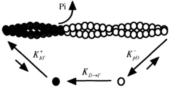
kinetics 1311.Turnover in its simplest form functions as a molecular treadmill (Fig. I-1) [15]. In this
scheme, unpolymerized actin monomers bound to ATP assemble onto the barbed
filament ends, hydrolyze ATP into an unstable ADP-Pi intermediate internally on the
filament, and, following inorganic phosphate dissociation, remain bound to ADP in the
core of the filament until they eventually disassemble from the pointed end [311. Oncefree from the filament, ADP-G-actin
monomers recycle their ADP for
ATP to repeat the cycle.The time required for an F-actin
*
~ '~
-
-
O
subunit to traverse a filament from
Fig. I-i1. Actin filament treadmilling. After assembly at the
barbed end, ATP-actin monomers () are hydrolyzed to
the barbed to the pointed end
ADPoPi-actin () as they flux through the actin filament,and disassemble as ADP-actin (O) at the pointed end.
provides
estimates
of
the
characteristic lifetime of the actin filament. Turnover is governed by the association and
dissociation kinetics of actin at the filament ends, properties that depend heavily on the
nucleotide profiles of subunits at the filament termini and internally on the polymer
chains [321. In purified actin networks, filament subunit recycling requires hours 1371, but
the pace can be accelerated to seconds or minutes in cells by the actions of associatedactin binding proteins [ 1, 11,38-411.
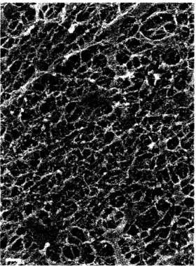
The actin cytoskeleton
In cells, actin filaments are linked together to form a three-dimensional support structure
that fills the cytoplasmic space (Fig. 1-2). This dense meshwork of actin polymer and
bound regulatory proteins forms the actin cytoskeleton, which gives the cell shape,
stiffness, and provides a mechanical scaffold that helps to fix the position of organelles
and facillitate pathways for intracellular transport. The inter-filament spacing of the
cortical actin cytoskeleton has been modeled as an orthogonal lattice with a pore size ofi
KD1
Z - 1tU nm 14z, 4)S1, tnrougn wnicn tne cell
transports cytoplasmic solute and proteins.
The cytoskeleton is organized by
cross-linking, bundling, and branching proteins into
functional subdomains based on their structure
and dynamics. Some of the structures are
relatively static, such as epithelial brush
border microvilli, hair cell stereocilia,
Drosophila neurosensory bristles, and
non-muscle cell stress fibers 1441, while others like
Fig. 1-2. The actin ctoseleton. Act i nfilaments are crosslinked together to form a
lamellipodia, filopodia, and membrane ruffles
dense meshwork of interconnected polymers.Many actin binding proteins are associated
with this insoluble cellular fraction. Bar, 200 are highly dynamic. The majority of cellular nm. Image courtesy of J. H. Hartwig.
actin filaments are fixed in space by
cross-linking proteins to form stiff gels 114]; however, there is some evidence that small actinfilaments are able to diffuse through cytoskeletal pores [401 and are mobile near the
plasma membrane [45].
Regulation of actin remodeling by actin binding proteins
In the cellular environment, a large cast of actin binding proteins establish spatial and
temporal regulation of actin by influencing the unpolymerized and polymerized phases inorder to tightly control the distribution, location, and kinetics of cytoplasmic actin 1461.
The control mechanisms utilized by actin binding proteins can be generalized according
to their mechanism of action: barbed (gelsolin, capping protein) and pointed
(tropomodulins,
the Arp2/3 complex) end capping, barbed end anti-capping
(Ena/VASPs), acceleration of filament depolymerization (ADF/cofilins), filament
severing (gelsolin, ADF/cofilins), filament nucleation (the Arp2/3 complex), nucleotide
exchange (profilin), and monomer sequestration (thymosins). Other actin binding
proteins regulate cytoskeletal geometry by forming filament bundles (c-actinin),
dendritic branches (the Arp2/3 complex), and orthogonal networks (filamins). To achieve
the dynamic cycling rates and localized structural control of actin, many of these
mechanisms behave synergistically [1, 32, 47, 48]. A few important actin binding
proteins will be discussed here as examples of these general mechanisms of actin
regulation.Gelsolin
Gelsolin is an actin filament severing and barbed end capping protein 149]. When
activated by M calcium concentration, gelsolin binds to the side of an actin filament,interdigitates, and severs it with unmatched potency. After severing, gelsolin remains
associated with the barbed filament end forming a tight cap. A severing/capping
mechanism rapidly dissolves cytoskeletal networks by creating an increased number of
short actin filaments that cannot anneal or rapidly elongate. Gelsolin can also initiate
rapid actin filament polymerization and membrane protrusion through interactions with
plasma membrane polyphosphoinositides that release gelsolin from the barbed ends 50,
511. Gelsolin null mice exhibit deficiencies in wound healing, inflammation, and
thrombosis 152, 531. Cells from these animals crawl poorly and organize pronounced
stress fibers consistent with a diminished capability to sever actin filaments 52, 531.
C)ver-expression of gelsolin produces the opposite effect leading to increased membrane
ruffling and chemotaxis 1541. Gelsolin also plays an important role in apoptosis as a
substrate for caspase-3, which cleaves gelsolin to constituitively activate its severing
function [55, 561. In support, apoptotic cell death is delayed in cells that do not express
gelsolin 1561..The Arp2/3 complex
The Arp2/3 complex binds to a preexisting actin filament, mimics a free barbed end, and
nucleates a daughter filament at a 70
°angle 571. Nucleation by the Arp2/3 complex is
activated by WASp/SCAR proteins 1581 and may require the exposure of binding sites atbarbed filament ends 159-611, although other reports maintain that nucleation can occur
on the side of the parent filament 162-641. The Arp2/3 complex also caps pointed
filament ends with nM affinity 1571, and is essential for rogue actin polymerization and
the rocketing propulsion of the bacterial pathogens Listeria and Shigella 651. Due to itsability to form a barbed end nucleation site, the Arp2/3 complex has been implicated in
numerous models of cell membrane protrusion involving the formation of branched,
dendritic actin networks 162, 66, 671. However, branched actin filament networks created
by the Arp2/3 complex alone are unlikely to account for the coherence of the leading
edge of crawling cells, as the dendritic structures formed by the Arp2/3 complex are
unable to gel filament networks in vitro unless the potent actin filament cross-linking
protein filamin is present [681.
ADF/cofilins
When barbed ends are capped, actin filament disassembly is slow, but it can be
accelerated by actions of the ADF/cofilin protein family [691. ADF/cofilin binds
cooperatively to the sides of filaments 1701, predominately at ADP-bound subunits whichare in excess near the pointed filament end, and enhances the rate of pointed end subunit
disassembly up to -25-fold in vitro 1711. The depolymerization activity of ADF/cofilins
is regulated by phosphorylation of serine-3. Phosphorylation of this residue by LIM
kinase inactivates ADF/cofilin by inhibiting its ability to bind to actin filaments 172, 731.This inhibition has been shown to be reversed by the Slingshot phosphatase 1741.
Membrane phospholipids can also inactivate ADF/cofilins by inhibiting its ability to bindF-actin 1751. Kinetic analysis of actin filament dynamics in vitro reveals that accelerated
filament turnover by ADF/cofilins produce net filament depolymerization in the presence
of sequestering proteins 171], which, in cooperation with barbed end capping, has beenpostulated to account for the high rates of actin filament turnover observed in vivo 1761].
ADF/cofilins have also been shown to be weak severing agents, fragmenting filaments
near the pointed end, which increases end numbers and accelerates depolymerization if
the newly formed barbed ends are capped [77-79] or polymerization if these ends remainuncaging of cofilin in cells increases barbed end exposure, F-actin content, locomotion,
and determines the direction of cell migration 811.Profilin
The intrinsic rate of nucleotide exchange on a free actin monomer can be accelerated by
profilin, which binds to monomers in solution with high affinity [821, exchanges ADP forATP -140x faster than a monomer alone, and shuttles ATP-bound monomer to uncapped
barbed ends 183, 841. Profilin is inactivated by the membrane polyphosphoinositide PIP,
1851. Since the intrinsic rate of nucleotide exchange by actin during rapid filament
turnover may be limiting in certain cases [321, profilin provides a mechanism to
overcome this energetic barrier.Ena/VASPs
Ena/VASPs interact with barbed filament ends at sites of actin assembly, such as the tips
of lamelllipodia and filopodia, where they antagonize the activity of barbed end capping
proteins, supporting F-actin assembly and causing filaments to grow longer and become
less branched [86, 871. This mechanism accounts for the diverse ability of these proteins
to negatively regulate motility in fibroblasts [881, but accelerate the movement of Listeria
in in vitro motility assays 165, 891. The activity of Ena/VASP proteins are regulated by
phosphorylation
at serine 157 by cAMP dependent protein kinase 189, 90].
Phosphorylation at this site increases Ena/VASP binding to F-actin by about 40-fold.
Tropormodulins
Although most widely studied for their function capping the thin filaments of muscle
sarcomeres, tropomodulins are also present in non-muscle cells where they cap actin
filament pointed ends, a process that is dramatically enhanced in the presence of
tropomyosins 191, 921. The association of tropomodulins with pointed ends is transient,
slowing polymerization at this end, which increases the levels of ADP-bound subunits at
filament termini and leads to net depolymerization [931. In endothelial cells,
tropomodulin 3 expression negatively regulates cell movement, and results in a decrease
in F-actin content and the number of free barbed ends [911, indicating that these events
are regulated in a coordinated fashion during cell locomotion.
Tlvmnosins
Thymosins are present in high concentrations in cells where they bind free monomers
stoichiometrically and inhibit polymerization
941. By sequestering cytoplasmic
monomer at concentrations much higher than the critical concentration of purified actin,
thymosins maintain large pools of unpolymerized actin available for the cell to
incorporate into filaments as needed. A sequestered monomer has a higher affinity for theunpolymerized state than for polymerization at pointed actin filament ends, but not at
barbed ends 1941. Therfore, as barbed ends are exposed, thymosin-sequestered monomerFilamins
Filamin family proteins are responsible for the creation and stabilization of actin filament
networks [131. Filamins are large, elongated, bivalent, flexible homodimers -160 nm inlength that bind and cross-link neighboring actin filaments into an orthogonal junction
195, 96]. Structurally, filamins are composed of an N-terminal actin binding domain
followed by 24 repeat motifs having a 13-barrel structure [97], where the terminal repeat
at the C-terminus forms the dimerization domain [95, 981. Filamins organize the
cytoskeletal architecture from sites deep within the cell body up to the leading edge
where they bind and affix the cytoskeleton to integral membrane proteins including 31and P7 integrins and GPlbc 198, 991. With a large and continually growing list of
binding partners already discovered, filamins are likely to play a unique role as
organizing centers for local cytoskeletal rearrangements [131.
Three filamin isoforms exist in humans, encoded by independent genes on different
chromosomes and differentially expressed depending on the tissue of origin 11001. In
non-muscle cells, filamin-A and filamin-B are the predominant isotypes 1131, which
abundantly localize to lamellipodia, promoting the formation of orthogonal filament
arrays [114, 961, and also localize at the base of filopodia 11011. The importance of
filamin-A in cytoskeletal organization and structure has been established in a natural
occurring line of human melanoma cells that lack this protein [102, 1031. Despite
expressing approximately wild-type levels of gelsolin, a-actinin, profilin, fodrin, and the
Arp2/3 complex, filamin-A deficient cells are unable to crawl and have a surface repletewith spherical aneurysms (blebs) indicating a lack of cortical stability in the absence of
FL,Na 1102, 1031. Rescuing these cells with filamin-A cDNA results in the reappearanceof lamellar protrusions and membrane ruffles and restores a normal motile phenotype
11031, as well as protecting them against apopototic cell death when mechanically
stimulated on their surface by pulling on magnetic beads 1104-1061. In humans,
mutations of filamin-A, which is located on the X chromosome, result in severe
congenital malformations of the brain, skeleton, viscera, and urogenital tract due to a
putative gain of function effect 11071. Complete deletion of filamin-A is male embryonic
lethal. Female filamin-A heterozygotes survive with the disease periventricular
heterotopia, which is characterized by abnormal brain development, seizures, and
vascular complications [11081.
Cell crawling
To move, a cell must selectively build, compress, and destroy its actin-based structures in
discrete regions. At the leading edge of a motile cell, actin polymerization drives
membrane protrusion 109, 1101. As polymerization continues, actin filaments formed at
the periphery transport into the cell's interior where they are depolymerized to recycle
actin monomers for assembly elsewhere 110, 11, 38, 111, 1121. At steady state, cells
crawl by balanced actin polymerization and depolymerization under the control of actinbinding proteins 116, 761. The ratio of actin monomer to polymer, the average
cytoskeleton filament length, and the filament turnover time are determining factors that
modulate the speed at which a cell moves across a substrate 11, 401. For example, asendothelial cells and fibroblasts crawl faster their filament turnover times and
polymerized actin fractions decrease 11, 401. The measured correlations between cell
speed and actin dynamics provide strong evidence supporting a direct link between
cytoskeleton remodeling and cell locomotion.
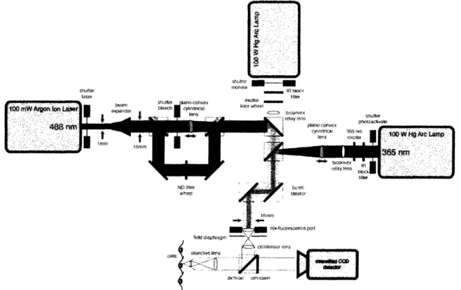
Fluorescence techniques for measuring actin dynamics
Actin dynamics can be measured in single cells with variable precision. Two analogous
fluorescence techniques provide some of the best estimates of parameters that describe
the dynamic nature of actin networks: photoactivation of fluorescence (PAF) and
Shuir lsel shut, - f, til fil" slitte p Sonu tae phtoavae ND filter wheel dinfi en-1sfslOn
Fig. 1-3. Schematic design of a l'A'/FRAP microscopy systenm. Sample photoactivation or photobleaching is performed by two opposing light sources focused to a narrow rectangular band. Global excitation of the fluorophore for monitoring purposes is achieved with a third, perpendicular light source. Fluorescence emission is collected by a computer controlled intensified CCD camera.
fluorescence recovery after photobleaching (FRAP) (Fig. 1-3). In PAF, a non-fluorescent,
caged precursor actin molecule is selectively uncaged by exposure to ultraviolet (UV)
light in a small spot or rectangular region within the cell 140, 113, 1141. FRAP is theinverse technique, in which the fluorescence of a labeled actin molecule is quenched with
high-intensity light at the excitation wavelength of the fluorophores 1115]. While PAF
has inherent signal-to-noise advantages over FRAP 1 16], the advent of green fluorescent
proteins and its colored variants are allowing new and exciting applications for FRAP
technology using molecular fusion proteins [1 171. Once the fluorescent actin derivative isuncaged or bleached, the fluorescence decay or recovery at the center of the
photoactivated or photobleached region, respectively, is monitored over time. Using an
interpretive model that describes the actin remodeling processes underlying the
fluorescence evolution, appropriate physical parameters can be extracted and analyzed
11161.
A mathematical description of actin dynamics
One mathematical model describing PAF- and FRAP-based actin dynamics, the Tardy
Model, provides simultaneous estimates of the translational diffusion coefficient of actin
monomer, the fraction of total actin polymerized, and the actin filament turnover time
11161, and has been used to quantitate actin dynamics in various cell types such asendothelium, fibroblasts, and melanoma cells [1, 401, yielding results consistent with
those previously published by other investigators [11, 38, 39, 411. Although this model
was developed to analyze cellular actin dynamics, the simplified rectangular cell
geometry and assumptions (e.g. homogeneous sample, no nucleus or organelles) inherent
in the theory are equally suited for probing the dynamics of purified actin preparations.
The general mathematical problem defined by the Tardy Model involve one-dimensional,
unsteady, coupled partial differential equations that describe actin monomer diffusion and
filament turnover in a model cell. The general solution is represented as an infinite series
for both actin monomer and filament concentrations. The result is a biphasic response,
where the short-term dynamics are primarily due to monomer diffusion and the long-termto filament turnover, that depend strongly the ratio of the monomer diffusion and filament
turnover times [1161. Under certain conditions when monomer diffusion is rapid
compared to filament turnover, as typically is the case in purified actin solutions, the
general solution to the long-term fluorescence can be simplified to a decaying
exponential, where the y-intercept represents the 'immobile' fluorescence fraction and
the decay constant is the filament turnover time [1161.Potential deleterious effects of photoactivation and photobleaching
While PAF and FRAP are generally non-invasive techniques that require only short
exposures to light to initiate experiments, phototoxicity, photodissolution, and local
heating of actin during uncaging, bleaching, and fluorescence excitation remain important
factors to consider. These destructive processes can potentially induce actin filament and
monomer crosslinking, denature actin protein structure, and/or break actin filaments.
Estimation of the photoactivation (100
kW/m
2) and excitation (10
kW/m
2)
fluorescence intensities at the sample [1 18] classifies these light levels as low excitation
intensities (< 5 MW/m2) according to Vigers and colleagues 11191. At low light excitationlevels, both actin and microtubules have the potential to fracture and dissolve after as
short as -1 min of continuous illumination i1 19, 1201. To mitigate these effects, light
exposure must be minimized during sampling. Surface heating of the actin solution is less
problematic, since even at the high laser intensities (> 5 MW/m2) used to photobleachsamples for FRAP experiments, the amount of sample heating is estimated to be less than
0.1 °C 11211. Finally, the best evidence that F-actin phototoxicity is minimal after light
exposure is measurements that photobleached or photoactivated actin filament lifetimes
are similar to previously published values using alternative methods [161.
Endothelial cells
Endothelial cells, which line the inner surface of the vasculature, depend on proper actin
cytoskeletal structure and dynamics. Situated at the barrier between flowing blood and
soft tissue, the endothelium senses fluid forces and extracellular, soluble chemical signalsin the blood to regulate macromolecule permeability, maintain vascular tone, and provide
a surface resistant to blood clot formation [122]. Damage to the endothelial lining
promotes thrombotic episodes, potentially resulting in myocardial infarction and stroke.
In response to vascular wounding and signals promoting angiogenesis, endothelial cells
are stimulated to move, redefining and remodeling their actin cytoskeletons. The fractionof total cellular actin incorporated into filaments, the lifetime of these filaments, the
structure of the actin cytoskeleton, and the linkages between actin and cell-substrate
adhesion sites determine the cell's shape, stiffness, potential to crawl, and integrity of
attachment to the artery wall, all of which affect endothelial function in vivo.
Endothelial cells are constantly subjected to mechanical forces that oscillate with variable
magnitude and direction as blood is pumped over them during each cardiac cycle [1221.
The hemodynamic environment and mechanical forces experienced by endothelial cells
in
vivo at different regions within the arterial system strongly influence their function
11221 . The wall shear stress, one component of the applied force, has been shown to be
particularly important in regulating endothelial cell function and has been postulated as a
key parameter that modulates atherogenesis 1122, 123]. Atherosclerotic lesions develop
predominately at regions within the arterial tree where large fluctuations and gradients inthe wall shear stress occur, such as highly curved or bifurcating blood vessels 11241.
Invitro models of the in vivo environment have been used successfully for many years in
order to decipher the mechanisms responsible for endothelial sensation and response to
fluid flow 1125-1291.The most striking aspect of the endothelial shear stress sensitivity is the marked shape
change that occurs when shear stress is chronically applied to its apical surface [1291. Instatic culture, endothelial cells form a tightly packed, cobblestone monolayer. Subjecting
these cells to laminar, steady fluid flow is sufficient to cause them to align and elongate
in the direction of applied flow forming torpedo shapes and developing similarly
oriented, prominent actin stress fibers 1129, 1301. This occurs through a cascade of eventsthat ultimately drives reorganization of the underlying actin filament network [122, 1311.
These morphological changes are the eventual result of mechanisms that transduce
mechanical surface perturbations into intracellular chemical and molecular signals that
impinge on the actin cytoskeleton 1122, 1231.Endothelial cell actin dynamics in static culture
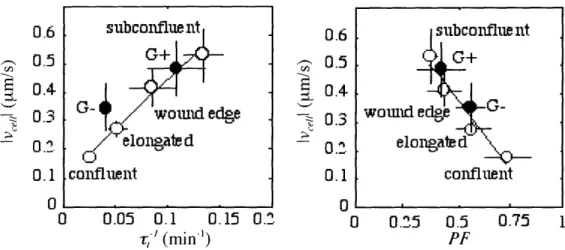
When a small region within an endothelial cell monolayer is denuded, adjacent cells
migrate into the wound at speeds proportional to their distance from the wound edge 11.
The fastest cells are situated within the wound unimpeded by surrounding cells, while theslowest are far from the wound edge in the intact confluent monolayer. Filament turnover
and polymer content correlate with cell speed such that the fastest cells contain the leastamount of polymerized actin and exhibit the most rapid rates of filament turnover (Fig.
I-4) 111. While depolymerization coupled with accelerated filament turnover has been
attributed to the ADF/cofilin family of actin binding proteins 69, 71], slower moving
endothelial cells in static culture contain more F-actin associated cofilin. Since lack of
0.6 v) 0.5 C.I E
.4
--
~ 0.3
- 0.2 0.1 0 0.6 0.5 E 0.4 -0.3
- 0.2
0.1 n 0 0.05 0.1 0.15 0.2 0 0.25 0.5 0.75 1,' (min
-')
PF
Fig. 1-4. Actin dynamics correlate with cell speed. Endothelial cells that crawl faster have more rapid filament turnover rates (,-) and less total actin polymerized (F). Subconfluent mouse fibroblasts () that express (G+) or lack (G-) gelsolin follow a similar correlation. Adapted from reference [1].
subconflue nt
G+
t
.coanIfluen
lt
co-ubnfluentwouand ege
elongtdP
\
coniflent
gelsolin in dermal fibroblasts from gelsolin knockout mice slows their speed and actin
dynamics in a similar fashion to endothelial cells, it has been postulated that the motile
transition in endothelial cells is controlled by a gelsolin-mediated severing mechanism
that liberates new pointed ends for ADF/cofilin to rapidly depolymerize 1 .References
1.
McGrath, J.L., et al., Regulation of the actin cycle in vivo by actin filament
severing. Proc Natl Acad Sci U S A, 2000. 97(12): p. 6532-6537.
2. Oster, G.F., On the crawling of cells. J Embryol Exp Morphol, 1984. 83 Suppl: p.
329-64.
3. Oster, G.F. and A.S. Perelson, The physics of cell motility. J Cell Sci Suppl, 1987. 8: p. 35-54.
4. Oster, G., Brownian ratchets: Darwin's motors. Nature, 2002. 417(6884): p. 25.
5..
Evans, E., New physical concepts for cell amoeboid motion. Biophys J, 1993.
64(4): p. 1306-22.
6.
Lauffenburger, D.A. and A.F. Horwitz, Cell migration: a physically integrated
molecular process. Cell, 1996. 84(3): p. 359-69.
7.
Lauffenburger, D.A., Molecules, mechanics, and migration of cells. Appl Mech
Rev, 1994. 47(6, pt 2): p. S287-S290.8. Bray, D. and C. Thomas, Unpolynzerized actin infibroblast and brain. J Mol Biol, 1976. 16: p. 1055-1069.
9.
Carlsson, L., et al., Profilin, a low molecular weight protein controlling actin
polymerisability, in Contractile systems in non-muscle tissues, S.V. Perry, A.
Margreth, and R.S. Adelstein, Editors. 1976, Elsevier/North-Holland Biomedical Press: Amsterdam. p. 39-49.
10.
Theriot, J.A. and T.J. Mitchison, Comparison of actin and cell surface dynamics
in motilefibroblasts. J Cell Biol, 1992. 119(2): p. 367-77.
11.
Theriot, J.A. and T.J. Mitchison, Actin microfilament dynamics in locomoting
cells. Nature, 1991. 352(633 1): p. 126-31.
12. Condeelis, J., Are all pseudopods created equal? Cell Motility and the Cytoskeleton, 1992. 22: p. 1-6.
13.
Stossel, T.P., et al., Filamins as integrators of cell mechanics and signalling. Nat
Rev Mol Cell Biol, 2001. 2(2): p. 138-45.14. Hartwig, J.H. and P. Shevlin, The architecture of actin filaments and the
ultrastructural
location of actin-binding
protein in the periphery of lung
macrophages. J Cell Biol, 1986. 103(3): p. 1007-20.15. Wegner, A., Head to tail polymerization of actin. J Mol Biol, 1976. 108(1): p. 139-50.
16.
Zigmond, S.H., Recent quantitative studies of actin filament turnover during cell
17.
Janmey, P.A. and C. Chaponnier, Medical aspects of the actin cytoskeleton. Curr
Opin Cell Biol, 1995. 7(1): p. 111-7.18.
Elzinga, M., et al., Complete amino-acid sequence of actin of rabbit skeletal
muscle. Proc Natl Acad Sci U S A, 1973. 70(9): p. 2687-91.
19. Hightower, R.C. and R.B. Meagher, The molecular evolution of actin. Genetics, 1986. 114(1): p. 315-32.
20. Pollard, T.D., Actin. Curr Opin Cell Biol, 1990. 2(1): p. 33-40.
21.
Vandekerckhove, J. and K. Weber, At least six different actins are expressed in a
higher mammal: an analysis based on the amino acid sequence of the
amino-terminal tryptic peptide. J Mol Biol, 1978. 126(4): p. 783-802.
22. Herman, I.M., Actin isoforms. Curr Opin Cell Biol, 1993. 5(1): p. 48-55. 23. Otterbein, L.R., P. Graceffa, and R. Dominguez, The crystal structure of
uncomplexed actin in the ADP state. Science, 2001. 293(5530): p. 708-11.
24.
Kabsch, W., et al., Atomic structure of the actin:DNase I complex. Nature, 1990.
347(6288): p. 37-44.
25. Carlier, M.F., D. Pantaloni, and E.D. Korn, Fluorescence measurements of the
binding of cations to high-affinity and low-affinity sites on ATP-G-actin. J Biol
Chem, 1986. 261(23): p. 10778-84.
26. Frieden, C., D. Lieberman, and H.R. Gilbert, A fluorescent probe for
conformational changes in skeletal muscle G- actin. J Biol Chem, 1980. 255(19):
p. 8991-3.
27. Carlier, M.F., D. Pantaloni, and E.D. Korn, The effects of Mg2+ at the
high-affinity and low-high-affinity sites on the polymerization of actin and associated ATP
hydrolysis. J Biol Chem, 1986. 261(23): p. 10785-92.
28. Oosawa, F., Size distribution of protein polymers. J Theor Biol, 1970. 27(1): p.
69-86.
29.
Wegner, A. and J. Engel, Kinetics of the cooperative association of actin to actin
filaments. Biophys Chem, 1975. 3(3): p. 215-25.
30.
Pollard, T.D., Rate constants for the reactions of ATP- and ADP-actin with the
ends of actin filaments. J Cell Biol, 1986. 103(6 Pt 2): p. 2747-54.
31. Korn, E.D., M.F. Carlier, and D. Pantaloni, Actin polymerization andATP
hydrolysis. Science, 1987. 238(4827): p. 638-44.
32.
Bindschadler, M., et al., A mechanistic model of the actin cycle in cells. Biophys
J, 2004.33. Bonder, E.M., D.J. Fishkind, and M.S. Mooseker, Direct measurement of critical
concentrations
and assembly rate constants at the two ends of an actin filament.
34.
Holmes, K.C., et al., Atomic model of the actin filament. Nature, 1990. 347(6288):
p. 44-9.35.
Milligan, R.A., M. Whittaker, and D. Safer, Molecular structure of F-actin and
location of surface binding sites. Nature, 1990. 348(6298): p. 217-21.
36.
Amos, L.A., Structure of muscle filaments studied by electron microscopy. Annu
Rev Biophys Biophys Chem, 1985. 14: p. 291-313.37.
Selve, N. and A. Wegner, Rate of treadmilling of actin filaments in vitro. J Mol
Biol, 1986. 187(4): p. 627-31.38.
Wang, Y.L., Exchange of actin subunits at the leading edge of livingfibroblasts:
possible role of treadmilling. J Cell Biol, 1985. 101(2): p. 597-602.
39.
Theriot, J.A. and T.J. Mitchison, Comparison of actin and cell surface dynamics
in motilefibroblasts. J Cell Biol, 1992. 119(2): p. 367-77.
40.
McGrath, J.L., et al., Simultaneous measurements of actin filament turnover,
filamentfraction, and monomer diffusion in endothelial cells. Biophys J, 1998.
75(4): p. 2070-8.
41. Kreis, T.E., B. Geiger, and J. Schlessinger, Mobility of microinjected rhodamine
actin within living chicken gizzard cells determined by fluorescence
photobleaching recovery. Cell, 1982. 29(3): p. 835-45.
42.
Luby-Phelps, K., D.L. Taylor, and F. Lanni, Probing the structure of cytoplasm. J
Cell Biol, 1986. 102(6): p. 2015-22.43. Satcher, R.L., Jr. and C.F. Dewey, Jr., Theoretical estimates of mechanical
properties of the endothelial cell cytosheleton Isee comments. Biophys J, 1996.
71(1): p. 109-18.
44.
Bartles, J.R., Parallel actin bundles and their multiple actin-bundling
proteins.
Curr O)pin Cell Biol, 2000. 12(1): p. 72-8.
45.
Sund, S.E. and D. Axelrod, Actin dynamics at the living cell submembrane
imaged by total internal reflection fluorescence photobleaching. Biophys J, 2000.
79(3): p. 1655-69.46.
dos Remedios, C.G., et al., Actin binding proteins: regulation of cytoskeletal
microfilaments. Physiol Rev, 2003. 83(2): p. 433-73.
47. Didry, D., M.F. Carlier, and D. Pantaloni, Synergy between actin depolymerizing
factor/cofilin and profilin in increasing actin filament turnover. J Biol Chem,
1998. 273(40): p. 25602-11.
48.
Dufort, P.A. and C.J. Lumsden, How profilin/barbed-end synergy controls actin
polymerization: a kinetic model of the ATP hydrolysis circuit. Cell Motil
49. Sun, H.Q., et al., Gelsolin, a multifunctional actin regulatory protein. J Biol Chem, 1999. 274(47): p. 33179-82.
50. Janmey, P.A., et al., Polyphosphoinositide micelles
andpolyphosphoinositide-containing vesicles dissociate endogenous gelsolin-actin complexes and promote
actin assembly
front the fast-growing end of actin filaments blocked by gelsolin. J
Biol Chem, 1987. 262(25): p. 12228-36.
51.
Hartwig, J.H., et al., Thrombin receptor ligation and activated Rac uncap actin
filament barbed ends through phosphoinositide
synthesis in permeabili ed human
platelets. Cell, 1995. 82(4): p. 643-53.52.
Witke, W., et al., Hemostatic, inflammatory, and fibroblast responses are blunted
in mice lacking gelsolin. Cell, 1995. 81(1): p. 41-51.
53.
Azuma, T., et al., Gelsolin is a downstream effector of racforfibroblast motility.
Embo J, 1998. 17(5): p. 1362-70.54. Cunningham, C.C., T.P. Stossel, and D.J. Kwiatkowski, Enhanced motility in NIH
3T3fibroblasts that overexpress gelsolin. Science, 1991. 251(4998): p. 1233-6.
55.
Kwiatkowski, D.J., Functions of gelsolin: motility, signaling, apoptosis, cancer.
Curr ()pin Cell Biol, 1999. 11(1): p. 103-8.56.
Kothakota, S., et al., Caspase-3-generatedfragment of gelsolin: effector of
morphological change in apoptosis. Science, 1997. 278(5336): p. 294-8.
57. Mullins, R.D., J.A. Heuser, and T.D. Pollard, The interaction of Arp2/3 complex
with actin: nucleation, high affinity pointed end capping, andfornmation of
branching networks offilaments. Proc Natl Acad Sci U S A, 1998. 95(11): p.
6181-6.
'58.
Machesky, L.M., et al., Scar, a WASp-relatedprotein, activates nucleation of
actin filaments by the Arp2/3 complex. Proc Natl Acad Sci U S A, 1999. 96(7): p.
3739-44.
59.
Falet, H., et al., Importance offree actin filanment barbed ends for Arp2/3 complex
firnction in platelets andfibroblasts. Proc Natl Acad Sci U S A, 2002. 99(26): p.
16782--7.
60.
Pantaloni, D., et al., The Arp2/3 complex branches filament barbed ends:
finctional antagonism with capping proteins. Nat Cell Biol, 2000. 2(7): p.
385-91.
61. Ichetovkin, I., W. Grant, and J. Condeelis, Cofilin produces newly polymerized
actin filaments that are preferredfor dendritic nucleation by the Arp2/3 complex.
Curr Biol, 2002. 12(1): p. 79-84.
62.
Amann, K.J. and T.D. Pollard, The Arp2/3 complex nucleates actinfilament
branches from the sides of pre-existing filaments. Nat Cell Biol, 2001. 3(3): p.
306-10.
63.
Amann, K.J. and T.D. Pollard, Direct real-time observation of actin filament
branching mediated by Arp2/3 complex using total internal reflection
fluorescence microscopy. Proc Natl Acad Sci U S A, 2001. 98(26): p. 15009-13.
64.
Carlsson, A.E., M.A. Wear, and J.A. Cooper, End versus Side Branching by
Arp2/3 Complex. Biophys J, 2004. 86(2): p. 1074-81.
65.
Loisel, T.P., et al., Reconstitution
of actin-based
notility of Listeria and Shigella
using pure proteins. Nature, 1999. 401(6753): p. 613-6.
66.
Pollard, T.D. and G.G. Borisy, Cellular motility driven by assembly and
disassembly of actinfilaments. Cell, 2003. 112(4): p. 453-65.
67.
Svitkina, T.M. and G.G. Borisy, Arp2/3 complex and actin depolymerizing
factor/cofilin in dendritic organization and treadmilling of actin filament array in
lamellipodia. J Cell Biol, 1999. 145(5): p. 1009-26.68.
Nakamura, F., et al., Comparison offilamin A-induced cross-linking and Arp2/3
complex- mediated branching on the mechanics of actin filaments. J Biol Chem,
2002. 277(11): p. 9148-54.
69.
Bamburg, J.R., Proteins of the ADF/cofilin
family: essential regulators of actin
dynamics. Annu Rev Cell Dev Biol, 1999. 15: p. 185-230.
70.
McGough, A., et al., Cofilin changes the twist of F-actin: implications for actin
filament dynamics and cellularfunction. J Cell Biol, 1997. 138(4): p. 771-81.
71.
Carlier, M.F., et al., Actin depolymerizing factor (ADF/cofilin) enhances the rate
offilament turnover: implication in actin-based motility. J Cell Biol, 1997.
136(6): p. 1307-22.72.
Arber, S., et al., Regulation of actin dynamics through phosphorylation of cofilin
by LIM- inase
Isee
comments!. Nature, 1998. 393(6687): p. 805-9.73.
Yang, N., et al., Cofilin phosphorylation by LIM-Ainase and its role in
Rac-mediated actin reorganization. Nature, 1998. 393(6687): p. 809-12.
74.
Niwa, R., et al., Control of actin reorganization by Slingshot, a family of
phosphatases that dephosphorylate ADF/cofilin. Cell, 2002. 108(2): p. 2.33-46.
75.
Yonezawa, N., et al., Inhibition of the interactions of cofilin, destrin, and
deoxyribonuclease I with actin by phosphoinositides. J Biol Chem, 1990. 265(15):
p. 8382-6.
76.
Carlier, M.F. and D. Pantaloni, Control of actin dynamics in cell motility. J Mol
Biol, 1997. 269(4): p. 459-67.77.
Du, J. and C. Frieden, Kinetic studies on the effect of yeast cofilin on yeast actin
78.
Maciver, S.K., H.G. Zot, and T.D. Pollard, Characterization of actin filament
severing by actophorin from Acanthanmoeba castellanii. J Cell Biol, 1991. 115(6):
p. 1611-20.
79.
Maciver, S.K., et al., The effect of two actin depolyn-erizing factors (ADF/cofilins)
on actin filament turnover: pH sensitivity of F-actin binding by human ADF, but
not of Acanthanmoeba actophorin. Eur J Biochem, 1998. 256(2): p. 388-97.80.
Condeelis, J., How is actin polymerization nucleated in vivo? Trends Cell Biol,
2001. 11(7): p. 288-93.81.
Ghosh, M., et al., Cofilin promotes actin polymerization and defines the direction
of cell motility. Science, 2004. 304(5671): p. 743-6.
82.
Perelroizen, I., et al., Role of nucleotide exchange and hydrolysis in the function
of profilin in actin assembly. J Biol Chem, 1996. 271(21): p. 12302-9.
83.
Selden, L.A., et al., Impact of profilin on actin-bound nucleotide exchange and
actin polymerization dynamics. Biochemistry, 1999. 38(9): p. 2769-78.
84.
Goldschmidt-Clermont, P.J., et al., Mechanism of the interaction of hunan
plateletprofilin with actin. J Cell Biol, 1991. 113(5): p. 1081-9.
85. Lassing, I. and U. Lindberg, Specific interaction between phosphatidylinositol
4,5-bisphosphate and profilactin. Nature, 1985. 314(6010): p. 472-4.
86. Krause, M., et al., The Ena/VASP enigma. J Cell Sci, 2002. 115(Pt 24): p. 4721-6.
87.
Bear, J.E., et al., Antagonism between Ena/VASP proteins and actin filament
capping regulates fibroblast motility. Cell, 2002. 109(4): p. 509-21.
88.
Bear, .J.E., et al., Negative regulation offibroblast motility by Ena/VASP proteins.
Cell, 2000. 101(7): p. 717-28.89.
Laurent, V., et al., Role of proteins of the Ena/VASP family in actin-based motility
of Listeria monocytogenes. J Cell Biol, 1999. 144(6): p. 1245-58.
90.
Gertler, F.B., et al., Mena, a relative of VASP and Drosophila Enabled, is
implicated in the control of microfilanment dynamics. Cell, 1996. 87(2): p. 227-39.
91. Fischer, R.S., K.L. Fritz-Six, and V.M. Fowler, Pointed-end capping by
tropomodulin3 negatively regulates endothelial cell motility. J Cell Biol, 2003.
161(2): p. 371-80.92.
Fowler, V.M., Tropomodulin: a cytoskeletal protein that binds to the end of
erythrocyte troponmyosin
and inhibits tropomyosin binding to actin. J Cell Biol,
1990. 111(2): p. 471-81.
93. Littlefield, R., A. Almenar-Queralt, and V.M. Fowler, Actin dynamics at pointed