The effect of treatment of skeletal open bite with two types
of bite-blocks
Robert Kuster and Bengt Ingervall
Orthodontic Clinic, University of Bern, SwitzerlandSUMMARY The treatment of anterior skeletal open bite was studied in two groups of children. The children of one group wore a removable spring-loaded bite-block in the lower jaw for one year. The bite-block exerted an intrusive force on the upper and lower posterior teeth. The children of the other group were treated for 3 months with bite-blocks with repelling magnets. These bite-blocks were cemented on the posterior teeth of both jaws.
The effects of treatment were monitored by measurement of the bite-force (group with spring bite-blocks only), by electromyographic recording of the activity of the temporal and masseter muscles, and by X-ray cephalometry. Recordings were made before, during, and at the end of the treatment, and at a follow-up observation.
The bite-force increased during the first months of treatment, but was then unchanged. The activity of the masseter muscle during maximal bite also increased in the first part of the period of treatment with a spring bite-block. In the group treated with magnetic bite-blocks, there was an increase in the resting activity of the masseter muscle and in the chewing activity of the anterior temporal muscle.
The effects of the treatment on bite and facial morphology were less marked in the group with spring bite-blocks than in the group with magnetic bite-blocks, with an average improvement of the overbite of 1.3 mm with the spring block therapy. In the group with magnetic bite-blocks, the average improvement in overbite was 3 mm. This was thought to be due to anterior rotation of the mandible and increased eruption of the incisors. The mandibular rotation was a result of intrusion of the upper and lower posterior teeth and possibly also increased mandibular growth. A follow-up of the cases treated with magnetic bite-blocks revealed a tendency for the beneficial effects of the treatment to relapse which possibly could be counteracted by a long phase of active retention.
Introduction
Individuals with a skeletal open bite have a long-face morphology characterized by a large anterior face height and a steep inclination of the mandible (Fields et al., 1984; Ellis et ai, 1985). The orthodontic treatment of this maloc-clusion is difficult. Camouflage of the underlying skeletal anomaly by the elongation of the incisors results in too much incisor exposure and does not improve the excessive anterior face height. A causal type of treatment, i.e. growth modification, has until recently been regarded as impossible. Attempts to treat a skeletal open bite by growth modification with the use of a posterior bite-block have, however, lately been described (Dellinger, 1986; Woodside
and Linder-Aronson, 1986) and shown promis-ing results. Dellpromis-inger used bite-blocks with repelling magnets on the upper and lower posterior teeth while Woodside and Linder-Aronson used bite-blocks fastened in the lower jaw and exerting an intrusive force on the upper and lower posterior teeth through a spring mechanism.
Bite-blocks could have several beneficial therapeutic effects. They could intrude the pos-terior teeth and, thus, make possible autorot-ation of the mandible to produce bite closure. This would be equivalent to the effect of a posterior maxillary set-up procedure in orthog-nathic surgery. In patients with ongoing vertical growth in the posterior part of the face (in the posterior cranial base and in the mandibular
condyles), retardation of the eruption of the posterior teeth would have the same effect. Another possibility is that the bite-blocks would increase the condylar growth. Such an effect would be conceivable through unloading of the temporomandibular joints and/or protrusion of the condyles during the wearing of the bite-blocks and would be comparable to the effect of an activator or a Herbst appliance. Increased vertical condylar growth would rotate the mandible anteriorly and tend to close the bite. A maximal effect of bite-block therapy would be achieved by simultaneous intrusion of the posterior teeth and an increased posterior ver-tical growth. All these effects of bite-blocks have been described in the relatively few reports hitherto published. Intrusion or retarded erup-tion of the posterior teeth was demonstrated in monkeys by McNamara (1977), Carlson and Schneiderman (1983), Altuna and Woodside (1985), and Woods and Nanda (1988). In humans, intrusion of posterior teeth induced by the use of bite-blocks was demonstrated by Dellinger (1986), Woodside and Linder-Aronson (1986), Kalra et al. (1989), Kiliaridis
et al. (1990), and Barbre and Sinclair (1991).
Enhancement of mandibular growth was dem-onstrated by Kalra et al. (1989), who treated patients with open bite with cemented magnetic bite-blocks for 4 months.
Another approach to the treatment of skeletal open bite has involved training of the masticat-ory muscles. It is known that individuals with a long-face morphology have weak masticatory muscles (Ringqvist, 1973; Ingervall and Helk-imo, 1978; Proffit et al., 1983; Ingervall et al., 1989). It was shown that training of the mastic-atory muscles in children with skeletal open bite by the chewing of a special type of tough chewing-gum increased the muscle strength. This resulted in an excessive anterior rotation of the mandible with closure of the open bite (Ingervall and Bitsanis, 1987; Bakke and Siersbaek-Nielsen, 1990).
Because of the difficulties in the treatment of skeletal open bite, the relatively new treatment method with bite-blocks should be further evaluated for its therapeutic possibilities. Not only the effect of the treatment on the morpho-logy of the face and the dentition, but also the possible effect on the muscle strength should be studied. An increase of the muscle strength would be a benefit of the treatment that could
help to maintain the treatment result. The aim of this investigation is to study the effect on facial morphology and on masticatory muscle strength of two types of bite-blocks in the treatment of skeletal open bite.
Subjects and methods
The investigation included two series of patients. One series comprised 22 children (11 boys and 11 girls) aged 7 years 5 months to 11 years 7 months (median age 9 years 4 months) who were treated with a bite-block with springs (spring bite-block) as described by Woodside and Linder-Aronson (1986). The 11 children of the other series (four boys and seven girls), aged 9 years 9 months to 14 years 5 months (median age 10 years 9 months), were treated with magnetic bite-blocks in the upper and lower jaws.
The children of both series were selected from among those enrolled for orthodontic treatment at the orthodontic clinic, University of Bern, Switzerland. The children had a long face mor-phology with a varying degree of anterior open bite which was treated with one of the two types of bite-blocks. Clinically, a neutral or a distal occlusion was present. Variables describing the facial morphology are given in Table 5.
During the first part of the time-span of the investigation, only spring bite-blocks were tried for the treatment of a skeletal anterior open bite. This type of treatment was abandoned and replaced by magnetic bite-blocks for the children enrolled at a later date.
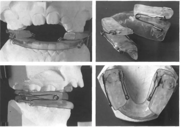
The spring bite-blocks (Fig. 1) were worn at night for 1 year. The height of the bite-blocks (with compressed springs) at the first permanent molars varied between 6.5 and 9 mm (median 7 mm). The upper and lower parts of the bite-blocks were joined by steel springs on the labial and lingual sides. The springs (wire diameter 0.9 mm) were designed to exert an intrusive force on the upper and lower posterior teeth. The force needed to compress the springs until contact between the two halves of the bite-blocks was measured in eight cases. The follow-ing forces (in g) were found: 264, 304, 339, 480, 501, 527, 560, and 672 (mean of right and left sides). The patients wearing the spring bite-blocks came to the clinic for monthly checks. The springs were then activated so that a pres-sure on the posterior teeth was exerted when
Figure 1 Spring bite-block.
the mandible was in rest position. Broken springs had to be replaced in 12 patients. No subject had any problem wearing the bite-block at night and all reported consistent use of the appliance. One patient who removed his bite-block from his mouth during the night was excluded from the study at an early stage.
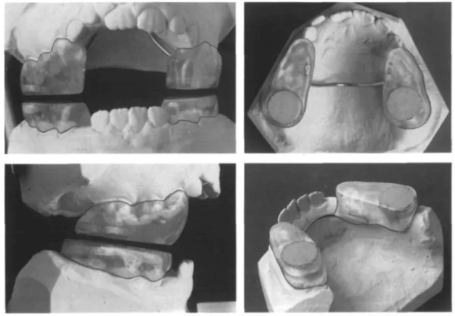
Samarium-cobalt magnets (Ugimag Recoma AG, Lupfig, Switzerland) were used for the magnetic bite-blocks. Two round repelling mag-nets, 2 mm thick and with a diameter of 12 mm, were embedded in the acrylic over the posterior teeth in the upper and lower bite-blocks (Fig. 2). The bite-blocks on the right and left sides of the dental arch were joined by a steel bar. The magnets were electrolytically covered with gold and thereafter with hardening glaze (Perma Link, G-C Dental Industrial Corp., Japan). This was to protect the magnets from corrosion and prevent the leakage of possibly poisonous prod-ucts. The combined height of the upper and lower bite-blocks at the first permanent molars varied between 6 and 9 mm (median 7 mm).
The theoretical maximum repelling force of the magnets was, because of the distance between them and their coverage, reduced. In six cases the repelling force of the bite-blocks was meas-ured when they were placed on dental casts mounted on an articulator (corresponding to their use in the mouth) and was found to be 299, 356, 413, 417, 440, and 483 g, respectively. The magnetic bite-blocks were cemented onto the upper and lower posterior teeth and were left in place 3 months. The patients were checked every 3-4 weeks during this period. Recordings
The sequence of the recordings is given in Table 1.
Measurement of bite-force
In the group with spring blocks, the bite-force at the right and left first permanent molars was measured as described earlier (Ingervall and Bitsanis, 1987) with the bite-force recorder of Floystrand et al. (1982). The mean of the
bite-Figure 2 Magnetic bite-blocks.
force on the two sides was calculated. Measure-ments were made at each control visit. Thus, altogether 13 measurements (at the start of the treatment and each month during the 12 months of treatment) were made.
Electromyography (EMG)
The activity of the anterior portion of the temporal muscle and of the masseter muscle was recorded bilaterally. Bipolar hook elec-trodes were used as described earlier (Ingervall and Egermark-Eriksson, 1979). Recordings were made as in an earlier study (Ingervall and Bitsanis, 1987) in the rest position of the mand-ible, during maximal bite in the intercuspal position and during chewing of apple and pea-nuts with a Disa electromyograph (Disa Elek-tronik, Copenhagen) and a Gould electrostatic writer (Gould Elektronik, Zurich). The mean voltage amplitude in the rest position and during maximal bite was measured. The maximal mean voltage amplitude during the closing phase of the chewing cycle was determined as the mean
of six randomly selected cycles during an act of chewing. The mean of the recordings from the muscles on the right and left sides was calculated.
Cephalometry
Cephalograms were taken with the mandible in the intercuspal position. The reference points and lines used in the analysis of the profile cephalogram (linear enlargement 3.6 per cent) are shown in Figs 3 and 4. The variables recorded are given in Table 5. The reference line OL was defined as a line from mo bisecting the distance is-ii. The point io was constructed as the perpendicular projection of the point ii on the line mo-is (upper occlusal line). The dis-tances is-io and ii—io give the overjet and overbite, respectively, and were measured from the central incisor in the most extreme position. The quotients s-tgo/n-mex 100 and n-sp'/sp'-me x 100 express the relationship between the posterior and anterior face heights, and the upper and lower anterior face heights,
respect-Table 1 Recordings at different times in the two series of patients.
Spring bite-blocks
Magnetic bite-blocks
Recording 1 Before the start of the treatment
Profile cephalogram EMG
Bite-force
Recording 1 Before the start of the treatment Profile cephalogram EMG Recording 2 After 6 months EMG Bite-force Recording 2 After 3 months (end of treatment) Profile cephalogram EMG Recording 3 After 12 months (end of treatment) Profile cephalogram EMG Bite-force Recording 3 After 15 months Profile cephalogram Recording 4 After 18 months EMG 9O Figure 3 analysis.
Reference points used in the cephalometric
ively. The total rotation of the mandible during the period of observation was analysed by the superimposition of the profile cephalograms using the natural reference structures of the mandible as described by Bjork and Skieller (1983). An arbitrary reference line (approxi-mately parallel to ML) was drawn on the mand-ible (Fig. 4) and transferred from the first to the second cephalogram by the superimposition on the natural reference structures of the mand-ible. The difference of the angle between the mandibular reference line (MRL) and the NSL between the two cephalograms expresses the degree of mandibular rotation during the time interval. The MRL was also used to record the
Figure 4 Reference lines used in the cephalometric
analysis.
vertical position of the lower molar cusp (lmc) and of the edge of the lower incisor (ii) by the measurement of the perpendicular distance to the MRL.
Statistical methods
Differences between observations on the different occasions and between the duplicate determinations were tested with Wilcoxon's matched-pairs, signed-ranks test. The accidental errors (si) were calculated with the formula
si= sf'Ld2/2n, where d is the difference between
the two determinations. Errors of the method
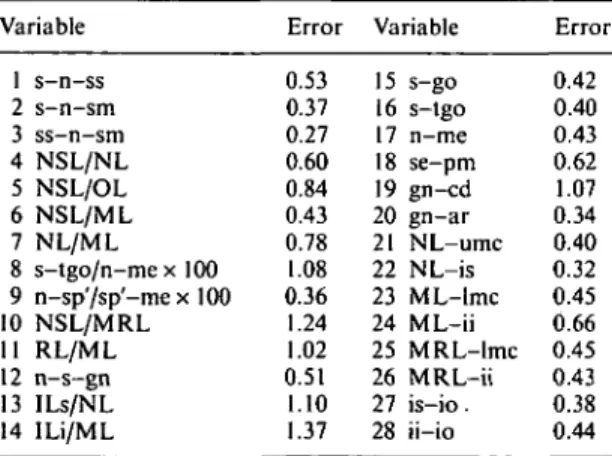
The errors of the cephalometric analysis, includ-ing the procedure of superimposition, were determined by duplicate measurements of 20 randomly selected cephalograms. A systematic difference between the two measurements was found for two variables. Variable 8 (s-tgo/ n - m e x 100) was greater at the second than at the first measurement (mean difference 0.97, 0.001 < P < 0 . 0 1 ) and variable 28 (ii-io) was likewise greater at the second measurement (mean difference 0.32 mm, 0.01</)<0.05). The accidental errors of the method are given. in Table 2.
Results
Bite-force
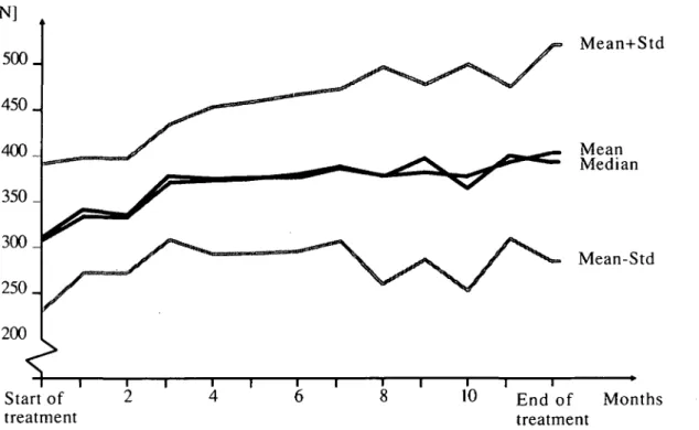
The bite-force increased during the first months of the treatment with the spring bite-blocks (Fig. 5), but did not change in the interval from 6 months to the end of the treatment (Table 3). Electromyography
The muscle activity in the rest position or during chewing did not change during the treatment with the spring bite-blocks (recordings 1-3, Table 2). The activity of the masseter muscle during maximal bite increased from the first to the second recording (a numerical increase was Table 2 Accidental errors of the method of the cephalometric variables in degrees (variables 1-7,
10-14) and in mm (variables 15-28). Variable 1 s-n-ss 2 s-n-sm 3 ss-n-sm 4 NSL/NL 5 NSL/OL 6 NSL/ML 7 NL/ML 8 s-tgo/n-me x 100 9 n-sp'/sp'-me x 100 10 NSL/MRL 11 RL/ML 12 n-s-gn 13 ILs/NL 14 ILi/ML Error 0.53 0.37 0.27 0.60 0.84 0.43 0.78 1.08 0.36 1.24 1.02 0.51 1.10 1.37 Variable 15 s-go 16 s-tgo 17 n-me 18 se-pm 19 gn-cd 20 gn-ar 21 NL-umc 22 NL-is 23 ML-lmc 24 ML-ii 25 MRL-lmc 26 MRL-ii 27 is-io. 28 ii-io Error 0.42 0.40 0.43 0.62 1.07 0.34 0.40 0.32 0.45 0.66 0.45 0.43 0.38 0.44
also found for the anterior temporal muscle) with no further change to the recording at one year.
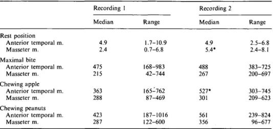
In the group treated with magnetic bite-blocks, electromyographic recordings were made before the start of the treatment and one week after the removal of the bite-blocks at 3 months. Between these two recordings, the activity of the masseter muscle at rest and of the anterior temporal muscle during chewing of apple increased (Table 4).
Cephalometry
The values of the cephalometric variables at the start of the treatment and the change to the following recording are given in Table 5. The table also gives the annual changes during nor-mal growth in the untreated longitudinal sample of Riolo et al. (1974). The values of Riolo et al. were matched with those of the present subjects with respect to sex and age, and reduced to the same degree of linear magnification.
In the group with spring bite-blocks, the mandibular prognathism (var. 2, Table 5) increased and the angle for sagittal jaw relation (var. 3) decreased slightly during the year of observation. The inclination of the jaws (vars. 4 and 6) and the vertical jaw relation (var. 7) did not change.
There were also no changes of the quotients between the posterior and anterior face heights (var. 8) or between the upper and lower anterior face heights (var. 9). The gonial angle (var. 11) increased and the j>-axis angle (var. 12) decreased slightly. The linear dimensions of the face (vars. 15-26) increased significantly with the exception of the distances from the lower molar cusp to ML (var. 23) and to MRL (var. 25), which were constant. The lower incisors uprighted (var. 14) and the overbite (var. 28) increased.
In the group with magnetic bite-blocks (observed for 3 months), there was a greater increase of the mandibular prognathism (var. 2) and a greater decrease of the sagittal jaw relation angle (var. 3, Table 5) than in the group with spring bite-blocks. In addition, there was in the group with magnetic bite-blocks a slight anterior rotation of the mandible expressed as a decrease of the angles NSL/ML and NSL/ MRL and an increase of the quotient between the posterior and anterior face heights (var. 8). The upper posterior face height (var. 18) and
TREATMENT OF SKELETAL OPEN BITE 495 [N] 5 0 0 . 450 _ 400_ 350 _ 300 _ 250 _ Mean+Std Mean Median Mean-Std 200 I Start of treatment 10 End of Months treatment
Figure 5 Bite-force values in the group with spring bite-blocks during the period of observation.
Table 3 Bite-force (in N) and muscle activity (in uV) in the rest position, during maximal bite and during chewing recorded on the various occasions in the group treated with spring bite-blocks.
Bite-force Rest position Anterior temporal m. Masseter m. Maximal bite Anterior temporal m. Masseter m. Chewing apple Anterior temporal m. Masseter m. Chewing peanuts Anterior temporal m. Masseter m. Recording Median 309 3.9 1.2 417 243 313 150 393 180 1 Range 170-471 0.7-12.2 0.0-3.8 98-1389 81-396 183-658 80-214 187-869 94-310 Recording Median 376** 4.1 1.4 598 301** 321 149 373 182 2 Range 249-520 0.7-12.2 0.0-5.4 194-1461 128-562 219-652 77-316 210-844 97-330 Recording Median 392 3.5 1.7 486 290 347 171 382 199 3 Range 199-616 0.7-18.2 0.0-7.4 117-1729 147-757 161-835 85-280 205-970 116-429 Recording 4 Median 4.8 3.4* 605 319 315 183* 398 240 Range 1.4-14.2 0.0-10.1 178-1299 78-1068 111-942 92-335 160-1147 48-401
*0.0I </><0.05, **0.001 </><0.0I; indicates significant difference from previous recording.
the mandibular length (vars. 19 and 20) increased and the incisors erupted (vars. 22, 24, 26). In contrast to the findings in the group with spring bite-blocks, there was in the group with magnetic bite-blocks a significant intrusion
of the upper and lower molars (vars. 21 and 23). In the group with magnetic bite-blocks, both the upper and the lower incisors uprighted (vars. 13 and 14) and there was a marked increase of the overbite (var. 28).
Table 4 Muscle activity (in u.V) in the rest position, during maximal bite, and during chewing recorded on two occasions in the group treated with magnetic bite-blocks.
Rest position Anterior temporal m. Masseter m. Maximal bite Anterior temporal m. Masseter m. Chewing apple Anterior temporal m. Masseter m. Chewing peanuts Anterior temporal m. Masseter m. Recording I Median 4.9 2.4 475 215 363 288 423 287 Range 1.7-10.9 0.7-6.8 168-983 42-744 165-762 87-469 187-1016 122-600 Recording 2 Median 4.9 5.4* 488 267 527* 301 561 356 Range 2.5-6.8 2.4-8.1 383-725 200-697 303-745 209-623 239-824 96-677
*0.01 </><0.05; indicates significant difference from previous recording.
Discussion
No adverse effects of the bite-blocks were seen. Thus, no pain or functional problems were noted. No child wearing a magnetic bite-block developed cross-bite, a problem described in some studies (Kalra et al., 1989; Kiliaridis et al., 1990), but not mentioned by other authors (Barbre and Sinclair, 1991).
Before the start of the treatment, the bite-force of the group of children treated with a spring bite-block was below normal (average in random 9-year old children 375 N, Ding and Kober, 1985). The bite-force increased during the period of observation. This might be due to normal development (average in 11-year-old chil-dren 424 N, Dine and Kober, 1985), but could also at least partly be an effect of the treatment. The latter explanation is supported by the fact that the increase occurred during the first part of the period of treatment with no further increase during the following months. The elec-tromyographically recorded muscle activity dur-ing maximal bite also increased durdur-ing the first part of the treatment and was thereafter constant. Similar results (increase of bite-force and muscle activity) were achieved by the training of the masticatory muscles in long-face children (Ingervall and Bitsanis, 1987) and were found to influence the facial morphology favourably. In the present investigation, the same effects on the muscles could contribute to the treatment results and be a positive factor for their
mainten-ance provided the muscle strength does not decline after treatment. The bite-force was not measured after the end of the treatment. This was due to various types of treatment (including the wearing of multiband appliances) of the children after the phase of bite-block treatment. From the electromyographic recordings at 18 months, there was, however, no evidence of a decline in muscle strength.
The changes in facial morphology in the group treated with spring bite-blocks were larg-ely in accordance with the annual changes in the control sample of Riolo et al. (1974). There was no evidence of intrusion of the posterior teeth or of increased mandibular growth and thus no sign of anterior rotation of the mand-ible. A slight increase of the gonial angle and an uprighting of the lower incisors were noted. The uprighting is probably an effect of tightening of the lips (or withdrawal of the tongue) due to the increased bite height caused by the wearing of the bite-blocks, similar to the effect on incisor position of mouth-breathing (Linder-Aronson, 1970). During the year, the overbite increased on average by 1.3 mm. This is more than would have been expected from normal development (Moyers et al., 1976, annual increase for matched controls 0.3 mm; Bergersen, 1988, annual increase for matched controls 0.1 mm). This improvement of the overbite might be due to small, but favourable contributions from several sources, slight inhibi-tion of the erupinhibi-tion of the posterior teeth,
497 Table 5 Median and range in degrees (variables 1-7, 10-14) and in mm (variables 15-28) of the cephalometric variables at the start of the treatment and median differences between subsequent recordings as well as annual changes in a control sample (Riolo et al., 1974).
Variable 1 s-n-ss 2 s-n-sm 3 ss-n-sm 4 NSL/NL 5 NSL/OL 6 NSL/ML 7 NL/ML 8 s-tgo/n-mex 100 9 n-sp'/sp'-me x 100 10 NSL/MRL 11 RL/ML 12 n-s-gn 13 ILs/NL 14 ILi/ML 15 s-go 16 s-tgo 17 n-me 18 se-pm 19 gn-cd 20 gn-ar 21 NL-umc 22 NL-is 23 ML-lmc 24 ML-ii 25 MRL-lmc 26 MRL-ii 27 is-io 28 ii-io Spring bite-blocks Recording 1 Med. 78.7 74.0 5.1 6.7 24.0 42.1 34.7 58.6 77.9 45.5 131.1 72.6 108.8 92.9 62.5 63.8 111.5 42.5 100.3 93.3 19.8 27.3 29.3 38.0 20.5 31.0 5.0 - 0 . 5 Range 73.1-84.0 67.6-78.9 -1.1-9.1 3.0-12.2 17.1-33.7 37.7-54.6 29.1-49.4 49.4-63.1 69.2-84.2 40.5-60.0 124.4-144.0 68.7-78.0 97.3-116.8 81.0-100.4 53.0-70.5 53.5-72.0 99.5-120.5 38.5-48.5 94.0-108.5 83.5-102.0 15.0-25.0 23.0-31.5 26.0-33.5 35.0-42.5 16.5-22.5 27.5-35.0 1.0-9.0 -6.0-2.0 Dif. rec 3 and 1 0.2 0.6* - 0 . 3 * - 0 . 1 -0.8** 0.2 0.4 0.2 0.4 0.0 1.2* - 0 . 3 * - 0 . 6 -2.6** 1.0** 1.3** 2.5** 1.0** 3.0** 2.0** 0.5* Riolo el al. 0.1 0.2 - 0 . 1 0.1 - 0 . 2 - 0 . 3 - 0 . 4 0.1 0.6 0.4 * 1.7 • 2.4 * 1.2 * 2.6 * 2.4 0.8 1.0*** 0.7 0.3 0.6 1.0*** 0.9 0.5 1.0*** - 0 . 3 1.3*** Magnetic bite-blocks Recording 1 Med. 82.1 76.1 5.2 6.2 20.0 37.1 31.1 64.4 75.3 46.0 126.0 71.0 110.8 96.8 70.0 72.0 113.0 44.0 102.5 98.0 20.5 28.5 30.0 40.0 19.0 32.0 6.5 - 2 . 0 Range 76.8-84.9 68.9-78.8 2.6-13.7 3.4-12.8 15.2-27.0 30.5-47.4 23.6-37.4 54.0-66.9 62.2-85.9 32.5-59.0 120.7-135.0 64.5-77.6 101.8-116.4 79.6-106.5 59.5-77.5 60.5-79.0 106.5-120.0 39.0-51.5 97.0-116.5 90.0-109.0 18.5-27.0 22.5-30.0 29.0-34.0 34.0-41.5 17.0-24.0 25.5-35.0 2.0-12.0 -4.0-0.0 Dif. rec. 2 and 1 - 0 . 5 0.9** -1.5** - 0 . 4 0.0 - 1 . 1 * - 0 . 6 0.6* 0.8 - 1 . 5 * 1.1* -1.0* - 3 . 1 * * -5.3** 0.5 0.5 0.5 0.5* 1.5** 1.0* -1.0** 0.5* - 0 . 5 * 0.5** - 0 . 5 0.5* - 1 . 5 * 3.0** Riolo et al. 0.2 0.4 - 0 . 2 0.2 - 0 . 5 - 0 . 7 - 1 . 0 - 0 . 1 0.1 - 0 . 1 1.8 1.9 0.6 2.3 2.2 0.7 0.4 0.7 0.6 *0.01</><0.05, **0.001</><0.01, ***/»<0.001.
slightly increased eruption of the incisors and retroclination of the incisors.
In the group treated with magnetic bite-blocks, in contrast, definite therapeutic effects were seen. Thus, during the 3 months the man-dibular prognathism increased twice as much as during one year in the control sample (var.
2, Table 5). There was also clear evidence of
anterior mandibular rotation. Thus, the angle NSL/ML decreased by twice the annual decrease in the control sample in three months. The anterior mandibular rotation of 1.5 degrees (change in the angle NSL/MRL) during three months is large in comparison with the normal annual anterior rotation of 1 degree found by Bjork and Skieller (1983). With the use of the magnetic bite-blocks, both the upper and the
lower molars intruded, and there was possibly also an increased rate of eruption of the incisors (large changes of the variables 22 and 24 com-pared to the annual changes in the control sample). The molar intrusion and possibly increased mandibular growth (comparatively large three-month changes of variables 19 and 20) led to an anterior rotation of the mandible. This, together with a marked eruption of the incisors, resulted in a favourable median increase of the overbite of 3 mm in 3 months. In the group with magnetic bite-blocks, there was a more marked uprighting of the incisors (which also tends to increase the overbite) than in the group with spring bite-blocks. This is probably due to the fact that the magnetic bite-blocks were worn day and night.
Several of the effects of the bite-blocks found in this study agree with the results of other investigations. There was thus no effect of either of the two bite-blocks on the maxilla. This is in agreement with the results of other studies in humans (Kalra et al., 1989; Barbre and Sinclair, 1991), but in contrast to the experi-ments in monkeys mentioned in the introduc-tion. In all the animal experiments, a marked maxillary displacement was found. The anterior mandibular rotation found by us in the group with magnetic bite-blocks agrees very well with the results of Kalra et al. (1989), but was somewhat greater than that found by Barbre and Sinclair (1991) in their studies of the effects of magnetic bite-blocks. Kalra et al. (1989) found an increased mandibular growth with the use of their cemented magnetic splints. The mandibular length increment in our study (vars. 19 and 20) was less than in the study of Kalra
et al., but still large compared to the control
sample of Riolo et al. No definite conclusions can be drawn, however, because of the lack of a specific control group in our study. The overbite correction found by us after the use of magnetic bite-blocks was of the same magnitude as reported by Kalra et al. (1989), and Barbre and Sinclair (1991). Like the latter authors, we found an increased eruption and uprighting of the incisors. An uprighting of the incisors was also noted by Dellinger (1986), and by Woodside and Linder-Aronson (1986).
In 9 of the 11 patients treated with magnetic bite-blocks, cephalograms were taken one year after the removal of the bite-blocks. During this interval, three patients had been treated with multi-band appliances. Two patients had had no appliances and four had worn an appliance for retention. Thus, in one case an activator was used and the three other patients wore an upper removable plate with bite platforms over the posterior teeth for 6-8 months. The six patients with no treatment or retention only underwent the following development: increase of the overbite by 3 mm during active treatment followed by a decrease by 1.5 mm after treat-ment, i.e. a 50 per cent relapse. Decrease of the angles NSL/ML and NL/ML by 1.1 degrees and 0.3 degree, respectively, during treatment, increase by 1.2 degrees and 0.4 degree, respect-ively, after treatment, i.e. a complete relapse. An anterior rotation of the mandible of 1.5 degrees (angle NSL/MRL) during treatment
followed by 0.5 degree posterior rotation after treatment. It is thus clear that the results achieved by treatment with magnetic bite-blocks tend to relapse. It therefore seems to be neces-sary to continue the treatment with so-called active retention for a long period. A combina-tion of cemented magnetic bite-blocks for the active treatment followed by a longer period of wearing of removable bite-blocks for part of the day and night might be successful in the correction of skeletal open bite.
It should be noted that the same effect on mandibular rotation as in the present study was found after training of the masticatory muscles in long-face children (Ingervall and Bitsanis, 1987). The increase in bite-force found during the first phase of the treatment with bite-blocks might thus be a positive factor to stabilize the treatment result. Further studies should be per-formed to determine whether it would be of advantage to increase the height of removable bite-blocks (without magnets) from time to time to achieve a greater increase in muscle strength.
Address for correspondence
Dr Robert Kuster
Klinik fur Kieferorthopadie Freiburgstrasse 7
CH-3010 Bern Switzerland
Acknowledgement
This study was supported by Schweizerischer Nationalfonds zur Forderung der wissen-schaftlichen Forschung, Grant No. 3.905-0.85
References
Altuna A, Woodside D G 1985 Response of the midface to treatment with increased vertical occlusal forces. Angle Orthodontist 55: 251-263
Bakke M, Siersbaek-Nielsen S 1990 Training of mandibular elevator muscles in subjects with anterior open bite. European Journal of Orthodontics 12: 502 (Abstract) Barbre R E, Sinclair P M 1991 A cephalometric evaluation
of anterior open bite correction with magnetic active vertical corrector. Angle Orthodontist 61: 93-102 Bergersen E O 1988 A longitudinal study of anterior vertical
overbite from eight to twenty years of age. Angle Ortho-dontist 58: 237-256
Bjork A, Skieller V 1983 Normal and abnormal growth of the mandible. A synthesis of longitudinal cephalometric implant studies over a period of 25 years. European Journal of Orthodontics 5: 1-46
499
Carlson D S, Schneiderman E D 1983 Cephalometric ana-lysis of adaptations after lengthening of the masseter muscle in adult rhesus monkeys Macacca mulatta. Archives of Oral Biology 28: 627-637
Dellinger E L 1986 A clinical assessment of the active vertical corrector—A nonsurgical alternative for skeletal open bite treatment. American Journal of Orthodontics 89: 428-436
Dine S, Kober M 1985 Bisskraftmessungen an 9-bis 11-jahrigen Kindern. Medical Dissertation, University of
Berne
Ellis E, McNamara J A, Jr, Lawrence T M 1985 Compon-ents of adult Class II open-bite malocclusion. Journal of Oral and Maxillofacial Surgery 43: 92-105
Fields H W, Promt W R, Nixon W L, Phillips C, Stanek E 1984 Facial pattern differences in long-faced children and adults. American Journal of Orthodontics 85: 217-223 Floystrand F, Kleven E, Oeilo G 1982 A novel miniature
bite force recorder and its clinical application. Acta Odontologica Scandinavica 40: 209-214
Ingervall B, Bitsanis E 1987 A pilot study of the effect of masticatory muscle training on facial growth in long-face children. European Journal of Orthodontics 9: 15-23 Ingervall B, Helkimo E 1978 Masticatory muscle force and
facial morphology in man. Archives of Oral Biology 23: 203-206
Ingervall B, Egermark-Eriksson I 1979 Function of tem-poral and masseter muscles in individuals with dual bite. Angle Orthodontist 49: 131-140
Ingervall B, Thuer U, Kuster R 1989 Lack of correlation between mouth-breathing and bite force. European Journal of Orthodontics 11: 43-46
Kalra V, Burstone C J, Nanda R 1989 Effects of a fixed magnetic appliance on the dentofacial complex. American Journal of Orthodontics and Dentofacial Orthopedics 95: 467-478
Kiliaridis S, Egermark I, Thilander B 1990 Anterior open bite treatment with magnets. European Journal of Orthodontics 13: 447-457
Linder-Aronson S 1970 Adenoids Their effect on mode of breathing and nasal air flow and their relationship to characteristics of the facial skeleton and the dentition. Acta Oto-Laryngologica Suppl 265
McNamara J A Jr 1977 An experimental study of increased vertical dimension in the growing face. American Journal of Orthodontics 71: 382-395
Moyers R E, van der Linden F P G, Riolo M L, McNamara J A Jr 1976 Standards of human occlusal development. Monograph Number 5, Craniofacial Growth Series, Center for Human Growth and Development, University of Michigan, Ann Arbor, Michigan
Proffit W R, Fields H W, Nixon W L 1983 Occlusal forces in normal and long-face adults. Journal of Dental Research 62: 566-571
Ringqvist M 1973 Isometric bite force and its relation to dimensions of the facial skeleton. Acta Odontologica Scandinavica 31: 35-42
Riolo M L, Moyers R E, McNamara J A Jr, Hunter W S 1974 An atlas of craniofacial growth. Cephalometric standards from the University School Growth Study, Monograph Number 2, Craniofacial Growth Series, Center for Human Growth and Development, University of Michigan, Ann Arbor, Michigan
Woods M G, Nanda R S 1988 Intrusion of posterior teeth with magnets. An experiment in growing Baboons. Angle Orthodontist 58: 136-150
Woodside D G, Linder-Aronson S 1986 Progressive increase in lower anterior face height and the use of posterior occlusal bite-block in its management. In: Graber L W (ed.) Orthodontics; state of the art, essence of the science. The CV Mosby Company, St Louis