Comp lete enzymatic synthesis of the mucin-type sLe x
epitope Zen g et al.
Complete enzymic synthesis of the mucin-type sialyl
Lewis x epitope, involved in the interaction between
PSGL-1 and P-selectin
Steffen Zeng
1†, Ricardo Gutiérrez Gallego
2†, Andre Dinter
1, Martine Malissard
1, Johannis P. Kamerling
2,
Johannes F.G. Vliegenthart
2and Eric G. Berger
1*
1Physiologisches Institut, Universität Zürich, Winterthurerstrasse 190, CH-8057 Zürich, Switzerland
2Bijvoet Center, Department of Bio-Organic Chemistry, Utrecht University, P.O. Box 80.075, NL-3508 TB Utrecht, The Netherlands
Sialyl Lewis x (sLex) is an established selectin ligand occurring on N- and O-linked glycans. Using a completely enzymic approach starting fromp-nitrophenyl N-acetyl-a-D-galactosaminide (GalNAc(a1-pNp as core substrate, the sLex -oligosac-charide Neu5Ac(a2-3)Gal(b1-4)[Fuc(a1-3)]GlcNAc(b1-6)[Gal(b1-3)]GalNAc(a1-pNp, representing the O-linked form, was syn-thesized in an overall yield of 32%. In a first step, Gal(b1-3)GalNAc(a1-pNp was prepared in a yield of 52% using UDP-Gal and an enriched preparation of b3-galactosyltransferase (EC 2.4.1.122) from rat liver. UDP-GlcNAc and a recombinant affinity-purified preparation of core 2b6-N-acetylglucosaminyltransferase (EC 2.4.1.102) fused to Protein A were used to branch the core 1 structure, affording GlcNAc(b1-6)[Gal(b1-3)]GalNAc(a1-pNp in a yield of⬎85%. The core 2 structure was galactosylated using UDP-Gal and purified human milk b4-galactosyltransferase 1 (EC 2.4.1.38) (yield of ⬎85%), then sialylated using CMP-Neu5Ac and purified recombinant a3-sialyltransferase 3 (EC 2.4.99.X) (yield of 87%), and finally fucosylated using GDP-Fuc and recombinant human a3-fucosyltransferase 6 (EC 2.4.1.152) produced in Pichia pastoris (yield of 100%). Overall 1.5lmol of product was prepared. MALDI TOF mass spectra, and 1D and 2D TOCSY and ROESY1H NMR analysis confirmed the obtained structure.
Keywords: sialyl Lewis x, enzymic synthesis, N-acetylglucosaminyltransferase, fucosyltransferase, galactosyltransferase, sialyltransferase, recombinant glycosyltransferases, P-selectin, PSGL-1
Abbreviations: b3-GalT, UDP-Gal:GalNAca-R b-1,3-galactosyltransferase, b3-galactosyltransferase; C2GnT, UDP-GlcNAc: Gal(b1-3)GalNAc(a1-R (GlcNAc to GalNAc)b-1,6-N-acetylglucosaminyltransferase,b6-N-acetylglucosaminyltransferase;b 4-GalT1, UDP-Gal:GlcNAcb-1,4-galactosyltransferase 1,b4-galactosyltransferase 1; ST3Gal3, CMP-Neu5Ac:Gal(b1-4)GlcNAc
a-2,3-sialyltransferase 3,a3-sialyltransferase 3; a3-FucT6, GDP-Fuc:Gal(b1-4)GlcNAc (Fuc to GlcNAc)a -1,3-fucosyltrans-ferase 6, a3-fucosyltransferase 6; Caco, sodium cacodylate (Na(CH3)2AsO2) - HCl buffer; CIAP, Calf Intestinal Alkaline Phosphatase; core 1, Gal(b1-3)GalNAc; core 2, GlcNAc(b1-6)[Gal(b1-3)]GalNAc; core 4, GlcNAc(b1-6)[GlcNAc(b1-3)]GalNAc; core 6, GlcNAc(b1-6)GalNAc; FPLC, Fast Protein Liquid Chromatography; HPLC, High Performance Liquid Chromatography; LacNAc, Gal(b1-4)GlcNAc; lacto-N-biose, Gal(b1-3)GlcNAc; MALDI TOF, Matrix Assisted Laser Desorption Ionisation Time of Flight; MES, 2-(N-Morpholino)ethanesulfonic acid - NaOH buffer; Me2SO, Dimethyl sulfoxide; MLEV, Malcolm Levitt; NMR, Nuclear Magnetic Resonance; pNp, p-Nitrophenyl; PSGL-1, P-Selectin Glycoprotein Ligand 1; ROESY, Rotating Frame Nuclear Overhauser Enhancement Spectroscopy; sLex, Sialyl Lewis x; TOCSY, Total Correlation Spectroscopy; WEFT, Water Eliminated Fourier Transform.
Introduction
Selectin-mediated cell adhesion via the sialyl Lewis x (sLex)
epitope (Neu5Ac(a2-3)Gal(b1-4)[Fuc(a1-3)]GlcNAc(b1-)
[1,2] is playing an important role in many pathophysio-logical processes, e.g. in cancer [3,4], inflammatory diseases (reviewed in [5,6]), and acute rejections of solid organ trans-plants [7]. Oligosaccharides containing the sLex structure
have been demonstrated in vivo [8] and in vitro [9] to inhibit E-, L- and P-selectin-mediated adhesive interactions. Up till now these components as well as their mimetics have been tested and demonstrated to be beneficial in lung injury [10], myocardial ischemia and reperfusion injury [11,12] as well as in the inhibition of angiogenesis [13].
© 2000 Kluwer Academic Publishers. Manufactured in The Netherlands
The description of a similar synthesis was published by Leppanen et al. (J Biol Chem1999 Aug 27᐀4(35):24838–48 after this paper was sub-mitted (Aug. 2, 1999).
†Both authors contributed equally.
*To whom correspondence should be addressed. Tel.: ⫹⫹41 1 635
Blocking sLex-selectin binding, therefore, represents a
highly relevant therapeutic target. To develop selectin blockers, different strategies have been followed. Since the first chemical synthesis of sLexin 1991 [14], several elegant
organic synthetic routes for this epitope have been de-scribed [14–16]. Alternatively, enzymic as well as chemo-enzymic approaches using glycosyltransferases and/or glycosidases have been investigated [17,18]. The quite low affinity of simple sLex oligosaccharides [19–21] as well as
the short life time in the circulation both led to the devel-opment of multiple sLex-containing structures [22,23] and
numerous mimetics (reviewed in [24]), showing enhanced binding properties compared to simple sLex[25,26]. While
the analogues may be toxic or antigenic, the synthesis of complex glycoconjugates based on naturally occurring se-lectin ligands seems to be a possible alternative. A promis-ing candidate is the hexasaccharide O-linked to Thr57 of the N-terminus of PSGL-1, one of the best characterized glycoproteins involved in P- and E-selectin binding [27].
Here, we report the completely enzymic synthesis of O-linked core 2 type sLex,
Neu5Ac(a2-3)Gal(b1-4)[Fuc(a1-3)] GlcNAc(b1-6)[Gal(b1-Neu5Ac(a2-3)Gal(b1-4)[Fuc(a1-3)]GalNAc(a1-pNp, based on the sequential transfer of appropriate monosaccharides from nucleotide donors using suitable glycosyltransferases as biocatalysts. The identify of this product is confirmed by MALDI TOF mass spectrometry, 1D and 2D TOCSY and ROESY1H NMR analysis.
Materials and methods
Chemicals
All reagents were commercially available and of highest purity unless otherwise noted. UDP-Gal, UDP-GlcNAc, CMP-Neu5Ac, GDP-Fuc, and GalNAc(a1-pNp were pur-chased from Sigma (Buchs, Switzerland). The correspond-ing 14C-labeled nucleotide sugars were obtained from
Amersham International plc (Zürich, Switzerland).2H 2O
was purchased from Isotec (Veenendaal, The Netherlands), HPLC-grade acetonitrile from Rathburn (Walkerburn, Scotland), and ammonium bicarbonate and 6-aza-2-thiothymine from Sigma (Zwijndrecht, The Netherlands). Scintillant Irga-Safe Plus was purchased from Packard (Zürich, Switzerland), and calf intestinal alkaline phos-phatase (CIAP) from Boehringer Mannheim (Mannheim, Germany).
Preparation of glycosyltransferases
b3-Galactosyltransferase (b3-GalT). Following the
meth-ods of Schachter and Brockhausen [28,29], 40 g of fresh rat liver taken from 7 month old Long evans male were rinsed, minced with scissors and homogenized in 80 ml of 50 mM MES, pH 6.5, containing 250 mM sucrose and 20 mM MgCl2, in a 1 l waring blendor (two 20 s burst at high settings, with 40 s rest). After centrifugation at 680 g for 10
min, the pellet was homogenized using a Potter-Elvehjem glass homogenizer with a motor driven pestle by making three passes at 800 rpm. Then, the homogenate was centri-fuged for 1 h at 10,000 g, and 22.5 ml of the pellet were extracted five times overnight with intermediate centrifu-gation at 100,000 g using equal volumes of 50 mM MES, pH 6.5, containing 5 mM MnCl2, 0.02% NaN3, 1% Triton X-100, and 0.1 M NaCl. The highest activity was found in the third extract after 3 days of incubation. All steps were performed at 4⬚C.
a3-Fucosyltransferase 6 (a3-FucT6). Human a3-FucT6
(GenBank Accession number M98825) was produced us-ing the Pichia pastoris expression system from Invitrogen (Groningen, The Netherlands). The generation of the
Pichia strain will be described elsewhere. Briefly, a strain
was used that secreted 1 U/l of a soluble form of a3-FucT6 into the supernatant, as measured by using N-acetyllac-tosamine (LacNAc) as acceptor. The enzyme was enriched by ultrafiltration and affinity-purified on GDP-hexano-lamine-agarose.
Other glycosyltransferases. Recombinant mouse core 2
b6-N-acetylglucosaminyltransferase (C2GnT) (GenBank Accession number U19265) was expressed as a Protein A tagged soluble enzyme in CHO cells and purified as de-scribed previously [30]. b4-Galactosyltransferase 1 (b4-GalT1) was purified from human milk as described previously [31]. Purified recombinant rat a3-sialyltrans-ferase 3 (ST3Gal3) (GenBank Accession number M97754), produced in Sf9 cells, was obtained as a gift from M. Streiff (Novartis Pharma, Basel, Switzerland).
Enzymic synthesis protocols
The experimental conditions for the various steps in the enzymic synthesis of Neu5Ac(a2-3) Gal(b1-4)[Fuc(a1-3)]GlcNAc(b1-6) [Gal(b1-3)]GalNAc(a1-pNp are pre-sented in Table 1. All incubations of saccharide acceptors and nucleotide sugar donors in the presence of glycosyl-transferases were carried out at 37⬚C in a water bath,except for the incubation related to b3-GalT (25⬚C).
Yields were determined via parallel incubations with
14C-labeled nucleotide sugars, using 6-13% of the
incuba-tion mixtures; 20-ll samples of these mixtures were taken at different time intervals. Reactions were stopped by add-ing 0.5 ml of ice-cold water. Separations were carried out on a Sep-Pak C18 cartridge (Waters, Milford, USA)
mounted on a vacuum chamber, equilibrated with 10 ml of MeOH followed by 10 ml of H2O. To remove unreacted
radiolabeled nucleotide sugar, the cartridge was washed with 15 ml of H2O. The non-radioactive acceptor and the
radiolabeled product were eluted with 5 ml of MeOH, and the eluate was mixed with 10 ml of scintillant Irga-Safe Plus. Typically, cpm values of 1,000–5,000 were counted.
The non-radioactive incubation mixtures were separated as mentioned above, using three sequentially coupled
Sep-Pak C18cartridges. In each case, the MeOH phase was
con-centrated in a Speed vac, and the residues were used directly in the next glycosylation step, except prior to the fucosyla-tion.
After the sialylation and fucosylation steps, the products were purified via gel filtration and HPLC. Reaction mix-tures were desalted on Sephadex G-25 (FPLC system; HiTrap, 4⫻ 5 ml bedvolume; Pharmacia, Uppsala, Sweden) using 5 mM ammonium hydrogen carbonate as eluent at a flow rate of 1.5 ml/min, and subsequent lyophilization. Ef-fluents were monitored by UV at 214 nm, and conductivity. Sephadex G-25 fractions were further purified by HPLC on a ChromSpher 5 C8 reversed phase column (10⫻ 250 mm, Chrompack, Bergen op Zoom, The Netherlands) at a flow rate of 2.0 ml/min using a Kratos SF 400 HPLC system (ABI Analytical, Kratos Division). The column was eluted isocratically with solvent A (aqueous 80% acetonitrile) during 5 min, followed by a gradient from 100% solvent A - 0% solvent B (aqueous 20% acetonitrile) to 65% solvent A - 35% solvent B in 18 min. The effluents were monitored at 280 nm using a 757 absorbance detector (ABI Analytical,
Kratos Division). The collected fractions were immediately lyophilized for further analysis.
Mass Spectrometry
Negative-ion mode MALDI TOF mass spectrometric analysis of the products was performed on a Voyager-DE (PerSeptive Biosystems) instrument operating at an accel-erating voltage of 22 kV (grid voltage 92%, ion guide wire voltage 0.1%) and equipped with a VSL-337ND-N2laser. The samples were dissolved in bidistilled water (1 lg/ll) and subsequently mixed in the sample well with 6-aza-2-thiothymine (10 mg/ml in water:acetonitrile 1:1, v/v) at a ratio of 1:3. Linear mass scans were recorded over 1000 Dalton using a pulse delay time of 90 ns. Recorded data were processed using GRAMS/386 software (v. 3.04, Galac-tic Industries Corporation).
NMR Spectroscopy
Prior to analysis the reaction products were repeatedly exchanged in 2H
2O (99.9 atom % 2H) with intermediate
Table 1. Reaction parameters used for the sequential build-up of the core 2 type sLexepitope
Temperature Time Step
Step Acceptor Donor Transferase (⬚C) Other Components (h) yield
1a 15 mM (⫽10.3 mg) 20 mM (⫽24.4 mg) enriched rat liver 25 1% Triton X-100 72 29%*
GalNAc(a1-pNp UDP-Gal b3-GalT (5.6 mU) 10% Me2SO
0.1 M MES pH 6.5
10 mM MnCl2
60 U CIAP
1b 10 mM (⫽10.3 mg) 20 mM (⫽24.4 mg) enriched rat liver 25 1% Triton X-100 72 23%*
GalNAc(a1-pNp UDP-Gal b3-GalT (3 mU) 10% Me2SO 52%
0.1 M MES pH 6.5 total
10 mM MnCl2
60 U CIAP
2 20 mM (⫽7.8 mg) 25 mM (⫽12.6 mg) recombinant mouse 37 0.1 M MES pH 6.5 32 85%*
Gal(b1-3)GalNAc(a1-pNp UDP-GlcNAc C2GnT (64 mU) 10 mM MgCl2
3 10 mM (⫽8.7mg) 15 mM (⫽11.3 mg) human milk 37 0.1 M Tris-HCl pH 7.4 5 85%*
GlcNAc(b1-6)[Gal UDP-Gal b4-GalT1 (200 mU) 10 mM MgCl2
(b1-3)]GalNAc(a1-pNp
4a 10 mM (⫽8.3 mg) 15 mM (⫽8.8 mg) recombinant human 37 0.1 M Caco pH 6.8 16 27%*
Gal(b1-4)GlcNAc(b1-6)[Gal CMP-Neu5Ac ST3Gal3 (170 mU) 10 mM MgCl2
(b1-3)]GalNAc(a1-pNp 2 mM CaCl2
20 U CIAP
4b 10 mM (⫽7.7 mg) 15 mM (⫽8 mg) recombinant human 37 0.1 M Caco pH 6.8 5 60%*
Gal(b1-4)GlcNAc(b1-6)[Gal CMP-Neu5Ac ST3Gal3 (72 mU) 10 mM MgCl2 87%
(b1-3)]GalNAc(a1-pNp 2 mM CaCl2 total*⫹
20 U CIAP
5 3.5 mM (⫽2 mg) 20 mM (⫽6 mg) recombinant human 37 25 mM Caco pH 6.2 6 100%*⫹
Neu5Ac(a2-3)Gal(b1-4) GDP-Fuc a3-FucT6 (70 mU) 5 mM ATP
GlcNAc(b1-6)[Gal 10 mM Fuc
(b1-3)]GalNAc(a1-pNp 10 mM MnCl2
20 U CIAP
*Calculated by radioactive labeled parallel incubations. Yields are given on the basis of acceptor used for the specific reaction.⫹Calculated from the
lyophilization and finally dissolved in 450 ll2H
2O (99.96
atom % 2H). Resolution-enhanced 1H 1D and 2D NMR
spectra were recorded on Bruker AMX-500, DRX-500 or DRX-600 (Department of NMR Spectroscopy, Utrecht University) spectrometers, at probe temperatures of 300 K. Chemical shifts (d) were expressed in parts per million relative to internal acetate (d 1.908; acetone d 2.225). HO2H signal suppression was achieved by applying a
WEFT pulse sequence [32] in 1D1H experiments and by
presaturation for 1 s in 2D experiments. 2D TOCSY spectra were recorded by using MLEV-17 mixing sequences [33] with effective spin-lock times between 20 and 100 ms. 2D ROESY [34] spectra were recorded with a mixing time of 250 ms. The spin-lock field strength corresponded to a 90⬚ pulse of about 120 ls. A 2D 600 MHz off-resonance ROESY spectrum of the final product was recorded ac-cording to [35]. The spin-lock field strength corresponded to a 90⬚ pulse of about 115 ls.1H 1D and 2D spectra were
processed on Silicon Graphics IRIS work stations (Indigo 2 and O2) using XINSP2 software (Bijvoet Center, Depart-ment of Bio-Organic Chemistry).
Results
Enzymic synthesis of the mucin-type sialyl Lewis x
epitope
A survey of the strategy and the amounts of donors, ac-ceptors, and glycosyltransferases used to build up the O-linked hexasaccharide, Neu5Ac(a2-3)Gal(b1-4)[Fuc(a1-3)] GlcNAc(b1-6) [Gal(b1-3)]GalNAc(a1-pNp, bearing the sLexepitope, are presented in Table 1. Yields were
deter-mined via parallel incubations with radioactive nucleotide sugar donors (see Materials and methods). During prelimi-nary studies, the individual glycosyltransfer reactions (ex-cept for the sialylation) were optimized, and the products Gal(b1-3)GalNAc(a1-pNp(1), Gal(b1-4)GlcNAc(b1-6) [Gal(b1-3)]GalNAc(a1-pNp (2), and Gal(b1-4)[Fuc(a1-3)] GlcNAc(b1-6)[Gal(b1-3)]GalNAc(a1-pNp (3) were puri-fied for characterization by mass spectrometry and 1H
NMR spectroscopy (Table 2). Therefore, in the complete enzymic synthesis, as described below, the steps 1–4 were carried out without intermediate chromatographic purifica-tions and detailed structural analysis.
GalNAc(a1-pNp was chosen as a starting core substrate. This compound was elongated to yield Gal(b1-3)Gal-NAc(a1-pNp by using UDP-Gal and a rat liver b3-GalT (EC 2.4.1.122; 0.5 U/g) Triton X-100 extract. To increase the yield, calf intestinal alkaline phosphatase (EC 3.1.3.1; 2500 U/mg) was added [36]. Even though the activity at 25⬚C was only 67% compared to an incubation at 37⬚C, 25 ⬚C was chosen because of a better stability of the transferase. Furthermore, it was found that at least 17% of UDP-Gal was degraded at 25⬚C within 24 h. For this reason, after 3 days of incubation (step 1a) fresh UDP-Gal and enzyme
preparation were added for another 3 days of incubation (step 1b). In step 1a the product yield was 29%; via step 1b an overall yield of 52% was reached.
For the branching of the core 1 structure Gal(b1-3)Gal-NAc(a1-pNp, yielding the core 2 structure, UDP-GlcNAc and purified mouse recombinant C2GnT (EC 2.4.1.102) were applied (step 2). Mouse C2GnT was expressed as a Protein A fusion in CHO cells [30]. GlcNAc(b1-6)[Gal(b1-3)]GalNAc(a1-pNp was obtained in a yield of 85% after 32 h of incubation.
The core 2 structure was further elongated at O4 of GlcNAc using UDP-Gal and purified human milk b4-GalT1 (step 3). Under the applied conditions Gal(b1-4) GlcNAc(b1-6) [Gal(b1-3)]GalNAc(a1-pNp was generated in a yield of⬎85% after 5 h of incubation.
To sialylate the tetrasaccharide, CMP-Neu5Ac and puri-fied recombinant rat ST3Gal3 (EC 2.4.99.X) were used [37]. In two sequential incubations of 16 h and 5 h (step 4; for conditions, see Table 1) Neu5Ac(a2-3)Gal(b1-4)GlcNAc (b1-6)[Gal(b1-3)]GalNAc(a1-pNp (4) was synthesized in a total yield of 87%. For the purification use was made of HiTrap chromatography and reversed phase HPLC.
Finally, the pentasaccharide (4) was fucosylated with GDP-Fuc in the presence of recombinant enriched human a3-FucT6 (EC 2.4.1.152) (step 5), affording the sLex
epi-tope-containing title compound 5 after 6 h of incubation in a yield of 100%, as observed by HPLC and supported by a radioactive parallel incubation. In contrast to insect cell supernatant [38] no exoglycosidase degrading the acceptor substrate for a3-FucT6 could be detected.
Taken together, 1.5 lmol of the mucin-type sLex
-con-taining hexasaccharide could be prepared in an overall yield of 32%, whereby the individual yield of each step is calculated on the basis of the acceptor used.
Structural analysis of synthesized products 4 and 5
The negative-ion mode MALDI TOF mass spectrum (Fig-ure 1a) of compound 4 showed one intense signal at m/z 1159.4, corresponding to the deprotonated pseudo-molecu-lar ion of a pentasaccharide with the brutoformula Neu5 AcHex2HexNAc2-pNp (calculated mass 1159.4)
The 1D1H NMR spectrum (Figure 2a) of compound 4
revealed three main resonances downfield of the HO2H
signal (d 4.766) at d 8.307 (3J
m,o9.0 Hz), 7.269 (3Jo,m9.0 Hz)
and 5.805 (3J
1,24.0 Hz)(Table 2). The two most downfield
signals were assigned to the m- and o-protons of the p-ni-trophenyl aglycon [39], respectively, while the remaining resonance was attributed to the anomeric proton of Gal-NAc (A; pyranose ring form), a-glycosidically linked to the
p-nitrophenyl aglycon. Upfield of the HO2H resonance
three additional anomeric resonances at d 4.538 (3J 1,27.5
Hz), 4.467 (3J
1,27.5 Hz) and 4.447 (3J1,27.5 Hz) were
iden-tified and assigned to b-1,3-linked Gal3(B; pyranose ring
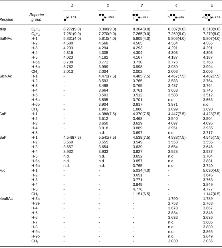
Table 2. 500 and 600-MHz1H-NMR chemical shifts of pNp-oligosaccharides recorded at 300K referenced to internal acetated1.908
(acetoned2.225). Compounds are represented by symbolic shorthand notation: , GalNAc;䊉, GlcNAc;䊏⫺, Gal4;䊏⁄, Gal3;ⵧ, Fuc; and䉭, Neu5Ac. 1 2 3 4 5 Reporter Residue group pNp C6H4⬘ 8.272(9.0) 8.306(9.0) 8.304(9.0) 8.307(9.0) 8.310(9.0) C6H4 7.281(9.0) 7.270(9.0) 7.265(9.0) 7.269(9.0) 7.270(9.0) GalNAc H-1 5.831(4.0) 5.810(4.0) 5.805(4.0) 5.805(4.0) 5.807(4.0) H-2 4.580 4.568 4.565 4.564 4.566 H-3 4.293 4.294 4.293 4.291 4.291 H-4 4.316 4.305 4.304 4.303 4.303 H-5 4.023 4.182 4.167 4.187 4.187 H-6a 3.738 3.771 3.730 3.776 3.763 H-6b 3.762 3.999 3.998 3.989 3.994 CH3 2.013 2.004 2.002 2.003 2.006 GlcNAc H-1 - 4.472(7.5) 4.485(7.5) 4.467(7.5) 4.482(7.5) H-2 - 3.593 3.765 3.583 3.764 H-3 - 3.498 3.765 3.487 3.764 H-4 - 3.664 3.761 3.663 3.745 H-5 - 3.503 3.512 3.588 3.512 H-6a - 3.595 3.701 n.d. 3.563 H-6b - 3.904 3.917 3.971 n.d. CH3 - 1.901 1.886 1.899 1.890 Gal4 H-1 - 4.386(7.5) 4.370(7.5) 4.447(7.5) 4.428(7.5) H-2 - 3.512 3.468 3.540 3.504 H-3 - 3.650 3.629 4.097 4.096 H-4 - 3.918 3.889 3.951 3.935 H-5 - n.d. 3.697 n.d. 3.717 Gal3 H-1 4.548(7.5) 5.541(7.5) 4.539(7.5) 4.538(7.5) 4.545(7.5) H-2 3.560 3.555 3.549 3.553 3.555 H-3 3.657 3.654 3.639 3.654 3.646 H-4 3.932 3.933 3.927 3.928 3.937 H-5 n.d. n.d. 3.652 n.d. 3.704 H-6a n.d. n.d. 3.857 n.d. 3.881 H-6b n.d. n.d. 3.765 n.d. 3.740 Fuc H-1 - - 5.034(4.0) - 5.030(4.0) H-2 - - 3.651 - 3.645 H-3 - - 3.771 - 3.763 H-4 - - 3.849 - 3.849 H-5 - - 4.776 - 4.777 CH3 - - 1.151(6.5) - 1.147(6.5) Neu5Ac H-3a - - - 1.790 1.789 H-3e - - - 2.753 2.763 H-4 - - - 3.670 3.667 H-5 - - - 3.834 3.848 H-6 - - - 3.636 3.636 H-7 - - - n.d. 3.605 H-8 - - - n.d. 3.889 H-9a - - - n.d. 3.865 H-9b - - - n.d. 3.649 CH3 - - - 2.030 2.036
b-1,4-linked Gal4 (D; pyranose ring form), respectively
(compare with compounds 1–3 in Table 2 and literature data [39]). The three N-acetyl resonances representing three protons each, at d 2.030, 2.003 and 1.899 could be attributed to Neu5Ac, GalNAc and GlcNAc, respectively [39]. The presence of only one Neu5Ac residue (E) in this oligosaccharide was confirmed by comparing the intensi-ties of the H3e and H3a resonances at d 2.753 and 1.790, respectively, with those of the discrete signals of the p-ni-trophenyl moiety.
By means of 2D TOCSY most of the resonances present in the 1D spectrum could be identified (Table 2, compound
4). In the TOCSY spectrum (100 ms, not shown) the
anomeric track of the Gal4residue D revealed three
cross-peaks at d 3.540 (H2), 4.097 (H3), and 3.951 (H4). When compared to the corresponding signals of precursor 2, these resonances showed downfield shifts of 0.028 ppm for Gal4
H2, 0.447 ppm for Gal4 H3, and 0.033 ppm for Gal4H4,
indicating that Neu5Ac is linked at O3 of Gal4. The
anomeric track of the Gal3residue B revealed three
cross-peaks at d 3.553 (H2), 3.654 (H3), and 3.928 (H4), in agree-ment with the terminal position of this residue (c.f. compound 2, Table 2), thereby excluding a possible sialyla-tion of the Gal3residue. The ST3Gal3-side activity on core 1
galactose as observed by Kono et al. [40] was absent in our case. The combined MS and NMR results justify the conclu-sion that oligosaccharide 4 has the structure Neu5Ac (a2-3) Gal(b1-4)GlcNAc(b1-6)[Gal(b1-3)]GalNAc(a1-pNp.
The negative-ion mode MALDI TOF mass spectrum (Figure 1b) of compound 5 showed one major peak at m/z 1305.0 corresponding to the deprotonated pseudo-molecu-lar ion of a hexasaccharide with the brutoformula Neu5Ac Hex2dHexHexNAc2-pNp (calculated mass 1305.4).
The 1D1H NMR spectrum (Figure 2b) of compound 5
revealed five anomeric signals at d 5.807 (3J
1,24.0 Hz), 5.030
(3J
1,2 4.0 Hz), 4.545 (3J1,2 7.5 Hz), 4.482 (3J1,27.5 Hz), and
4.428 (3J
1,27.5 Hz), which could be identified on guidance
of the NMR data of compounds 1–4 (Table 2). The addi-tional anomeric resonance at d 5.030 in compound 5 be-longed to the incorporated a-Fuc residue (G;pyranose ring form). 2D TOCSY NMR spectroscopy allowed the identi-fication of most of the signals (Table 2). The introduction of Fuc at O3 of GlcNAc (C) in 4 resulted in distinct downfield shifts of GlcNAc H1 (0.015 ppm), H2 (0.181 ppm), H3
(0.277 ppm), and H4 (0.082 ppm), a feature which is also observed when the NMR data of compounds 2 and 3 are compared (Table 2).
In order to confirm the various glycosidic linkages in compound 5 a 2D off-resonance ROESY (Figure 3) experi-ment was performed. The interresidual cross-peaks be-tween Gal3H1 and Ga1NAc H3 (d 4.545/4.291), GlcNAc
H1 and GalNAc H6a (d 4.482/3.763), Fuc H1 and GlcNAc
Figure 2. One-dimensional 500 MHz or 600 MHz1H NMR spectra of (a) Neu5Ac(a2-3)Gal(b1-4) GlcNAc(b1-6)[Gal(b1-3)]GalNAc(a1-pNp (4) (500
MHz) and (b) Neu5Ac(a2-3)Gal(b1-4) [Fuc(a1-3)]GlcNAc(b1-6)[Gal(b1-3)]GalNAc(a1-pNp (5) (600 MHz) in2H2O at 300 K (referenced to internal
Figure 3. Two-dimensional 600 MHz ROESY spectrum (mixing time 200 ms) of Neu5Ac(a2-3)Gal(b1-4)[Fuc(a1-3)]GlcNAc(b1-6)[Gal(b
H3 (d 5.030/3.764), and between Gal4H1 and GlcNAc H4
(d 4.428/3.745) proved the presence of the Gal(b1-3) GalNAc, GlcNAc(b1-6)GalNAc, Fuc(a1-3)GlcNAc, and Gal(b1-4)GlcNAc linkages, respectively. It should be noted that the interresidual cross-peak between Neu5Ac H3e and Gal4H3 as reported by Ball et al. [41] was not observed in
our study. The combined MS and NMR data indicate that oligosaccharide 5 has the structure Neu5Ac(a2-3)Gal(b1-4)[Fuc(a1-3)]GlcNAc(b1-6)[Gal(b1-3)]GalNAc(a1-pNp.
Discussion
The production of selectin blockers seems to be a reward-ing task with respect to several therapeutic targets, such as inflammatory diseases, transplant rejection and metastasis. This new class of anti-adhesion compounds can be enzymi-cally generated by taking advantage of the high stereo- and regioselectivity of glycosyltransferases. This approach which ascertains an easy access to the naturally occurring PSGL-1 type sLexepitope, in relatively high yields, opens
the possibility for the synthesis of glycopeptides by making use of the four cloned animal polypeptide GalNAc-trans-ferases [42–46]. The structural identity with the natural ligand would not only render these compounds highly com-patible in the competition with PSGL-1, but also assure a prolonged life-time and reduced antigenicity when com-pared with the non-natural ligands.
To demonstrate the suitability of glycosyltransferases for the synthesis of complex oligosaccharides, we used Gal-NAc(a1-pNp as the starting substrate for the elongation of O-linked glycans. As shown previously [30] the branching C2GnT has a strict requirement for a 3-substitution of Gal-NAc. Even though a b3-GalT, involved in the GM1/GD1 synthesis, transferring Gal to lipid-linked GalNAc [47], has been cloned, yet no recombinant enzyme is available elon-gating peptide-bound GalNAc. The rapid degradation of UDP-Gal in our system correlates well with the observation [48] that UDP-Gal decomposes rapidly in the presence of Mn2⫹, a metal ion needed to maintain b3-GalT activity.
Therefore, a two step incubation was carried out, resulting in a final yield of 52% for Gal(b1-3)GalNAc(a1-pNp (core 1 structure).
To date three different C2GnTs, of use for the branching of the core 1 structure, have been cloned [49–51]. Two of the three transferases [50,51] are also capable of synthesiz-ing the core 4 structure (GlcNAc(b1-6)[GlcNAc(b1-3)] GalNAc). As shown recently, the core 1 disaccharide can alternatively be branched with the b-N-acetyl-D-hexos-aminidase from Nocardia orientalis [52]; unfortunately, the yield is only around 6%. Applying a crude mouse kidney C2GnT preparation, the branching of a core 1 structure was realized in a yield of 74% [53]. By the use of a recom-binant purified mouse C2GnT we achieved a yield of ⬎85% for GlcNAc(b1-6)[Gal(b1-3)]GalNAc(a1-pNp (core 2 structure).
Recently, b-galactosidase from bovine testes was used in a one pot reaction together with a recombinant b-1,6-GlcNAc transferase. The galactosidase, which reversibly links galac-tose via a (b1-3) linkage to N-acetylgalactosamine, provides the substrate for the GlcNAc transferase in situ [54]. Since this synthesis ended up with a mixture of core 2 and core 6 (GlcNAc(b1-6)GalNAc) structures, and can only be driven towards high yields of the core 6 structure (yield⬎90%),we preferred the use of a crude b3-GalT preparation in combi-nation with C2GnT.
The core 2 structure can be galactosylated at O4 of GlcNAc by various b4-GalTs. By now, a whole family of b4-GalTs has been identified and cloned (reviewed in [55]). Although b4-GalT1 has been reported to be inefficient for the elongation of the core 2 structure [56], the purified hu-man milk enzyme, known as b4-GalT1, was successfully ap-plied to extend the core 2 structure, yielding Gal(b1-4) GlcNAc(b1-6)[Gal(b1-3)]GalNAc(a1-pNp. In spite of the earlier reported data that a strong inhibition of b4-GalT1 should exist for core 2 acceptor concentrations of 5 mM [53], we were able to reach a yield of⬎85% with 200 mU of enzyme at an acceptor concentration of 10 mM. It should be noted that within 5 h of incubation we did not achieve the earlier reported 100% yield with the enzyme used in [53].
The tetrasaccharide as generated above, was further ex-tended by sialic acid using ST3Gal3. Out of the four to date cloned enzymes only ST3Gal3 and ST3Gal4 have been demonstrated to sialylate N-acetyllactosamine (LacNAc) [40]. In our studies we used purified recombinant rat ST3Gal3, despite the preference of this enzyme for Gal (b1-3)GlcNAc (lacto-N-biose). Both ST3Gal3 and ST3Gal4 have a side activity in sialylating the Gal residue in a core 1 structure [40]. However, Gal(b1-4)GlcNAc (b1-6)[Neu5Ac(a2-3)Gal(b1-3)] GalNAc(a1-pNp was not found in our incubation mixture. This indicates that the side activity is strictly confined to the absence of a Gal(b1-4)X and the presence of a Gal(b1-3)X acceptor. Using a similar substrate in an earlier study, a yield of 68% was obtained with the partially purified recombinant rat ST3Gal3 [53]. Here, we obtained Neu5Ac(a2-3)Gal(b1-4)GlcNAc(b1-6)[Gal(b1-3)]GalNAc(a1-pNp in a yield of 87% via a two step incubation using 170 and 72 mU of the purified en-zyme.
To obtain the sLexepitope, the foregoing structure was
fucosylated at O3 of GlcNAc. At present five cloned a3-FucTs (a3-FucT3 - a3-FucT7), displaying different acceptor specificities, are known. a3-FucT4 fucosylates nearly ex-clusively LacNAc, whereas a3-FucT7 only acts on (a2-3)-sialylated LacNAc [57,58]. a3-FucT6, used in this study, fucosylates both sialylated and non-sialylated LacNAc. This enzyme, which has been shown to be highly active in vivo [59], is also of interest for the preparation of difucosyl sLex
structures on poly-LacNAc chains [60]. The expression sys-tem for a3-FucT6, used in our study, prevails over the syssys-tem
described by Licari et al. [38], since no exoglycosidases are present in the supernatant which cause degradation of the acceptor substrate. In a 6 h incubation with 70 mU of re-combinant a3-FucT6, Neu5Ac(a2-3)Gal(b1-4)[Fuc(a1-3)] GlcNAc(b1-6)[Gal(b1-3)]GalNAc(a1-pNp was synthe-sized in 100% yield.
In summary, we performed the first full enzymic synthe-sis of a core 2 type sLex-containing hexasaccharide in an
overall yield of about 32%. Once recombinant b3-GalT forming the core 1 structure will become available, scaling-up of the procedure described here should be feasible.
Acknowledgments
This work was supported by grant BIO4-CT95-0138 ‘Engi-neering O-Glycosylation’ of the EU to EGB/JFGV and grant 5002-46084 (SPP-Biotech) of the Swiss National Sci-ence Foundation to EGB. The authors also thank Markus Streiff (Novartis Pharma, Basel, Switzerland) and Frank Bootz (University of Zürich, Switzerland) for providing us with ST3Gal3 and fresh rat liver, respectively.
References
1 Walz G, Aruffo A, Kolanus W, Bevilacqua M, Seed B (1990)
Science 250: 1132–35.
2 Foxall C, Watson SR, Dowbenko D, Fennie C, Lasky LA, Kiso M, Hasegawa A, Asa D, Brandley BK (1992) J Cell Biol 117: 895–902. 3 Kim YJ, Borsig L, Varki NM, Varki A (1998) Proc Natl Acad Sci
USA 95: 9325–30.
4 Yamaguchi A, Ding K, Maehara M, Goi T, Nakagawara G (1998)
Oncology 55: 357–62.
5 Varki A (1997) J Clin Invest 99: 158–62. 6 Kansas GS (1996) Blood 88: 3259–87.
7 Turunen JP, Paavonen T, Majuri ML, Tiisala S, Mattila P, Mennan-der A, Gahmberg CG, Hayry P, Tamatani T, Miyasaka M, Renk-onen R (1994) Eur J Immunol 24: 1130–36.
8 McEver RP, Moore KL, Cummings RD (1995) J Biol Chem 270: 11025–28.
9 Han KT, Sharar SR, Phillips ML, Harlan JM, Winn RK (1995) J
Immunol 155: 4011–15.
10 Mulligan MS, Paulson JC, DeFrees S, Zheng ZL, Lowe JB, Ward PA (1993) Nature 364: 149–51.
11 Buerke M, Weyrich AS, Zheng Z, Gaeta FC, Forrest MJ, Lefer AM (1994) J Clin Invest 93: 1140–48.
12 Murohara T, Margiotta J, Phillips LM, Paulson JC, DeFrees S, Zalipsky S, Guo LS, Lefer AM (1995) Cardiovasc Res 30: 965–74. 13 Nguyen M, Eilber FR, DeFrees S (1996) Biochem Biophys Res
Commun 228: 716–23.
14 Kameyama A, Ishida H, Kiso M, Hasegawa A (1991) Carbohydr
Res 209: C1–C4.
15 Kiyoi T, Nakai Y, Kondo H, Ishida H, Kiso M, Hasegawa A (1996)
Bioorg Med Chem 4: 1167–76.
16 Baba K, Iwata N, Hamajima H, Ikami T, Ishida H, Hasegawa A, Kiso M (1998) Biosci Biotechnol Biochem 62: 590–92.
17 Lin CH, Shimazaki M, Wong C-H, Koketsu M, Juneja LR, Kim M (1995) Bioorg Med Chem 3: 1625–30.
18 Ichikawa Y, Lin YC, Dumas DP, Shen GJ, Garcia-Junceda E, Williams MA, Bayer R, Kretcham C, Walker LE, Paulson JC, Wong C-H (1992) J Am Chem Soc 114: 9283–98.
19 Nelson RM, Cecconi O, Roberts WG, Aruffo A, Linhardt RJ, Bevilacqua MP (1993) Blood 82: 3253–58.
20 Mulligan MS, Polley MJ, Bayer RJ, Nunn MF, Paulson JC, Ward PA (1992) J Clin Invest 90: 1600–07.
21 Polley MJ, Phillips ML, Wayner E, Nudelman E, Singhal AK, Hakomori S, Paulson JC (1991) Proc Acad Natl Sci USA 88: 6224–28.
22 Renkonen O, Toppila S, Penttila L, Salminen H, Helin J, Maahe-imo H, Costello CE, Turunen JP, Renkonen R (1997)
Glycobiol-ogy 7: 453–61.
23 Miyauchi H, Yuri M, Tanaka M, Kawamura N, Hayashi M (1997)
Bioorg Med Chem Lett 7: 989–92.
24 Simanek EE, McGarvey GJ, Jablonowski JA, Wong C-H (1998)
Chem Rev 98: 833–62.
25 Hiruma K, Kajimoto T, Weitz-Schmidt G, Ollmann I, Wong C-H (1996) J Am Chem Soc 118: 9265–70.
26 Wittmann V, Takayama S, Gong KW, Weitz-Schmidt G, Wong C-H (1998) J Org Chem 63: 5137–43.
27 Liu W, Ramachandran V, Kang J, Kishimoto TK, Cummings RD, McEver RP (1998) J Biol Chem 273: 7078–87.
28 Brockhausen I, Moller G, Pollex-Kruger A, Rutz V, Paulsen JC, Matta KL (1992) Biochem Cell Biol 70: 99–108.
29 Bendiak B, Schachter H (1987) J Biol Chem 262: 5775–83. 30 Zeng S, Dinter A, Eisenkrätzer D, Biselli M, Wandrey C, Berger
EG (1997) Biochem Biophys Res Commun 237: 653–58. 31 Gerber AC, Kozdrowski I, Wyss SR, Berger EG (1979) Eur J
Biochem 93: 453–60.
32 Hård K, van Zadelhoff G, Moonen P, Kamerling JP, Vliegenthart JFG (1992) Eur J Biochem 209: 895–915.
33 Bax A, Davis J (1985) J Magn Reson 65: 355–60. 34 Bax A, Davis J (1985) J Magn Reson 63: 207–13.
35 Desvaux H, Berthault P, Birlirakis N, Goldman M, Piotto M (1995) J Magn Reson 113: 47–52.
36 Unverzagt C, Kunz H, Paulson JC (1990) J Am Chem Soc 112: 9308–09.
37 Tsuji S, Datta AK, Paulson JC (1996) Glycobiology 6: R5–7 38 Licari PJ, Jarvis DL, Bailey JE (1993) Biotechnol Prog 9: 146–52. 39 Pollex-Kruger A, Meyer B, Stuike-Prill R, Sinnwell V, Matta KL,
Brockhausen I (1993) Glycoconjugate J 10: 365–80.
40 Kono M, Ohyama Y, Lee YC, Hamamoto T, Kojima N, Tsuji S (1997) Glycobiology 7: 469–79.
41 Ball GE, O’Neill RA, Schultz JE, Lowe JB, Weston BW, Nagy JO, Brown EG, Hobbs CJ, Bednarski MD (1992) J Am Chem Soc 114: 5449–51.
42 Clausen H, Bennett EP (1996) Glycobiology 6: 635–46.
43 Homa FL, Hollander T, Lehman DJ, Thomsen DR, Elhammer AP (1993) J Biol Chem 268: 12609–16.
44 White T, Bennett EP, Takio K, Sorensen T, Bonding N, Clausen H (1995) J Biol Chem 270: 24156–65.
45 Bennett EP, Hassan H, Clausen H (1996) J Biol Chem 271: 17006–12.
46 Hagen FK, Ten Hagen KG, Beres TM, Balys MM, VanWuyck-huyse BC, Tabak LA (1997) J Biol Chem 272: 13843–48. 47 Amado M, Almeida R, Carneiro F, Levery SB, Holmes EH,
No-moto M, Hollingsworth MA, Hassan H, Schwientek T, Nielsen PA, Bennett EP, Clausen H (1998) J Biol Chem 273: 12770–78.
48 Nunez HA, Barker R (1976) Biochemistry 15: 3843–47.
49 Bierhuizen MFA, Fukuda M (1992) Proc Natl Acad Sci USA 89: 9326–30.
50 Yeh JC, Ong E, Fukuda M (1999) J Biol Chem 274: 3215–21. 51 Schwientek T, Nomoto M, Levery SB, Merkx G, van Kessel AG,
Bennett EP, Hollingsworth MA, Clausen H (1999) J Biol Chem
274: 4504–12.
52 Murata T, Itoh T, Usui T (1998) Glycoconjugate J 15: 575–82. 53 Oehrlein R, Hindsgaul O, Palcic MM (1993) Carbohydr Res 244:
149–59.
54 Dudziak G, Zeng S, Berger EG, Gutiérrez Gallego R, Kamerling JP, Kragl U, Wandrey C (1998) Bioorg Med Chem Lett 8: 2595–98. 55 Breton C, Bettler E, Joziasse DH, Geremia RA, Imberty A (1998)
J Biochem 123: 1000–09.
56 Ujita M, McAuliffe J, Schwientek T, Almeida R, Hindsgaul O, Clausen H, Fukuda M (1998) J Biol Chem 273: 34843–49. 57 Niemelä R, Natunen J, Majuri M-L, Maaheimo H, Helin J, Lowe
JB, Renkonen O, Renkonen R (1998) J Biol Chem 273: 4021–26. 58 Grabenhorst E, Nimtz M, Costa J, Conradt HS (1998) J Biol Chem
273: 30985–94.
59 Kimura H, Shinya N, Nishihara S, Kaneko M, Irimura T, Nari-matsu H (1997) Biochem Biophys Res Commun 237: 131–37. 60 Weston BW, Smith PL, Kelly RJ, Lowe JB (1992) J Biol Chem 267:
24575–84.
![Figure 3. Two-dimensional 600 MHz ROESY spectrum (mixing time 200 ms) of Neu5Ac(a2-3)Gal(b1-4)[Fuc(a1-3)]GlcNAc(b1-6)[Gal(b1-3)]Gal- Neu5Ac(a2-3)Gal(b1-4)[Fuc(a1-3)]GlcNAc(b1-6)[Gal(b1-3)]Gal-NAc(a1-pNp (5) in 2 H 2 O at 300 K (referenced to internal aceto](https://thumb-eu.123doks.com/thumbv2/123doknet/14842165.625601/8.892.171.729.109.987/figure-dimensional-roesy-spectrum-glcnac-glcnac-referenced-internal.webp)