HAL Id: hal-02538539
https://hal.archives-ouvertes.fr/hal-02538539
Submitted on 9 Apr 2020HAL is a multi-disciplinary open access archive for the deposit and dissemination of sci-entific research documents, whether they are pub-lished or not. The documents may come from teaching and research institutions in France or abroad, or from public or private research centers.
L’archive ouverte pluridisciplinaire HAL, est destinée au dépôt et à la diffusion de documents scientifiques de niveau recherche, publiés ou non, émanant des établissements d’enseignement et de recherche français ou étrangers, des laboratoires publics ou privés.
A New Light-Emitting, Fabric-Based Device for
Photodynamic Therapy of Actinic Keratosis: Protocol
for a Randomized, Controlled, Multicenter,
Intra-Individual, Phase II Noninferiority Study (The
Phosistos Study)
Anne-Sophie Vignion-Dewalle, Henry Abi Rached, Elise Thecua, Fabienne
Lecomte, Pascal Deleporte, Hélène Béhal, Theresa Hommel, Alain Duhamel,
Rolf-Markus Szeimies, Laurent Mortier, et al.
To cite this version:
Anne-Sophie Vignion-Dewalle, Henry Abi Rached, Elise Thecua, Fabienne Lecomte, Pascal Deleporte, et al.. A New Light-Emitting, Fabric-Based Device for Photodynamic Therapy of Actinic Kerato-sis: Protocol for a Randomized, Controlled, Multicenter, Intra-Individual, Phase II Noninferiority Study (The Phosistos Study). JMIR Research Protocols, JMIR publications, 2019, 8 (4), pp.e12990. �10.2196/12990�. �hal-02538539�
A
NEW LIGHT-
EMITTING,
FABRIC-
BASED DEVICE FOR PHOTODYNAMIC THERAPY OF ACTINIC KERATOSIS:
PROTOCOL FOR A RANDOMIZED,
CONTROLLED,
MULTICENTRE,
INTRA-
INDIVIDUAL,
PHASEII
NON-
INFERIORITYSTUDY(
THEP
HOSISTOS STUDY)
AS. Vignion-Dewalle1, H. Abi-Rached1, 2, E. Thecua1, F. Lecomte1, P. Deleporte1, H. Béhal3, T. Hommel4, A. Duhamel3, RM. Szeimies4, *, L. Mortier1, 2, *, S. Mordon1
1
Univ. Lille, INSERM, CHU Lille, U1189 – ONCO-THAI – Image Assisted Laser Therapy for Oncology, F-59000 Lille, France
2
Department of Dermatology, CHU Lille, F-59000 Lille, France
3
Univ. Lille, CHU Lille, EA 2694 – Santé Publique: épidémiologie et qualité des soins, Unité de Biostatistiques, F-59000 Lille, France
4
Department of Dermatology and Allergology, Klinikum Vest GmbH, D-45657 Recklinghausen, Germany
*
RM. Szeimies and L. Mortier contributed equally to this work.
Email addresses: anne-sophie.vignion@inserm.fr henry.abirached@gmail.com elise.thecua@inserm.fr fabienne.lecomte@inserm.fr pascal.deleporte@inserm.fr helene.behal2@chru-lille.fr theresa.hommel@klinikum-vest.de alain.duhamel@univ-lille2.fr rolf-markus.szeimies@klinikum-vest.de laurent.mortier@chru-lille.fr serge.mordon@inserm.fr
Corresponding author: Anne-Sophie Vignion-Dewalle INSERM U1189 ONCO-THAI,
1, avenue Oscar Lambret, F-59037 LILLE Cedex, France
Trial sponsor: Centre HospitalierUniversitaire de Lille, Direction de la Recherche et de l’Innovation, 6, rue PrLaguesse,
F-59037 LILLE Cedex, France
Phone: +33 (0)3 20 44 59 69 Fax: +33 (0)3.20.44.57.11
Abstract-
Background:
Actinic keratosis (AK) is a common early in situ skin carcinomacaused by long-term sun exposure and usually develops on sun-exposed skin areas. Left untreated, AK may progress to squamous cell carcinoma. In order to prevent such risk, most clinicians routinely treat AK. Therapy options for AK include cryotherapy, topical treatments, curettage, excision surgery and photodynamic therapy (PDT).
Objective:
The aim of this study is to assess the non-inferiority, in terms of efficacy at 3 months, of a PDT protocol involving a new light-emitting device (P-PDT) compared to the conventional protocol (C-PDT) in the treatment of AK.
Methods:
In this randomized, controlled, multicentre, intra-individual,phase II non-inferiority clinical study, subjects with AK of the forehead and scalp are treated with P-PDT on one area and with C-PDT on the contralateral area. In both areas, lesions are prepared and methyl aminolevulinate (MAL) is applied. Thirty minutes after MAL application, the P-PDT area is exposed to red light at low irradiance (1.3 mW/cm2) for 2h30 so that a light dose of 12 J/cm2 is achieved. In the control area (C-PDT area), a 37 J/cm2 red light irradiation is performed 3 hours after MAL application. Recurrent AK at three months are retreated. The primary endpoint is the lesion complete response rate at three months. Secondary endpoints includepain scores at one day, local tolerance at seven days, lesion complete response rate at six months, cosmetic outcome at three and six months, and patient-reported quality of life and satisfaction throughout the study. Forty-five patients need to be recruited.
Results:
Clinical investigations are complete: 46 patients were treated with P-PDT on one area (n=285 AK) and with C-PDT on the contralateral area (n=285 AK). Data analysis is ongoing and statistical results will be available in the first half of 2019.
Conclusion:
In case of non-inferiority in efficacy and superiority in tolerability of P-PDT compared to C-PDT, P-PDT could become the treatment of choice for AK.
Trial registration:
The study was registered in ClinicalTrials.gov under identifier NCT03076892 (date of registration: March 10, 2017).
Keywords:
Photodynamic therapy, protocols, actinic keratosis, Aktilite CL 128 lamp, light-emitting fabric-based device.
Introduction
Actinic keratosis (AK) is a common early in situ skin carcinoma caused by long-term sun exposure and usually develops on sun-exposed skin areas such as the face, ears, scalp, neck, forearms, and back of the hands. Left untreated, AK will progress to invasive squamous cell carcinoma (SCC) in approximately 10% of patients [1]. In order to reduce the risk of developing SCC, consensus guidelines recommend that clinicians routinelytreatAK[2]. Treatment options include cryotherapy, topical treatments, curettage, surgical excisionand photodynamic therapy (PDT).
PDT is a cancer treatment modality combining light of appropriate wavelengths, a nontoxic photosensitizer, and sufficient molecular oxygen to generate reactive oxygen species and destroy target cells[3]. Over the last 15 years, PDT using 5-aminolevulinic acid (ALA) and PDT using methyl aminolevulinate (MAL) have been extensively investigated for the treatment of AK [4-8]. Topical application and incubation of ALA or MAL leads to selective accumulation of the endogenous photosensitizer protoporphyrin IX (PpIX) in the AK cells, and subsequent activation of PpIX by light of appropriate wavelengths induces, in the presence of oxygen, photochemical reactions leading to cell death [3].
Activation by red light using a total light dose of 37 J/cm2 after 3 hours of incubation with MAL is a conventional protocol, usually referred to as C-PDT, that is approved and likely the most widely used in Europe for PDT of AK [9-11]. This protocol has been reported to be an effective PDT treatment optionfor AK and to result in similar response rates and improved cosmetic outcomes compared with standard therapies [9]. However, high pain scores havebeen demonstrated with this protocol and concurrent use of cold air analgesia may be required to preventdiscomfort [12, 13].
Recently, several protocols involving an incubation with MAL for a maximum of 30 minutes followed by an activation by daylight for between 1h30 and 2h30 have been investigated [14-17]. From an European consensus [18], using a 2 hour daylight activation within 30 minutes after MAL application leads to a protocol (D-PDT) as effective as and better tolerated by patients than C-PDT. This better tolerability results from the maximum of 30 minutes for MAL incubation and the subsequent continuous activation of small amounts of PpIX.
Nonetheless, using daylight as the irradiation source is not realistic for all weather conditions [19].
New protocols designed to be as effective as C-PDT, as nearly painless as D-PDT and usable all year round are therefore emerging. Among these alternative protocols are the Flexitheralight protocol that we have recently published [20, 21]and the Phosistos protocol (P-PDT) that is discussed in this study. Developed within the Phosistos project supported by the European Commission under the Competitiveness and Innovation Framework Programme (CIP) (Project identifier: CIP-ICT-PSP-2013-7-621103), P-PDT uses a 30-minute MAL incubation followed by 2h30 of irradiation with a light-emitting, fabric-based device. Due to the short incubation time, P-PDT should be as nearly painless as D-PDT. Furthermore, from a recent study that discusses potential PDT overtreatment when using some protocols including C-PDT[22], P-PDTwith a total light dose almost three times lower than that of C-PDT could prove non-inferior in efficacy. Moreover, the high flexibility of the light-emitting, fabric-based device ensures an optimal irradiation of the treatment area, which is not the case with the rigid flat light sources used in C-PDT.
The aim of thisrandomized, controlled, multicentre, intra-individual, non-inferiority study is to assess the efficacy and tolerability of P-PDTcompared to that of C-PDT in treating patients with AK of the forehead and scalp.
Methods
Study design
This study is a randomized, controlled, multicentre, intra-individual, non-inferiority study comparing P-PDT versus C-PDT in the treatment of AK of the forehead and scalp. The study was conducted at two investigational sites: the department of dermatology at the Lille University Hospital in France and the Klinikum Vest in Germany.
Study status
Recruitment is closed and data collectionis completed. Data analysis is ongoing and is expected to be completed in the first half of 2019.
Ethical Approval
This study wasperformed in accordance with the ethical principles of the Declaration of Helsinki (2008) and the International Conference on Harmonization Good Clinical Practice guidelines. The study design was reviewed and approved by the French National Agency for the Safety of Medicines and Health Products (AgenceNationale de Sécurité du Médicament et des Produits de Santé, ANSM) (authorization number: 2016-A00010-51), the French Ethics Committee (Comités de Protection des Personnes, CPP) (authorization number: CPP 03/008/2016), the Federal Institute for Drugs and Medical Devices (BundesinstitutfürArzneimittel und Medizinprodukte, BfArM) (authorization number: 2015_79 1.1) and the Ethics committee of the University of Münster(Ethik-Kommission der ÄrztekammerWestfalen-Lippe und der WestfälischenWilhelms-Universität) (Approval number: 2016-513-f-M).
Study population
Patients were recruited from the patient population of the investigational sites.
Patients were eligible to be included in the study if they met all of the following criteria: They had a clinical diagnosis through visual inspection and palpation of 10-14 previously
untreated, non-pigmented, non-hyperkeratotic, grade I or II (according to the classification of Olsen et al. [23]), AK on the forehead and scalp (in case of more than 14 AK, only 14 AK were considered),
These AK had to be distributed in two non-coalescing areas with a similar number and grade of AK according to the following conditions:
o a minimum distance of 2 mm between two AK in the same area,
o a minimum distance of 10 mm between two AK, each in a different area,
Other AK treatment options were considered as unacceptable or medically less appropriate,
They did not have any AK treatment in the previous 30 days,
Patients were not eligible for inclusion in the study if they fulfilled one or more of the following criteria:
They had a clinical diagnosis of porphyria, They were immunosuppressed,
They used topical corticosteroids on the forehead or scalp in the previous two weeks, They received local treatment (including cryotherapy, curettage/electrocoagulation,
topical treatments with imiquimod, 5-flurouracil, diclofenac, or ingenol mebutate, or PDT) on the face or scalp in the previous 30 days,
They used topical retinoids, alpha-hydroxy acids, systemic retinoids, chemotherapy or immunotherapy in the previous 30 days,
They had pigmented AK,
They had known allergy to MAL or to any other ingredient of the MAL cream, peanut or soya,
They participated within the last 30 days in other clinical studies, They were pregnant,
They had any condition with a risk of poor protocol compliance, They currently received regular ultraviolet radiation therapy,
They were protected by a legal regime, in emergency situations or kept in detention. All patients received oral and written information before signing informed consent forms and subsequently entering the study.
The Phosistos protocol (P-PDT)
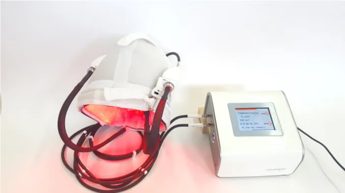
P-PDT includes application of MAL cream under transparent occlusive dressing immediately followed by the installation on the patient's head and turn-on of a light-emitting, fabric-based device for 3 hours. This device consists of a power control unit delivering 635 nm red light to a fibre optic-based fabric lining the inside of a cap (Texinov, Saint-Didier-de-la-Tour, France) (Figure 1). The device, classified as exempt risk group according to IEC 60601-2-57/2012, is configured to automatically start a 1.3 mW/cm2 irradiation 30 minutes after it is turned on (resulting in an incubation time of 30 minutes) and to stop 2h30 later (resulting in a light dose of 12 J/cm2).
Figure 1: The light-emitting, fabric-based device involved in P-PDT: 635 nm red light is emitted at 1.3 mW/cm2 by a fibre optic based fabric that lines the inside of a cap.
Study objectives/outcomes
The primary objective of the study is to assess the non-inferiority, in terms of efficacy at 3 months, of P-PDT compared to C-PDT. Outcome for the primary objective is the lesion complete response rate at three months.
The secondary objectives are:
To evaluate the treatment tolerance including pain at the end of treatment and adverse effects at seven days,
To evaluate the complete response rate at 6 months, To evaluate the cosmetic results at 3 and 6 months,
To estimate the number of patients with AK reduction higher than 75% at 3 and 6 months, To evaluate the patient’s quality of life and satisfaction throughout the study.
The corresponding outcomes are:
The pain score reported by the patient using a visual analogue scale ranging from 0 (no pain) to 10 (worst pain) at the end of treatment,
The adverse effects/reactions including erythema, skin exfoliation, skin burning sensation and skin oedema reported by the patient at seven days,
The complete response rate at 6 months,
The skin appearance(3 stands for “excellent”, 2 for “good”, 1 for “fair”, or 0 for “poor”) at one day, 3 months and 6 months: the cosmetic outcome at 3 months (respectively, at 6 months) is defined as the change in skin appearance between one day and 3 months (respectively, 6 months) and has the following possible values : {-3 ; -2 ; -1 ; 0 ; 1 ; 2 ; 3}, The Dermatology Life Quality Index(DLQI) and the standard satisfaction questionnaire
both completed by the patient throughout the study.
The lesion complete response (“complete response”, “incomplete response”) and the skin appearancewere clinically assessed by the investigators.
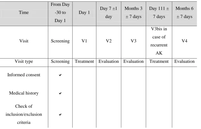
Study schema
The study flow chart is shown in Table 1. After screening, patients entering the study had to come to the investigational site for one treatment visit (V1) and three evaluation visits (V2, V3 and V4). In case of recurrent AK at the 3-month evaluation visit (V3), patients hada second treatment visit within the three following weeks (V3bis).
Table 1: Study flow chart.
Time From Day -30 to Day 1 Day 1 Day 7 ±1 day Months 3 ± 7 days Day 111 ± 7 days Months 6 ± 7 days Visit Screening V1 V2 V3 V3bis in case of recurrent AK V4
Visit type Screening Treatment Evaluation Evaluation Treatment Evaluation
Informed consent Medical history Check of inclusion/exclusion criteria
Documentation of AK including location,
number and grade
Photo-documentation of AK Separation of AK in two areas Randomization
Pain score during
treatment Adverse effects/reactions Skin appearance/cosmetic outcome Completion of the DLQI Completion of the satisfaction questionnaire Documentation of adverse events and serious adverse events
Pregnancy test
On the day of treatment (V1), 10 to 14 AKwere located, graded, photographed and divided into two areas (area A and area B)similar to each other in terms of number and grade of AK.The location of each AK was marked on plastic sheets.Randomization was then performed by opening the next envelope in sequence. This envelope specified the protocol that each area had to receive: either P-PDT for area A and C-PDT for area B, or C-PDT for area A and P-PDT for area B.In both cases, P-PDT (30-min MAL incubation followed by 2.5 h of irradiation) was performed first,so that the 3-hour MAL incubation required for C-PDT was achieved after P-PDT was completed.
Both the areas were prepared by removing crusts, gently scraping the lesion surface and applying MAL cream (Metvixia, Galderma, France) under a transparent occlusive dressing (Tegaderm, 3M, London Ontario, Canada) to the AK and surrounding normal skin (5-10 mm margin). An aluminium foil was placed over the transparent occlusive dressing, which covered the area randomized to receive C-PDT. The device involved in P-PDT was immediately set up and turned-on. After 3 hours, P-PDT was completedas described in paragraph II.D. The device involved in P-PDT was removed and the MAL cream washed off with saline solution. The patient rated his pain on a pain scale. The area that just received P-PDT was then protected with aluminium foil, while an Aktilite CL128 lamp (Galderma SA, Lausanne, Switzerland) was placed 5 to 8 cm from the other area and programmed to deliver 37 J/cm2 in seven to 10 minutes. At the end of C-PDT, the corresponding pain levelwas rated by the patient, who alsocompleted the DLQI and the satisfaction questionnaire.
Seven days after the treatment day (V2), patients were invited to report adverse effects/reactionsand to complete the DLQI and the satisfaction questionnaire.
The treatment responsewas assessed three months after the treatment (V3) by the investigators by comparison with the photographs at the treatment day (V1). An investigator's assessment of the skin appearance followed by the determination of the resulting cosmetic outcome was also performed.The DLQI and the satisfaction questionnaire were completed by patients. In case of recurrent AK, these latter were counted, graded and photographed and a second treatment visit, identical to the above-described first treatment (V1),was scheduled within three weeksafter V3 (V3bis).
The last follow-up visit (V4) was performed six months after V1. During this visit, the treatment response and the cosmetic outcome were investigator-assessed by comparison with the photographs and the skin appearance at V1, respectively. The patientswere asked to complete the DLQI and the satisfaction questionnaire.
Note that any AK appearing between V1 and V4 was not included in the assessment of the study outcomes.
Patients were randomly allocated to one of the two treatment options (either P-PDT for area A and C-PDT for area B or C-PDT for area A and P-PDT for area B) in a 1:1 ratio. The randomization sequence with stratification by treatment centre in blocks of four was generated by an independent statistician using the PROC PLAN procedure of SAS (SAS Institute Inc., Cary, North Carolina, USA) and transferred to a sequence of sealed, opaque, consecutively numbered envelopes. When a patient entered the study, randomization was performed by opening the next envelope in sequence.
The study is unblinded, both investigators and patients are aware of the treatment allocation.
Statistical methodology
Study hypothesis
The study primary hypothesis is the non-inferiority of P-PDTcompared to C-PDT in terms of the lesion complete response rate at three months.
Sample size determination
The study was designed to have a statistical power of 80% with a one-sided alpha level of 0.05to demonstrate non-inferiority in terms of lesion complete response rate at three months of P-PDT compared to C-PDT.Assuming a lesion complete response rate at three months of 75% in both areas, a correlation between lesions within the same patient, a correlation between lesions within the same area, an absolute non-inferiority margin of -10%, a mean lesion number per patient per area of six and a possible sample loss of 10%, 270 lesions per area (i.e., 45 patients) are required.
Statistical analysis of the primary outcome
Continuous variables will be expressed as mean and standard deviation, and categorical variables will be expressed as frequency and percentage. The normality of distribution will be assessed graphically and using the Shapiro-Wilk test.
The lesion complete response rate at three months will be analysed according to the protocol using the generalized linear mixed model to take into account the patient cluster effect (a
correlation between the complete responses of lesions within a same patient may exist) with adjustment on the area period (all lesions within a same area will receive the same protocol). The one-sided 95% confidence interval (CI) of the absolute difference in lesion complete response rate at three months between the two protocols will be calculated (D=P-PDT - C-PDT). In case of a lower limit of the one-sided 95% CI higher than -10%, P-PDT will be declared non-inferior to C-PDT and a two-sided superiority test will be performed at an alpha level of 5%.
All statistical analyses will be performed using SAS software version 9.4 (SAS Institute Inc., Cary, North Carolina, USA).
Statistical analysis of the secondary outcomes
The lesion complete response rate at six months will be processed using the same statistical analysis as the lesion complete response data at three months (previous paragraph).The differences in pain scores at the end of treatment between P-PDT and C-PDT will be assessed using a linear mixed model with patients as random effects (the significance level will be set at a two-sided alpha level of 0.05). The cosmetic outcomes at 3 and 6 months and the DLQI scores throughout the study will be compared between C-PDT and P-PDT using the Wilcoxon signed-rank test.
Data management
All patient data were collected using an electronic Case Report Form (eCRF)according to Good Clinical Practice and Standard Operating Procedures. Data collection was regularly monitored by a clinical research associate. Any deviation from the protocol was noted and the reason for the deviation documented. Any data inconsistency was brought to the attention of the clinical team and investigational site personnel (if required, data queries were send). Resolutions to these data inconsistencies were reflected in the database.
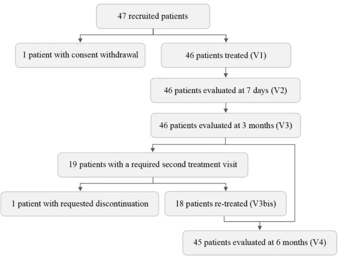
Results
The recruitment is closed and the clinical investigations are complete (Figure 2). Of the 47 recruited patients, one withdrew consent and did not receive any treatment protocol. Forty-six
patients were therefore treated with P-PDT on one area (for a total of 285 AK) and with C-PDT on the contralateral area (for a total of 285 AK). All these patients were evaluated at 3 months. Due to recurrent AK, 19 patients were required to undergo a second treatment visit. One of these patients dropped out for fear of pain as intense as that experienced with C-PDT during the first treatment visit. As a result, 18 patients were retreated and 45 patients completed the study at 6 months.
All treated patients were men, and their mean age was 72.2 years. Sixty-three percent of these patients had a Fitzpatrick skin type of II. Whatever the protocol, approximately 45% of the AK were grade I and 55% were grade II (Table 2).
Data analysis is ongoing and statistical results are expected to be available in the first half of 2019.
Figure 2: Study flow diagram.
Total (n=46 patients) Age (years) Mean ± SD 72.2 ± 9.1 Sex (%) Male 46 (100) Female 0 (0)
Fitzpatrick skin phototype (%)
I 8 (17) II 29 (63) III 8 (17) IV 1 (2) C-PDT (n=285 AK) P-PDT (n=285 AK) Grade of lesions (%) Grade I 130 (45.6) 128 (44.9) Grade II 155 (54.4) 157 (55.1) Discussion
C-PDT that has been proven to be effective in many studies [9-11] is likely the most widely used approved protocol in Europe for PDT of AK. The major adverse effect of C-PDT is pain during treatment, which has been described as a burning and stinging sensation localized to the treatment area [24-26].
Several studies has recently showed that the Europe-approved D-PDT is as effective as C-PDT but better tolerated and nearly painless[18, 27]. This painless characteristic comes from the short MAL incubation, which results in a continuous activation of small amounts of PpIX. Unfortunately, PDT using daylight activation depends on weather conditions [19]and cannot be performed in rainy, windy or cold conditions unless a greenhouse is used[28]. Moreover, due to the varying intensity of daylight depending on the weather conditions and the locations, it is impossible to control the light dose.
New PDT protocols including the Flexitheralight protocol[20, 21] have been designed to be as effective as C-PDT, as nearly painless as D-PDT, usable all year round and associated with a
known light dose. Consisting of a 30-minute incubation with MAL followed by 2.5 hours of activation with a quite cumbersome, light-emitting, fabric-based device, which delivers 37 J/cm2 at an irradiance of 12.3 mW/cm2, the Flexitheralight protocolhas been shown to be non-inferior to C-PDTwhile being nearly pain-free[21].We have revised downward the irradiation parameters of the Flexitheralight protocol: the new version of the Flexitheralight protocol, referred to as P-PDT, involves an irradiance of 1.3 mW/cm2 and a light dose of 12 J/cm2.The choice of such a light dose was based on a study that demonstrated the ability of two light sources with light doses lower than 15 J/cm2 to completely photobleach PpIX [28]. Regarding the irradiance, the value of 1.3 mW/cm2was selected on the basis of the study of Ibbotson et al. that reported effective PDT treatment when using a 7 mW/cm2 red light source [29]. These choices are in line with studies reporting similar efficacy for different irradiances [30] and light doses [22].With these new irradiation parameters, the light-emitting, fabric-based device has been significantly modified to be more user friendly in terms of dimensions and ergonomics.
The present study aims to assess the non-inferiority in efficacy at 3 months (primary objective) and superiority in tolerability (secondary objective) of P-PDT compared to C-PDT in the treatment of AK of the forehead and scalp.
Data collection is completed, and data analysis is ongoing. The results are expected in the first half of 2019. In case of a positive assessment, P-PDT could be preferred to the conventionally-used C-PDT. Moreover, as P-PDT can be performed in all weather conditions, in any geographic location and year-round, it could also be preferred to D-PDT. Hence, PDTcould become the treatment of choice for AK. Furthermore, an ambulatory version of P-PDT could be further investigated.
Acknowledgements
The authors thank all the project staff for their input into the design and conduct of the study.
S. Mordon, AS. Vignion-Dewalle, H. Abi-Rached, E. Thecua, F. Lecomte, C. Vicentini, P. Deleporte, H. Béhal, D. Kerob, T. Hommel and A. Duhamel declare that they have no competing interest.
RM. Szeimies is vice president of EURO-PDT. He has been a member of advisory boards for Almirall, Biofrontera, Galderma, ISDIN, LEO Pharma, photonamic and Pierre-Fabre, and has received speakers’ honoraria from the aforementioned companies.
L. Mortier has been a member of advisory boards for BMS, Roche, GSK, Novartis, LEO Pharma and MSD. He has received travel grants for attending congresses from BMS, Roche, GSK, Novartis and LEO Pharma. He has been the principal investigator of clinical trials performed for BMS, Roche, GSK, Novartis, LEO Pharma and MSD.
Funding
The clinical study described in this manuscript is part of the Phosistos project. This project, which aims to develop a flexible light-emitting fabric-based device for PDT of AK (from design to clinical trial), was financially supported by the European Commission under the Competitiveness and Innovation Framework Programme (CIP) (Project identifier: CIP-ICT-PSP-2013-7-621103).
Metvixia cream (168 mg of MAL/g) was graciously supplied by Galderma R&D (France). Galderma R&D (France) was not further involved.
Author’s contributions
The manuscript was written by ASVD. The study protocol was created by SM. SM, ET, FL, ASVD and PD contributed to the design of the study and to the development of the device. HB and AD were responsible for statistical advice and planning. HAR, TH, RMS and LM contributed with the clinical aspects of the study. SM, ET, FL critically reived the manuscript. All authors read and approved the final manuscript.
References
1. Glogau RG: The risk of progression to invasive disease. J Am Acad Dermatol 2000, 42(1 Pt 2):23-24.
2. Stockfleth E, Ferrandiz C, Grob JJ, Leigh I, Pehamberger H, Kerl H, European Skin A: Development of a treatment algorithm for actinic keratoses: a European Consensus. Eur J Dermatol 2008, 18(6):651-659.
3. Plaetzer K, Krammer B, Berlanda J, Berr F, Kiesslich T: Photophysics and photochemistry of photodynamic therapy: fundamental aspects. Lasers Med Sci 2009, 24(2):259-268.
4. Braathen LR, Szeimies RM, Basset-Seguin N, Bissonnette R, Foley P, Pariser D, Roelandts R, Wennberg AM, Morton CA, International Society for Photodynamic Therapy in D: Guidelines on the use of photodynamic therapy for nonmelanoma skin cancer: an international consensus. International Society for Photodynamic Therapy in Dermatology, 2005. J Am Acad Dermatol 2007, 56(1):125-143.
5. Ericson MB, Wennberg AM, Larko O: Review of photodynamic therapy in actinic keratosis and basal cell carcinoma. Ther Clin Risk Manag 2008, 4(1):1-9.
6. Morton CA, McKenna KE, Rhodes LE, British Association of Dermatologists Therapy G, Audit S, the British Photodermatology G: Guidelines for topical photodynamic therapy: update. Br J Dermatol 2008, 159(6):1245-1266.
7. Morton C, Szeimies RM, Sidoroff A, Wennberg AM, Basset-Seguin N, Calzavara-Pinton P, Gilaberte Y, Hofbauer G, Hunger R, Karrer S et al: European Dermatology Forum Guidelines on topical photodynamic therapy. Eur J Dermatol 2015, 25(4):296-311.
8. Wiegell SR: Update on photodynamic treatment for actinic keratosis. Curr Probl Dermatol 2015, 46:122-128.
9. Morton C, Campbell S, Gupta G, Keohane S, Lear J, Zaki I, Walton S, Kerrouche N, Thomas G, Soto P et al: Intraindividual, right-left comparison of topical methyl aminolaevulinate-photodynamic therapy and cryotherapy in subjects with actinic keratoses: a multicentre, randomized controlled study. Br J Dermatol 2006, 155(5):1029-1036.
10. Pariser D, Loss R, Jarratt M, Abramovits W, Spencer J, Geronemus R, Bailin P, Bruce S: Topical methyl-aminolevulinate photodynamic therapy using red light-emitting diode light for treatment of multiple actinic keratoses: A randomized, double-blind, placebo-controlled study. J Am Acad Dermatol 2008, 59(4):569-576.
11. Szeimies RM, Matheson RT, Davis SA, Bhatia AC, Frambach Y, Klovekorn W, Fesq H, Berking C, Reifenberger J, Thaci D: Topical methyl aminolevulinate photodynamic therapy using red light-emitting diode light for multiple actinic keratoses: a randomized study. Dermatol Surg 2009, 35(4):586-592.
12. Tyrrell J, Campbell SM, Curnow A: The effect of air cooling pain relief on protoporphyrin IX photobleaching and clinical efficacy during dermatological photodynamic therapy. J Photochem Photobiol B 2011, 103(1):1-7.
13. Stangeland KZ, Kroon S: Cold air analgesia as pain reduction during photodynamic therapy of actinic keratoses. J Eur Acad Dermatol Venereol 2012, 26(7):849-854.
14. Wiegell SR, Haedersdal M, Philipsen PA, Eriksen P, Enk CD, Wulf HC: Continuous activation of PpIX by daylight is as effective as and less painful than conventional photodynamic therapy for actinic keratoses; a randomized, controlled, single-blinded study. Br J Dermatol 2008, 158(4):740-746.
15. Wiegell SR, Haedersdal M, Eriksen P, Wulf HC: Photodynamic therapy of actinic keratoses with 8% and 16% methyl aminolaevulinate and home-based daylight exposure: a double-blinded randomized clinical trial. Br J Dermatol 2009, 160(6):1308-1314.
16. Wiegell SR, Fabricius S, Stender IM, Berne B, Kroon S, Andersen BL, Mork C, Sandberg C, Jemec GB, Mogensen M et al: A randomized, multicentre study of directed daylight exposure times of 1(1/2) vs. 2(1/2) h in daylight-mediated photodynamic therapy with methyl aminolaevulinate in patients with multiple thin actinic keratoses of the face and scalp. Br J Dermatol 2011, 164(5):1083-1090.
17. Wiegell SR, Fabricius S, Gniadecka M, Stender IM, Berne B, Kroon S, Andersen BL, Mork C, Sandberg C, Ibler KS et al: Daylight-mediated photodynamic therapy of moderate to thick actinic keratoses of the face and scalp: a randomized multicentre study. Br J Dermatol 2012, 166(6):1327-1332.
18. Wiegell SR, Wulf HC, Szeimies RM, Basset-Seguin N, Bissonnette R, Gerritsen MJ, Gilaberte Y, Calzavara-Pinton P, Morton CA, Sidoroff A et al: Daylight photodynamic therapy for actinic keratosis: an international consensus: International Society for Photodynamic Therapy in Dermatology. J Eur Acad Dermatol Venereol 2012, 26(6):673-679.
19. Wiegell SR, Fabricius S, Heydenreich J, Enk CD, Rosso S, Baumler W, Baldursson BT, Wulf HC: Weather conditions and daylight-mediated photodynamic therapy: protoporphyrin IX-weighted daylight doses measured in six geographical locations. Br J Dermatol 2013, 168(1):186-191.
20. Lecomte F, Vignion-Dewalle AS, Vicentini C, Thecua E, Deleporte P, Duhamel A, Mordon S, Mortier L: A phase II study evaluating the non-inferiority of a photodynamic therapy protocol involving the Flexitheralight device compared to the conventional protocol (the FLEXITHERALIGHT study).JMIR research protocols 2018, Accepted.
21. Vicentini C, Vignion-Dewalle AS, Thecua E, Lecomte F, Maire C, Deleporte P, Behal H, Kerob D, Duhamel A, Mordon S et al: Photodynamic therapy for actinic keratosis of the forehead and scalp: a randomized, controlled, phase II clinical study evaluating the non-inferiority of a new protocol involving irradiation with a light-emitting, fabric-based device (the Flexitheralight protocol) compared with the conventional protocol involving irradiation with the Aktilite CL 128 lamp. Br J Dermatol 2018.
22. Vignion-Dewalle AS, Baert G, Thecua E, Lecomte F, Vicentini C, Abi-Rached H, Mortier L, Mordon S: Comparison of 10 efficient protocols for photodynamic therapy of actinic keratosis: How relevant are effective light dose and local damage in predicting the complete response rate at 3 months?Lasers Surg Med 2018, 50(5):576-589.
23. Olsen EA, Abernethy ML, Kulp-Shorten C, Callen JP, Glazer SD, Huntley A, McCray M, Monroe AB, Tschen E, Wolf JE, Jr.: A double-blind, vehicle-controlled study evaluating masoprocol cream in the treatment of actinic keratoses on the head and neck. J Am Acad Dermatol 1991, 24(5 Pt 1):738-743.
24. Wiegell SR, Skiveren J, Philipsen PA, Wulf HC: Pain during photodynamic therapy is associated with protoporphyrin IX fluorescence and fluence rate. Br J Dermatol 2008, 158(4):727-733.
25. Arits AH, van de Weert MM, Nelemans PJ, Kelleners-Smeets NW: Pain during topical photodynamic therapy: uncomfortable and unpredictable. J Eur Acad Dermatol Venereol 2010, 24(12):1452-1457.
26. Serra-Guillen C, Hueso L, Nagore E, Vila M, Llombart B, Requena Caballero C, Botella-Estrada R, Sanmartin O, Alfaro-Rubio A, Guillen C: Comparative study between cold air analgesia and supraorbital and supratrochlear nerve block for the management of pain during photodynamic therapy for actinic keratoses of the frontotemporal zone. Br J Dermatol 2009, 161(2):353-356.
27. Rubel DM, Spelman L, Murrell DF, See JA, Hewitt D, Foley P, Bosc C, Kerob D, Kerrouche N, Wulf HC et al: Daylight photodynamic therapy with methyl aminolevulinate cream as a convenient, similarly effective, nearly painless alternative to conventional photodynamic therapy in actinic keratosis treatment: a randomized controlled trial. Br J Dermatol 2014, 171(5):1164-1171.
28. Lerche CM, Heerfordt IM, Heydenreich J, Wulf HC: Alternatives to Outdoor Daylight Illumination for Photodynamic Therapy--Use of Greenhouses and Artificial Light Sources. Int J Mol Sci 2016, 17(3):309.
29. Ibbotson SH, Ferguson J: Ambulatory photodynamic therapy using low irradiance inorganic light-emitting diodes for the treatment of non-melanoma skin cancer: an open study. Photodermatol Photoimmunol Photomed 2012, 28(5):235-239.
30. Apalla Z, Sotiriou E, Panagiotidou D, Lefaki I, Goussi C, Ioannides D: The impact of different fluence rates on pain and clinical outcome in patients with actinic keratoses treated with photodynamic therapy. Photodermatol Photoimmunol Photomed 2011, 27(4):181-185.
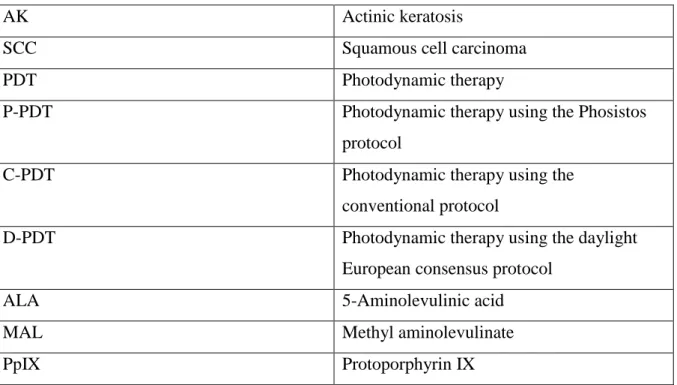
List of abbreviations
Table 3: Abbreviations.
AK Actinic keratosis
SCC Squamous cell carcinoma
PDT Photodynamic therapy
P-PDT Photodynamic therapy using the Phosistos
protocol
C-PDT Photodynamic therapy using the
conventional protocol
D-PDT Photodynamic therapy using the daylight
European consensus protocol
ALA 5-Aminolevulinic acid
MAL Methyl aminolevulinate