HAL Id: hal-02953922
https://hal.archives-ouvertes.fr/hal-02953922
Submitted on 30 Sep 2020
HAL is a multi-disciplinary open access
archive for the deposit and dissemination of sci-entific research documents, whether they are pub-lished or not. The documents may come from teaching and research institutions in France or abroad, or from public or private research centers.
L’archive ouverte pluridisciplinaire HAL, est destinée au dépôt et à la diffusion de documents scientifiques de niveau recherche, publiés ou non, émanant des établissements d’enseignement et de recherche français ou étrangers, des laboratoires publics ou privés.
Hydroxyl Groups on Cellulose Nanocrystal Surfaces
form Nucleation Points for Silver Nanoparticles of
Varying Shapes and Sizes
Dafne Musino, Camille Rivard, Gautier Landrot, Bruno Novales, Thierry
Rabilloud, Isabelle Capron
To cite this version:
Dafne Musino, Camille Rivard, Gautier Landrot, Bruno Novales, Thierry Rabilloud, et al.. Hydroxyl Groups on Cellulose Nanocrystal Surfaces form Nucleation Points for Silver Nanoparticles of Vary-ing Shapes and Sizes. Journal of Colloid and Interface Science, Elsevier, 2021, 584, pp.360-371. �10.1016/j.jcis.2020.09.082�. �hal-02953922�
Journal Pre-proofs
Regular Article
Hydroxyl Groups on Cellulose Nanocrystal Surfaces form Nucleation Points for Silver Nanoparticles of Varying Shapes and Sizes
Dafne Musino, Camille Rivard, Gautier Landrot, Bruno Novales, Thierry Rabilloud, Isabelle Capron
PII: S0021-9797(20)31271-6
DOI: https://doi.org/10.1016/j.jcis.2020.09.082
Reference: YJCIS 26987
To appear in: Journal of Colloid and Interface Science
Received Date: 20 July 2020
Revised Date: 15 September 2020
Accepted Date: 21 September 2020
Please cite this article as: D. Musino, C. Rivard, G. Landrot, B. Novales, T. Rabilloud, I. Capron, Hydroxyl Groups on Cellulose Nanocrystal Surfaces form Nucleation Points for Silver Nanoparticles of Varying Shapes and Sizes, Journal of Colloid and Interface Science (2020), doi: https://doi.org/10.1016/j.jcis.2020.09.082
This is a PDF file of an article that has undergone enhancements after acceptance, such as the addition of a cover page and metadata, and formatting for readability, but it is not yet the definitive version of record. This version will undergo additional copyediting, typesetting and review before it is published in its final form, but we are providing this version to give early visibility of the article. Please note that, during the production process, errors may be discovered which could affect the content, and all legal disclaimers that apply to the journal pertain.
Graphical abstract
Hydroxyl Groups on Cellulose Nanocrystal Surfaces form
Nucleation Points for Silver Nanoparticles of Varying Shapes and
Sizes
Dafne Musino,a Camille Rivard, b,c Gautier Landrot,b Bruno Novales,a Thierry Rabilloud d and Isabelle
Caprona*
a INRAE, BIA, 44316 Nantes, France
b SOLEIL Synchrotron, L’Orme des Merisiers, Gif-sur-Yvette, 91192 Saint-Aubin, France c INRAE, TRANSFORM, 44316 Nantes, France
d Univ. Grenoble Alpes, CNRS, CEA, IRIG, SYMMES, Laboratoire de Chimie et Biologie des Métaux, 38000 Grenoble, France
* Author for correspondence: isabelle.capron@inrae.fr, INRAE, BIA, Rue de la Geraudiere, 44316
Nantes, France. Tel : +33 2 40 67 50 43
dafne.musino@inrae.fr camille.rivard@synchrotron-soleil.fr gautier.landrot@synchrotron-soleil.fr
bruno.novales@inrae.fr thierry.rabilloud@cnrs.fr
Hydroxyl Groups on Cellulose Nanocrystal Surfaces form
Nucleation Points for Silver Nanoparticles of Varying Shapes and
Sizes
Dafne Musino,a Camille Rivard, b,c Gautier Landrot,b Bruno Novales,a Thierry Rabilloud d and Isabelle
Caprona*
a INRAE, BIA, 44316 Nantes, France
b SOLEIL Synchrotron, L’Orme des Merisiers, Gif-sur-Yvette, 91192 Saint-Aubin, France c INRAE, TRANSFORM, 44316 Nantes, France
d Univ. Grenoble Alpes, CNRS, CEA, IRIG, SYMMES, Laboratoire de Chimie et Biologie des Métaux, 38000 Grenoble, France
*Author for correspondence: isabelle.capron@inrae.fr, INRAE, BIA, Rue de la Geraudiere, 44316
Nantes, France. Tel : +33 2 40 67 50 43
dafne.musino@inrae.fr camille.rivard@synchrotron-soleil.fr gautier.landrot@synchrotron-soleil.fr
bruno.novales@inrae.fr thierry.rabilloud@cnrs.fr
Abstract. In this study, we investigate the interactions between the cellulose surface and Ag
nanoparticles (AgNPs) for the purpose of manufacturing hybrid nanomaterials using bacterial cellulose nanocrystals (BCNs) as a model substrate. We focus on the role of the BCN surface chemistry on the AgNP nucleation obtained by chemical reduction of Ag+ ions. Homogeneous hybrid
suspensions of BCN/AgNP are produced, regardless of whether the BCNs are quasi-neutral, negatively (TBCNs) or positively charged (ABCNs). The characterization of BCN/AgNP hybrids identifies the –OH surface groups as nucleation points for AgNPs, of about 20 nm revealing that surface charges only improve the accessibility to OH groups. X-ray Absorption technics (XANES and EXAFS) revealed a high metallic Ag0 content ranging from 88% to 97%. Moreover, the grafting of
hydrophobic molecules on a BCN surface (HBCNs) does not prevent AgNP nucleation, illustrating the versatility of our method and the possibility to obtain bifunctional NPs. A H2O2 redox post-treatment
on the hybrid induces an increase in AgNPs size, up to 90 nm as well as a shape variation (i.e., triangular). In contrast, HO induces no size/shape variation for aggregated hybrids, emphasizing that
Keywords. Bacterial cellulose nanocrystal (BCN), hydroxyl groups, surface modification, silver
1. Introduction
Cellulose is an almost inexhaustible biopolymer extracted from wood, cotton, algae, tunicates or bacteria [1–3], leading to variation in dimensions and structural organization [4]. Cellulose displays interesting properties such as high water retention capacity, high wet strength, low density, bio-compatibility, non-toxicity and biodegradability, which make it very appropriate for several applications in biomedicine and pharmacology [5–7], cosmetics [8], paper and textiles [9]. Cellulose can also be hydrolyzed to increase its crystallinity, commonly by acid hydrolysis, leading to the so-called cellulose nanocrystals (CNCs) [10]. While the extraction of plant-sourced cellulose often requires the use of hazardous chemicals, bacterial cellulose (BC) is mainly secreted extracellularly by certain bacteria such as Gluconacetobacter xylinu [1]. Therefore, bacterial cellulose nanocrystal (BCN) contain no functional groups other than hydroxyl groups [11] and thanks to its high chemical purity, they are particularly interesting for food and medical applications.
To confer antimicrobial properties to CNCs, as to BCNs, it is quite common to graft or nucleate metallic nanoparticles (e.g., Cu, Ag, ZnO, Au) on their surface [12–15]. Among others, silver nanoparticles (AgNPs) have been widely studied. A silver precursor is generally incorporated by addition of a salt such as silver nitrate (AgNO3), and several experimental methods (e.g., UV radiation
[16,17], hydrazine reduction [18]) have been proposed to nucleate AgNPs on the cellulose nanocrystal surface, thus obtaining a hybrid material (i.e., CNC/AgNPs). Chemical reduction is one of the most common ways to synthesize metallic NPs, and sodium borohydride (NaBH4) [19–22] is reported to be
one of the most efficient reducing agents, inducing the rapid formation of 2-3-nm AgNPs [23]. NH4OH [24] and ascorbic acid [25] can also be used to reduce Ag+ ions into AgNPs. To carry out this
chemical AgNP synthesis, capping agents or stabilizers (e.g., trisodium citrate: TSC; polyvinylpirrolidine: PVP; Cetrimonium bromide: CTAB) are often introduced to better control the morphological properties of AgNPs, preventing their aggregation [26–28]. In other cases, nanocellulose is proposed as both a support and a reducer for the generation of metallic nanoparticles [29]. Xiong et al. [30] claim that cellulose promotes the generation of AgNPs and dendritic Ag nanostructures thanks to the extensive presence of surface hydroxyl groups. Furthermore, various surface modifications of CNCs can be performed (e.g., amidation, oxidation, esterification, etherification) [10], possibly affecting the reduction of AgNPs on the CNC surface. Most of the studies concerning CNC/AgNP hybrids [31–33] indicate how the introduction of negative surface charges (i.e., via TEMPO-mediated oxidation [32]) can provide the high-binding capability for the transition of metal species such as Ag+ ions. Moreover, other surface modifications can be performed
on the CNC surface to extend the possible application fields. For example, cationized cellulose can be produced by grafting different molecules on the cellulose surface (e.g., HPTMAC: hydroxypropyltrimethylammonium chloride [34], BriBBr: a-bromoisobutyryl bromide [35], HDTMA:
modification of cellulose cotton fibers by 3-chloro-2-hydroxypropyl trimethyl ammonium chloride (CHPTAC) affects the absorption of AgNPs on the fiber surface. Indeed, such an adsorption is greater on cationized cellulose fibers than on untreated ones. To obtain these cationized fibers, Dong et al. [40] propose to graft ammonium ions onto the surface, thus creating two different pathways for the deposition of metal NPs (e.g., Au, Pt, Pd): the first one based on the electrostatic assembly of metal nanoparticles capped with negative citrate ions, and a second one where negative metal complex ions are adsorbed onto the cationic substrate and then reduced. Both approaches lead to a high surface coverage of the CNC surface by metallic nanoparticles.
CNCs can also be made hydrophobic to allow their dispersion in non-aqueous solvents. To do this, one of the most widely used approaches is amidation, where coupling agents are grafted onto TEMPO-oxidized nanocrystals [10]. Araki et al. [41] propose the grafting of poly(ethylene glycol) with a terminal amino group onto the surface of TEMPO-oxidized CNCs. Zhang et al. [42] use a similar experimental approach to covalently functionalize CNCs via peptide coupling chemistry with different alkylamines (e.g., propylamine, n-butylamine, amylamine, hexylamine, heptylamine). Hu et al. [43] propose using tannic acid to covalently attach primary amine with a long alkyl chain (i.e., decylamine) to the CNC surface. In addition, Cunha et al. [44] produced inverse emulsions, tailoring the hydrophobicity of CNCs and nanofibrillated cellulose by chemical modification with lauroyl chloride (C12). However, no studies have proposed bifunctional hybrids, using hydrophobically-modified CNCs
as substrates for AgNP nucleation.
As for nanocellulose surface modification, AgNP characteristics (e.g., size, shape, crystalline structure) can strongly influence the final properties of the CNC/AgNP hybrid system. Some works report simple techniques for a shape- and size-selective synthesis of Ag nanostructures (e.g., from spherical AgNPs to triangular-shaped AgNPs or Ag nanoprisms, denoted AgNPrisms [45]) based on the use of hydrogen peroxide (H2O2) redox post-treatment [20,21,46,47], as well as photo-induced [48]
or solution-phase [49] methods. In particular, the size-shape transformation of AgNPs achieved with H2O2 relies to redox capabilities linked to the autocatalytic decomposition on the Ag surface. These
AgNPrisms are anisotropic (i.e., their lateral dimension is greater than their thickness) and are thus characterized by a strong localized surface plasmon resonance (LSPR). Their resulting size- and shape-dependent optical properties make them suitable for sensors [50,51] and biological imaging [52], catalysts [53], nanophotonic devices and circuits [54,55]. Parnklang et al. [46] show the shape transformation from spherical AgNPs to AgNPrisms when H2O2 oxidizes the AgNPs to Ag+ ions and
thus reduces them into Ag atoms in nanometric silver particles. The H2O2 redox post-treatment has
also been applied to hybrid systems. Jiang et al. [20] propose the fabrication of films made of AgNPrisms and TEMPO-oxidized cellulose nanofibrils, which work as capping and shape-regulating agents. The presence of a predominant (111) peak in the XRD pattern allows them to affirm that AgNPrisms are bounded to cellulose nanofibers.
The result is that hybrids made of AgNPs fixed on the CNC surface have been produced, whereas the interactions and binding mechanisms have not been studied in detail [29,31,56]. In this paper, we propose to perform different surface modifications on BCNs in order to shed light on the effective role of cellulose nanocrystal surface chemistry on the nucleation of AgNPs. We also prepared bifunctional NPs that combine a hydrophobic surface and AgNPs. Moreover, we investigated the impact of the CNC surface modification on the H2O2 redox post-treatment, linking the size and shape variation of
AgNPs to their physicochemical characteristics (i.e., AgNP structure and oxidation state).
2. Materials and Methods
Chemicals. Food grade nata de coco was purchased from AROY-D (Thailand). All the other products
were purchased from Sigma-Aldrich (France) and used as received without further purification: silver nitrate (AgNO3 ≥ 99%), sodium borohydride (NaBH4 ≥ 96%), 2,2,6,6-Tetramethylpiperidine 1-oxyl
(TEMPO: 98.8%), sodium bromide (NaBr: 99%), sodium hypochlorite solution (NaClO: 10% RT), cholaminchloride hydrochloride (AETMA: 99%; pKa = 7), octylamine (C8: 99%; pKa = 10),
N-hydroxysuccinimide (NHS), N-(3-dimethylaminopropyl)-N′-ethylcarbodiimide (EDC), hydrogen peroxide 30% (H2O2). For all the aqueous suspensions, ultra-pure water was used.
Synthesis of uncharged BCNs. For the preparation of BCNs, the experimental procedure proposed by
Kalashnikova et al. [57] was followed. Briefly, nata de coco cubes were dialyzed in ultra-pure water for 9 days, after which they were ground in a Waring blender, suspended in 0.5 N NaOH solution, and stirred for 2 h at 70°C. Finally, the sample was rinsed using ultra-pure water. A whitening treatment was performed twice, dispersing the slurry in a NaClO2 solution (8.5 g/L) in a sodium acetate buffer
(pH = 4.5) and stirring it for 2 h at 70°C. The bleached cellulose was rinsed with distilled water and then hydrolyzed with 2.5 M HCl solution at 70°C for 2 h. The suspension was centrifuged three times (10 min., 10000 g) and dispersed in ultra-pure water up to pH > 5. The final BCN suspension was again dialyzed against ultra-pure water for one week and finally homogenized by ultrasound (amplitude = 20; 5 min). The BCN content in the suspension (around 2 g/L) was determined by a drying test.
Synthesis of anionic BCNs mediated via TEMPO oxidation (TBCNs). The experimental protocol
for the BCN surface carboxylation via TEMPO-mediated oxidation was adapted from the method proposed by Araki et al. [58]. A quantity of 100 mL of BCN suspension (2 g/L) was mixed with 0.25 g of NaBr and 0.05 g of TEMPO, after which 2.5 mL of NaClO were added to start the BCN carboxylation, leading to a pH increase. The resulting suspension was stirred for 24 h at room temperature while maintaining the pH at 10.3 through the controlled addition of 0.1 M NaOH solution using an automatic titrator (Metrohm 901 Titrando, France). The final suspension was centrifuged twice (10 min; 10000 g), dispersed in ultra-pure water while maintaining the BCN concentration at 2
against ultra-pure water for one week to remove any residual contaminants (dialysis bath volume to sample volume = 10:1).
Synthesis of cationic BCNs (ABCNs). For the BCN surface modification by AETMA, the coupling
method proposed by Zhang et al. [42] was used with several modifications, working in a H2O medium
instead of N,N-dimetilformammide (DMF). A quantity of 100 mL of TBCN suspension (2 g/L) was mixed for 5 min at room temperature with 230 mg of EDC. Then, 180 mg of NHS were added and the suspension was stirred for a further 30 min. Finally, a quantity of 210 mg of AETMA were introduced and the sample was stirred for 24 h at room temperature while maintaining the pH at 7 through the controlled addition of 0.1 M NaOH solution using an automatic titrator (Metrohm 901 Titrando, France). The final suspension was centrifuged to remove unreacted residual molecules and dispersed in ultra-pure water to be homogenized by ultra-sound (amplitude = 10; 5 min). The suspension was dialyzed for one day against a KNO3-saturated solution to promote ion exchange from chloride to
nitrate, and then against ultra-pure water for one week (dialysis bath volume to sample volume = 10:1).
Synthesis of hydrophobic BCNs (HBCNs). To obtain HBCNs, the TBCNs were functionalized with
alkyl chains (C8) using a coupling method similar to the one previously proposed for the preparation of
the ABCNs in suspension. A quantity of 60 mL of TBCN suspension (2 g/L) was stirred for 5 min at room temperature with 230 mg of EDC and for an additional 30 min after the addition of 180 mg of NHS. In another beaker, a volume of 270 μL of octylamine (C8) were dissolved in 40 mL of absolute
ethanol and added drop-by-drop to the TBCN suspension. The sample was stirred for 24 h at room temperature while maintaining the pH at 10 by the addition of 0.1 M NaOH solution controlled by an automatic titrator (Metrohm 901 Titrando, France). The suspension was then dialyzed against a 60/40 %v/v water/ethanol mixture for 3 days to remove the unreacted residual molecules. The final HBCN concentration was found to be around 2 g/L.
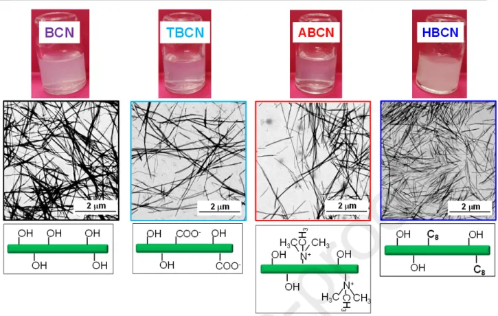
A schematic representation of the various BCN surface modifications and the corresponding scanning transmission electron microscopy (STEM) images are reported in Fig. 1.
Fig. 1. BCN, TBCN, ABCN and HBCN suspensions: visual appearance (top line), STEM images
(middle line) and scheme of various surface modifications (bottom line).
Synthesis of BCN/AgNP hybrids. Both AgNO3 and NaBH4 solutions were freshly prepared just
before the experimental session, and the NaBH4 aqueous solution was placed in ice to minimize its
decomposition. AgNO3 aqueous solution (300 μL, 50 mM) was added to 10 mL of BCN water
suspension and mixed for 1 min at room temperature. The NaBH4 aqueous solution (500 μL, 100 mM)
was then introduced to reduce Ag+ ions to AgNPs, immediately turning the suspension from
translucent to light-yellow. The final hybrid suspension was stirred at room temperature for 24 h, protected by light using aluminum foil to prevent silver oxidation, and then dialyzed against water for 24 h. The procedure produced hybrid suspensions with BCNs, independently of prior surface modifications of the BCN.
H2O2 redox post-treatment. The modification of AgNP characteristics was performed by adding H2O2 to the BCN/AgNP initial hybrid suspension. From now on, the AgNPs obtained after the addition
of H2O2 will be generically indicated as AgNPs_H2O2 and, more specifically, as AgNPrisms when
they reach a triangular shape. Immediately after the reduction by NaBH4, various amounts of H2O2
(i.e., 0, 40, 80, 120, 160, 250 μL) were added to obtain various H2O2/AgNP mass ratios, (i.e., 0,
0.08, 0.17, 0.25, 0.33, 0.52). The H2O2 introduction induces an exothermic reaction associated with the
gas bubbles formation associated to the H2O2 decomposition [21]. The color of the sample gradually
ultra-Characterization. For FTIR measurements, the pure and surface-modified BCN suspensions were
freeze-dried using a LYO GT2 lyophilizer (SRK System Technik, Germany; T = - 90°C; 1.5 10-2
mbar). A quantity of 2 mg of freeze-dried sample was homogenized with 120 mg of KBr and the resulting powder was compressed (5 tons; 5 s) to obtain a circular pellet. The FTIR analysis were performed using a Nicolet iS50 FTIR spectrometer (Thermo Fisher Scientific, USA) and the spectra were determined to be in the 400 – 4000 cm-1 range, with a resolution of 4 cm-1. Each spectrum was
the average of 200 scans collected in absorbance mode and examined using OMNICTM software
(Thermo Fisher Scientific, USA).
The conductivity of BCN, TBCN, ABCN and HBCN aqueous suspensions was automatically measured by a Metrohm 856 Conductivity Module (France). The suspensions were titrated using 0.1 M aqueous NaOH (addition rate of 0.1 mL/30 s) [58]. The data were recorded by TiamoTM Titration
software and the conductivity values were corrected from dilution effects and thus plotted against the added volume of sodium hydroxide solution. The inflection point was graphically determined from the intersection of the least squares regression lines fit and the data points in the distinct regions of the titration curves, evaluating the NaOH volume required for neutralization and, thus, the surface charge density [59,60], ρ (mol/g). Using ρ values, the degree of oxidation [61] (DO) was calculated as:
DO = nCOOH × MAGU mcellulose― nCOOH × MCOOH
where nCOOH represents COOH moles (mol) deduced from the surface charge density, ; MAGU was the molecular weight of an anhydroglucose unit (162 g/mol); mcellulose corresponded to BCN mass and
was the molecular weight of a carboxylic group. MCOOH
The zeta potential values were measured using a ZetaSizer Nano ZS (Malvern, UK). BCN suspensions were diluted at 0.1 g/L and filtered (pore diam. = 5 μm). Five measurements at T = 20°C were performed for each sample. For the case of HBCNs suspended in a H2O/EtOH mixture, the residual
ethanol was removed by dialysis of several milliliters of suspension against water for 2 days. All the suspensions were sonicated just before the measurements.
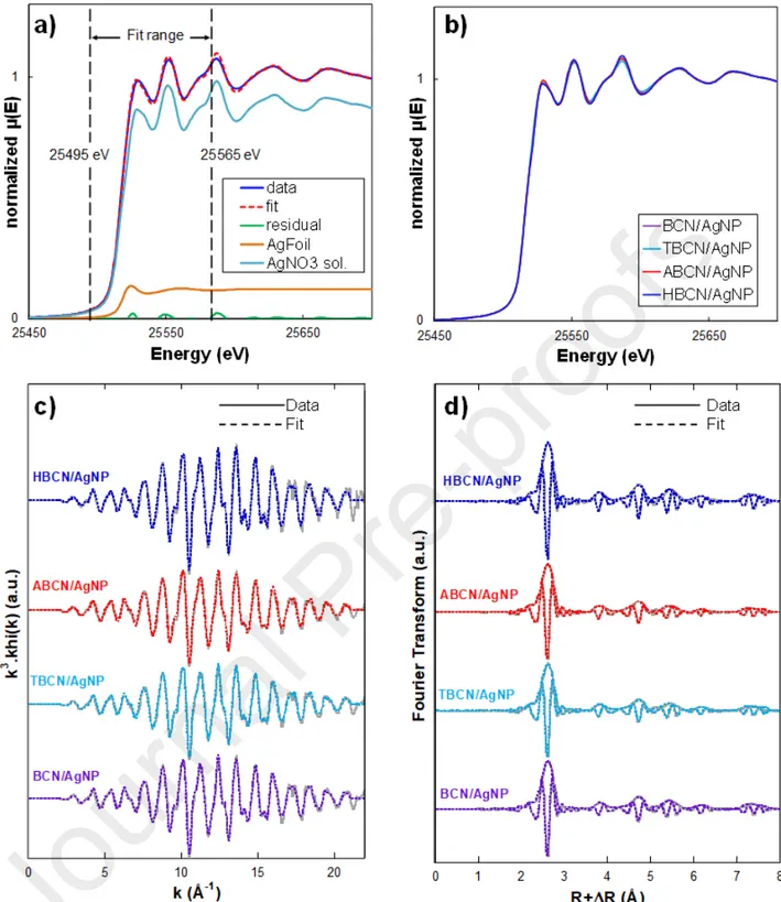
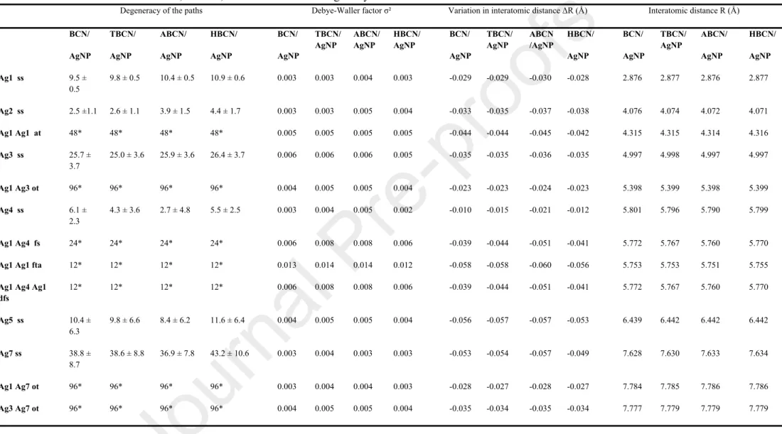
X-ray Absorption Near-Edge Structure (XANES) measurements were performed to investigate the AgNP oxidization state (i.e., metallic silver, Ag0; ionic silver, Ag+) while Extended X-ray Absorption
Fine Structure (EXAFS) data made it possible to shed light on the AgNP bulk atomic structure (e.g., bond length, interatomic distance). Both XANES and EXAFS spectra were simultaneously recorded in transmission mode at the Ag K-edge (25250 to 27750 eV) on the SAMBA beamline at the SOLEIL synchrotron (Saint Aubin, France). The Si (220) monochromator was calibrated to 25515.6 eV at the first inflection point of the Ag foil XANES spectrum. To be analyzed, all the hybrids were freeze-dried and then compressed to obtain a circular pellet of 6 mm where the quantity of AgNP was high enough to reach an absorption edge jump close to 1. These pellets were placed on a sample rod,
quenched in liquid nitrogen and then introduced into the He cryostat (T = 20 K). Silver foil (Agfoil) and AgNO3 aqueous solution with 1 wt% glycerol (AgH2O) were used as standards. One continuous
scan was recorded for each hybrid sample in the 25250 to 27750 eV energy range with a monochromator velocity of 5 eV/s and an integration time of 0.08 s/point. The obtained scans were then normalized and background-subtracted using the Athena software package [62]. The XANES spectra were analyzed by a linear combination fitting (LCF) procedure in the E0 - 20 eV, E0 + 50 eV
energy range, with E0 set to 25514 eV and using Agfoil and AgH2O standards as components. All
component weights were forced to be positive and the relative proportions of the components were forced to add up to 100%. Concerning the EXAFS oscillation, a background subtraction was performed before applying an autobk algorithm (Rbkb = 1, k-weight = 3). The Fourier transform of the k3-weighted EXAFS spectra was then calculated over a k range of 2.5-19.5 Å-1, using a Hanning
apodization window (width of the transition region window parameter = 1). The k3 EXAFS fitting was
performed in the 2.35–7.7 Å distance range with the Artemis interface [62] to IFEFFIT using least-squares refinements. Paths used for fitting standards and samples were obtained from a metallic silver crystallographic model [63] using the FEFF6 algorithm included in the Artemis interface. Only paths with a rank higher than 7% were considered, and the E0 value was set to 25520 eV. The amplitude
reduction factor S0² was determined to be equal to 0.978 by fitting the 1st coordination sphere of the
Agfoil spectrum over a range of 2.30-2.83 Å. This value was used in all the fitting procedures. Degeneracy of the paths, energy shift ΔE0, radial distance shift ΔR, and thermal and static disorder σ²
were fitted for each of the selected paths for a total of 57 independent points and 19 variables. All R-factors were lower than 0.02.
The pellets prepared for XANES-EXAFS measurements were then analyzed by X-ray powder diffraction (XRD). The XRD diffractograms were recorded in 10 min on a Bruker D8 Discover diffractometer (France). Cu-Kα1 radiation (Cu Kα1, 1.5405 Å) produced in a sealed tube at 40 kV and 40 mA was selected and parallelized using a Gobël mirror parallel optics system and collimated to produce a 500-mm beam diameter. The data were collected in the 3°-70° 2 range. XRD θ measurements were also performed for initial BCN suspensions with different surface modifications. The crystallinity index (CrI, %) was calculated as a function of the maximum intensity of the diffraction peak from the crystalline region (I200) at 2 = 22.5° and the minimum intensity from the θ
amorphous region (Iam) at 2 = 18°, according to the Segal equation [64]:θ
CrI =I200― Iam I200
CS = Kλ βcosθ
where is the shape factor (0.9), is the X-ray wavelength (1.54 Å), is the full-width at half-K λ β maximum (FWHM) and is the angle of the diffraction peak of the crystalline phase (Bragg’s angle). θ The FWHM was determined considering the characteristic peak at 2 = 22.5° for initial BCN θ suspensions and the peak at 2 = 38° for the AgNPs in hybrids.θ
The light-visible absorbance of hybrid suspensions was measured in the 300–800 nm range using a Mettler-Toledo UV7 (USA) spectrophotometer equipped with a 10-mm quartz cell. All the samples were water-diluted (1:10) and ultra-pure water was used as a blank reference.
To determine the real content of AgNPs in hybrid suspensions, a volume of 1 mL of sample was digested by 40 mL water/aqua regia mixture (i.e., 30% v aqua regia, HCl/HNO3 : 3/1). The resulting
suspensions were then analyzed by atomic absorption spectroscopy, AAS (ICE 3300 AAS, Thermo Fisher Scientific, USA). A calibration curve was obtained by the measurement of a digested sample of silver standard solution (1000 μg/mL, Chem-Lab NV, Belgium) at different concentrations, from 0.5 to 10 ppm. Two independent measurements were repeated for each sample.
For scanning transmission electron microscope (STEM) observations, the as-synthesized suspensions were water-diluted at 0.5 g/L in BCN content and 10 μL were then deposited onto glow-discharged carbon coated grids (200 meshes, Delta Microscopies, France) for two minutes, removing the excess by touching the edge of the drop with Whatman filter paper. The grids were dried overnight in air and then coated with a platinum layer (thickness = 0.5 nm) by an ion-sputter coater (LEICA EM ACE600, Germany). Brightfield images were recorded using a field emission gun scanning electron microscope (Quattro S, Thermo Fischer Scientific, USA) operating at 10 kV with a STEM detector. For each sample, the acquired images were analyzed by ImageJ software. The AgNP Feret diameter (i.e., the largest distance between two tangents to the contour of the measured particle) was determined considering the largest possible number of AgNPs (from 35 to 100, depending on the sample).
3. Results
Characteristics of surface-modified BCNs. To elucidate the role of functional groups on the surface
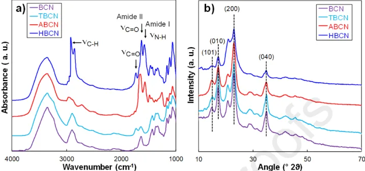
of the BCNs in AgNP nucleation, different surface modifications were performed. Firstly, a suspension of neutral BCNs was obtained from HCl acid hydrolysis. BCNs were then carboxylated via the TEMPO oxidation method (TBCN) obtaining a DO of 0.1 estimated from conductometric data. FTIR spectroscopy assessed the effective surface-modifications. As shown in Fig. 2a, the unmodified BCNs showed well-defined peaks at 1000 cm-1 and 3000-3500 cm-1, typical of–OH and –C–O–C–
groups, respectively [66]. The TBCN spectrum displayed a strong absorption band between 1630 cm-1
(carboxylate) and 1730 cm-1 (acid), which was characteristic of the carboxyl groups in their acidic
form, validating a successful TEMPO oxidation of BCNs [61]. TBCNs were further surface-modified to obtain two types of surface chemistry. Firstly, a cholamine chloride derivative (AETMA) was used
to synthesize aminated cationic BCNs (ABCNs). For this sample, the FTIR spectrum revealed the presence of two new bands at around 1580 cm-1 assigned to the C=O stretching of the amide I band
and to the N–H vibration of the amide II band, respectively [67]. Secondly, an octylamine C8 was
covalently grafted onto the carboxyl group of TBCNs using NHS/EDC, leading to a hydrophobic BCN (HBCN). The good degree of coupling between TBCNs and C8 was proved by the very low surface
charge density measured by conductometric titration (i.e., 0.010 mmol/g). The detection of these same bands in the spectrum of HBCNs and the presence of asymmetrical and symmetrical CH2 stretching
from the C8 alkyl chain at 2850 and 2930 cm-1 indicated the successful octylamine grafting [43].
Nevertheless, a residual peak at 1730 cm-1 was still visible probably due to remaining free COOH
groups . FTIR results were corroborated by the variation of the zeta potential (), implying that a modification of the electrical charge environment of the BCN was induced by the different chemical surface modifications. While pure BCNs were quasi-neutral, TBCNs showed a negative value of -25 mV, correlated to the variation of surface charge density (ρ). On the other hand, a clear increase in to +16 mV was observed for the aminated ABCNs due to the substitution of COO- groups by the N+ of
the AETMA molecules. The slightly negative value of -9 mV was measured for the alkyl hydrophobized HBCNs because of the grafting of octylamine onto the surface carboxyl groups. This low value indicated that HBCNs were highly substituted and no longer electrostatically but, instead, sterically stabilized. These experimental evidences were consistent with a stable suspension of highly-charged colloids for TBCNs and ABCNs with respect to BCNs.
The surface modifications affected the nanocrystal dispersions, as shown from STEM images of dry samples in Fig. 1. Even though the global morphology of the nanocrystals remained unchanged after modification, it could be observed that TBCNs and ABCNs were better dispersed in comparison to quasi-neutral unmodified BCNs and HBCNs. Furthermore, TBCN and ABCN suspensions were less opaque than those of pure BCNs and HBCNs (Fig. 1). Analysis of STEM images indicated an average length of between 850 and 1750 nm and a width of between 20 and 40 nm for all surface-modification treatments, which was in agreement with several studies in the literature [57,68,69]. All the outcomes are summarized in Table 1, indicating that the BCN surface was successfully modified for all of the cases considered.
Fig. 2. Characterization of unmodified BCNs and BCNs with TEMPO oxidation, aminated or
hydrophobic surface modifications: (a) FTIR spectra; (b) XRD patterns.
Table 1. Crystalline and surface charge characteristics of native and modified BCNs Sample Crystallinity, CrI (%)1 Crystallite size, CS (nm) 1 Surface charge density, (mmol/g)𝛒 2 Zeta potential, (mV) BCN 59 6.1 0.006 ± 0.001 -11.2 ± 1.1 TBCN 57 5.8 0.630 ± 0.003 -25.4 ± 0.8 ABCN 62 5.7 0.540 ± 0.002 +15.9 ± 0.7 HBCN 58 6.1 0.010 ± 0.002 -9.3 ± 1.9
- 1 by XRD; 2 by conductometric titration, two measurements were performed for each sample.
The XRD patterns of unmodified BCNs and surface-modified BCNs (Fig. 2b) showed 2 diffraction θ peaks at 14.5°, 16.4°, 22.5° and 34.8°, related to (101), (010), (200) and (040) crystalline planes, respectively, according to the triclinic indexation of Nishiyama et al.[70]. As reported in Table 1, the crystallinity was unchanged around 60% after correction of the amorphous for all the samples, and the chemical treatment did not affect the size of crystallites, which was 5.9 ± 0.2 nm for all the samples, in agreement with the value found by Vasconcelos et al. [71]. These results are consistent with the fact that TEMPO oxidation and other post treatments do not affect the crystalline part of the samples [72,73].
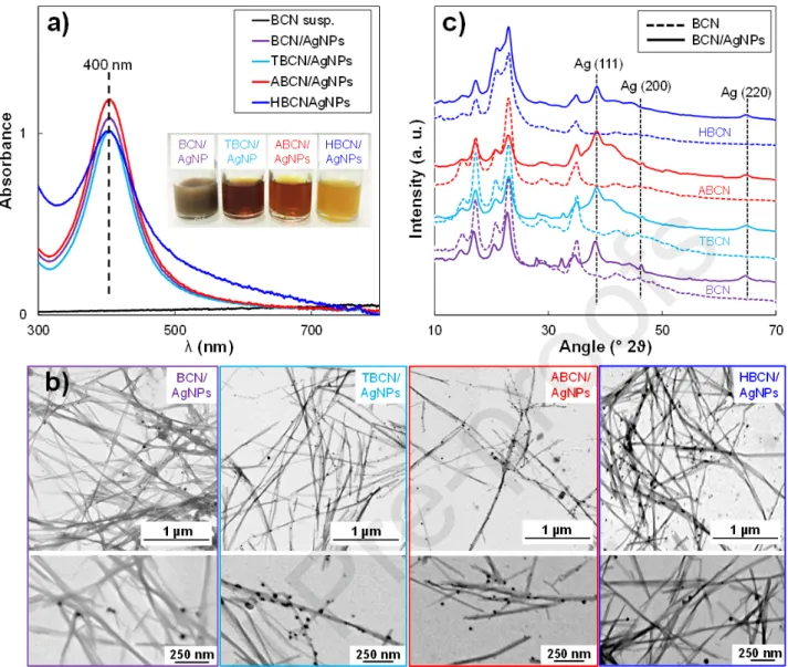
AgNP nucleation on BCNs with various surface modifications. The nucleation of AgNP was
investigated on the four BCN types of interest (i.e., native BCNs, TBCNs, ABCNs and HBCNs). Firstly, AgNO3 aqueous solution was added to the BCN suspension and NaBH4 aqueous solution was
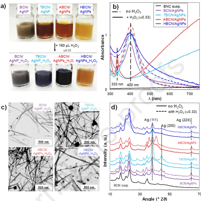
translucent aqueous suspension turned light or dark yellow (inset, Fig. 3a), giving different shades of color, probably because of the initial dispersion state of the type of BCN used. However, such a color variation was not reflected in the UV-Vis spectra (Fig. 3a) since the effective AgNP content was very similar in all the hybrids (i.e., 7.9 wt% AgNP in BCN/AgNP; 6.8 wt% AgNP in TBCN/AgNP; 8.5 wt% AgNP in ABCN/AgNP; 8.6 wt% AgNP in HBCN/AgNP) with a same dominant in-plane absorption peak at λmax~ 400 nm. It confirmed the synthesis of well-dispersed AgNPs derived from the coalescence of monomeric Ag particles obtained by a reduction to a zero-valence Ag atom [20]. Even if the surface modification did not seem to affect the intensity of the spectra (i.e., similar AgNP content for all the hybrids), the full width of the main peak and the background intensity increased for HBCN/AgNP. This type of behavior is attributed to the synthesis of HBCN/AgNP in a H2O/EtOH
mixture since the AgNO3 is not well-soluble in ethanol and could affect AgNP nucleation, leading to a
larger distribution of AgNP sizes or heterogeneities. However, the use of EtOH was essential since it allowed the C8 dissolution and helped hydrophobic nanocrystal dispersion after the synthesis, as was
the presence of H2O, which ensured the correct dissolution of silver precursor and, consequently,
AgNP formation.
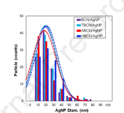
STEM images of dry BCN/AgNP, TBCN/AgNP, ABCN/AgNP and HBCN/AgNP hybrids are shown in Fig. 3b. The analysis of these images confirmed that all the AgNPs had the same particle size, which was thus not affected by the different surface modifications (i.e., BCN/AgNP = 17.1 ± 12 nm; TBCN/AgNP = 17.5 ± 12 nm; ABCN/AgNP = 16.1 ± 13 nm; HBCN/AgNP = 18.0 ± 12 nm). Complete size distributions are reported in Fig. S1. The nucleation growing method resulted in well-grafted AgNPs on the cellulose surface for the neutral and modified CNCs. In contrast, AgNPs synthesized in the same conditions without BCN rapidly aggregated. This proved that BCNs form a perfect substrate for the nucleation point to obtain well-dispersed quite-monodispersed AgNPs without the need for any other capping agents or stabilizers [74].
Nevertheless, these results suggested that AgNP nucleation on the cellulose nanocrystal surface non-specifically occurred on hydroxyl groups and/or on the additional negative surface charges. The aim was to determine if these surface charges represent an additional nucleation point that interacts with Ag+ ions or if they just acted as promoters of nanocrystal dispersion, facilitating the accessibility to the
hydroxyl surface groups. The TEMPO carboxylated TBCNs were then compared to aminated ABCNs. In this case, the functionalization by the positive surface charges preserved the electrostatic repulsion and, consequently, the good dispersion. At the same time, these positive charges could not interact with Ag+ ions and thus did not represent a possible nucleation point for AgNPs. The STEM image of
the ABCN/AgNP hybrid (Fig. 3b) showed that AgNPs were well-nucleated on both surfaces, indicating that the –OH surface groups represented the effective AgNP nucleation point. The OH groups and Ag+ ions are complexed throught ion-dipole interactions [22] and the extensive number of
To our knowledge, such a result represents the first experimental proof that hydroxyl surface groups on cellulosic surfaces are the real nucleation points for metallic nanoparticles and that additional negative surface charges just improve the dispersion state, thereby increasing the accessibility to the nucleation sites. This result was corroborated by the fact that for the HBCN/AgNP hybrid, most of the AgNPs were well-grafted onto the HBCN surface as well. In this case, the only real possible nucleation point was represented by –OH surface groups since most of the carboxylate groups were removed by grafting of C8 molecules. The presence of several AgNPs not anchored at the HBCN
surface was visible in STEM images and could be linked to the suboptimal AgNO3 dissolution due to
the presence of EtOH in the suspending medium.
Concerning the structural characterization, all the XRD patterns of BCN/AgNPs (Fig. 3c) showed characteristic peaks of crystalline silver, especially at 37.9° which is usually attributed to the (111) lattice plane of face-centered cubic (fcc) silver (JCPDS Card No. 89-3722). Other characteristic silver diffraction peaks at 46.2° and 64.4° related to other lattice planes (i.e., (200) and (220) crystalline planes) were detected with a weaker intensity. The presence of fcc crystal facets confirmed the isotropic nature of the crystals [20]. For all the samples, the same average AgNP diameter of 3 nm was estimated from XRD patterns. The nucleation mechanism of AgNPs could affect their growth on the nanocrystal surface and, thus, their final characteristics. Therefore, the oxidation state of AgNPs grafted onto BCN with different surface modifications (i.e., Ag0 and Ag+ contents) was determined by
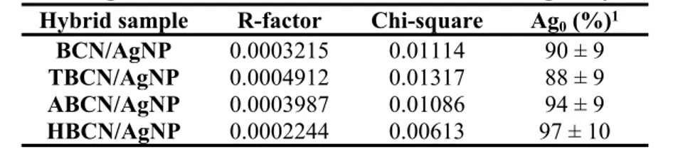
a linear combination fitting (LCF) of XANES spectra of the hybrids (Fig. S2a and Fig. S2b). For each sample, the R-factor and the Chi-square values of the fits are reported in Table S1. A quantity of 92 ± 9% of Ag0 was found for all the hybrids, showing the efficiency of the AgNP reduction and nucleation,
independently of the surface treatment of the initial BCNs. All Fourier transform spectra of BCN/AgNP (Fig. S2c and Fig. S2d) were fitted with the crystallographic structure of metallic silver with an R-factor systematically lower than 0.015. The shifts in R values (i.e., interatomic distance) obtained from the fits were systematically negligible (< 0.06 Å-1; all of the values are presented in
Table S2), showing that the interatomic distances in BCN/AgNP case did not significantly change in comparison to the metallic silver distances and that the space group of BCN/AgNP sample still corresponded to the fcc silver structure, as suggested by XRD. It follows that the initial and final crystal structural organization of AgNPs were not affected by the various surface-modification treatments. These results showed the NaBH4 reduction with –OH surface groups as nucleation points
represented an efficient nucleation mechanism where the speciation and the crystalline structure of AgNPs were not affected by the surface modifications performed on cellulose nanocrystals.
Fig. 3. Characterizations of BCN-, TBCN-, ABCN-, HBCN-/AgNP hybrid suspensions: (a) UV-Vis
spectra; inset: sample pictures; (b) STEM images; (c) XRD patterns.
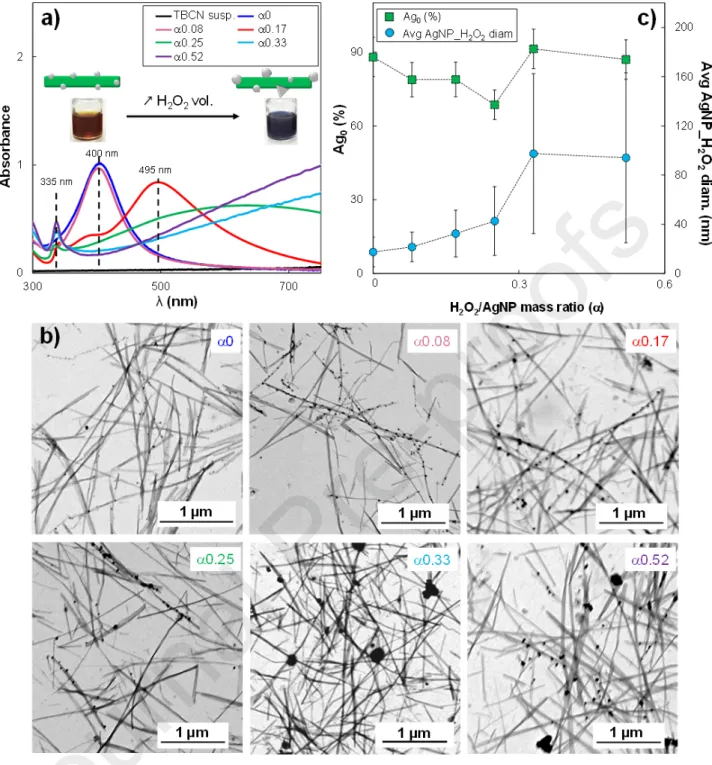
AgNP redox post-treatment by H2O2. H2O2 was defined as an efficient etching agent producing
shape and structural modifications of AgNP into larger and more robust AgNPs (e.g., AgNPrisms) isolated or even attached to surface-carboxylated cellulose nanofibrils [20]. In this case, we proposed a more detailed study on the effect of the H2O2 redox post-treatment on properties of AgNPs (i.e.,
AgNP_H2O2) in the different BCN/AgNP hybrids. The outcomes reported in this section will support
the understanding of the impact of the H2O2 action for BCN/AgNP where BCN was surface-modified.
Even if all the prepared samples were able to undergo the same H2O2-induced structural change, the
TBCN/AgNP hybrid system was chosen as a case study since the TBCNs led to good dispersion. Such a TBCN/AgNP hybrid suspension at constant initial AgNP content (i.e., 7 wt%) was mixed with different amounts of H2O2 (40 – 250 μL), thus obtaining a variation of the H2O2/AgNP mass ratio, ,
4a). The color change was associated with a modification of the UV-Vis spectra of the H2O2
-containing suspensions with respect to the reference one (i.e., 0 μL H2O2), as shown in Fig. 4a. The
addition of H2O2 induced aredshift of the in-plane dipole surface plasmon resonance peak at 400 nm.
This was already visible at equal to 0.17, since the absorption peak became broader and shifted to 495 nm, showing the presence of two shoulders at 345 nm and 385 nm related to an early modification of primary AgNPs. At = 0.25, the in-plane plasmon peak was further shifted to 638 nm and finally moved out of the measurement window for higher values. In this case, a lower intensity peak was detected at λmax = 335 nm that is usually associated with an out-of-plane quadrupole resonance peak, thus representing a good indicator for general prismatic architectures since it strongly depends on aspect ratio [48]. Such a peak became sharper for values of 0.33 and 0.52 (i.e., 160 μL and 250 μL of H2O2, respectively). The presence of a low-intensity peak at 335 nm, at the same time as the shift of
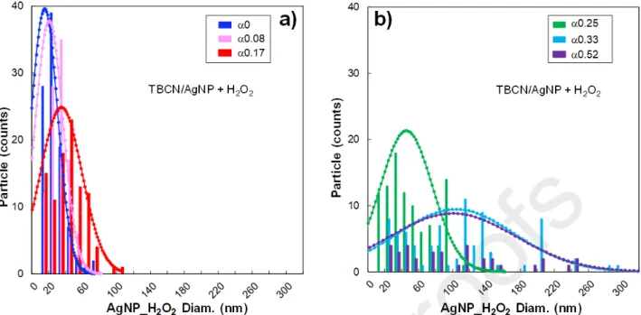
the in-plane resonance peak out of the measurement window, and the absence of the primary AgNP peak is proof that an AgNP size-shape variation is induced, as already observed in literature [20]. To better describe the impact of the H2O2 redox post-treatment on the AgNP morphology STEM
acquisition were performed (Fig. 4b). The analysis of STEM images indicated that AgNPs initially had an average diameter of 17.5 ± 12 nm that regularly increased to reach 94.1 ± 69 nm for equal to 0.52, where several AgNPs_H2O2 with quite irregular edges and a sometimes vaguely triangular shape
could be observed. These results agreed with the experimental evidence reported in a recent study of our group [76], in which we investigated the effect of the H2O2 redox treatment in hybrid suspensions
where primary 10-nm AgNPs are nucleated on wood cellulose NCs, proving the efficient size-shape transition from 10-nm spherical AgNPs into 300 nm AgNPrisms. In this work, we proposed a H2O2
redox mechanism where H2O2 induced the oxidative dissolution of primary AgNPs, generating Ag+
ions. When equal to or greater than 0.20, the H2O2 oxidation interest most of the AgNPs, except the
contact location where AgNPs are effectively grafted onto the NC surface which could actually work as nucleation sites for the formation of newly formed AgNPs with a triangular shape. Finally, all the images clearly showed that AgNPs_H2O2, even the bigger ones, were still attached to the TBCN
surface, exactly as for the reference sample before the H2O2 post-treatment, showing the importance of
cellulose as a support for AgNPs_H2O2 nucleation, growth and stabilization. The formation of
non-perfectly prismatic silver objects and the increase in the average diameter associated with an increase in the polydispersity in size (Fig. S3a and Fig. S3b, Table S3) could be linked to the absence of any additional stabilizer, which implied that the structural change was heavily affected by the aggregation of newly-formed AgNPs_H2O2 [46].
To establish how the H2O2 post-treatment affected the oxidation state of AgNPs, XANES spectra were
recorded and then fitted using the LCF procedure (Fig. S4a and Table S4). Data showed that the amount of Ag0 in AgNPs_H2O2 slightly decreased with respect to the reference case, from 88% before
variation (corresponding to from 0.08 to 0.25), while remaining equal to 91% and 87% for values of 0.33 and 0.52, respectively (Table S4). The formation of AgNPs_H2O2 mostly composed of Ag0
silver was also corroborated by the presence in the XRD patterns of characteristic (111), (200) and (220) Ag0 peaks, confirming the presence of a lattice plane of a face-centered cubic structure (Fig.
S4b). It could be observed that the H2O2 post-treatment did not induce a structural modification of
AgNPs_H2O2 with respect to the primary AgNPs before the H2O2 treatment and that a H2O2/AgNP
mass ratio equal to 0.33 and 0.52 determined an increase in the intensity of the characteristic fcc Ag (111) peak, which corresponded to the highest recorded values of Ag0. Such a result suggested that a
parameter at least equal to 0.33 represented the optimum value necessary to reach an effective size-shape variation that maintains the conversion of the initial Ag+ to Ag
0 ratio. Finally, the EXAFS
Fourier transform spectra (Fig. S4c and Fig. S4d) of the AgNP_H2O2 in hybrids were fitted with the
crystallographic structure of metallic silver, (R-factor systematically lower than 0.020), presenting systematically negligible shifts in R space (< 0.06 Å-1; all values are in Table S5). This result
confirmed that the interatomic distances in AgNP_H2O2 did not significantly change in comparison to
the metallic silver distances and that the space group of the samples still corresponded to the fcc silver structure, as observed by XRD. The final crystal structural organization was not affected by the H2O2
redox post-reaction nor by the particle size variation, as observed by Ma et al. [77] Conversely, the AgNP diameter measured by XRD was about 10 nm, which was consistently smaller than the 40-80 nm measured by STEM. Such a difference could be explained by the fact that XRD makes it possible to evaluate the size of the coherent diffraction domains in AgNPs, i.e., perfect repetition in single-crystal particles, twinned or imperfect particles that have more than a single diffraction domain [77]. The characteristics of all the investigated samples are reported in Table 2.
All these results, coupled with the experimental proof that -OH groups on the CNC surface are the AgNP nucleation points, allowed us to conclude that our approach to tune the morphology and the oxidation state of the AgNP grafted onto cellulosic support may be extended to all the hybrid systems where metallic NPs are anchored on a polysaccharide substrate (e.g., cellulose fibers, films or chitin NCs).
Fig. 4. (a) UV-Vis spectra and (b) STEM images of TBCN/AgNP hybrid suspension mixed with
different volumes of H2O2 and, thus, at various values. (c) Ag0 content of AgNP_H2O2 by XANES
fitting, and average AgNP_H2O2 diameter determined by STEM as a function of the parameter in
TBCN/AgNP_H2O2 hybrids. For Ag0 value, standard error as considered as the 10% of the measured
value.
Table 2. Summary of characteristics of BCN/AgNP hybrids with and without H2O2 redox
post-treatment.
Sample H2O2/AgNP mass ratio () Avg diam. (nm)1 Ag0 (%)2
TBCN/AgNP 0 17.5 ± 12 88 ± 9 ABCN/AgNP 0 16.1 ± 13 94 ± 9 HBCN/AgNP 0 18.0 ± 12 97 ± 10 0.08 21.6 ± 12 79 ± 8 0.17 32.5 ± 19 79 ± 8 0.25 42.7 ± 28 69 ± 7 0.33 97.5 ± 65 91 ± 9 TBCN/AgNP_H2O2 0.52 94.1 ± 69 97 ± 10 BCN/AgNP_H2O2 0.33 32.5 ± 19 93 ± 9 ABCN/AgNP_H2O2 0.33 95.4 ± 87 97 ± 10 HBCN/AgNP_H2O2 0.33 29.4 ± 22 97 ± 10
1 by STEM; 2 by XANES, the standard error as 10% of the measured value.
Impact of BCN surface modification on H2O2 redox post-treatment. Hybrids prepared with various BCN types were submitted to the H2O2 redox post-treatment, keeping the H2O2/AgNP ratio, equal
to 0.33, the value at which an effective size-shape modification of the initial AgNPs was found in the case of TBCN/AgNP (Table 2). In the case of aminated surfaces, the color variation of the ABCN/AgNP_H2O2 from yellow to blue was observed after the addition of H2O2, as observed for the
TBCN/AgNP_H2O2 (Fig. 5a). This suspension showed an in-plane absorption peak shifted to higher
(i.e., out of the measurement window), and a sharp out-of-plane quadrupole resonance peak was λmax
visible at 333 nm (Fig. 5b). This result agreed with the transformation of the primary AgNPs of 16.1 ± 13 nm into bigger AgNPs_H2O2. of 95.4 ± 87 nm, as confirmed by STEM (Fig. 5c). On the other hand,
the addition of H2O2 to the BCN/AgNPturned the suspension a darker gray color, and the particle size
slightly changed with respect to the reference case (i.e., 32.5 ± 19 nm). Moreover, the λmax in the UV-Vis spectra remained at 402 nm and a poorly-defined low-intensity broad peak appeared at 340 nm. These results indicated an uncomplete transformation of the AgNPs, suggesting that the BCN surface modification affected the H2O2 transformation of primary AgNPs since the BCN surface modification
influenced the BCN dispersion rather than its chemical structure.
When the surface charges provided a good repulsion between nanocrystals (the cases of TBCN and ABCN), the addition of H2O2 made it possible to obtain the formation of AgNPs_H2O2 of about 100
nm, whereas the size of AgNPs_H2O2 for the almost neutral surface charge in the BCN case remained
constant (i.e., about 20 nm). It appeared that well-dispersed nanocrystals facilitate the accessibility to the surface groups, thereby promoting the H2O2 redox action.
Furthermore, the UV-Vis spectrum of the HBCN/AgNP_H2O2 suspension did not change with respect
to the reference HBCN/AgNP. In this case, not only the absence of repulsion prevented good nanocrystal dispersion, but the suspending medium containing EtOH could promote aggregation and affect the efficiency of the H2O2 redox reaction as well. To check the influence of EtOH on the H2O2
treatment, we compared aqueous TBCN/AgNPs suspension (highly charged nanocrystals to ensure dispersion) to the same TBCN/AgNPs diluted in a H2O/EtOH (60/40 v/v) mixture, like in the case of
the HBCN/AgNPs. In pure water, the addition of H2O2 induced a color variation from yellow to blue,
whereas the hybrid prepared in the H2O/EtOH mixture just turned a dark yellow (Fig. S5). This result
confirmed that the presence of ethanol limited the formation of AgNPs_H2O2 that could be linked to a
weak oxidation of AgNPs to Ag+ and the subsequent reduction in a non-aqueous medium. Size
distributions of AgNPs_H2O2 in BCN hybrids are reported in Fig. S6 and the average diameters are
summarized in Table S6.
Interestingly, all the reformed AgNPs_H2O2 appeared to be nucleated on nanocrystal surfaces,
irrespective of the applied surface modification. This means that the BCN still serves as a substrate for AgNP_H2O2 formation, ensuring adequate AgNP dispersion. We hypothesized that the H2O2 did not
completely oxidize the AgNPs, leaving hooks available as a large amount of re-nucleation points for AgNP_H2O2 nanoparticles in addition to the –OH surface groups, as reported in another work of our
Fig. 5. (a) Color variation of hybrid suspensions; (b) UV-Vis spectra; (c) STEM images; (d) XRD
patterns of BCN-, TBCN-, ABCN-, HBCN-/AgNP_H2O2 hybrid suspensions with the addition of 160
μL of H2O2 ( = 0.33).
Moreover, the analysis of XANES spectra (Fig. S7a, Table S7) showed that the addition of H2O2 did
not affect the Ag0 content (i.e., 95 ± 10% compared to 92 ± 9% of the reference cases), indicating that
the oxidation state did not change. Finally, the XRD patterns of hybrid suspensions without ( = 0) and with the addition of H2O2 ( = 0.33), Fig. 5d., indicating the presence of the Ag (111), Ag (200)
and Ag (220) peaks, characteristic of the fcc silver model. This confirmed the formation of metallic AgNPs_H2O2 well-grafted onto the BCN substrate, irrespective of the surface modification. Moreover,
S8b, data in Table S8). These studies confirmed that all native and modified BCN/AgNP samples were composed quasi-exclusively of Ag0 with similar crystallographic organization.
4. Conclusion
In this study, we investigated the interactions involving well-dispersed AgNPs grafted on a bio-based substrate, focusing on the role of polysaccharide surface chemistry. Working with bacterial cellulose nanocrystals (BCNs) as model substrate, we experimentally showed that the hydroxyl groups serve as nucleation points for AgNPs through ion-dipole interaction and that the surface charges only promoted nanocrystal dispersion, improving accessibility to the OH groups. To our knowledge, this is the first experimental identification of the effective nucleation point for metallic nanoparticles on a solid cellulose surface. This was investigated modifying native quasi-neutral BCNs dispersed in aqueous suspension into negatively or positively charged, and hydrophobic BCNs then used to prepare hybrid nanomaterials (i.e., BCN/AgNP, TBCN/AgNP, ABCN/AgNP, HBCN/AgNP) where highly controlled AgNPs are anchored on the BCN surface. This work proved that no surface modification of cellulose (e.g., TEMPO oxidation [32]) is necessary to efficiently nucleate and anchor AgNPs on their surface as long as the reduction step is carried out. Furthermore, we prove that the grafting of hydrophobic molecules on BCNs does not prevent the nucleation and the growth of AgNPs, thus opening the way to the formulation of bifunctional nanoparticles in non-aqueous media.
The investigated hybrid suspensions were subjected to an H2O2 redox post-treatment. An optimal
H2O2/AgNP mass ratio ( = 0.33) was defined at which a size-shape transition of AgNPs, from 10-nm
spherical NPs to 100-nm NPs with a triangular shape, while maintaining the initial fcc crystal structure and increasing the Ag0 content in AgNPs. Here, we showed that the dispersion of the BCNs improved
the efficiency of the H2O2 post-treatment, with an increase in AgNP size detected only in presence of
repulsive surface charges (i.e., TBCNs and ABCNs)
As a result, this highly controlled synthesis of AgNPs well-grafted onto the cellulose NC surface can promote the development and design of new bio-based hybrid materials, such as sensors and biological imaging, catalysts, conductive material. Furthermore, a controlled release of Ag+ ions from
well-characterized AgNPs would allow producing bio-based nanomaterials for biocidal properties that can be used for several applications, such as food packaging, paints or surface treatment.
CRediT authorship contribution statement
D. Musino: Conceptualization, methodology, investigation, formal analysis, visualization, data
curation, writing – original draft. C. Rivard: Formal analysis, investigation, writing – review and editing. G. Landrot: Formal analysis, investigation, writing – review and editing. B. Novales: Investigation, writing – review and editing. T. Rabilloud: Supervision, investigation, writing – review and editing. I.Capron: Conceptualization, investigation, supervision, project administration, writing - review & editing.
Declaration of competing interest
The authors declare that they have no known competing financial interests or personal relationships that could have appeared to influence the work reported in this paper;
Acknowledgments
We acknowledge SOLEIL for providing synchrotron radiation facilities on the SAMBA beamline. We are grateful to S. Durand (BIA-Nantes) for her support with FTIR measurements, F. X. Lefevre and N. Guichard (Université de Nantes) for their support with AAS experiments, B. Pontoire (BIA-Nantes) for performing XRD acquisitions, and S. Haouache (BIA-Nantes) for her assistance during SAMBA beam time. This work is a contribution to the Labex SERENADE (n° ANR-11-LABX-0064), funded by the "Investissement d'Avenir" French government program of the French National Research Agency (ANR) through the A*MIDEX project (n° ANR-11-IDEX-0001-02).
Appendix A. Supplementary data
Supplementary data to this article can be found online at: 10.1016/x0xx00000x
References
[1] E. Board, L.T.E. Long, T.B. Voit, Polyscaccharides II, 2006. https://doi.org/10.1007/978-3-540-85103-5.
[2] S. Iwamoto, A.N. Nakagaito, H. Yano, M. Nogi, Optically transparent composites reinforced with plant fiber-based nanofibers, Appl. Phys. A Mater. Sci. Process. 81 (2005) 1109–1112. https://doi.org/10.1007/s00339-005-3316-z.
[3] I. Siró, D. Plackett, Microfibrillated cellulose and new nanocomposite materials: A review, Cellulose. 17 (2010) 459–494. https://doi.org/10.1007/s10570-010-9405-y.
[4] P. Ross, R. Mayer, M. Benziman, Cellulose biosynthesis and function in bacteria., Microbiol. Rev. 55 (1991) 35–58.
http://www.ncbi.nlm.nih.gov/pubmed/2030672%0Ahttp://www.pubmedcentral.nih.gov/articler ender.fcgi?artid=PMC372800.
[5] W. Czaja, A. Krystynowicz, S. Bielecki, R.M. Brown, Microbial cellulose - The natural power to heal wounds, Biomaterials. 27 (2006) 145–151.
https://doi.org/10.1016/j.biomaterials.2005.07.035.
[6] Y. Hu, J.M. Catchmark, Y. Zhu, N. Abidi, X. Zhou, J. Wang, N. Liang, Engineering of porous bacterial cellulose toward human fibroblasts ingrowth for tissue engineering, J. Mater. Res. 29 (2014) 2682–2693. https://doi.org/10.1557/jmr.2014.315.
[8] P. Boonme, Amnuaikit, Chusuit, Raknam, Effects of a cellulose mask synthesized by a bacterium on facial skin characteristics and user satisfaction, Med. Devices Evid. Res. (2011) 77. https://doi.org/10.2147/mder.s20935.
[9] K.C. Cheng, J.M. Catchmark, A. Demirci, Effects of CMC addition on bacterial cellulose production in a biofilm reactor and its paper sheets analysis, Biomacromolecules. 12 (2011) 730–736. https://doi.org/10.1021/bm101363t.
[10] S. Eyley, W. Thielemans, Surface modification of cellulose nanocrystals, Nanoscale. 6 (2014) 7764–7779. https://doi.org/10.1039/c4nr01756k.
[11] D. Klemm, F. Kramer, S. Moritz, T. Lindström, M. Ankerfors, D. Gray, A. Dorris,
Nanocelluloses: A new family of nature-based materials, Angew. Chemie - Int. Ed. 50 (2011) 5438–5466. https://doi.org/10.1002/anie.201001273.
[12] J. Ramyadevi, K. Jeyasubramanian, A. Marikani, G. Rajakumar, A.A. Rahuman, Synthesis and antimicrobial activity of copper nanoparticles, Mater. Lett. 71 (2012) 114–116.
https://doi.org/10.1016/j.matlet.2011.12.055.
[13] V.K. Sharma, R.A. Yngard, Y. Lin, Silver nanoparticles: green synthesis and their antimicrobial activities., Adv. Colloid Interface Sci. 145 (2009) 83–96.
https://doi.org/10.1016/j.cis.2008.09.002.
[14] S. Gunalan, R. Sivaraj, V. Rajendran, Green synthesized ZnO nanoparticles against bacterial and fungal pathogens, Prog. Nat. Sci. Mater. Int. 22 (2012) 693–700.
https://doi.org/10.1016/j.pnsc.2012.11.015.
[15] X. Li, S.M. Robinson, A. Gupta, K. Saha, Z. Jiang, D.F. Moyano, A. Sahar, M.A. Riley, V.M. Rotello, Functional gold nanoparticles as potent antimicrobial agents against multi-drug-resistant bacteria, ACS Nano. 8 (2014) 10682–10686. https://doi.org/10.1021/nn5042625. [16] R.J.B. Pinto, P.A.A.P. Marques, C.P. Neto, T. Trindade, S. Daina, P. Sadocco, Antibacterial
activity of nanocomposites of silver and bacterial or vegetable cellulosic fibers, Acta Biomater. 5 (2009) 2279–2289. https://doi.org/10.1016/j.actbio.2009.02.003.
[17] C.H. Bae, S.H. Nam, S.M. Park, Formation of silver nanoparticles by laser ablation of a silver target in NaCl solution, Appl. Surf. Sci. 197–198 (2002) 628–634.
https://doi.org/10.1016/S0169-4332(02)00430-0.
[18] U. Nickel, A.Z. Castell, K. Pöppl, S. Schneider, Silver colloid produced by reduction with hydrazine as support for highly sensitive surface-enhanced Raman spectroscopy, Langmuir. 16 (2000) 9087–9091. https://doi.org/10.1021/la000536y.
[19] G. Yang, J. Xie, F. Hong, Z. Cao, X. Yang, Antimicrobial activity of silver nanoparticle impregnated bacterial cellulose membrane: Effect of fermentation carbon sources of bacterial cellulose, Carbohydr. Polym. 87 (2012) 839–845.
https://doi.org/10.1016/j.carbpol.2011.08.079.
enhanced raman scattering, Biomacromolecules. 15 (2014) 3608–3616. https://doi.org/10.1021/bm5011799.
[21] M. Tsuji, S. Gomi, Y. Maeda, M. Matsunaga, S. Hikino, K. Uto, T. Tsuji, H. Kawazumi, Rapid transformation from spherical nanoparticles, nanorods, cubes, or bipyramids to triangular prisms of silver with PVP, citrate, and H 2O 2, Langmuir. 28 (2012) 8845–8861.
https://doi.org/10.1021/la3001027.
[22] A.R. Lokanathan, K.M.A. Uddin, O.J. Rojas, J. Laine, Cellulose nanocrystal-mediated synthesis of silver nanoparticles: Role of sulfate groups in nucleation phenomena, Biomacromolecules. 15 (2014) 373–379. https://doi.org/10.1021/bm401613h.
[23] D.L. Van Hyning, C.F. Zukoski, Formation Mechanisms and Aggregation Behavior of Borohydride Reduced Silver Particles, Langmuir. 14 (2002) 7034–7046.
https://doi.org/10.1021/la980325h.
[24] G. Ling, J. He, L. Huang, Size control of silver nanoparticles deposited on silica dielectric spheres by electroless plating technique, J. Mater. Sci. 39 (2004) 2955–2957.
https://doi.org/10.1023/b:jmsc.0000021490.52788.62.
[25] Y. Qin, X. Ji, J. Jing, H. Liu, H. Wu, W. Yang, Size control over spherical silver nanoparticles by ascorbic acid reduction, Colloids Surfaces A Physicochem. Eng. Asp. 372 (2010) 172–176. https://doi.org/10.1016/j.colsurfa.2010.10.013.
[26] Y. Sun, B. Mayers, Y. Xia, Transformation of silver nanospheres into nanobelts and triangular nanoplates through a thermal process, Nano Lett. 3 (2003) 675–679.
https://doi.org/10.1021/nl034140t.
[27] S. Padalkar, J.R. Capadona, S.J. Rowan, C. Weder, Y.H. Won, L.A. Stanciu, R.J. Moon, Natural biopolymers: Novel templates for the synthesis of nanostructures, Langmuir. 26 (2010) 8497–8502. https://doi.org/10.1021/la904439p.
[28] K. Patel, B. Bharatiya, T. Mukherjee, T. Soni, A. Shukla, B.N. Suhagia, Role of stabilizing agents in the formation of stable silver nanoparticles in aqueous solution: Characterization and stability study, J. Dispers. Sci. Technol. 38 (2017) 626–631.
https://doi.org/10.1080/01932691.2016.1185374.
[29] M. Kaushik, A. Moores, Review: Nanocelluloses as versatile supports for metal nanoparticles and their applications in catalysis, Green Chem. 18 (2016) 622–637.
https://doi.org/10.1039/c5gc02500a.
[30] R. Xiong, C. Lu, W. Zhang, Z. Zhou, X. Zhang, Facile synthesis of tunable silver
nanostructures for antibacterial application using cellulose nanocrystals, Carbohydr. Polym. 95 (2013) 214–219. https://doi.org/10.1016/j.carbpol.2013.02.077.
[32] S. Ifuku, M. Tsuji, M. Morimoto, H. Saimoto, H. Yano, Synthesis of silver nanoparticles templated by TEMPO-mediated oxidized bacterial cellulose nanofibers, Biomacromolecules. 10 (2009) 2714–2717. https://doi.org/10.1021/bm9006979.
[33] L. Johnson, W. Thielemans, D.A. Walsh, Nanocomposite oxygen reduction electrocatalysts formed using bioderived reducing agents, J. Mater. Chem. 20 (2010) 1737–1743.
https://doi.org/10.1039/b922423h.
[34] M. Hasani, E.D. Cranston, G. Westman, D.G. Gray, Cationic surface functionalization of cellulose nanocrystals, Soft Matter. 4 (2008) 2238–2244. https://doi.org/10.1039/b806789a. [35] H. Rosilo, J.R. McKee, E. Kontturi, T. Koho, V.P. Hytönen, O. Ikkala, M.A. Kostiainen,
Cationic polymer brush-modified cellulose nanocrystals for high-affinity virus binding, Nanoscale. 6 (2014) 11871–11881. https://doi.org/10.1039/c4nr03584d.
[36] A. Kaboorani, B. Riedl, Surface modification of cellulose nanocrystals (CNC) by a cationic surfactant, Ind. Crops Prod. 65 (2015) 45–55. https://doi.org/10.1016/j.indcrop.2014.11.027. [37] S. Lombardo, W. Thielemans, Thermodynamics of the interactions of positively charged
cellulose nanocrystals with molecules bearing different amounts of carboxylate anions, Phys. Chem. Chem. Phys. 20 (2018) 17637–17647. https://doi.org/10.1039/c8cp01532e.
[38] L. Jasmani, S. Eyley, R. Wallbridge, W. Thielemans, A facile one-pot route to cationic
cellulose nanocrystals, Nanoscale. 5 (2013) 10207–10211. https://doi.org/10.1039/c3nr03456a. [39] M. Shateri Khalil-Abad, M.E. Yazdanshenas, M.R. Nateghi, Effect of cationization on
adsorption of silver nanoparticles on cotton surfaces and its antibacterial activity, Cellulose. 16 (2009) 1147–1157. https://doi.org/10.1007/s10570-009-9351-8.
[40] B.H. Dong, J.P. Hinestroza, Metal nanoparticles on natural cellulose fibers: Electrostatic assembly and in situ synthesis, ACS Appl. Mater. Interfaces. 1 (2009) 797–803.
https://doi.org/10.1021/am800225j.
[41] J. Araki, M. Wada, S. Kuga, Steric stabilization of a cellulose microcrystal suspension by poly(ethylene glycol) grafting, Langmuir. 17 (2001) 21–27. https://doi.org/10.1021/la001070m. [42] Y. Zhang, V. Karimkhani, B.T. Makowski, G. Samaranayake, S.J. Rowan, Nanoemulsions and
Nanolatexes Stabilized by Hydrophobically Functionalized Cellulose Nanocrystals, Macromolecules. 50 (2017) 6032–6042. https://doi.org/10.1021/acs.macromol.7b00982. [43] Z. Hu, R.M. Berry, R. Pelton, E.D. Cranston, One-Pot Water-Based Hydrophobic Surface
Modification of Cellulose Nanocrystals Using Plant Polyphenols, ACS Sustain. Chem. Eng. 5 (2017) 5018–5026. https://doi.org/10.1021/acssuschemeng.7b00415.
[44] A.G. Cunha, J.B. Mougel, B. Cathala, L.A. Berglund, I. Capron, Preparation of double pickering emulsions stabilized by chemically tailored nanocelluloses, Langmuir. 30 (2014) 9327–9335. https://doi.org/10.1021/la5017577.
[45] J.E. Millstone, S.J. Hurst, G.S. Métraux, J.I. Cutler, C.A. Mirkin, Colloidal gold and silver triangular nanoprisms, Small. 5 (2009) 646–664. https://doi.org/10.1002/smll.200801480.
[46] T. Parnklang, B. Lamlua, H. Gatemala, C. Thammacharoen, S. Kuimalee, B. Lohwongwatana, S. Ekgasit, Shape transformation of silver nanospheres to silver nanoplates induced by redox reaction of hydrogen peroxide, Mater. Chem. Phys. 153 (2015) 127–134.
https://doi.org/10.1016/j.matchemphys.2014.12.044.
[47] G.S. Métraux, C.A. Mirkin, Rapid thermal synthesis of silver nanoprisms with chemically tailorable thickness, Adv. Mater. 17 (2005) 412–415. https://doi.org/10.1002/adma.200401086. [48] R. Jin, Y. Cao, C. Mirkin, K. Kelly, G. Schatz, J. Zheng, Photoinduced Conversion of Silver
Nanospheres to Nanoprisms, 1901 (2013) 1901–1904. https://doi.org/10.1126/science.1066541.
[49] S. Chen, D.L. Carroll, For Evaluation Only . Synthesis and Characterization of Truncated Triangular Silver Nanoplates, Nano. (2008) 2005–2008.
[50] A.J. Haes, R.P. Van Duyne, A nanoscale optical biosensor: Sensitivity and selectivity of an approach based on the localized surface plasmon resonance spectroscopy of triangular silver nanoparticles, J. Am. Chem. Soc. 124 (2002) 10596–10604. https://doi.org/10.1021/ja020393x. [51] A.D. McFarland, R.P. Van Duyne, Single silver nanoparticles as real-time optical sensors with
zeptomole sensitivity, Nano Lett. 3 (2003) 1057–1062. https://doi.org/10.1021/nl034372s. [52] K.A. Homan, M. Souza, R. Truby, G.P. Luke, C. Green, E. Vreeland, S. Emelianov, Silver
nanoplate contrast agents for in vivo molecular photoacoustic imaging, ACS Nano. 6 (2012) 641–650. https://doi.org/10.1021/nn204100n.
[53] P. Christopher, S. Linic, Engineering selectivity in heterogeneous catalysis: The impact of Ag surface structure on ethylene epoxidation selectivity, AIChE Annu. Meet. Conf. Proc. (2008) 11264–11265.
[54] A.P. Kulkarni, K.M. Noone, K. Munechika, S.R. Guyer, D.S. Ginger, Plasmon-enhanced charge carrier generation in organic photovoltaic films using silver nanoprisms, Nano Lett. 10 (2010) 1501–1505. https://doi.org/10.1021/nl100615e.
[55] C. Yang, H. Gu, W. Lin, M.M. Yuen, C.P. Wong, M. Xiong, B. Gao, Silver nanowires: From scalable synthesis to recyclable foldable electronics, Adv. Mater. 23 (2011) 3052–3056. https://doi.org/10.1002/adma.201100530.
[56] X. Lin, M. Wu, D. Wu, S. Kuga, T. Endo, Y. Huang, Platinum nanoparticles using wood nanomaterials: Eco-friendly synthesis, shape control and catalytic activity for p-nitrophenol reduction, Green Chem. 13 (2011) 283–287. https://doi.org/10.1039/c0gc00513d.
[57] I. Kalashnikova, H. Bizot, B. Cathala, I. Capron, New pickering emulsions stabilized by bacterial cellulose nanocrystals, Langmuir. 27 (2011) 7471–7479.
https://doi.org/10.1021/la200971f.