HAL Id: hal-01229277
https://hal.archives-ouvertes.fr/hal-01229277
Submitted on 16 Nov 2015
HAL is a multi-disciplinary open access archive for the deposit and dissemination of sci-entific research documents, whether they are pub-lished or not. The documents may come from teaching and research institutions in France or abroad, or from public or private research centers.
L’archive ouverte pluridisciplinaire HAL, est destinée au dépôt et à la diffusion de documents scientifiques de niveau recherche, publiés ou non, émanant des établissements d’enseignement et de recherche français ou étrangers, des laboratoires publics ou privés.
Cell wall thickening in developing tension wood of
artificially bent poplar trees
Raoufeh Abedini, Bruno Clair, Kambiz Pourtahmasi, Françoise Laurans,
Olivier Arnould
To cite this version:
Raoufeh Abedini, Bruno Clair, Kambiz Pourtahmasi, Françoise Laurans, Olivier Arnould. Cell wall thickening in developing tension wood of artificially bent poplar trees. IAWA Journal, Brill publishers, 2015, 36 (1), pp.44-57. �10.1163/22941932-00000084�. �hal-01229277�
Pre-print: Abedini, R., Clair, B., Pourtahmasi, K., Laurans, F., Arnould O. (2015) Cell wall
thickening in developing tension wood of artificially bent poplar trees. IAWA Journal. 36(1): 44-57.
CELL WALL THICKENING IN DEVELOPING TENSION WOOD OF ARTIFICIALLY BENT POPLAR
TREES
Raoufeh Abedini1,2 , Bruno Clair1,3*, Kambiz Pourtahmasi2, Françoise Laurans4, Olivier Arnould1 1 Laboratoire de Mécanique et Génie Civil (LMGC), Université Montpellier 2, CNRS, Montpellier,
France
2 Department of Wood & Paper Science and Technology, Faculty of Natural Resources, University of Tehran, Karaj, Iran.
3 CNRS, UMR Ecologie des Forêts de Guyane (EcoFoG), Kourou, France. 4 INRA, UR588 Amélioration, Génétique et Physiologie Forestières, Orléans, France.
*Corresponding author; e-mail: bruno.clair@ecofog.gf
Abstract
Trees can control their shape and resist gravity thanks to their ability to produce wood under tensile stress. This stress is known to be produced during the maturation of wood fibres but the mechanism of its generation remains unclear. This study focuses on the formation of the secondary wall in tension wood produced in artificially tilted poplar saplings. Thickness of secondary wall layer (SL) and gelatinous layer (GL) were measured from cambium to mature wood in several trees sampled at different times after tilting. Measurements on wood fibres produced before tilting show the progressive increase of secondary wall thickness during the growing season. After the tilting date, SL thickness decreased markedly from normal wood to tension wood while the total thickness increased compared to normal wood, with the development of a thick GL. However, even after GL formation, SL thickness continues to increase during the growing season. GL thickening was observed to be faster than SL thickening. The development of the unlignified GL is proposed to be a low cost, efficient strategy for a fast generation of tensile stress in broadleaved trees.
Keywords: gelatinous layer, secondary wall layer, developing xylem, maturation stress, tree biomechanics
The ability of trees to regulate their shape and maintain their trunk vertically is performed thanks to an asymmetrical distribution of mechanical stresses around the tree circumference (Archer 1986). When the axes of hardwood species need a strong reorientation or reaction to weight, a high tensile stress can be produced on the upper side of the leaning stem by the production of the so-called tension wood (Fournier et al. 2014). The cell wall structure of tension wood can exhibit important changes compared with normal wood (Onaka 1949, Ruelle 2014). Normal wood cells are composed of a middle lamella, a thin primary wall and a large secondary wall layer (SL) divided into three sub-layers, called S1, S2 and S3. In numerous species, such as poplar, tension wood is characterized by fibres with a specific morphology and chemical composition due to the development of a so-called gelatinous layer (GL), replacing the S3 and a part of or the whole S2 layer (Saiki and Ono 1971, Andersson-Gunneräs et al. 2006). The GL is known to have a high cellulose content (Norberg and Meier 1966; Côté et al. 1969) with microfibrils oriented nearly parallel to the cell axis (Fujita et al. 1974, Prohdan et al. 1995), embedded in an un-lignified matrix (Pilate et al. 2004) made of numerous specific non-cellulosic polysaccharides (Mikshina et al. 2013). At the tissue level, poplar tension wood is also characterized by a reduced number and a lower diameter of vessel elements (Jourez et al. 2001).
Although the GL is the most important structural change of tension wood in most temperate eudicots, numerous tropical species do not produce a GL (Okuyama et al. 1994; Yoshida et al. 2000; Clair et al. 2006, Sultana et al. 2010). In tension wood with G-fibres, the GL is recognized as the driving force of tensile stress as its amount is directly related to the mechanical stress level (Clair et al. 2003, Washusen et al. 2003, Fang et al. 2008) and tensile stress in cellulose microfibrils has been identified to occur synchronously with their deposition in the GL during cell maturation (Clair et al. 2011). However, the clear mechanism of tensile stress generation remains unclear, and it is therefore interesting to focus research on the development of the GL of tension wood.
Tension wood is often formed at a higher rate compared with normal wood (Andersson-Gunneräs 2006). Growth speed and developmental decisions regarding the cell type formed are determined in the meristematic cambial zone, whereas the formation of the GL takes place later during xylem differentiation (Timell, 1986). The perception of the need of reaction is very fast; Jourez and Avella-Shaw (2003) observed that reaction is visible several hours after tree inclination but the GL is only visible after 1 to 2 days, depending on the trees. However, the development at a finer scale, and especially the balance between SL and GL production, has never been studied and could be of special interest for the understanding of maturation stress generation. Indeed, several observations have been done on the decrease of the SL thickness when the GL thickness increases (visible but not discussed in Clair et al. 2011, Yoshinaga et al. 2012 and Chang et al. 2014). The starting point of this study was therefore to identify whether GL formation could be partially due to a modification of SL during maturation.
In this study, the growth of cell wall layers of tension wood is investigated in poplar grown under artificial conditions. The study focuses on GL formation during the secondary wall formation stage (i.e., excluding the cambial zone and early stages of xylem cell expansion). This study aims to answer the question how GL and SL thickness change during the reaction process. What is the relationship between GL and SL thickening? Does GL formation reduce SL thickness in the tension wood cell wall?
MATERIAL AND METHODS Material
Poplar saplings (hybrid Populus tremula x P. alba (clone INRA 717-1B4)) were grown in a greenhouse at the INRA centre in Orléans, France. Trees were tilted and attached to a tilted pole in the middle of the growing season on 25th June, 2012. Then, trees were sampled after one day (T1), three (T3), seven (T7), fourteen (T14) and 25 days (T25) after tilting. Sampling was performed on three trees at each sampling date. Trees were 1.3 m high and had a basal diameter of around 10 to 12 mm at the beginning of the experiment. Small blocks were cut from tension wood sides (upper side) and opposite wood side (lower side) of the basal part of the tilted stems. All samples were dehydrated through ethanol series and embedded in LR white resin (London Resin) according to standard methodology (two exchanges of resin/ethanol mixture for one hour, followed by two exchanges in pure resin for one hour, kept one day at room temperature, then kept overnight in a capsule mould at 65ºC). Thin transverse sections (0.5 µm in thickness) were obtained using a rotary microtome (Leica RM2265) with diamond knife (Diatome Histo). The use of dehydrated and embedded samples may affect the quantitative determination of the cell wall thickness compared to the native state. This sample preparation is expected to produce a slight shrinkage or swelling of the wall. For example, Chang et al. (2012) showed that ethanol dehydration produces a macroscopic swelling of 0.2%. However, this preparation is necessary to avoid the observation of the GL in a swollen state due to the border artefact described in Clair et al. (2005). This artefact has been shown to swell the GL by around 60% (Clair et al. 2005, Fang et al. 2008). In order to avoid observation of GL in a swollen state, sections were taken at least at 50 µm below the trimming surface of the embedded samples. Sections were mounted in EukittTM on glass slides without staining.
Measurement of cell wall thickness
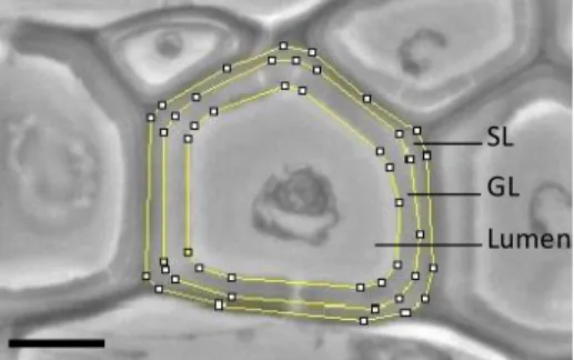
Cell wall layer thickness of wood fibres was measured using the phase contrast mode with a Leica DMLP microscope with immersion oil lenses. Phase contrast is preferable to bright field microscopy when high magnifications (400x, 1000x) are needed especially as the specimen is colourless or the details are so fine that colour does not show up well. Light microscopy allows for the measurement of all the cells along a radial line and an average measurement on each whole cell, which is not possible using TEM technique that requires the deposition of the sections on a grid thus hiding part of the sample. Several images were captured using a digital camera (Leica DFC320) from cambium cells to ring boundary with a sufficient overlap to allow for the repositioning of each image with the previous one in order to accurately measure the distance of each cell to the cambium. Preliminary testing on another sampling (data not shown here) indicated that the thickness of the compound middle lamella (CML) is constant all along the maturation sequence and does not show significant variation with SL and GL thickness changes. Therefore, CML thickness was not measured in this study. Thickness of GL and SL were measured in three radial lines of cells per sample using image analysis software ImageJ (National Institutes of Health, Bethesda, MD, USA). In order to handle the variability of cell wall layer thickness around the cell, and to increase the precision of the measurement, a mean cell wall thickness was calculated according to the method proposed by Yoshinaga et al (2012). External contours of the GL, SL and lumen were plotted manually from images (Fig. 1) and the average thickness was calculated according to following formula:
GL mean thickness = 2 × AGL/(PGL + PLumen), and
SL mean thickness = 2 × ASL/(PSL + PGL),
where AGL is the area of GL, ASL the area of SL, PGL the external perimeter of GL, PSL the external perimeter of SL and PLumen the lumen perimeter. Finally, the mean cell diameter was evaluated as D = PSL/π. This method integrates the whole fibre and thus allows for a better precision in the thickness measurement than when performed only in some points. A reproducibility test, made by measuring 30 times the same wall thickness, yielded a confidence interval at 95% of 0.015 µm.
Each measured cell was later recognized by its distance to the cambium both in µm and in number of the cells (when a vessel interrupted the radial line, the number of cells was counted on the adjacent radial line), its cell dimensions (equivalent mean diameter) and its mean cell wall thicknesses (SL and GL). As mentioned in Fang et al. (2008), GL thickness is positively related to the cell diameter (the higher the fibre diameter, the thicker the GL) due to the reduced wall thickness near the end of the fibres, as shown by Okumura et al. (1977). Therefore, in order to make the progressive changes in the wall thickness comparable from fibre to fibre, thicknesses are presented as relative thicknesses by dividing them by the mean cell diameter.
Figure 1. Detail of an optical image used for the measurement of cell walls parameters with ImageJ software. Scale bare = 5 µm
SL GL Lumen
RESULTS
As it is not possible to follow the thickening of a single cell, several cells in a single radial line from cambium to mature wood are considered as a good proxy of the maturing cell all along this study. On each sample, the 3 radial lines measured were very similar to each other, only disturbed by the presence of vessels. Therefore, for the sake of clarity, only one radial line is presented in our graphs, for a given sample.
Stimulus duration before GL formation
The presence of fibres with a GL (G-fibre) was not detectable one day after tilting. 3 days after tilting, GL was observable only in 1 tree among the 3 sampled trees. One week after tilting, all trees exhibit a GL in almost all of the tension wood fibres.
Identifying the tilting date in sections
To better understand the cell wall thickness changes in response to the gravitropic stimulus, it is necessary to identify which cells were produced before and after tilting. This date is clearly identifiable thanks to the appearance of the GL at the tilting date followed by a strong decrease in SL thickness. In the transition zone, SL is first as thick as before tilting but with a thin GL (Fig. 2d); then SL thickness gradually decreases. Whereas SL has a thickness of around 1.56 µm before the tilting date (average of all samples), it is reduced to around 0.6 µm when the GL thickness remains stable. One can consider that the gradual change in SL thickness is due to the sudden change in the signal that modified the function of the cell, stopping the development of the SL to start the deposition of a GL. Therefore, cells with SL of intermediate thickness correspond to cells that already differentiated but were not mature at the tilting date, whereas cells with a thinner SL and a thick GL were differentiated after the tilting date.
Growth rate
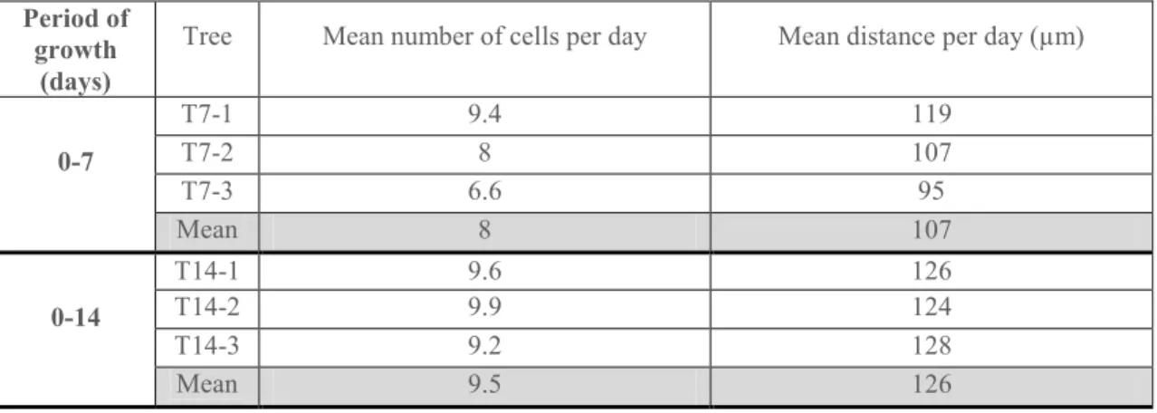
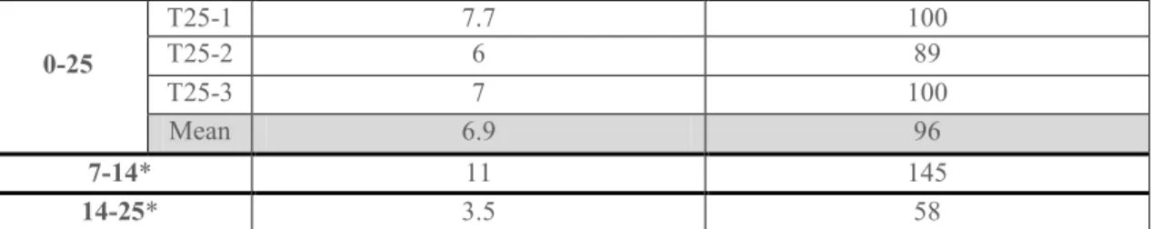
Growth rate is obtained by counting the number of cells and the distance from the tilting position to the cambium. Growth rates are given in Table 1. The number of cells formed per day slightly increased after 7 days to 14 days after tilting, and then decreased strongly during the last period.
Table 1. Radial growth rates, expressed as the number of cells formed during a tilting period, and as the mean distance of the cell produced during this period. Each value is an average of three radial lines measured for each tree. Gray background corresponds to the average of the three trees sampled at a given date. * indicates that the growth rate was estimated according to the previous period.
Period of growth
(days)
Tree Mean number of cells per day Mean distance per day (µm)
0-7 T7-1 9.4 119 T7-2 8 107 T7-3 6.6 95 Mean T7? 8 107 0-14 T14-1 9.6 126 T14-2 9.9 124 T14-3 9.2 128 Mean T14 9.5 126
0 0.1 0.2 0 1000 2000 3000 4000 R e l. c e ll w a ll th ic k n e s s
Distance from cambium (µm)
a b c d e e a b c d 0-25 T25-1 7.7 100 T25-2 6 89 T25-3 7 100 Mean T25 6.9 96 7-14* 11 145 14-25* 3.5 58
SL thickness before tilting
Table 2 summaries the mean values and the significance of relationships between changes in thickness and distance to the cambium for each measured sample. The average thickness of SL before tilting (Fig. 2 stage e) was measured on all the trees sampled at T7, T14 and T25 with a mean value of 1.56 µm (standard deviation (SD): 0.13) and a mean relative thickness of 0.097 (SD: 0.004). However, this thickness is not constant as it increases from ring boundary to tilt position (Fig. 2 stage e). This increase was statistically significant in 7 samples out of 9 (Table 2). In the two samples where the correlation was not significant, some GL are observed before tilting (as in Fig. 2 stage e). The presence of GL can largely affect the SL thickness and explain the disturbed relationship. The presence of GL in upright trees is commonly observed and can be considered as a normal behaviour considering the need for the tree to stay up-right. Some other trees presented a thin GL before tilting, which weakly affected the present relationship (Fig. 2).
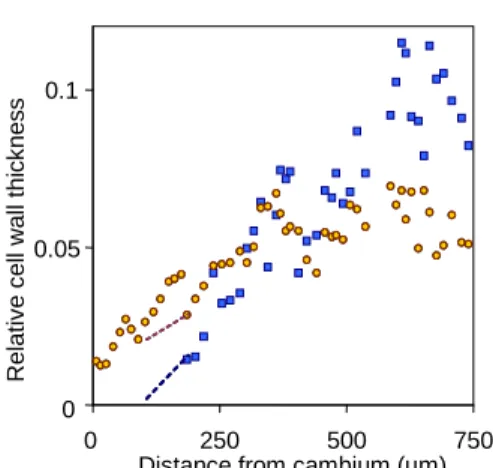
Figure 2. GL and SL relative thickness change from the cambium to the ring boundary in a tree sampled 25 days after tilting. -- a: SL thickening, -- b: GL thickening, -- c: GL constant thickness, -- d: tilting, -- e: before tilting. Squares: GL, circles: SL. Dotted line: left: end of SL thickening, center left: end of G thickening, center right: end of the transition, right: tilting date.
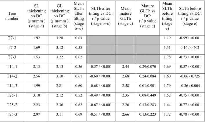
Table 2: Mean value of the measured thickness and statistical analysis of the change in thickness versus distance to the cambium (DC) in the different stages presented on Fig. 2. r: correlation coefficients calculated of the relative thickness vs. distance from cambium. SLTh and GLTh: respectively SL and GL thickness (in µm). Slopes were measured using the “real” thickness to illustrate the thickening in µm/mm. Grey background indicates that measurements were not performed as it was meaningless to measure a mature GLTh when it cannot be certified that GL reached its final thickness. Seven days after tilting, SLTh after tilting (stage b+c) was too small to compute the correlation between SL thickness and distance to the cambium.
Tree number SL thickening vs DC (µm/mm ) (stage a) GL thickening vs DC (µm/mm ) (stage b) Mean SLTh after tilting (stage b+c) SLTh after tilting vs DC: r / p value (stage b+c) Mean mature GLTh (stage c) Mature GLTh vs DC: r / p value (stage c) Mean SLTh before tilting (stage e) SLTh before tilting vs DC: r / p value (stage e) T7-1 1.92 3.28 0.63 1.19 -0.59 / <0.001 T7-2 1.69 3.12 0.58 1.31 0.16 / 0.402 T7-3 1.53 3.22 0.62 1.78 -0.73 / <0.001 T14-1 2.13 3.13 0.56 -0.57 / <0.001 2.44 0.29/0.070 1.69 -0.57 / <0.001 T14-2 2.56 3.10 0.61 -0.60 / <0.001 2.68 0.24/0.084 1.60 -0.06 / 0.725 T14-3 1.99 2.81 0.60 -0.68 / <0.001 2.58 0.01/0.901 1.79 -0.36 / 0.004 T25-1 3.10 2.12 0.52 -0.49 / <0.001 2.35 0.08/0.449 1.52 -0.75 / <0.001 T25-2 2.23 2.36 0.62 -0.67 / <0.001 2.26 0.13/0.283 1.44 -0.77 / <0.001 T25-3 2.97 3.11 0.69 -0.51 / <0.001 2.66 0.13/0.223 1.72 -0.78 / <0.001
Change in SL thickness after tilting
After the tilting date, SL has a rather constant thickness in mature wood (i.e., except in the differentiating zone near the cambium), around 2.6 times thinner than before tilting. A careful investigation of SL thickness in this stable zone shows however that SL thickness slightly increases when the distance to the cambium decreases with a significant negative correlation between SL thickness and distance from cambium (Table 2). This negative correlation is significant in trees sampled at T14 and T25. In trees sampled after 7 days, the relationship is meaningless as the stable zone is too short.
Kinetics of GL deposition vs. SL thickening
In most of the recorded radial lines, GL thickening occurs synchronously or soon after completion of the SL thickening. Three radial lines (out of the 27 measured ones) presented a peculiar behaviour as the GL deposition started slightly before the end of SL thickening, so that SL and GL thickness continue to increase simultaneously (Fig. 3). The late increase in the
thickness of SL is only observed after a long time of tilting (T25). Moreover, it has to be noticed that the GL is generally not observable or measurable in its early stage of deposition, as attested by the absence of measured thickness between 0 and 0.3 µm (Fig. 3). This induces an over-estimation of SL in this portion as attested by the steep decrease in SL thickness at the position where the first GL is detected in Fig. 3 where an empirical correction is proposed. It can therefore be supposed that GL deposition starts slightly earlier than what is recorded in some other samples and therefore this behaviour could be more common than the few cases observed. After the end of its thickening, final GL thickness was found to be 2.5 µm (SD: 0.17) on average. This value takes into account only radial lines from trees sampled at T14 and T25 as it is difficult to be sure that the GL thickening period was finished for trees sampled at T7 and before.
Fig. 3: detailed view of sample T25-1 where the GL (squares) deposition starts before the end of SL (circles) thickening. Lower dotted line is an empirical extrapolation to correct the lack of GL measurement when its thickness is too thin. This lack induces an over-estimation of SL in this portion as attested by the steep decrease in SL thickness at the position where the first GL is detected. The upper dotted line thus proposes a corrected trend of SL thickness in this area taking the linear extrapolation of the GL thickness into account.
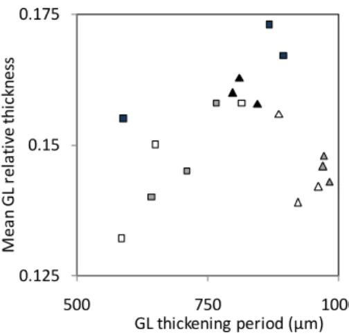
The GL thickening period is considered to be the distance between the first occurrence of GL in young cells near the cambium to where they reached a constant thickness (stage b in Fig. 2). GL thickening period varies from 570 to 982 µm (Fig. 4), with, as a general trend, a longer period for trees sampled at T25 compared to trees sampled at T14 (Fig. 4). No general trend can be detected for the relationship between mean GL thickness (or relative thickness) and GL thickening period (Fig. 4).
0 0.05 0.1
0 250 500 750
Distance from cambium (µm)
R e lat iv e c e ll w a ll th ic k n e s s
Fig. 4: relationship between the mean GL thickness (plotted as relative thickness) and the GL thickening period. Squares: T14, triangles: T25. Different shades represent different trees.
GL and SL growth rates
Table 2 gives the slope of the SL and GL thickening vs. the distance to cambium. This slope can be expressed as an increase in the wall thickness in µm by distance to the cambium in mm. It gives an estimate of the growth rate at the cell wall level under the assumption of a constant cell division speed. Although the validity of this assumption is uncertain, it is interesting to compare these stages of cell wall building in a single tree and even more in a single radial line of cells. In 8 of the 9 measured trees, the slope of the SL thickening was lower than the slope of GL thickening, whatever the growth rate in the considered period as presented in Table 1.
Cell wall thickening in opposite wood during tension wood formation
The marking of the tilting position in opposite wood (OW) side is much less clear than in tension wood (TW) as there is no GL development or steep decrease in SL. Moreover, TW and OW have different growth rates; it is therefore problematic to compare the cell wall thickening on both side of the stem from their distance to the cambium, as cells are not produced at the same date. Therefore, tilting position was determined assuming a similar growth rate all around the stem before tilting. In order to compare cell wall thickness at the same date in TW and OW, the remaining cells produced after tilting (from the tilting position to the cambium) are then assumed to grow at different but proportional speed on both sides.
Before tilting, cell wall thickness of OW fibres shows a similar trend as observed in TW side (Fig. 5 stage e) as, at that time, trees were upright and no differences are expected on both sides. After tilting (stage b+c), the trend observed before tilting is disturbed in all trees. In several trees, an increase of the SL thickness is observed consecutively to the tilting. However, no general trend was present for all trees.
In T25 samples, average SL thickness in OW after tilting (stage b+c) was 1.78 µm whereas it was 1.64 µm before tilting. This should be compared to the total cell wall thickness on the TW side, which measured 3.01 µm after the GL had been formed.
0.125 0.15 0.175 500 750 1000 M ea n G L rel at iv e th ic kn es s GL thickening period (µm)
Figure 5. SL thickness variation in opposite wood (OW) of the sample T25-2 from the cambium to the ring boundary. The different stages of TW cell wall development as defined in Fig. 2 are indicated in the top of this figure to emphasise the change in OW due to tilting (at d).
DISCUSSION
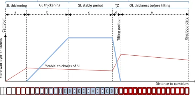
The present study allows us to reconstruct the general developmental pattern of fibre cell wall layers in tilted trees before and after tilting as presented in Fig. 6. Developing cells and mature cells are two main stages in this diagram. The developing fibres part contained young cells in which cell wall thickening is in progress with first the deposition of CML (not presented) followed by the formation of SL and GL.
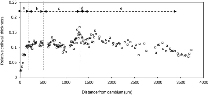
Measurements of wood cell wall thickness before tilting show the progressive increase of SL during the growing season. An increase in cell wall thickness is commonly observed in softwoods (e.g., Mork 1928 in Denne 1988) and the change in wall thickness at the ring transition is also reported for hardwoods (IAWA Committee, 1989) but, we were unable to find reference reporting quantitative measurements of changes in fibre wall thickness during the growing season. It may be noticed that the seasonal SL thickness change in our poplar samples is very slight compared to what is observed in softwood, such as Abies alba, Picea abies or Pinus
sylvestris, where a factor of more than 2.5 is observed for the tracheids wall thickness change
during a season (Cuny et al. 2014).
Interestingly, this trend of increasing SL thickness remains true in tension wood with the presence of GL. This observation, always observed in our sampling, confirms some earlier unrecorded observations (Clair et al. 2011, Yoshinaga et al. 2012, Chang et al. 2014) and proves that this increase is not linked to the development of the GL but is a common trend throughout the growing season both in normal wood and tension wood in poplar trees.
0 0.05 0.1 0.15 0.2 0.25 0 500 1000 1500 2000 2500 3000 3500 4000 R e la ti v e c e ll w a ll th ic k n e s s
Distance from cambium (µm)
The GL thickness remains nearly constant in mature wood. This stability may be attributed to the constant stimulus as the stem is attached to a pole. However, the secondary layer increases significantly during the growing season. This would indicate that, whatever the needs for up-righting, poplar trees cannot allocate all of their resources to the production of the GL and some trade-off is needed to ensure its sustainability all along the season.
Figure 6. Schematic of the development pattern of fibre cell wall layers in a tilted tree producing GL. Thick line: GL, thin line: SL. -- TZ: transition zone is the zone where already differentiated fibres change in function and develop a GL later.
Transition zone at the tilting date
Around the tilting position different type of fibres are observable: 1- Fibres without GL in normal wood zone, considered to have already finished their maturation before tilting, in these cells the tilting has no effect on their cell wall thickness (Figs. 2, 6: e); 2- Fibres with decreasing SL thickness and increasing GL thickness in the transition zone (tilting), these cells already started their SL thickening before tilting then received the reaction signal and changed in function during their development, halting their SL formation in a rather thick state to start a GL at the end of the maturation (Fig. 2, 6: d). 3- Fibres that were in the cambial zone at tilting time, these cells have a thin SL and constantly thick GL (Fig. 2, 6: c). This interpretation of the timing of cell wall deposition assumes that a signal is received soon after the tilting. Jourez and Avella-Shaw (2003) have shown that a stimulus is perceived in poplar after only few hours, however modifications of the wood cell wall was more visible in opposite wood than in tension wood, thanks to a dedicated protocol using double tilting. A G-layer was observed in their study from 6 h to 48 h after tilting, depending on the trees. In our sampling, the reaction may have been slightly slower as GL was visible in one of the trees cut after 3 days and all of the trees sampled after 7 days of tilting.
a b c d Ti lti n g p o si ti o n 'Stable' thickness of SL Developing fibres R in g b o u n d ary Mature fibres Fi b re w all laye r th ic kn e ss Distance to cambium Cambi u m e
OL thickness before tilting GL stable period
GL thickening
Concerning the modification of the cell during maturation to adapt to the new mechanical needs of the plant, it is interesting to note that this behaviour is much more reactive in poplar than what was observed by Yoshizawa et al. (1985) in softwood. In their study on artificially tilted Taxus cuspidata, they have shown that only small modifications in cell wall structure occur in tracheids that were already in the enlargement or thickening zones when the stimulus starts. We could therefore propose that this ability to transform fibres already formed is an efficient way to react to tilting and may allow poplar trees to be more efficient to react with tension wood formation than T. cuspidata with compression wood formation .
SL thickness decreased markedly from normal wood to tension wood confirming that in tension wood, GL replaces part of the S2 layers (Saiki and Ono 1971). Total thickness also increased compared to normal wood cells before tilting and opposite wood cells that were formed after tilting. This shows that the GL is thicker than the replaced layer that exist in normal fibres, as already observed by Fang et al. (2007).
Timing of GL versus SL deposition
In most of the samples, GL formation start after SL reached its maximum thickness. It means that SL thickening was completed before GL formation. However, in some samples, especially 25 days after tilting, SL thickness seems to increase even after the start of GL formation. This would mean that the SL is able to continue thickening when GL deposition already started. The building of the GL before the end of SL thickening is surprising as layers are supposed to be deposited one after one. Considering the presupposed assumption of the study that several cells along a radial line are considered as a single cell during maturation one could suspect a misinterpretation. In order to verify it, let us consider a second interpretation: as soon as the GL starts to be observable, the SL may be supposed to have reached its final thickness. As we observed an increasing SL thickness after GL deposition, the pattern of thickening would indicate a decrease in SL thickness from ring to cambium. This pattern has never been observed in our samples; reversely the opposite pattern was statistically significant in all our samples. Moreover, at the end of the growing season, near the ring boundary, SL tends to be thicker with no more GL (data not shown from other samples) as previously shown by Jourez and Avella-Shaw (2003). Thus, even if surprising, our observations indicate that in some samples, the SL continues its thickening after GL started to be deposited. This observation can be discussed in the light of what has been observed by Yoshinaga et al. (2012). These authors observed a continuation of the lignification of the SL after the start of the GL deposition. They suggest that monolignols may be transported through the developing GL during the lignification of the SL or propose an alternative hypothesis of an external synthesis in the rays. Our observation of an increase in SL thickness could be the result of the swelling of the wall during this lignification process.
GL thickening period
Our results indicate that GL thickening distance (i.e., the number of cells from the beginning of GL deposition to the end of GL thickening) is higher in trees sampled after 25 days than in trees sampled after 14 days (Fig. 4). This observation can be interpreted in two ways: the time to mature is longer or the cell production is faster (higher growth rate). According to the computation of the number of cells produced after tilting and the distance of the tilting position to the cambium (Table 1), growth rate was more than 3 times higher during the period from 7 to
14 days than during the period from 14 to 25 days. It is therefore clear that this increase in GL thickening period refers to a longer maturation process. Tension wood is reported to have a longer maturation process than normal wood (Bollhöner et al., 2012). However, the reason for this great change in maturation time remains enigmatic. No relationship was observed between the time of thickening and the final GL thickness. This indicates that thickness is not directly linked to the time of maturation.
The slope of the GL or SL thickening vs. distance to the cambium can be interpreted as thickening speed of the layers and be expressed as µm/mm. This allows us to compare cell wall layer thickening under the assumption of a constant cell division speed. It is therefore not possible to compare layers thickening at different dates or to compare TW to OW, but it remains reasonable to compare SL and GL thickening in a single TW sample. GL appears to grow faster than SL, i.e., 1.9 and 1.4 times faster in T7 and T14 respectively. At T25, around the same thickening speed was measured on average in the 3 trees in SL and GL because in one of the trees, the slope was lower for GL than for SL. This could be attributed to limited resources such as a decrease in light or temperature between T14 and T25 that affect cell division and growth rate for that tree in T25. Regarding the faster growth rate of GL compared to SL, it may be speculated that this would be a consequence of its lower carbon cost due to the lack of lignin (Pilate et al. 2004) and the high mesoporosity (Clair et al. 2008) in the GL. This would allow a faster production of thick GL, which has been recognized to be the driving force of the maturation stress generation (Fang et al. 2008, Clair et al. 2011). Following this idea, the lack of lignin in GL would be a strategy for a fast recovery when reaction is needed. It will be interesting in the future to test this hypothesis, especially comparing species producing TW with a GL to species where the GL is absent in TW.
ACKNOWLEDGMENTS
Special thanks are due to Prof. Joseph Gril (LMGC, CNRS, Université de Montpellier). This study received the support of the SCAC (Service de Coopération et d’Action Culturelle) of French embassy in Tehran and the Iranian center of Excellency on applied management of fast growing wooden species. Part of this work was performed in the framework of the project “StressInTrees” funded by the French National Research Agency (ANR-12-BS09-0004).
REFERENCES
Archer R. 1986. Growth stresses and strains in trees. Timell E (ed.) Springer-Verlag,Berlin, Heidelberg, New York, 240 pp. Andersson-Gunneräs S, Mellerowicz EJ, Love J, Segerman B, Ohmiya Y, Coutinho PM, Nilsson P, Henrissat B, Moritz Th &
Sundberg B. 2006. Biosynthesis of cellulose-enriched tension wood in Populus: global analysis of transcripts and metabolites identifies biochemical and developmental regulators in secondary wall biosynthesis. The Plant Journal, 45: 144–165.
Bollhöner B, Prestele J & Tuominen H. 2012. Xylem cell death: emerging understanding of regulation and Function, Journal of Experimental Botany, 63: 1081–1094.
Chang SS, Quignard F, Di Renzo F, & Clair B 2012. Solvent polarity and internal stresses control the swelling behavior of green wood during dehydration in organic solution. BioResources 7: 2418-2430.
Chang SS, Salmén L, Olsson AM & Clair B. 2014. Deposition and organisation of cell wall polymers during maturation of poplar tension wood by FTIR microspectroscopy. Planta, 239: 243-254.
Clair B, Ruelle J, Thibaut B. 2003. Relationship between growth stress, mechanical–physical properties, and proportion of fibre with gelatinous layer in chestnut (Castanea sativa Mill.). Holzforschung 57:189–195.
Clair B, Gril J, Baba K, Thibaut B & Sugiyama J. 2005. Precautions for the structural analysis of the gelatinous layer in tension wood. IAWA Journal,26: 189-195.
Clair B, Ruelle J, Beauchêne J, Prévost MF & Fournier Djimbi M. 2006. Tension wood and opposite wood in 21 tropical rain forest species. 1. Occurrence and efficiency of the G-layer. IAWA Journal 27: 329-338.
Clair B, Gril J, Di Renzo F, Yamamoto H & Quignard F. 2008. Characterization of a gel in the cell wall to elucidate the paradoxical shrinkage of tension wood. Biomacromolecules, 9: 494-498.
Clair B, Alméras T, Pilate G, Jullien D, Sugiyama J & Riekel C. 2011. Maturation stress generation in poplar tension wood studied by synchrotron radiation microdiffraction. Plant Physiology, 155: 562-570.
Côté WA, Day AC & Timell TE 1969. A contribution to the ultrastructure of tension wood fibers. Wood Science and Technology, 3: 257–271.
Cuny HE, Rathgeber CB, Frank D, Fonti P & Fournier M. 2014. Kinetics of tracheid development explain conifer tree-ring structure. New Phytologist, 203: 1231-41.
Denne MP. 1988. Definition of latewood according to Mork (1928). IAWA Bulletin n.s., 10: 59-62.
Fang CH, Clair B, Gril J & Alméras T. 2007. Transverse shrinkage in G-fibers as a function of cell wall layering and growth strain. Wood Science and Technology, 41: 659-671.
Fang CH, Clair B, Gril J & Liu Sh. 2008. Growth stresses are highly controlled by the amount of G-layer in Poplar tension wood. IAWA Journal, 29: 237–246.
Fujita M, Saiki H & Harada H. 1974. Electron microscopy of microtubules and cellulose microfibrils in secondary wall formation of poplar tension wood fibers. Mokuzai Gakkaishi, 20: 147-156.
Fournier M, Alméras T, Clair B, Gril J. 2014 Biomechanical action and biological functions. In: Gardiner B, Barnett J, Saranpää P & Gril J. eds. The biology of reaction wood, Springer Series in Wood Science, Berlin Heidelberg: 139-170.
IAWA Committee. 1989. IAWA list of microscopic features for hardwood identification. Wheeler, EA, Baas, P & Gasson, PE Eds). IAWA Journal n.s. 10: 219-332. pp.234-235
Jourez B, Riboux A & LeclercqA. 2001. Anatomical characteristics of tension wood and opposite wood in young inclined stems of poplar (Populus euramericana cv ‘Ghoy’). IAWA Journal, 22: 133-157.
Jourez B & Avella-Shaw T. 2003. Effet de la durée d’application d’un stimulus gravitationnel sur la formation de bois de tension et de bois opposé dans de jeunes pousses de peuplier (Populus euramericana cv ‘Ghoy’). Annals of Forest Science, 60: 31-41.
Mikshina P, Chernova T, Chemikosova S, Ibragimova N, Mokshina N & Gorshkova T. 2013. Cellulosic Fibers: Role of Matrix Polysaccharides in Structure and Function, in: van de Ven T & Godbout L eds. Cellulose - Fundamental Aspects, InTech, 91-112.
Norberg PH & Meier H. 1966. Physical and chemical properties of the gelatinous layer in tension wood fibre of aspen (Populus
tremula L.). Holzforschung, 20: 174-178.
Okumura S, Harada H & Saiki H. 1977. Thickness variation of the G-layer along a mature and a differentiating tension wood fiber in Populus euramericana. Wood Science and Technology, 11: 23-32.
Okuyama T, Yamamoto H, Yoshida M, Hattori Y & Archer RR. 1994. Growth stresses in tension wood: role of microfibrils and lignification. Annals of Science Forest, 51: 291-300.
Onaka F. 1949. Studies on compression and tension wood. Wood research, Bulletin of the Wood research Institute, Kyoto University, Japan, 24(3): 1-88.
Pilate G, Chabbert B, Cathala B, Yoshinaga A, Leplé JC, Laurans F, Lapierre C & Ruel K. 2004. Lignification and tension wood. C. R. Biol., 327: 889–901.
Prodhan AKMA, Funada R, Ohtani J, Abe H & Fukazawa K. 1995. Orientation of microfibrils and microtubules in developing tension-wood fibres of Japanese ash (Fraxinus mandshurica vat. japonica). Planta, 196: 577-585.
Ruelle J. 2014 Morphology, Anatomy and ultrastructure of reaction wood. In: Gardiner B, Barnett J, Saranpää P & Gril J. (eds). The biology of reaction wood, Springer, Berlin Heidelberg: 13-35.
Saiki H & Ono K. 1971. Cell wall organization of gelatinous fibers in tension wood. Bull. Kyoto Univ. For., 42: 210–220. Sultana RS, Ishiguri F, Yokota S, Iizuka K, Hirawa T & Yoshizawa N. 2010. Wood anatomy of nine Japanese hard wood species
forming reaction wood without gelatinous fibers. IAWA Journal 31: 191-202. Timell TE. 1986. Compression wood in gymnosperms. Springer-Verlag, Heidelberg.
Washusen R, Ilic J & Waugh G. 2003. The relationship between longitudinal growth strain and the occurrence of gelatinous fibers in 10 and 11-year-old Eucalyptus globulus Labill. Holz Roh- Werkstoff 61: 299–303.
Yoshida M, Okuda T & Okuyama T. 2000. Tension wood and growth stress induced by artificial inclination in Liriodendron
tulipifera Linn. and Prunus spachiana Kitamura f. ascendens Kitamura. Annals of Forest Science, 57: 739-746.
Yoshinaga A, Kusumoto H, Laurans F, Pilate G & Takabe K. 2012. Lignification in poplar tension wood lignified cell wall layers. Tree Physiology, 32:1129–1136.
Yoshizawa N, Koike S & Idei T. 1985. Formation and structure of compression wood tracheids induced by repeated inclination in Taxus cuspidata. Mokusai Gakkaishi, 31:325–333.