HAL Id: hal-01812668
https://hal.archives-ouvertes.fr/hal-01812668
Submitted on 6 Jul 2020
HAL is a multi-disciplinary open access
archive for the deposit and dissemination of
sci-entific research documents, whether they are
pub-lished or not. The documents may come from
teaching and research institutions in France or
abroad, or from public or private research centers.
L’archive ouverte pluridisciplinaire HAL, est
destinée au dépôt et à la diffusion de documents
scientifiques de niveau recherche, publiés ou non,
émanant des établissements d’enseignement et de
recherche français ou étrangers, des laboratoires
publics ou privés.
derived from 4-substituted-1,2,3-triazoles as IMP/GMP
mimics: synthesis and biological evaluation
Tai Nguyen Van, A. Hospital, C. Lionne, L.P. Jordheim, C. Dumontet, C.
Perigaud, L. Chaloin, S. Peyrottes
To cite this version:
Tai Nguyen Van, A. Hospital, C. Lionne, L.P. Jordheim, C. Dumontet, et al..
Beta-hydroxyphosphonate ribonucleoside analogues derived from 4-substituted-1,2,3-triazoles as IMP/GMP
mimics: synthesis and biological evaluation. Beilstein Journal of Organic Chemistry, Beilstein-Institut,
2016, 12, pp.1476–1486. �10.3762/bjoc.12.144�. �hal-01812668�
from 4-substituted-1,2,3-triazoles as IMP/GMP mimics:
synthesis and biological evaluation
Tai Nguyen Van
1, Audrey Hospital
1, Corinne Lionne
2, Lars P. Jordheim
3,
Charles Dumontet
3, Christian Périgaud
1, Laurent Chaloin
2and Suzanne Peyrottes
*1Full Research Paper
Open AccessAddress:
1Institut des Biomolécules Max Mousseron (IBMM), UMR 5247 CNRS – Université de Montpellier - ENSCM, Campus Triolet, cc1705, Place Eugène Bataillon, 34095 Montpellier, France, 2Centre d'études d'agents Pathogènes et Biotechnologies pour la Santé (CPBS), FRE 3689 CNRS - Université de Montpellier, 1919 route de Mende, 34293 Montpellier, France and 3Université de Lyon, Université Claude Bernard Lyon 1, INSERM 1052, CNRS 5286, Centre Léon Bérard, Centre de Recherche en Cancérologie de Lyon, 69008 Lyon, France Email:
Suzanne Peyrottes* - [email protected] * Corresponding author
Keywords:
cancer; cN-II inhibitors; nucleotide; phosphonate; triazole
Beilstein J. Org. Chem. 2016, 12, 1476–1486. doi:10.3762/bjoc.12.144
Received: 12 April 2016 Accepted: 27 June 2016 Published: 18 July 2016 Associate Editor: I. R. Baxendale
© 2016 Nguyen Van et al.; licensee Beilstein-Institut. License and terms: see end of document.
Abstract
A series of seventeen β-hydroxyphosphonate ribonucleoside analogues containing 4-substituted-1,2,3-triazoles was synthesized and fully characterized. Such compounds were designed as potential inhibitors of the cytosolic 5’-nucleotidase II (cN-II), an enzyme involved in the regulation of purine nucleotide pools. NMR and molecular modelling studies showed that a few derivatives adopted similar structural features to IMP or GMP. Five derivatives were identified as modest inhibitors with 53 to 64% of cN-II inhibition at 1 mM.
Introduction
Nucleotidases are an important family of enzymes involved in the metabolism of nucleotides [1]. In particular, human cytosolic 5’-nucleotidase II (cN-II) catalyses the dephosphory-lation of purine 5’-monophosphate derivatives to their corre-sponding nucleosides [2,3]. Recent studies on patients affected by haematological malignancies, such as acute myeloid leukaemia (AML) and myelodysplasic syndrome (MS), have
demonstrated the involvement of cN-II in the resistance to cyto-toxic drugs such as mercaptopurines and suggested the effec-tiveness of cN-II inhibitors in the treatment of these diseases [4,5]. As a result of our interest in this area, we and others [6] have investigated a number of structure–activity relationships (SAR) [7,8] and various medicinal chemistry approaches [9,10] to identify potential cN-II inhibitors. As part of a research
Figure 1: Previous (UA1776, UA2201 and UA2209 [7,8]) and new 1a–q phosphonate derivatives designed as potential cN-II inhibitors.
program on phosphonate derivatives of nucleosides as mimics of 5’-monophosphate nucleosides, we explored the SAR of various beta-hydroxyphosphonate analogues. Such derivatives have been extensively studied and exhibited Ki values in the same range as the known natural substrates (IMP, inosine 5’-monophosphate and GMP, guanosine 5’-monophosphate) [6,7]. In the particular context of cN-II, which is also known as High-Km nucleotidase owing to the high substrate concentra-tion required for activity (in the mM range), the Ki values of approximately 1 mM previously obtained for cytidine-contain-ing derivatives are biologically interestcytidine-contain-ing (Figure 1) [7]. In ad-dition, molecular docking studies have been performed and highlighted the importance of three binding areas within the active site of the protein: a hydrophobic clamp (Phe157, His209 and Tyr210) interacting with the nucleobase, a hydrophilic pocket (Ser251 and Lys215) where the hydroxy groups of the sugar interact and a phosphonate binding site close to the magnesium ion located in the substrate binding site. Thus, as few cytosine-containing analogues were equipotent in terms of cN-II inhibition to their purine counterparts (Figure 1) [7], we were interested in studying the effect of replacing the nucleo-base by a 1,2,3-triazole moiety, linked to various substituents, with the aim to retain or modulate essential elements for en-zyme recognition. Indeed, the assembly of the triazole ring with various substituents confers to the final compound high flexi-bility. Initially reported by K. Seley-Radtke’s group, the replacement of the nucleobase by a “flexi-moiety” where the imidazole ring and a six-membered heterocycle is linked through a C–C bond lead to new derivatives behaving as nucleosides and even as enzyme inhibitors [11-13]. The inherent flexibility of the corresponding nucleobase-mimics allows to accommodate steric and electronic clashes encoun-tered in protein binding sites and to interact with other amino
acids. Another type of fleximers, obtained by a click chemistry approach, was developed by R. H. E. Hudson to obtain “click-fleximer” as expanded nucleobase mimicking the purine [14]. “Click-fleximer” nucleoside analogues are easily accessible de-rivatives using copper-catalysed alkyne–azide cycloaddition (CuAAC) and this synthetic methodology allows generating a small library of derivatives depending on the nature of the alkyne employed.
Herein, we report the results of the synthesis and in vitro bio-logical evaluation on the purified human recombinant cN-II of a series of beta-hydroxyphosphonate ribonucleosides including as nucleobases 4-substituted-1,2,3-triazoles (Figure 1).
Results and Discussion
Chemistry
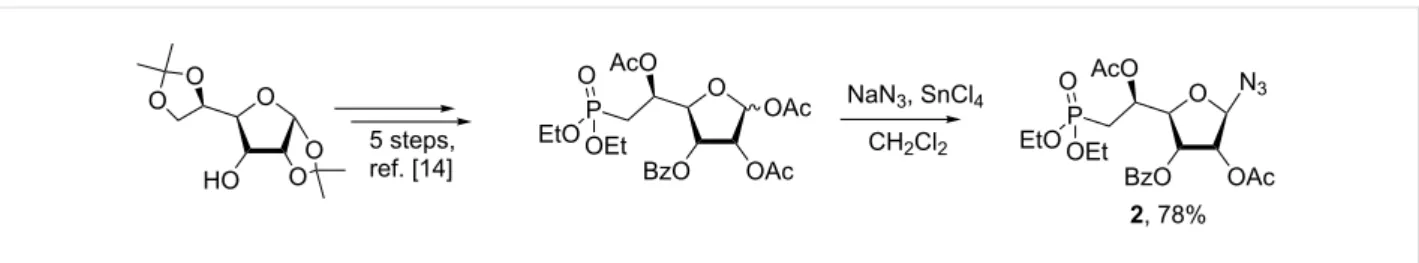
The synthesis of 1,2,3-triazolonucleotides (1a–o, Figure 1) was envisaged using the Huisgen 1,3-dipolar cycloaddition of the azido-sugar-phosphonate key-intermediate 2 (Scheme 1) and selected commercially available alkynes. Thus,
(1-azido-2,5-di-
O-acetyl-3-O-benzoyl-6-deoxy-6-diethylphosphono)-β-ribo-(5S)-hexofuranose (2) was obtained in good yield from 1,2,5- tri-O-acetyl-3-O-benzoyl-6-deoxy-6-diethylphosphono-(α,β)-ribo-(5S)-hexofuranose [15] following a glycosylation proce-dure using sodium azide as nucleophilic entity and tin(IV) chlo-ride as Lewis acid. Under these conditions, the reaction appeared highly stereoselective and only the β-anomer was ob-served, isolated and characterized on the basis of 1H NMR ex-periments (see section related to NMR studies).
The selection of alkynes was based on their commercial avail-ability as well as the diversity of the chemical group(s) to be introduced. They can be divided in two categories: aromatic
Scheme 1: Synthesis of (1-azido-2,5-di-O-acetyl-3-O-benzoyl-6-deoxy-6-diethylphosphono)-β-ribo-(5S)-hexofuranose 2 from commercially available
diacetone D-allofuranose [15].
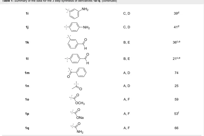
Table 1: Summary of the data for the 3 step synthesis of derivatives 1a–q.
Compound R Conditionsa Total yield (%)b
1a A, D 66 1b A, D 44 1c A, D 73 1d A, D 54 1e A, D 40c 1f A, D 71 1g A, D 77 1h C, D 45d
alkynes with various substituents (methoxy, amino, formyl…) in ortho, meta or para position, and short alkyne chains such as 3-butyn-2-one or methylpropiolate.
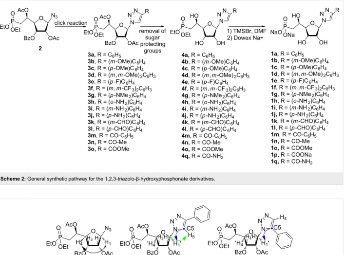
Starting from intermediate 2, the CuAAC reaction was either catalysed by CuI or CuSO4 (Table 1) and gave rise to the fully-protected nucleotides 3a–o (Scheme 2) in moderate to good
yields. Removal of the sugar protecting groups (acetyl and benzoyl) in basic conditions resulted in the formation of the nucleotides 4a–q (Scheme 2), which were then treated by tri-methylsilyl bromide (TMSBr) to generate the corresponding phosphonic acids. Thus, nucleoside phosphonate analogues
1a–q (Scheme 2) were isolated as their sodium salts with yields
Table 1: Summary of the data for the 3 step synthesis of derivatives 1a–q. (continued) 1i C, D 39d 1j C, D 41d 1k B, E 36c,e 1l B, E 21c,e 1m A, D 74 1n A, D 25 1o A, F 59 1p A, F 53f 1q A, F 66
aConditions for click reaction: (A) DIPEA, CuI, THF reflux, N,N-diethylenediamine; (B) CuI, CH3CN reflux; (C) CuSO4, sodium triascorbate, DMF; Conditions for removal of sugar protecting groups: (D) CH3OH, NH3; (E) Tetramethylguanine, CH2Cl2/CH3OH (1:1); (F) NaOMe/CH3OH. bAll interme-diates and the final compounds were isolated after chromatographic purification; cConversion rate during the click reaction was not total; ddegradation was observed during purification step using RP-chromatography; edue to side-reaction with ammonia, reaction conditions D were replaced by E;
fCompound 1p was obtained by saponification of derivative 1o in presence of sodium hydroxide.
compounds were unambiguously confirmed on the basis of NMR (1H, 13C and 31P) and MS (MS and HRMS) data analysis (see Supporting Information File 1).
NMR studies
To establish the stereochemistry and/or regiochemistry of the azidation and CuAAC steps, homo- or heteronuclear 2D NMR experiments (see Supporting Information File 2) were per-formed on compounds 2 and 3a (Figure 2). In addition, we syn-thesized compound 5 (resulting from an 1,5-addition) through Ru-catalysed cycloaddition using an adapted procedure from the literature [16].
As the α- and β-anomers of furanoses are diastereomeric, in principle they show distinguishable NMR spectra. In conse-quence, the proportions of both anomers may be determined by NMR. In our case, the main compound isolated from the glyco-sylation reaction corresponds to a single anomer as only one signal for the anomeric proton was detectable in the 1H NMR spectra. The beta-configuration of the azido-sugar-phosphonate
intermediate 2 was established on the basis of the 1H,1H NMR spectra showing a cross-peak between the anomeric proton H1 and the H4 (Figure 2). Consequently, both protons are located on the same face of the furanose ring and to the opposite face of the C4–C5 bond.
The formation of the triazole ring was confirmed through the presence of a single signal of the triazolyl proton at δ = 7.94 ppm and two signals for the olefin carbon atoms at δ = 148.4 ppm and δ = 119.3 ppm for the C4 and the C5, re-spectively. The regiochemistry of the cycloaddition was unam-biguously established using heteronuclear multiple bond corre-lation (HMBC) experiments to identify long-range (generally 2-or 3-bonds) coupling between protons and carbons. In com-pound 3a, the 1,4-substitution was proven by the fact that the H1‘(δ 6.26 ppm) signal shows a cross-peak with the C-5 (δ 119 ppm), and the C1’ (δ 89.8 ppm) cross-correlates with the signal of the H5 (δ 7.95 ppm). In the case of compound 5 (re-sulting from an 1,5-addition), the 1H NMR spectrum shows sig-nificant differences with the one of compound 3a, especially an
Scheme 2: General synthetic pathway for the 1,2,3-triazolo-β-hydroxyphosphonate derivatives.
Figure 2: Black arrow indicates 1H,1H-COSY correlations for compound 2. Green (C1’ and H5) and blue (H1’ and C5) arrows indicate 1H,13C-HMBC correlations for compounds 3a and 5.
Figure 3: Arrows indicate 1H,1H-NOESY (blue) and 1H,13C-HMBC (green) correlations for compound 3h. aromatic shielding effect on the H1’ signal. The corresponding
HMBC 2D-experiment also exhibits a cross-peak between C-5 and H-1’ (Figure 2). Thus, comparative analysis of all NMR data for compounds 3a and 5 demonstrates that cycloaddition of terminal alkynes catalysed by Cu(I) were highly regioselective and led to 4-substituted-1,2,3-triazoles in the β-hydroxyphos-phonate series.
Then, on the basis of the study reported by Hudson et al., we performed extensive 2D-NMR experiments on compounds 3h and 3i, respectively. Indeed, these two derivatives may be able to form an H-bond between the exocyclic amino group and the nitrogen atom in position 3 of the triazole ring, in particular for compound 3h, where the amino group is in the ortho position (Figure 3 and Supporting Information File 2). Thus, the
Figure 4: Arrows indicate 1H,1H-NOESY (blue) and 1H,13C-HMBC (green) correlations for compound 3i. NOESY spectrum showed correlation between the H5 of the
tri-azole ring and the aromatic proton in ortho (Ho), whereas rota-tion about the C–C bond linking the triazole and the aromatic rings would lead to the disappearance of this correlation peak. In addition, the HMBC spectrum is showing only one correla-tion peak between the C5 and the anomeric proton (H1’), indi-cating that rotation around the glycosidic bond may be limited. Therefore, we hypothesized that compound 3h behaved as a favoured anti-fleximer.
Similar analysis of the NOESY and HMBC spectra of deriva-tive 3i indicates that this compound behaves as several conformers in solution (Figure 4). Indeed, the NOESY experi-ment shows correlations between the two protons in the ortho position (Ho and Ho’) of the phenyl ring and the H5 of the tri-azole. In this case, the position of the exocyclic amino group in meta position does not allow the formation of a strong H-bond and the C–C bond linking the triazole and the aromatic rings is free to rotate. To our surprise, a cross-peak between the H5 of the triazole and the H3’ was observed (as well as with the anomeric proton), indicating that both syn- and anti-conformers may be present.
Enzyme inhibition and molecular modelling
studies
All derivatives were assayed for their ability to inhibit the human 5’-nucleotidase activity in comparison to previously published derivative (UA1776), using a rapid in vitro assay. Briefly, the enzyme catalyzes the hydrolysis of IMP into inosine and inorganic phosphate (IMP + H2O ↔ inosine + PO42−). This latter product is detected by using a malachite green assay and was quantified by the absorbance according to the phosphate concentration range.
Most of the compounds were found to reduce the enzyme activ-ity at high concentrations (>1 mM). However, only a moderate inhibition (Figure 5) was observed for these derivatives indicat-ing that they may not bind very tightly to the enzyme bindindicat-ing site. Nevertheless, five compounds 1i, 1h, 1n, 1o and 1q
exhib-Figure 5: Inhibition of the nucleotidase activity in presence of
repre-sentative triazole-based derivatives.
ited a more pronounced inhibition at 1 mM (Table 2) with at least 50% of inhibition. The nature, size and orientation of the substituent on the triazole ring seem to play a major role in the inhibitory activity as they should mimic a purine nucleobase, especially hypoxanthine for compounds 1n, 1o and 1q. The synthesized compounds may also interact with allosteric binding sites present in cN-II. Indeed, two potential binding sites have been described in the literature [17,18]. According to the X-ray crystallography, kinetics and mutagenesis studies, four effectors have been identified for binding in the effector site 1. The activation of the site 2 is more questionable as solid data is still required to clearly ascertain the biological effect of such activation. However, both sites may be occupied by one common activator (Ap4P). One way to evaluate the effect of the synthesized ribonucleoside analogues on the inhibition of allosteric activation will be to include the Ap4A effector in the enzymatic assay. Although allosteric inhibition was not ex-pected since these compounds bring only one phosphonate group (compared to more negatively charged activators) such enzymatic assay will be envisaged in future studies.
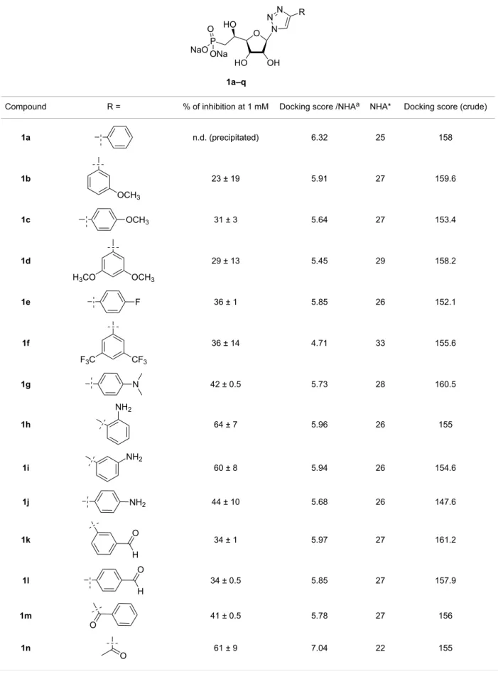
Table 2: Summary of the in vitro inhibition assays performed on human recombinant cN-II in the presence of the various derivatives and docking
scores obtained from molecular modelling calculation.
Compound R = % of inhibition at 1 mM Docking score /NHAa NHA* Docking score (crude)
1a n.d. (precipitated) 6.32 25 158 1b 23 ± 19 5.91 27 159.6 1c 31 ± 3 5.64 27 153.4 1d 29 ± 13 5.45 29 158.2 1e 36 ± 1 5.85 26 152.1 1f 36 ± 14 4.71 33 155.6 1g 42 ± 0.5 5.73 28 160.5 1h 64 ± 7 5.96 26 155 1i 60 ± 8 5.94 26 154.6 1j 44 ± 10 5.68 26 147.6 1k 34 ± 1 5.97 27 161.2 1l 34 ± 0.5 5.85 27 157.9 1m 41 ± 0.5 5.78 27 156 1n 61 ± 9 7.04 22 155
Table 2: Summary of the in vitro inhibition assays performed on human recombinant cN-II in the presence of the various derivatives and docking
scores obtained from molecular modelling calculation. (continued)
1o 53 ± 6 6.69 23 153.9
1p 41 ± 6 7.09 22 155.9
1q 57 ± 13 6.88 22 151.4
IMPb – 6.56 23 150.9
UA1776 85 ± 5 6.64 22 146.1
aNumber of heavy atoms (or non-hydrogen atoms); binosine 5’-monophosphate.
Figure 6: Comparison of the docking poses obtained for two active derivatives in the substrate binding site of cN-II. Main interactions between (A)
de-rivative 1n or (B) dede-rivative 1q and cN-II residues.
The most active compounds were further studied in details in order to decipher the mechanism by which they block the en-zyme activity. For this purpose, a SAR study was carried out by molecular docking. Attempts to make a direct correlation be-tween the inhibitory activity and the gold-computed docking score (Table 2) were unsuccessful. Indeed, all compounds exhibited a similar score with a value very close to that of IMP or to the previously characterized inhibitor, UA1776. This may be explained by the presence of a phosphonate chain in all com-pounds which binds to the protein by electrostatic interactions through the magnesium ion and thus largely contributing to the final score. As the substituent varies mainly in size, the docking score was normalized by dividing it by the number of heavy atoms (NHA, non-hydrogen atoms). By this mean and in
respect to IMP (used as a control as it is the natural substrate of cN-II) one group of derivatives is predicted as good binder (1n,
1o, 1p and 1q) with a normalized score above 6.5 (Table 2).
This result is in good agreement with the activity for deriva-tives 1n, 1o and 1q, even so for compounds 1i and 1h predic-tions were less favourable or over-estimated for derivative 1p. This observation is not so surprising as docking is dedicated to predict binding affinity and not the activity.
Nevertheless, we focused on the most active compounds and determined the main interactions with the target protein that will be required for lead optimization. We first analysed com-pounds with the smallest substituent on the triazole nucleobase (derivatives 1n–q). As shown in Figure 6, the presence of amide
function in position 4 of the triazole ring seems favourable for the inhibitory activity whereas a carboxylic or ester group is less advantageous. As expected, strong ionic interactions be-tween the magnesium ion and the phosphonate oxygen were ob-served. In addition, our binding predictions for compounds 1n and 1q indicated that the ribose moiety is linked to Lys215 by hydrogen bonding between the hydroxy groups and the lysine terminal nitrogen. As the ribose is being stabilized, the triazole ring is correctly positioned between the surrounding hydro-phobic residues Phe157 and His209 (at right angle to Phe157). Surprisingly, for these two derivatives the substituents in posi-tion 4 of the triazole ring (either an amide or an acetyl group) did not seem to interact with protein residues.
As the carbonyl group in position 6 of IMP is interacting with Arg202 in the crystal structure (distance of 3.0 Å in the crystal structure and 3.7 Å with derivative 1n), we expected that this functionality would constitute a good mimic of the hypoxan-thine nucleobase (for IMP) as shown below (Figure 7) with the superimposition of IMP (pink sticks) and derivatives 1n (cyan sticks) and 1q (green sticks) in the substrate binding site of cN-II.
Figure 7: Superimposition of the docking poses obtained for IMP (pink
sticks), derivatives 1n (cyan sticks) and 1q (green sticks) in the sub-strate binding site.
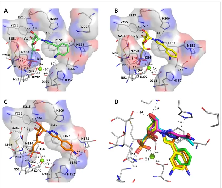
We then compared the three analogues bearing an aminophenyl substituent on the triazole ring with all possible orientations (ortho, meta or para) for the amino group (derivatives 1h, 1i and 1j). Interestingly, all of them showed very similar binding poses with respect to the positions of the oxygens linked to the phosphorus atom (strong ionic interactions with the magnesium ion), the ribose moiety (formation of hydrogen bonds between the hydroxy groups and Lys215) and the triazole ring oriented towards the hydrophobic residues Phe157 and His209 (Figure 8). However, the position of the phenyl group for deriv-ative 1h (amino group in the ortho position) is clearly different than the one of derivatives 1i and 1j (these last being very simi-lar to each other) and the rotation of the phenyl group appears to be dependent on the orientation of the amino group. According
to the inhibition results, derivative 1j was less potent than ex-pected (in view of the interaction of the para-amino phenyl with Asn158) and derivative 1h was found to be more active. This last may be explained by the interaction of the ortho-aminophenyl with His352 residue of cN-II as it represents the only difference with the others (Figure 8A). One should note that in comparison to smallest substituents on the triazole ring (compounds 1n, 1o and 1q) in compounds 1h, 1i and 1j the position of the five-membered ring is rotated by 90° (Figure 8D).
Conclusion
A small library of seventeen 1’-triazolyl beta-hydroxyphos-phonate ribonucleoside analogues was synthesized using conve-nient Cu(I)-catalysed cycloaddition. These derivatives were evaluated as potential cN-II inhibitors on the purified enzyme. Two derivatives including either an aminophenyl or an amido-substituent in the 4-position of the triazole ring were identified as modest inhibitors. Based on this study and previous SARs on cN-II inhibitors, we believe that optimized derivatives should be able to interact at least with: Phe157 and His209 for the nucleobase, Ser251 and Lys215 for the hydroxy groups of the sugar, and finally with Met53 and Lys292 for the phosphonate group, within the IMP-nucleotide binding site of cN-II.
Experimental
General procedure A for click reaction: The azido-sugar 2
(1 equiv) was dissolved in dry THF (45 mL/mmol) and the re-quired alkyne derivative (5.4 equiv), diisopropylethylamine (1.9 equiv), CuI (0.57 equiv) and DMEDA (5.2 equiv) were added. The reaction mixture was heated to reflux until TLC in-dicated complete consumption of 2, then the solvent was re-moved. The residue was dissolved in EtOAc and washed with H2O twice and once with an aqueous solution of EDTA (1%, m/v). The organic layer was dried over MgSO4, filtered and the solvent removed. Purification of the crude material on column chromatography on silica gel (CH2Cl2/EtOAc) afforded the desired product.
General procedure B for click reaction: The azido-sugar 2
(1 equiv) was dissolved in acetonitrile (25 mL/mmol) and then the required alkyne derivative (2.5 equiv) and CuI (0.4 equiv) were added. The reaction mixture was heated to reflux until TLC indicated complete consumption of 2. Then, the reaction mixture was filtered on celite and the solvent was removed. The residue was dissolved in EtOAc and washed with H2O twice and once with an aqueous solution of EDTA (1%, m/v). The organic layer was dried over with MgSO4, filtered and the sol-vent was removed. Purification of the crude material on column chromatography on silica gel (CH2Cl2/EtOAc) afforded the desired product.
Figure 8: Comparison of the docking poses obtained for three active derivatives in the substrate binding site of cN-II. Main interactions between
deriv-atives (A) 1h (green stick) or (B) 1i (yellow sticks) or (C) 1j (orange sticks) and cN-II residues (depicted in thin stick representation). (D) Superimposi-tion of the docking poses obtained for derivatives 1n (cyan sticks), 1q (pink sticks), 1h (green stick), 1i (yellow sticks), 1j (orange sticks) in the sub-strate binding site.
General procedure C for click reaction: The azido-sugar 2
(1 equiv) was dissolved in DMF (0.8 mL/mmol), the alkyne de-rivative (5.1 equiv), CuSO4 (0.03 equiv) and sodium ascorbate (0.1 equiv) were added to the solution. The reaction mixture was heated at 70 °C until TLC indicated complete consumption of 2. Then, it was diluted with water and extracted with EtOAc, the organic layers were combined and dried over with MgSO4 and concentrated under reduced pressure. The crude material was purified by flash chromatography (CH2Cl2/EtOAc) to give the desired product.
General procedure D for removal of sugar protecting groups: The protected derivative was dissolved in methanolic
ammonia (20 mL/mmol) at room temperature and stirred overnight, and then the reaction mixture was concentrated under
vacuum. The crude material was purified by flash chromatogra-phy (CH2Cl2/MeOH) to give the desired product.
General procedure E for diethyl phosphonate removal: The
protected phosphonate (1 equiv) was dissolved in anhydrous DMF (20 mL/mmol) and trimethylsilyl bromide (15 equiv) was added dropwise at 0 °C. The reaction mixture was stirred at room temperature until completion of the reaction was indicat-ed by TLC (isopropanol/water/ammonia, 7:2:1, v/v/v). Then, the reaction was stopped by adding triethylammonium bicar-bonate buffer (TEAB 1 M, pH 7) and concentrated to dryness under high vacuum. Column chromatography of the crude ma-terials on reverse phase (gradient: water to methanol 100%) gave the expected phosphonic acid (as triethylammonium salt), which was passed through a Dowex Na+ ion exchange column,
the desired fractions were collected and freeze dried leading to the title compound as sodium salt.
Supporting Information
Supporting Information File 1
Description of the materials and methods, and the preparation and characterization of new compounds. [http://www.beilstein-journals.org/bjoc/content/ supplementary/1860-5397-12-144-S1.pdf]
Supporting Information File 2
Copies of spectra for final compounds and NMR studies. [http://www.beilstein-journals.org/bjoc/content/ supplementary/1860-5397-12-144-S2.pdf]
Acknowledgements
This work was supported by Institutional funds from the Agence Nationale de la Recherche (ANR Programme Blanc 2011-SIMI7, projet cN-II Focus). T.N.V. and A.H. are grateful for their doctoral fellowships to the Vietnamese Government & the USTH, and the Ministère National de l’Enseignement et de la Recherche, respectively. We thank Miss M. Lagacherie for technical assistance and Mrs M.-C. Bergogne for dedicated editorial assistance.
References
1. Bianchi, V.; Spychala, J. J. Biol. Chem. 2003, 278, 46195–46198. doi:10.1074/jbc.R300032200
2. Jordheim, L. P.; Chaloin, L. Curr. Med. Chem. 2013, 20, 4292–4303. doi:10.2174/0929867311320340008
3. Tozzi, M. G.; Pesi, R.; Allegrini, S. Curr. Med. Chem. 2013, 20, 4285–4291. doi:10.2174/0929867311320340007
4. Meyer, J. A.; Wang, J.; Hogan, L. E.; Yang, J. J.; Dandekar, S.; Patel, J. P.; Tang, Z.; Zumbo, P.; Li, S.; Zavadil, J.; Levine, R. L.; Cardozo, T.; Hunger, S. P.; Raetz, E. A.; Evans, W. E.; Morrison, D. J.; Mason, C. E.; Carroll, W. L. Nat. Genet. 2013, 45, 290–294.
doi:10.1038/ng.2558
5. Tzoneva, G.; Perez-Garcia, A.; Carpenter, Z.; Khiabanian, H.; Tosello, V.; Allegretta, M.; Paietta, E.; Racevskis, J.; Rowe, J. M.; Tallman, M. S.; Paganin, M.; Basso, G.; Hof, J.;
Kirschner-Schwabe, R.; Palomero, T.; Rabadan, R.; Ferrando, A. Nat. Med. 2013, 19, 368–371. doi:10.1038/nm.3078
6. Cividini, F.; Pesi, R.; Chaloin, L.; Allegrini, S.; Camici, M.; Cros-Perrial, E.; Dumontet, C.; Jordheim, L. P.; Tozzi, M. G. Biochem. Pharmacol. 2015, 94, 63–68. doi:10.1016/j.bcp.2015.01.010 7. Gallier, F.; Lallemand, P.; Meurillon, M.; Jordheim, L. P.; Dumontet, C.;
Périgaud, C.; Lionne, C.; Peyrottes, S.; Chaloin, L. PLoS Comput. Biol.
2011, 7, e1002295. doi:10.1371/journal.pcbi.1002295
8. Meurillon, M.; Marton, Z.; Hospital, A.; Jordheim, L. P.; Béjaud, J.; Lionne, C.; Dumontet, C.; Périgaud, C.; Chaloin, L.; Peyrottes, S. Eur. J. Med. Chem. 2014, 77, 18–37.
doi:10.1016/j.ejmech.2014.02.055
9. Jordheim, L. P.; Marton, Z.; Rhimi, M.; Cros-Perrial, E.; Lionne, C.; Peyrottes, S.; Dumontet, C.; Aghajari, N.; Chaloin, L.
Biochem. Pharmacol. 2013, 85, 497–506. doi:10.1016/j.bcp.2012.11.024
10. Marton, Z.; Guillon, R.; Krimm, I.; Preeti; Rahimova, R.; Egron, D.; Jordheim, L. P.; Aghajari, N.; Dumontet, C.; Périgaud, C.; Lionne, C.; Peyrottes, S.; Chaloin, L. J. Med. Chem. 2015, 58, 9680–9696. doi:10.1021/acs.jmedchem.5b01616
11. Zimmermann, S. C.; O'Neill, E.; Ebiloma, G. U.; Wallace, L. J. M.; De Koning, H. P.; Seley-Radtke, K. L. Molecules 2014, 19, 21200–21214. doi:10.3390/molecules191221200
12. Zimmermann, S. C.; Sadler, J. M.; Andrei, G.; Snoeck, R.; Balzarini, J.; Seley-Radtke, K. L. Med. Chem. Commun. 2011, 2, 650–654. doi:10.1039/c1md00094b
13. Zimmermann, S. C.; Sadler, J. M.; O'Daniel, P. I.; Kim, N. T.; Seley-Radtke, K. L. Nucleosides, Nucleotides Nucleic Acids 2013, 32, 137–154. doi:10.1080/15257770.2013.771187
14. St Amant, A. H.; Bean, L. A.; Guthrie, J. P.; Hudson, R. H. E. Org. Biomol. Chem. 2012, 10, 6521–6525. doi:10.1039/c2ob25678a 15. Gallier, F.; Peyrottes, S.; Périgaud, C. Eur. J. Org. Chem. 2007,
925–933. doi:10.1002/ejoc.200600562
16. Wrobel, M.; Aubé, J.; König, B. Beilstein J. Org. Chem. 2012, 8, 1027–1036. doi:10.3762/bjoc.8.115
17. Pesi, R.; Allegrini, S.; Careddu, M. G.; Filoni, D. N.; Camici, M.; Tozzi, M. G. FEBS J. 2010, 277, 4863–4872.
doi:10.1111/j.1742-4658.2010.07891.x
18. Walldén, K.; Nordlund, P. J. Mol. Biol. 2011, 408, 684–696. doi:10.1016/j.jmb.2011.02.059
License and Terms
This is an Open Access article under the terms of the Creative Commons Attribution License
(http://creativecommons.org/licenses/by/2.0), which permits unrestricted use, distribution, and reproduction in any medium, provided the original work is properly cited. The license is subject to the Beilstein Journal of Organic
Chemistry terms and conditions:
(http://www.beilstein-journals.org/bjoc)
The definitive version of this article is the electronic one which can be found at:
![Figure 1: Previous (UA1776, UA2201 and UA2209 [7,8]) and new 1a–q phosphonate derivatives designed as potential cN-II inhibitors.](https://thumb-eu.123doks.com/thumbv2/123doknet/14270149.490316/3.892.91.805.821.1113/figure-previous-ua-phosphonate-derivatives-designed-potential-inhibitors.webp)