Cellular Compartmentation Effects in Receptor-mediated Signal Transduction
by
Jason Michael Haugh B.S., Chemical Engineering North Carolina State University, 1994
Submitted to the department of Chemical Engineering in partial fulfillment of the requirements for the degree of
Doctor of Philosophy in Chemical Engineering at the
Massachusetts Institute of Technology September 1999
© 1999 Massachusetts Institute of Technology" All rights reserved
Signature of Author ...
Department of Chei 1apgineering e 2, 1999 /:7 -.
Certified by...
Professor of Chemical Engineering Thesis Supervisor
/
/11
A ccepted by ... ...
Robert e. Cohen St. Laurent Professor of Chemical Engineering Chairman, Committee for Graduate Students
7M
STITUTECellular Compartmentation Effects in Receptor-mediated Signal Transduction by
Jason Michael Haugh
Submitted to the Department of Chemical Engineering on June 2, 1999 in partial fulfillment of the requirements for the degree of
Doctor of Philosophy in Chemical Engineering Abstract
Cells survey their environment primarily through the engagement of specific cell surface receptor proteins. Ligated receptors participate in signal transduction by initiating intracellular reactions involving heterologous proteins and metobolites. A quantitative understanding of these signaling processes is the key to controlling cell functional
responses, including survival and growth, migration, differentiation, and secretion. One issue of particular importance is how the structural organization of the cell can affect these events, by regulating the subcellular localization of signaling molecules. For example, many signaling receptors are internalized into the cell upon activation, leading to their delivery from the plasma membrane to intracellular organelles called endosomes.
Receptors in complex with cytosolic proteins almost invariably target membrane-associated molecules, including certain membrane lipids, Ras and related small GTPases, heterotrimeric G-proteins, and Src family tyrosine kinases, to carry out signaling functions. Based on theoretical modeling efforts, it is asserted that the membrane localization of these molecules impacts the organization of signaling interactions in two major ways: 1) it allows amplification of signaling via recruitment of enzymes from the cytosol to the membrane, which is expected to completely synergize with allosteric effects and covalent modifications like phosphorylation, and 2) it allows variations in membrane component concentrations and formation of microdomains to arise based on the chemical interactions between different membrane lipids. The latter would also affect the composition of endosomes relative to the plasma membrane, environments that are physically separated. For these reasons, unstructured models of intracellular signal transduction pathways are likely to be inadequate.
Experimental work involved the investigation of how the internalization of epidermal growth factor receptor (EGFR), a prominent receptor tyrosine kinase in mammals, affects the magnitude of signaling through distinct pathways involving
phospholipase C (PLC) and the Ras GTPase. For both pathways, the binding and tyrosine phosphorylation of cytosolic proteins (PLC-yl for the PLC pathway and Shc for the Ras pathway) were not affected by receptor compartmentation in endosomes for a given level of total receptor activation. However, at the level of membrane target modification,
endosome-associated PLC-yl could not hydrolyze its lipid substrate phosphatidylinositol (4,5)-bisphosphate (PIP,), while the membrane-associated protein Ras was efficiently activated by internal EGFR in the same cell line. This provides evidence that receptors in different compartments may not have access to the same membrane-associated signaling molecules, and that internalization can select for the activation of certain pathways. Thesis Supervisor: Douglas A. Lauffenburger
Acknowledgments
I am extremely grateful to my advisor, Douglas Lauffenburger, for his wisdom,
encouragement and patience during the completion of this project. I would also like to thank all DAL lab members past and present, especially Cartikeya Reddy, Dave Schaffer, Lily Chu, Marti Ware, Sean Palecek, Mark Powers, Chase Orsello, Eric Fallon, Anand
Asthagiri, Anne Dewitt, Casim Sarkar, Michael Lassle, and Doug "Pointspread" Osborne for fostering a lighthearted atmosphere on the 3rd floor and for helpful discussions. I will miss you guys, but I will still be in P.P.E.with picks ready to go. I am indebted to our collaborator on the project, Alan Wells, for immeasurable help and advice, as well as Kiran
Gupta, Mark Van Epps-Fung, and Heng Xie from his lab for technical assistance.
Excellent assistance in lab was also provided by Alarice Huang, an undergraduate student from the Biology department, who helped with this project. More great advice and encouragement was provided by the other members of my thesis committee: Professors William Deen, Gregory Stephanopoulos, and Lawrence Stern. While I owe a great debt to
a lot of people, I was personally kept out of debt by the National Science Foundation Graduate Fellowship Program, the National Science Foundation Biotechnology Program, and the Merck/MIT Collaboration, who funded my research. Finally, I want to thank my parents, Martin and Deborah Haugh, and especially my wife, Ruthie, for their support and
Table of Contents
Chapter 1: Introduction and Background ... 11
1.1 Epidermal Growth Factor Receptor (EGFR) and its Ligands ... 11
1.2 Regulation of EGFR Function by Phosphorylation . ... .... 13
1.3 EGFR-mediated Signal Transduction Pathways . ... 15
1.4 Intracellular Trafficking of EGFR and its Ligands ... 20
1.5 Thesis Topic ... 26
1.6 Previous Studies Relating EGFR Trafficking and Signaling ... 27
1.7 Thesis Overview 28 1.8 References 29 Chapter 2: Receptor-mediated Recruitment of Intracellular Signaling Proteins to the Plasma Membrane: Theoretical Analysis ... 38
2.1 Introduction 38 2.2 Theory of Reaction and Diffusion in Two and Three Dimensions .. 40
2.2.1 General Considerations 40 2.2.2 Association of a Cytosolic Protein with a Membrane Target.... 41
2.2.3 Interaction Between Two Membrane Species ... 44
2.3 Results 46 2.3.1 Enhancement of Association Rates 46 2.3.2 Upregulation of a Membrane Messenger by a Membrane Receptor-recruited Activator ... 50
2.3.3 Responsive Activation of a Cytosolic Messenger by a Membrane Receptor ... 55 2.3.4 Experimental Relevance ... 58 2.4 2.5 Discussion References 62 64 Chapter 3: Generalized Mathematical Model of Membrane- compartmentalized Receptor-mediated Signaling ... 67
3.1 Introduction 67
3.2 Mathematical Model 69
3.2.3 The Kinetic Approximation ... 76
3.2.4 The Smear Model... 79
3.2.5 Overall Signaling Activity ... 81
3.3 Results 82 3.3.1 Parameter Estimation 82 3.3.2 Static Representation of Compartmentalized RTK Signaling 85 3.3.3 Dynamic RTK Signaling: Insensitive to Substrate Phosphorylation ... 88
3.3.4 Dynamic RTK Signaling: Substrate Phosphorylation Required ... 91
3.3.5 Cellular Organization and the Selectivity Parameter A ... 91
3.4 Discussion 95 3.5 References 98 Chapter 4: Epidermal Growth Factor Receptor Trafficking and Regulation of Phospholipase C Signaling ... 101
4.1 Introduction 101 4.2 Experimental Procedures ... 104
4.2.1 Cell Culture and Quiescence Protocol ... 104
4.2.2 Receptor Binding and Internalization Studies ... 104
4.2.3 Removal of Surface-bound Ligand by Mild Acid Strip ... 105
4.2.4 EGFR-phosphotyrosine Sandwich ELISA ... 105
4.2.5 Immunoprecipitation and Western Blotting ... 106
4.2.6 Determination of Internal EGFR-phosphotyrosine ... 106
4.2.7 PIP, Hydrolysis Assay ... 107
4.3 Results 107 4.3.1 Binding, Activation, and Internalization of the EGFR ... 107
4.3.2 Stoichiometry of EGFR Tyrosine Autophosphorylation . 108 4.3.3 Dose Responses of pY-EGFR and PIP2 Hydrolysis ... 114
4.3.4 Spatial Restriction of PIP, Hydrolysis to the Cell Surface . 117 4.3.5 Tyrosine Phosphorylation of PLC-yl ... 123
4.4 Discussion 125
Chapter 5: Influence of Epidermal Growth Factor Receptor Trafficking
and Feedback Desensitization on the Activation of Ras 131
5.1 Introduction...3.... ... 11
5.2 Experimental Procedures 135
5.2.1 Cell Culture and Quiescence Protocol ... 135 5.2.2 Use of Pharmacological Inhibitor PD098059 ... 135
5.2.3 Surface Titration Protocol 135
5.2.4 EGFR Autophosphorylation ... 136 5.2.5 Shc Tyrosine Phosphorylation and
Coprecipitation with the EGFR ... 136
5.2.6 Ras Immunoprecipitation and
Elution of Guanine Nucleotides 137
5.2.7 Ras Guanine Nucleotide Exchange ... 137
5.2.8 GTP and GDP Determination 137
5.3 Results 138
5.3.1
... . . . .
Feedback Desensitization of Ras Guanine Nucleotide Exchange
5.3.2 Compartmentalization and Desensitization of pY-EGFR .
5.3.3 Tyrosine Phosphorylation of Shc ... ... 5.3.4 Complexation of Shc with EGFR ... 5.3.5 Activation of Ras.
5.4 Discussion 5.5 References
Chapter 6: Receptor-mediated Supply and Hydrolysis of Phosphoinositide Lipids: a Second Generation Mathematical Model
6.1 Introduction.
6.2 Mathematical Model.
6.2.1 General Considerations
6.2.2 Case 1.1: E binding saturated, T and C binding linear ... 6.2.3 Case 1.2: E and C binding saturated, T binding linear ... 6.2.4 Case 2.1: T binding saturated, E and C binding linear ... 6.2.5 Case 2.2: T and C binding saturated, E binding linear ... 6.2.6 Case 3.1: All protein-receptor interactions linear
6.2.7 Case 3.2: E and T binding linear, C binding saturated ...
138 139 141 144 147 154 158 162 162 166 166 169 169 170 170 170 171
6.3.1 Qualitative Assessment of Special Cases ... 172 6.3.2 Parameter Analysis: Linear Receptor-protein Interactions .. 175 6.3.3 Parameter Analysis: T and C Binding Saturable ... 176
6.4 Discussion 179
6.5 References 180
Appendices ... 182 A. Mean Diffusion Time within a Bounded Cone to a Sink
at the Center of its Top Surface ... 182 B. Receptor-ligand Binding and Internalization Kinetics 186 C. Membrane Recruitment and Zero-order Sensitivity ... 189 D: Simplified Receptor Trafficking and Membrane Dynamics ... 190 E. Secondary Effects on Substrate Phosphorylation State 193
Tables and Figures
Table 1.1 Signaling Functions of EGFR-binding Proteins ... 17
Table 3.1 Model Parameter Estimates 84 Table 6.1 Summary of Adjustable Model Parameters ... 173
Table 6.2 Molecular Requirements for Model Agreement with Experiment ... 174
Figure 1.1 Structure/Function of the Epidermal Growth Factor Receptor (EGFR) 12 Figure 1.2 Mechanisms of RTK-mediated Activation of Protein Substrates ... 16
Figure 1.3 Endocytic Trafficking... ... 22
Figure 1.4 Saturation of Internalization in EGFR-expressing NR6 Fibroblasts . 23 Figure 1.5 Mechanistic Analysis of Endosomal Sorting ... 25
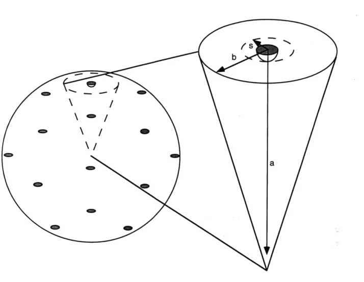
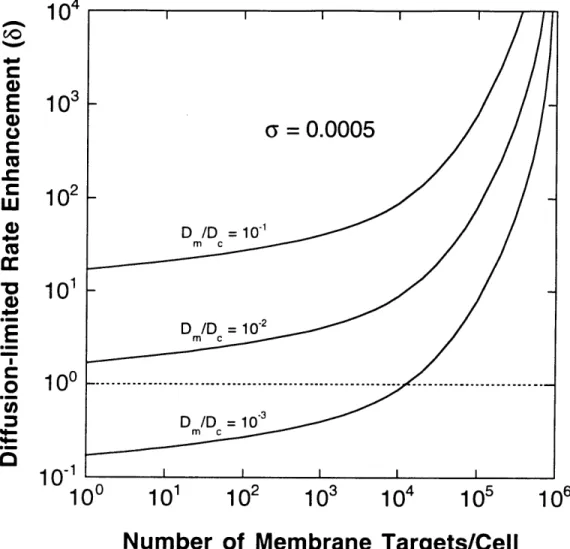
Figure 2.1 Geometry of the Area and Volume Afforded each Membrane Sink . 43 Figure 2.2 Enhancement of Diffusion-limited Target Association Rate Proferred by Recruitment to the Plasma Membrane ... 48
Figure 2.3 Enhancement of Target Association Rate Proferred by Recruitment to the Plasma Membrane: Reaction and Diffusion Limitations 49 Figure 2.4 Two Highly Simplified Signal Transduction Models ... 51
Figure 2.5 Activation of a Membrane Signaling Target ... 54
Figure 2.6 Regulation of a Cytosolic Signaling Protein ... ... 57
Figure 2.7 Sensitivity of Model 1 Signal Magnitude to Aspects of Membrane Recruitment 60 Figure 2.8 Sensitivity of Model 2 Signal Responsiveness to Aspects of Membrane Recruitment 61 Figure 3.1 Points of Regulation in RTK-mediated Signal Transduction ... 70
Figure 3.2 General Model Solution Approach ... 71
Figure 3.3 General Reaction-Diffusion RTK Signaling Model ... 73
Figure 3.4 Two Model Variations ... 77
Figure 3.5 Static RTK Signaling ... 87
Figure 3.8 Receptor Internalization and Target Availability 4.1 4.2 4.3 4.4 4.5 4.6 4.7 4.8 4.9 4.1( Figure 4.1 Figure 4.1 5.1 5.2 5.3 5.4 5.5 5.6 5.7 5.8 5.9 Figure Figure Figure Figure Figure Figure Figure Figure Figure Figure
The Ras/Erk Pathway ... MEK-dependent Desensitization of Ras
Guanine Nucleotide Exchange ... Compartmentalization of Activated EGFR
Quantitative Immunoblotting of Tyrosine-phosphorylated Shc Compartmentalization of Shc Tyrosine Phosphorylation ...
Coprecipitation of Shc with Surface and Internal EGFR ... GTP and GDP Determination...
Participation of Surface and Internal EGFR in the Activation of Ras . Two Models of EGFR-mediated Activation of Ras
134 140 142 143 145 148 151 153 157
Figure 6.1 Experimental Activation-response Relationships for
Wild-type EGFR-expressing NR6 Fibroblasts ... 165
Figure 6.2 Schematic of Model Signaling Pathway ... 167
Figure 6.3 Analysis of Presursor Hydrolysis: All Receptor Interactions Linear 177 Figure 6.4 Analysis of Precursor Hydrolysis: Transfer Protein- and Competitive Enzyme-receptor Interactions Saturable ... 178
The Phospholipase C Pathway ... ... 103
Kinetics of EGFR Internalization ... 109
Time Course of EGFR Tyrosine Phosphorylation ... 110
Analysis of EGFR Tyrosine Phosphorylation ... 112
Tyrosine Phosphorylation of Internalized EGFR ... 113
Dose Response of EGFR Tyrosine Phosphorylation ... 115
Dose Response of EGFR-mediated PIP2 Hydrolysis ... 116
Surface Titration Protocol .. ... 118
Results of Surface Titration Experiments ... 119
Inhibition of Recycled Receptor/ligand Complex Signaling by an anti-EGFR Antibody ... 121
1 Plot of PIP2 Hydrolysis versus pY-EGFR for Dose Response and Surface Titration Experiments ... 122
2 Analysis of PLC-yl Tyrosine Phosphorylation ... 124
Figure Figure Figure Figure Figure Figure Figure Figure Figure 94
Figure A. 1 Computational Analysis of Equation A.7... ... 185
Figure B. 1 Time Profiles of C,, the Level of Surface
Receptor-ligand Complexes per Cell ... 188
Figure D. 1 Simplified RTK Binding and Trafficking Kinetics ... 192
Figure E. 1 Secondary Substrate Phosphorylation/Dephosphorylation
CHAPTER 1
Introduction and Background
Living cells react to perturbations in their environment. Such responses include survival and proliferation, migration, or differentiation of cell function. The goal of cell
engineering is to quantitatively control these behaviors through manipulation of the cellular
environment and the ability of the cell to respond to it. Realization of this goal would allow rational design of pharmaceuticals and gene therapies at the cellular level, use of engineered cells themselves as therapeutic agents, tailoring of biomaterials for tissue engineering applications, and optimization of commercial protein production. The primary way in which cells survey their surroundings is through reversible binding of cell surface receptors. These proteins span the cell membrane and present docking sites for specific ligand molecules. Once a complex of ligand and receptor has formed, the receptor is able to initiate multiple chemical reactions. These reactions eventually trigger the activation of genes and expression of protein products, changes in cell metabolism, and modification of cellular structure. Thus, this process of signal transduction, or signaling, is the
biochemical integration of all the information perceived by the cell.
1.1 Epidermal Growth Factor Receptor (EGFR) and its Ligands
The 170 kDa epidermal growth factor receptor (EGFR) is the best-characterized of a class of signaling receptors called receptor tyrosine kinases (RTKs), which contain a domain with intrinsic enzymatic activity that is able to selectively phosphorylate protein
substrates on tyrosine residues. Other RTKs include the receptors for insulin, insulin-like growth factor, platelet-derived growth factor (PDGF), fibroblast growth factor, and nerve growth factor (van der Geer et al., 1994). The relavent structural details of the EGFR are illustrated in Figure 1.1. The functional domains of the EGFR include the extracellular ligand-binding (ecto) domain, which contains dual cysteine-rich regions that cooperate to bind growth factor ligands, short transmembrane and juxtamembrane regions, the kinase domain, and a cytosolic regulatory domain. Early studies of EGFR function indicated that
its tyrosine kinase activity is fully responsible for its biological function (Chen et al., 1987). EGFR is the first member of the erbB receptor family of RTKs. The other members are erbB-2, erbB-3, and erbB-4.
EGF
DOMAINSRECEPTOR
E C -958 T 0 -973-y* -- -992 622 -: 2 644 TM 663 -1022 K721 K II N A R y* - 1068 EY* 1086 G 3 957 -U L A 1114 TY - 1148 Y 1173 1186 1186Figure 1.1 Structure/Function of the Epidermal Growth Factor Receptor
(EGFR). The sites of tyrosine autophosphorylation are indicated by asterisks. Each of the regions designated 1, 2, and 3 provide gain of function when added onto truncated, internalization-deficient receptors. TM, transmembrane segment. Adapted from Chang et al., 1993.
The proto-oncogene erbB-2 is found to be overexpressed in multiple human tumors (particularly those of the breast), has no known ligand, and is transactivated by activated EGFR (Hynes and Stem, 1994; Dougall et al., 1994). ErbB-3 and -4 have differing ligand specificities from EGFR, and, interestingly, erbB-3 lacks intrinsic tyrosine kinase activity (Carraway and Cantley, 1994; Chen et al., 1996b).
Epidermal growth factor (EGF) is synthesized as a 1207 amino acid precursor, while the functional secreted form is 53 amino acids (Scott et al., 1983; Bell et al., 1986). EGF is capable of stimulating a spectrum of responses, including proliferation and
migration, in many cells of epithelial origin, including keratinacytes and fibroblasts. Discovered more than 30 years ago, roles for this cytokine have been identified in tissue organization during development and wound healing (Carpenter and Wahl, 1990;
Pittelkow, 1992). EGF binds reversibly to the EGFR with 1:1 stoichiometry (Weber et al., 1984), stimulating receptor tyrosine kinase activity, homo- and hetero-dimerization, and endocytic internalizaton, with the outcome of multiple signaling pathways regulating the cell behavioral response (Lund et al., 1990b; Lemmon and Schlessinger, 1994; van der Geer et al., 1994). TGFo, amphiregulin, heparin-binding EGF, and betacellulin are among other ligands which belong to the EGF family and bind EGFR; the EGF family member neu differentiation factor (NDF) does not bind to EGFR, but rather to erbB-3 and
-4 (Carpenter and Wahl, 1990; Kimura et al., 1990).
1.2 Regulation of EGFR Function by Phosphorylation
EGFR kinase activity, associated with residues 663-957, is activated upon ligand association, and the first substrate to be phosphorylated is the receptor itself. The kinase machinery autophosphorylates tyrosines in the C-terminal regulatory region (Figure 1.1), which can be considered to occur instantaneously upon receptor ligation (Hunter and Cooper, 198 1 ): the kinetics of EGFR kinase activity (Cheng and Koland, 1996) are very rapid compared to those of ligand binding. The autophosphorylation sites of EGFR compete with other substrates for the kinase, suggesting a mechanism by which
phosphorylation relieves an inhibition of kinase activity by the regulatory domain of the EGFR (Bertics and Gill, 1985; Bertics et al., 1985; Walton et al., 1990). Tyrosine phosphorylation is reversed by protein tyrosine phosphatases (PTPs) (Tonks and
Charbonneau, 1989; Fischer et al., 1991; Sun and Tonks, 1994; Neel and Tonks, 1997), although much less is known about phosphatases than kinases in general. Some PTPs that
modulate RPTK activity are membrane proteins (Kulas et al., 1996b; Kulas et al., 1996a), implying a high frequency of potentially reactive collisions with RTKs.
Y 1173 is the preferred autophosphorylation site of the EGFR, with nearly 1:1 phosphorylation per bound receptor. Y 1148 and Y1068 are secondary sites, while Y992 and 1086 are phosphorylated at very low stoichiometry (Downward et al., 1984; Margolis et al., 1989; Walton et al., 1990). Unoccupied receptors are either not able to be
phosphorylated or are summarily dephosphorylated by phosphatases, so there is a
stoichiometric relationship between ligated and tyrosine-phosphorylated EGFR (Lund and Wiley, 1994).
A large body of evidence suggests that EGFR signaling is attenuated by
heterologous phosphorylation on serine and threonine residues. In particular, T654, T669, S67 1, and S 1046/7 have been identified as phosphorylation sites that regulate EGFR function, although the relative importance of each is debatable. Activation of protein kinase C (PKC) has been shown to correlate with T654 phosphorylation, affinity
downmodulation, and abrogation of EGF-induced responses, representing a negative feedback loop that can be selectively blocked by the T654A mutation (Fearn and King,
1985; Lin et al., 1986; Welsh et al., 1991). PDGF agonization of its receptor also
potentiates phosphorylation of EGFR T654, but in a PKC-independent manner (Davis and Czech, 1987). Interestingly, depletion of PKC in human fibroblasts by long-term
exposure to phorbol esters does not affect the observed modulation of affinity, indicating other mechanisms (Wiley et al., 1989). T654 phosphorylation in response to phorbol ester treatment significantly attenuates EGFR kinase activity (Lund et al., 1990a). Thus, while affinity may be only modestly reduced, the biological activity of receptors desensitized in this manner may be seriously compromised in some cases. Another study examined T669 and S671 phosphorylation by mutating these sites to alanine. While these receptors bound ligand and were autophosphorylated normally, they were impaired in their ability to
internalize (see below) inducibly (Heisermann et al., 1990). Interestingly, EGFR T669 is phosphorylated by a mitogen-activated protein kinase (MAPK) in vitro, suggesting a feedback loop (Northwood et al., 199 1; Takishima et al., 1991). Finally, alanine mutation of S 1046/7 also inhibited internalization and prevented desensitization of EGFR-mediated phosphorylation of an exogenous substrate in vitro (Countaway et al., 1992; Theroux et al., 1992). Taken together, serine/threonine phosphorylation can compromise the biological activity of EGFR in varying ways.
1.3 EGFR-mediated Signal Transduction Pathways
Intracellular proteins that participate in signaling cascades recognize
autophosphorylated residues on EGFR and other RTKs. These physical interactions are achieved through the src homology 2 (SH2) and phosphotyrosine-binding (PTB) modular domains of signaling proteins (Pawson, 1995; van der Geer and Pawson, 1995), which derive specificity from the primary structure of the receptor around the target tyrosine (Songyang and Cantley, 1995; Zhou et al., 1995). The three major potential implications of these binding events, as illustrated in Figure 1.2, are: 1) allosteric activation of the protein substrate, 2) an increase in the frequency of phosphorylation of the bound substrate by the receptor kinase, and 3) recruitment to the membrane that enhances associations with downstream targets there. Allostery, covalent modification, and membrane localization are all likely to play a role in any given signaling pathway at the receptor level, and these contributions can be synergistic.
EGFR mediates the activation of three major signaling pathways: the phospholipase C (PLC), the Ras/mitogen-activated protein kinase (Ras/MAPK), and phosphatidylinositol 3-kinase [PI(3)K] pathways (Table 1.1). EGFR elicits activation of the yl isoform of PLC in fibroblasts, which catalyzes the hydrolysis of the acidic phospholipid
phosphatidylinositol (4,5)-bisphosphate (PIP2) (Rhee and Choi, 1992). The products of
this reaction are diacylglycerol (DAG), which remains in the membrane and activates the serine/threonine kinase protein kinase C (PKC), and inositol (1,4,5)-triphosphate (IP3), which cooperatively releases calcium from intracellular stores (Toker, 1998). Also, proteins with pleckstrin homology (PH) domains bind to PIP2 or other lipids with high
affinity (Lemmon et al., 1996; Lemmon et al., 1997), and PLC-mediated hydrolysis releases associated proteins into the cytosol. Binding of PLC-yl to autophosphorylated EGFR enhances the tyrosine phosphorylation of PLC-yl by the receptor kinase, predictably by reducing the Michaelis constant Km (Margolis et al., 1990; Rotin et al.,
1992: Zhu et al., 1992). Phosphorylation by EGFR activates the enzyme in vivo, apparently by allowing it to hydrolyze PIP, bound by other proteins (Goldschmidt-Clermont et al., 1991). Since PIP, is a membrane lipid, recruitment to activated EGFR would presumably also modulate PLC by membrane localization (Fig. 1.2).
Previous studies using EGFR-expressing NR6 fibroblasts have identified the molecular requirements for PLC activation and its role in cellular function. Using signaling-restrictive EGFR mutants, it was determined that EGFR kinase activity and autophosphorylation sites are strictly required for modulation of PLC activity in vivo (Margolis et al., 1990; Vega et al., 1992; Chen et al., 1994b).
--P Pmo- TARGET(S) Conformational Change ________ ATP k -TARGET(S) Phosphorylated Substrate MEMBRANE Localized
IGESubstrate
Figure 1.2 Mechanisms of RTK-mediated Activation of Protein Substrates. A. allostery. Upon association of a substrate molecule with an autophosphorylated
receptor. a conformational change is transmitted from the binding domain of the substrate to its catalytic domain. The activity of the catalytic domain towards downstream targets is affected by this structural change. B, increase in phosphorylation frequency. Receptor binding brings the substrate in prolonged proximity to the active kinase domain of the receptor, resulting in a significant increase in the rate at which the substrate is
phosphorylated. The activity of the substrate's catalytic domain towards downstream targets is affected by the phosphorylation state of the substrate. C, membrane localization. The catalytic domain of the substrate is already fully active per se, but the localization of a substrate molecule to a cellular membrane enhances its search for targets residing there.
Binding
Tyrosine-Domains Phosphorylated? Target Product(s)
Ras-GDP Grb2-Sos/ Ras-GDP DAG + IP 3 Ras-GTP Ras-GTP PIP3
Table 1.1 Signaling Functions of EGFR-binding Proteins. The enzymes phospholipase C-yl (PLC-yl) and phosphatidylinositol 3-kinase [PI(3)K] target the membrane lipid phosphatidylinositol (4,5)-bisphosphate (PIP2) for hydrolysis and
phosphorylation, respectively. The Grb2-Sos complex, aided by tyrosine-phosphorylated Shc. targets the membrane-tethered Ras-GTPase for nucleotide exchange (GTP for GDP) and activation. Substrate PLC-yl Grb2-Sos Shc PI(3)K 2xSH2 SH2 SH2, PTB 2xSH2 Y N Y Y
PLC-restrictive receptor mutants also failed to elicit cell migration in response to EGF, and inhibition of PLC activity by PLC-specific drug and PLC antisense treatments blocked cell motility (Chen et al., 1994a; Chen et al., 1994b). In these cells, PLC-mediated PIP, hydrolysis leads to liberation of the actin-modifying protein gelsolin into the cytosol. Inhibition of PLC blocked gelsolin release from the membrane, and treatment with
antisense gelsolin inhibited cell migration, linking EGFR-mediated PLC activity to dynamic changes in the cytoskeleton that affect migration (Chen et al., 1996a).
The Ras GTPase (21 kDa) is highly conserved in eukaryotes (Bourne et al., 1991), and is targeted for insertion in the plasma membrane by a series of posttranslational
modifications (Willumsen et al., 1984; Willumsen et al., 1996). In the GTP-bound active state, Ras initiates the Raf/MEK/MAPK kinase cascade and activates other GTPases to control cell growth, differentiation, and cytoskeletal organization in various cell types
(Vojtek and Der, 1998). Ras hydrolyzes GTP to GDP, which shuts off these signals, and the rate of hydrolysis is regulated by GTPase activating proteins (GAPs) (Zhang et al.,
1990; McCormick, 1996). Mutations which hinder the ability of Ras to hydrolyze GTP are transforming, and such mutations are implicated in a high percentage of human tumors (Bos, 1989). In fibroblasts, activation of the Ras pathway by EGFR is mediated by a complex of Grb2 and Sos proteins. Grb2 is a -23 kDa protein comprised almost entirely of one SH2 and two SH3 domains (Chardin et al., 1995), which ubiquitously
coprecipitates with EGFR and PDGF receptor via its SH2 domain. Grb2 is not
phosphorylated by RTKs and does not have enzymatic activity; its function is to serve as an adaptor that links other proteins such as Sos to RTKs via its SH3 domains (Lowenstein et al., 1992; Olivier et al., 1993; Simon et al., 1993; Buday and Downward, 1993a; Chardin et al.. 1993: Egan et al., 1993; Gale et al., 1993; Li et al., 1993; Rozakis-Adcock et al.,
1993). Homologs of Sos (Son of Sevenless) are guanine nucleotide exchange factors (GEFs) for the Ras GTPase (Wolfman and Macara, 1990; Bonfini et al., 1992; Bowtell et al., 1992).
Sos, as a GNP exchange factor, mediates the activation of Ras by stimulating the dissociation of GDP and GTP from Ras (Feig, 1994). The exchange is completed when Ras binds another GNP molecule from the pool of free nucleotides; this favors the active state in vito since GTP is in > 10-fold excess over GDP in the cytosol (Lai et al., 1993). The activation of Ras in response to RTK ligands, characterized almost exclusively in
fibroblasts, involves an increase in the exchange of nucleotides on Ras (Buday and Downward, 1993b; Medema et al., 1993). On the other hand, stimulated cells of hematopoietic lineage do not show such an increase in nucleotide exchange activity even
et al., 1990; Torti et al., 1992). In vitro studies demonstrate that Grb2 and mammalian homologs of Sos can form highly stable complexes; the SH3 domains of Grb2 both contribute to tight binding of proline rich sequences in Sos independently of Grb2 SH2 occupation (Cussac et al., 1994; Lemmon et al., 1994). While these studies suggest a precoupled heterodimer, coprecipitation of Sos and Grb2 is increased significantly in response to EGF in Rat 1 cells, suggesting that the interaction is regulated to some degree
in vivo (Buday and Downward, 1993a). Importantly, the in vitro exchange activity of Sos
does not depend on whether the protein is purified from quiescent or EGF-stimulated cells, nor is it affected by the presence of EGFR and/or Grb2 in vitro, suggesting that the Grb2-Sos or EGFR-Grb2-Grb2-Sos complexes by themselves do not have enhanced exchange activity (Buday and Downward, 1993a). Sos is not tyrosine-phosphorylated by RTKs and does not seem to be activated allosterically in a direct fashion by RTK and Grb2 interactions alone, begging the question of how the activity of Sos might be regulated by RTKs. A change in localization of Grb2-Sos from cytosol to membrane is observed in response to EGF, suggesting that the presence of Sos in complexes with EGFR has the effect of bringing GEF activity closer to the constitutively membrane-associated Ras (Buday and Downward, 1993a). Indeed, it was found that constitutive targeting of Sos to the
membrane is transforming in a Ras-dependent and RTK-independent fashion (Aronheim et al., 1994; Quilliam et al., 1994), confirming that location at the membrane is sufficient for
modulating Ras activation.
The 110 kDa mammalian phosphatidylinositol 3-kinases [PI(3)Ks] phosphorylate phosphatidylinositol (PI) lipids on the D3 position, producing PI 3-P, PI(3,4)P2, and PIP3.
The last two products are upregulated in response to RTK ligands (Auger et al., 1989) and seem to be instrumental in triggering cell proliferation, cytoskeletal organization, and survival (Carpenter and Cantley, 1996; Vanhaesebroeck et al., 1997; Carpenter, 1996; Toker and Cantley. 1997). Induction of PI(3)K activity associated with the a and P isoforms of p I 10 is observed in lysates of PDGF- and EGF-stimulated cells in vitro (Auger et al., 1989: Bjorge et al., 1990), and this activity coprecipitates with an 85 kDa protein recognized by anti-phosphotyrosine antibodies in PDGF-stimulated cells (Kaplan et al., 1987). Upon cloning of p85, it was discovered that there are two isoforms (a and p). These form stable heterodimers with p I 10 in vivo and have dual SH2 domains that
associate with autophosphorylated RTKs upon ligand stimulation in vitro (Escobedo et al., 1991; Skolnik et al., 1991; Hu et al., 1992; McGlade et al., 1992). While specific high affinity binding to PDGF receptors is primarily controlled by the C-terminal SH2 domain of p85 (Klippel et al., 1992), binding of both SH2 domains likely proffers stable binding and/or maximal PI(3)K activity in vivo (Cooper and Kashishian, 1993; Rordorf-Nikolic et
al., 1995). While EGF clearly stimulates PI(3)K in vivo, and binding to
autophosphorylated EGFR can be demonstrated in vitro, whether p85 can directly bind to EGFR in vivo has been questioned.
Binding of p85 SH2 domains induces a conformational change in p85, which is associated with an increase in p1 10 activity (Shoelson et al., 1993). This is accomplished via the p110 binding domain of p85, located between the SH2 domains; binding of this domain by itself is sufficient to activate p1 10 (Cooper and Kashishian, 1993; Klippel et al., 1993). A chimera of this domain fused with pl 10 is a constitutively active mutant (pl 10*), allowing study of PI(3)K-mediated downstream signaling (Hu et al., 1995). As previously discussed, RTK binding also localizes substrates to cellular membranes. Since the direct targets of PI(3)K are membrane lipids, a consequence of receptor binding would be that these target lipids would see a higher concentration of enzyme, yielding enhancement of the observed PI(3)K activity over and above that caused by the conformational change in p85 (allosteric effects). To test this, p110 constructs were modified with post-translational lipid modification signal peptides that target proteins to the plasma membrane, in tandem with the activating chimera; while the lipid-modified p1 10* has - 50% of the in vitro PI(3)K activity as p110*, it is significantly more potent in producing 3'-phosphorylated lipids and activating downstream effectors in vivo (Klippel et al., 1996). Membrane targeting alone is also sufficient to increase downstream signaling in vivo, albeit to a level lower than the membrane-localized, activated p110* (Reif et al., 1996; Klippel et al., 1996). These results suggest that allosteric and locational effects are synergistic in the potentiation of PI(3)K activity.
1.4 Intracellular Trafficking of EGFR and its Ligands
A complicating issue in the regulation of EGFR-mediated signaling is that ligated receptors do not necessarily stay at the plasma membrane the whole time they are activated. Many of these receptor-ligand complexes are inducibly internalized (receptor-mediated endocytosis) on the time scale of minutes by entrapment in specialized, clathrin-coated pits in the membrane (Trowbridge et al., 1993). This entrapment is mediated by adaptor proteins, which link endocytic motifs exposed in the cytoplasmic domain of a ligated receptor to the clathrin cage. These pits invaginate and pinch off to form vesicles (- 50
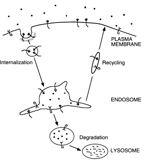
nm), which deliver receptor and ligand molecules to intracellular trafficking organelles known as sorting endosomes (100-500 nm). It is in endosomes that receptor and ligand molecules are sorted for recycling back to the cell surface or destruction in proteolytic
lysosomes (Figure 1.3). This allows the cell to control the number of receptor and ligand molecules available for long-term signaling (Mellman, 1996).
The specific rate of receptor internalization is characterized by the endocytic rate constant k, (Wiley and Cunningham, 1982):
t CS + CS'
C= keJ Csdt ~ ke I 2 .(t, - ti) (1.1),
where C and CS are the amount of ligand inside and on the surface of cells, respectively, and t is time. The value of ke can be determined by measuring C, and C, experimentally, employing the addition of radioiodinated ligand at time zero and construction of an InSur (Internal-Surface) plot, in which internal radioactivity C, is graphed versus the numerical integral of surface-associated radioactivity Cs with respect to time (eqn. 1.1). For EGFR, factors other than ligand binding influence the entrapment of receptors in clathrin-coated pits and therefore ke. Deletion of three regions of the EGFR cytosolic regulatory domain (Figure 1.3) yield successive loss of endocytic function (Chang et al., 1993). For
example, cells expressing a c'973 truncated EGFR internalize EGF at the same basal rate as that of non-activating anti-EGFR antibody internalization by wild-type EGFR. This basal rate is consistent with random inclusion in coated pits (Wiley, 1985). EGFR kinase activity is also required for internalization (Chen et al., 1987; Lamaze and Schmid, 1995). However, while receptor autophosphorylation has a positive effect on internalization, it is not a strict requirement for induced endocytosis, suggesting a role for a heterologous substrate of the EGFR kinase (Lund and Wiley, 1994).
As mentioned above, receptor-mediated endocytosis is strongly influenced by ligand-dependent interactions of the receptor with cytoplasmic adaptor molecules. Thus, the cell's ability to downregulate its ligated receptors is "saturable" in cell lines
overexpressing EGFR, presumably since the accessory proteins that immobilize receptors in clathrin-coated pits become stoichiometrically limiting (Wiley, 1988; Lund et al., 1990b). This effect can be assessed by plotting the endocytic rate constant ke versus the average C, for different ligand concentrations. Saturation is characterized by a drop in k, for increasing C'. Experiments of this nature were performed using radioiodinated EGF and EGFR-expressing NR6 fibroblasts. The parental NR6 line is devoid of EGFR mRNA and protein (Pruss and Herschman, 1977). The results (Figure 1.4) show that EGF internalization mediated by wild-type EGFR is saturable. The basal internalization rate, assessed for c'973 truncated EGFR, exhibits a constant, reduced value of ke.
0 0 * S * S * 0 * S 0 0
Recycling
ENDOSOME
Degradation
'
1
'
LYSOSOME
Figure 1.3 Endocytic Trafficking. The binding of cytokines to cognate receptors is often concomitant with the specific entrapment of the receptor in specialized clathrin-coated pits in the plasma membrane. Upon invagination the coated pit pinches off, and the resulting endocytic vesicle fuses with an early endosome, delivering receptor-ligand complexes and other components of the plasma membrane. It is here that receptors and
ligands are sorted for either recycling back to the cell surface or degradation. Long protruding endosomal tubules collect molecules for return to the surface, while molecules that remain in the vesicular portion of the endosome are routed for degradation in
lysosomes, which employ proteolytic enzymes to break down ligands and receptors. 0
Internali zation
e ,- -PLASMA
0.25
E,
0.2-40) A A AA -a U
0.15
-0 ~0*1
M
0.1 Is -0 NC~
0.05
00
00
0
0 1 1 1I ''ill 1 11 1 1102
10
3104
10
5 106Average Surface Binding CS (cell')
Figure 1.4 Saturation of Internalization in EGFR-expressing NR6
Fibroblasts. EGF was radioiodinated using lodobeads (Pierce), according to the
manufacturer's protocol. NR6 fibroblasts expressing wild-type (closed symbols) or c'973-truncated (open symbols) EGFR were exposed to "'I-EGF at various concentrations for times of 1. 2, 3, 4, and 5 minutes, and surface and internal ligand were discriminated by pH 3 acid washing (Wiley and Cunningham, 1982). The endocytic rate constant k. was calculated as described in the main text. The solid curve is the fit of the wild-type data to eqn. 1.2.
This plot can be analyzed theoretically by imposing a model for the interaction between ligated EGFR and a finite number of internalization components (Lund et al., 1990b):
(XPT +kK2)+k Cs
ke = (12)
(PT+ K-)+ Cs
where X and k, are the rates of coated-pit invagination and basal endocytosis, respectively, K, is an affinity constant characterizing the interaction with internalization components at steady state, and P is the total number of these components.
During the intracellular trafficking of EGFR, the sorting and degradative
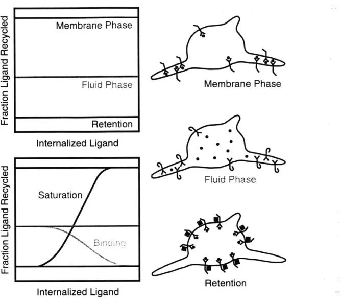
compartments become increasingly acidified. While the physiological pH encountered in the extracellular environment of most cells is 7.4, sorting endosomes and lysosomes contain proton pumps that decrease the pH of the lumen to roughly 5.5-6.5 and 4, respectively (Mellman et al., 1986). Acidification can affect the activity of proteolytic enzymes, as well as the ability of the ligand to remain complexed with the receptor after internalization. For this reason, sorting endosomes have been called the compartment for uncoupling of receptor and ligand (CURL) (Dunn and Hubbard, 1984). Both the geometry of endosomes and specific interactions with endosomal proteins are believed to have a strong influence on the sorting of ligands and receptors. Lipids and other "membrane phase" components are predominantly routed into recycling tubules, while soluble "fluid phase" components in the endosomal lumen are predominantly retained in the vesicular portion of an endosome and routed for degradation. For example, transferrin remains tightly complexed with its receptor in endosomes, and this ligand is constitutively recycled along with its receptor. In contrast, low-density lipoprotein dissociates from its receptor at endosomal pH, and this ligand is degraded while its receptor is free to return to the cell
surface (Ghosh et al., 1994). The EGFR ligands EGF and TGFax exhibit a marked difference in their receptor-binding affinities at pH 6.0, implying that EGF can occupy more receptors in endsomes than TGFx (French et al., 1995). This is likely due to the fact that TGFa contains five histidine residues, whereas EGF only has two, giving TGFa the much higher isoelectric point. This impacts the differential intracellular processing of these ligands (Ebner and Derynck, 1991), as detailed below.
The relative fluxes of ligand to recycling and degradative fates can be assessed experimentally by measuring the ligand recycling fraction (the fraction of radiolabeled ligand exocytosed by the cell that is intact) under steady state conditions (Figure 1.5). Nondissociative ligands can achieve recycling fractions even lower than fluid or membrane phase ligands if the ligated receptor is recognized by accessory proteins in the vesicular portion of an endosome, yielding endosomal retention. In this case, the receptor is also
Membrane Phase 0 U) CD a: C 0: 0 Ca 1L. U-Membrane Phase Fluid Phase Retention
Figure 1.5 Mechanistic Analysis of Endosomal Sorting. Lipids and other "membrane phase" components are predominantly routed into recycling tubules, while soluble "fluid phase" components in the endosomal lumen are predominantly retained in the vesicular portion of an endosome and routed for degradation. The relative fluxes of ligand to recycling and degradative fates can be assessed experimentally by measuring the ligand
recycling fraction (the fraction of radiolabeled ligand exocytosed by the cell that is intact)
under steady state conditions. Nondissociative cytokines can achieve recycling fractions even lower than fluid or membrane phase ligands if the ligated receptor is recognized by accessory proteins in the vesicular portion of an endosome, yielding endosomal retention (top graph). As the level of internalized ligand increases, a dissociative ligand such as TGFx will bind some receptors (leading to retention of ligand and receptor), and a nondissociative ligand such as EGF will occupy enough receptors to saturate the limited number of accessory retention proteins (bottom graph).
Fluid Phase
Retention
Internalized Ligand
Saturatio/n e
For example, EGF is retained in endosomes in an occupancy-dependent manner (Herbst et al., 1994; French et al., 1994). This process requires regions of the EGFR cytosolic domain distinct from those regulating endocytosis (Opresko et al., 1995) and has been linked to the interaction of ligated EGFR with an accessory sorting nexin (Kurten et al.,
1996).
Intermediate behaviors have been observed for EGF and TGFa in B82 fibroblasts; when the ligand recycling fraction is plotted versus the level of total intracellular ligand, the TGFa curve exhibits a slightly negative slope while the EGF curve clearly exhibits a positive slope. Counterintuitively, TGFa exhibits a higher recycling fraction at low levels of internal ligand, while EGF exhibits a higher recycling fraction at high levels of internal ligand (French et al., 1995). The current model adequately explains these subtle aspects of sorting (Figure 1.5): as the level of internalized ligand increases, a dissociative ligand such as TGFa will bind some receptors (leading to retention of ligand and receptor), and a nondissociative ligand such as EGF will occupy enough receptors to saturate the limited number of accessory retention proteins (French and Lauffenburger, 1996). Therefore, both receptor endocytosis and intracellular degradation are saturable in cells overexpressing EGFR, which has been linked to cell transformation and tumorigenesis (Wells et al., 1990; Masui et al., 1991; Reddy et al., 1994; Lenferink et al., 1998; Worthylake et al., 1999). Display of high receptor numbers leads to significant signal generation at low agonist concentrations, conditions under which only a small fraction of surface receptors are ligated and therefore subject to internalization and downregulation. When receptor overexpressors experience chronic agonist stimulation, the normal attenuation of signaling by receptor downregulation is mitigated by saturation of both specific internalization and intracellular degradation.
1.5 Thesis Topic
Acidification of endosomes causes many ligands, including TGFa, to dissociate from their receptors in endosomes, allowing the receptor to be sorted by default towards recycling to the plasma membrane. Unlike TGFa, EGF remains tightly complexed to EGFR in endosomes of multiple cell types (Kay et al., 1986; Carpentier et al., 1987). While this adequately explains the observed difference in postendocytic processing of the two ligands, it was not known whether internal receptor-ligand complexes could participate in signaling reactions. Receptor trafficking certainly downregulates the total numbers of receptors and ligands available for signaling in the long term, but internalization could also
have a short-term effect, either positive or negative, on signaling through the localization of a receptor pool in internal compartments.
The specific aims of my thesis project were 1) to assess whether the
compartmentalization of EGFR in endosomes versus the plasma membrane can affect the magnitude of signaling through specific pathways, and 2) to mechanistically determine the molecular bases for any observed differences. This line of investigation basically tackles the following fundamental question: how structured should a model of receptor-mediated signaling (and therefore cell function) be (Bailey, 1998)?
1.6 Previous Studies Relating EGFR Trafficking and Signaling
Knauer and colleagues proposed a model in which there is a linear relationship between cell growth rate (or DNA synthesis rate) and the level of EGFR-ligand complexes on the surface Cs (Knauer et al., 1984). Using a mathematical model validated previously (Wiley and Cunningham, 1981), they related C, to the extracellular ligand concentration [L] at pseudo-steady state:
Response = ,yC= yVskf (1.3),
keR(kr +keC) +keCkf[L]
where VS is the rate of de novo receptor synthesis, kf and k, are association and dissociation
rate constants for the receptor-ligand interaction, respectively, and keR and ke are basal and induced endocytic rate constants, respectively. y can be considered the intrinsic mitogenic
signal generation coefficient, a phenomenological parameter. The above model agreed well with experimental data obtained for the DNA synthesis rate (measured by incorporation of 'H-thymidine) of human foreskin fibroblasts stimulated with EGF (Knauer et al., 1984).
However, an interesting feature of the trafficking model is that internal complexes C is related to C, at steady state by a proportionality constant, such that the following model would be equally valid:
Response = ysCs + yC, = 7, + 'Yec CS (1.4),
where k is the rate constant describing lysosomal degradation of internal complexes. Other models have also been developed with a similar flavor, but including effects such as saturation of internalization (Starbuck and Lauffenburger, 1992).
As mentioned above, it has been established that the interaction of EGF with the EGFR is relatively insensitive to endosomal acidification. In multiple cell types, the
internal pool of EGF-ligated EGFR remains phosphorylated on tyrosine, and these autophosphorylation sites are accessible to cytosolic proteins (Lai et al., 1989; Sorkin and Carpenter, 1991; Wada et al., 1992). This suggested that EGFR could carry out signaling functions in endosomes. In fact, in the case of Shc in rat liver parenchyma, the endosome-associated pool of the protein adaptor Shc exhibited a phosphorylation stoichiometry at maximal internalized EGF/EGFR exceeding that seen at the plasma membrane for any receptor density (Di Guglielmo et al., 1994). However, internalization of EGFR does not seem to lead to a uniform up- or down-regulation of substrate phosphorylation, indicating some degree of specificity (Vieira et al., 1996).
Interestingly, previous studies relating receptor internalization and signaling have not assessed how endosomal localization affects the function of cytosolic enzymes recruited by the EGFR. This function is to modify their substrates, which are withfew exceptions
membrane-associated molecules. This implies that the composition of endosomal
membranes, compared to that of the plasma membrane, can be an important factor in determining the potential role of internal EGFR in signaling. In other words, this would in part determine the relative values of ys and y in eqn. 1.4.
1.7 Thesis Overview
The possible role of internalized EGFR in signal transduction was examined both theoretically and experimentally. To answer the question of interest, it was first important to address the more fundamental issue of how subcellular location in general can influence signaling reactions. The depiction of Fig. 1.2C implies that recruitment to a membrane, be it the plasma membrane or endosomal membranes, can affect the dynamics of signaling. This was addressed theoretically by analyzing reaction and diffusion of molecules in two
and three dimensions, to see how receptor-mediated membrane recruitment of a cytosolic enzyme can impact the association rate with a membrane-associated target (Chapter 2). To expand that effort to examine internalization of RTKs, another model was formulated that takes into account the binding of a cytosolic enzyme to both surface and internal receptors, phosphorylation/dephosphorylation of the enzyme, and the concentration profile of
phosphorylated molecules in the cytosol (Chapter 3). Thus, signal transduction was analyzed using theory of reaction kinetics and transport phenomena, both fundamentals of chemical engineering.
Experimentally, quantitative measurements of signaling pathways were made using EGFR-expressing NR6 fibroblasts. The overall methodology was to construct a
relationship between signaling and the total level of EGFR autophosphorylation (surface and internal), the activation-response relationship. Unlike a dose-response curve, in which the cellular response is plotted versus ligand concentration, this methodology accounts for differing ligand affinities and feedback modulation of the EGFR. This analysis was performed under conditions that manipulated the relative numbers of EGFR at the surface and in internal compartments. If the location of the receptor does not affect signaling, then all points on an activation-response plot will fall on the same curve. If receptor location does matter, then internal receptor activation shifts the curve to the left (internal superior to surface) or right (internal inferior to surface). Two distinct signaling pathways were investigated: the phospholipase C (PLC) pathway (Chapter 4) and the Ras/MAPK pathway (Chapter 5).
Finally, a second generation mathematical model was formulated to address specific aspects of the PLC pathway (Chapter 6). This was motivated by experimental observations that could not be explained using the generalized analysis of Chapter 3. The model
accounted for not only modulation of the enzyme PLC-yl by the EGFR, but also the regulation of the level of its lipid substrate PIP2, and it can mimic effectively the
experimental results. Importantly, the agreement of the model with experiment allowed the included molecular mechanisms, some controversial, to be scrutinized. Models of this type should be useful in the design of drug therapies and other biomedical intervention
strategies.
1.8 References
Aronheim, A., Engelberg, D., Li, N., Al-Alawi, N., Schlessinger, J. and Karin, M. (1994). Membrane targeting of the nucleotide exchange factor Sos is sufficient for
activating the Ras signaling pathway. Cell, 78: 949-961.
Auger. K.R.. Serunian. L.A., Soltoff. S.P., Libby, P. and Cantley, L.C. (1989). PDGF-dependent tyrosine phosphorylation stimulates production of novel
phosphoinositides in intact cells. Cell, 57: 167-175.
Bailey. J.E. (1998). Mathematical modeling and analysis in biochemical engineering: past accomplishments and future opportunities. Biotechnol. Prog., 14: 8-20.
Bell. G.I., Fong, N.M., Wornstead, M.A., Caput, D.F., Ku, L., Urdea, M.S., Rall, L.B. and Sanchez-Pescador, R. (1986). Human epidermal growth factor precursor: cDNA sequence, expression in vitro and gene organization. Nucleic
Acids Res., 14: 8427-8446.
Bertics, P.J. and Gill, G.N. (1985). Self-phosphorylation enhances the protein-tyrosine kinase activity of the epidermal growth factor receptor. J. Biol. Chem., 260:
14642-14647.
Bertics, P.J., Weber, W., Cochet, C. and Gill, G.N. (1985). Regulation of the epidermal growth factor receptor by phosphorylation. J. Cell. Biochem., 29: 195-208.
Bjorge, J.D., Chan, T., Antczak, M., Kung, H. and Fujita, D.J. (1990). Activated type I phosphatidylinositol kinase is associated with the epidermal growth factor (EGF) receptor following EGF stimulation. Proc. Natl. Acad. Sci. USA, 87: 3816-3820. Bonfini, L., Karlovich, C., Dasgupta, C. and Banerjee, U. (1992). The Son of Sevenless
gene product: a putative activator of Ras. Science, 255: 603-605.
Bos, J.L. (1989). Ras oncogenes in human cancer: a review. Cancer Res., 49: 4682-4689.
Bourne, H.R., Sanders, D.A. and McCormick, F. (1991). The GTPase superfamily: conserved structure and molecular mechanism. Nature, 349: 117-127.
Bowtell, D., Fu, P., Simon, M. and Senior, P. (1992). Identification of murine
homologues of the Drosophila Son of Sevenless gene: potential activators of Ras.
Proc. Natl. Acad. Sci. USA, 89: 6511-6515.
Buday, L. and Downward, J. (1993a). Epidermal growth factor regulates p2lras through the formation of a complex of receptor, Grb2 adapter protein, and Sos nucleotide exchange factor. Cell, 73: 611-620.
Buday, L. and Downward, J. (1993b). Epidermal growth factor regulates the exchange rate of guanine nucleotides on p2lras in fibroblasts. Mol. Cell. Biol., 13: 1903-1910.
Carpenter, C.L. (1996). Intracellular signals and the cytoskeleton: the interactions of phosphoinositide kinases and small G proteins in adherence, ruffling and motility.
Sem. Cell Developmental Biol., 7: 691-697.
Carpenter, C.L. and Cantley, L.C. (1996). Phosphoinositide kinases. Curr. Opin. Cell
Biol., 8: 153-158.
Carpenter, G. and Wahl, M.I. (1990). "The epidermal growth factor family." in Peptide
Growth Factors and Their Receptors, eds. Sporn, M. B. and Roberts, A. B. New
York: Springer-Verlag, pp. 69-171.
Carpentier, J., White, M.F., Orci, L. and Kahn, R.C. (1987). Direct visualization of the phosphorylated epidermal growth factor receptor during its internalization in A-431 cells. J. Cell. Biol., 105: 2751-2762.
Carraway, K.L. and Cantley, L.C. (1994). A neu acquaintance for erbB3 and erbB4: a role for receptor heterodimerization in growth signalling. Cell, 78: 5-8.
Chang, C., Lazar, C.S., Walsh, B.J., Komuro, M., Collawn, J.F., Kuhn, L.A., Tainer, J.A., Trowbridge, I.S., Farquhar, M.G., Rosenfeld, M.G., Wiley, H.S. and Gill, G.N. (1993). Ligand-induced internalization of the epidermal growth factor
receptor is mediated by multiple endocytic codes analogous to the tyrosine motif found in constitutively internalized receptors. J. Biol. Chem., 268: 19312-19320. Chardin, P., Camonis, J.H., Gale, N., Van Aelst, L., Schlessinger, J., Wigler, M.H. and
Bar-Sagi, D. (1993). Human Sos-l: a guanine nucleotide exchange factor for Ras that binds to Grb2. Science, 260: 1338-1343.
Chardin, P., Cussac, D., Maignan, S. and Ducruix, A. (1995). The Grb2 adaptor. FEBS
Letters, 369: 47-51.
Chen, P., Gupta, K. and Wells, A. (1994a). Cell movement elicited by epidermal growth factor receptor requires kinase and autophosphorylation but is separable from mitogenesis. J. Cell Biol., 124: 547-555.
Chen, P., Murphy-Ullrich, J.E. and Wells, A. (1996a). A role for gelsolin in actuating epidermal growth factor receptor-mediated cell motility. J. Cell Biol., 134: 689-698.
Chen, P., Xie, H., Sekar, M.C., Gupta, K. and Wells, A. (1994b). Epidermal growth factor receptor-mediated cell motility: phospholipase C activity is required, but mitogen-activated protein kinase activity is not sufficient for induced cell movement. J. Cell Biol., 127: 847-857.
Chen, W.S., Lazar, C.S., Poenie, M., Tsien, R.Y., Gill, G.N. and Rosenfeld, M.G. (1987). Requirement for intrinsic protein tyrosine kinase in the immediate and late actions of the EGF receptor. Nature, 328: 820-823.
Chen, X., Levkowitz, G., Tzahar, E., Karunagaran, D., Lavi, S., Ben-Baruch, N., Leitner, 0., Ratzkin, B.J., Bacus, S.S. and Yarden, Y. (1996b). An
immunological approach reveals biological differences between the two
NDF/heregulin receptors, ErbB-3 and ErbB-4. J. Biol. Chem., 271: 7620-7629. Cheng, K. and Koland, J.G. (1996). Nucleotide binding by the epidermal growth factor
receptor protein-tyrosine kinase. J. Biol. Chem., 271: 311-318.
Cooper, J.A. and Kashishian, A. (1993). In vivo binding properties of SH2 domains from GTPase-activating proteins and phosphatidylinositol 3-kinase. Mol. Cell.
Biol., 13: 1737-1745.
Countaway, J.L., Nairn, A.C. and Davis, R.J. (1992). Mechanism of desensitization of the epidermal growth factor receptor protein-tyrosine kinase. J. Biol. Chem., 267:
1129-1140.
Cussac, D., Frech, M. and Chardin, P. (1994). Binding of the Grb2 SH2 domain to phosphotyrosine motifs does not change the affinity of its SH3 domains for Sos proline-rich motifs. EMBO J., 13: 4011-4021.
Davis, R.J. and Czech, M.P. (1987). Stimulation of epidermal growth factor receptor threonine 654 phosphorylation by platelet-derived growth factor in protein kinase C-deficient human fibroblasts. J. Biol. Chem., 262: 6832-6841.
Di Guglielmo, G.M., Baass, P.C., Ou, W., Posner, B.I. and Bergeron, J.J.M. (1994). Compartmentalization of SHC, GRB2 and mSOS, and hyperphosphorylation of Raf-I by EGF but not insulin in liver parenchyma. EMBO J., 13: 4269-4277. Dougall, W.C., Qian, X.L., Peterson, N.C., Miller, M.J., Samanta, A. and Greene, M.I.
(1994). The neu-oncogene: signal transduction pathways, transformation mechanisms and evolving therapies. Oncogene, 9: 2109-2123.
Downward, J., Graves, J.D., Warne, P.H., Rayter, S. and Cantrell, D.A. (1990). Stimulation of p2lras upon T-cell activation. Nature, 346: 719-723.
Downward, J., Parker, P. and Waterfield, M.D. (1984). Autophosphorylation sites on the epidermal growth factor receptor. Nature, 311: 483-485.
Dunn, W.A. and Hubbard, A.L. (1984). Receptor-mediated endocytosis of epidermal growth factor by hepatocytes in the perfused rat liver: ligand and receptor dynamics. J. Cell Biol., 98: 2148-2159.
Ebner, R. and Derynck, R. (1991). Epidermal growth factor and transforming growth factor-a: differential intracellular routing and processing of ligand-receptor complexes. Cell Regulation, 2: 599-612.
Egan. S.E., Giddings, B.W., Brooks, M.W., Buday, L., Sizeland, A.M. and Weinberg, R.A. (1993). Association of SOS Ras exchange protein with GRB2 is implicated in tyrosine kinase signal transduction and transformation. Nature, 363: 45-50. Escobedo, J.A., Navankasattusas, S., Kavanaugh, W.M., Milfay, D., Fried, V.A. and
Williams, L.T. (1991). cDNA cloning of a novel 85 kd protein that has SH2 domains and regulates binding of P13-kinase to the PDGF beta-receptor. Cell, 65: 75-82.
Fearn, J.C. and King, A.C. (1985). EGF receptor affinity is regulated by intracellular calcium and protein kinase C. Cell, 40: 991-1000.
Feig. L.A. (1994). Guanine-nucleotide exchange factors: a family of positive regulators of Ras and related GTPases. Curr. Opin. Cell Biol., 6: 204-211.
Fischer, E.H., Charbonneau, H. and Tonks, N.K. (1991). Protein tyrosine phosphatases: a diverse family of intracellular and transmembrane enzymes. Science, 253: 401-406.
French, A.R. and Lauffenburger, D.A. (1996). Intracellular receptor/ligand sorting based on endosomal retention components. Biotech. Bioeng., 51: 281-297.


![Table 1.1 Signaling Functions of EGFR-binding Proteins. The enzymes phospholipase C-yl (PLC-yl) and phosphatidylinositol 3-kinase [PI(3)K] target the membrane lipid phosphatidylinositol (4,5)-bisphosphate (PIP 2 ) for hydrolysis and](https://thumb-eu.123doks.com/thumbv2/123doknet/14213288.482346/17.918.129.783.273.553/signaling-functions-proteins-phospholipase-phosphatidylinositol-phosphatidylinositol-bisphosphate-hydrolysis.webp)