HAL Id: tel-01650126
https://tel.archives-ouvertes.fr/tel-01650126
Submitted on 28 Nov 2017HAL is a multi-disciplinary open access
archive for the deposit and dissemination of sci-entific research documents, whether they are pub-lished or not. The documents may come from teaching and research institutions in France or abroad, or from public or private research centers.
L’archive ouverte pluridisciplinaire HAL, est destinée au dépôt et à la diffusion de documents scientifiques de niveau recherche, publiés ou non, émanant des établissements d’enseignement et de recherche français ou étrangers, des laboratoires publics ou privés.
From cellular variability to shape reproducibility :
mechanics and morphogenesis of Arabidopsis thaliana
sepal
Mathilde Dumond
To cite this version:
Mathilde Dumond. From cellular variability to shape reproducibility : mechanics and morphogenesis of Arabidopsis thaliana sepal. Morphogenesis. Université de Lyon, 2017. English. �NNT : 2017LY-SEN047�. �tel-01650126�
Numéro National de Thèse : 2017LYSEN047
THÈSE de DOCTORAT DE L’UNIVERSITÉ DE LYON
opérée parl’École Normale Supérieure de Lyon École Doctorale N°340
Biologie Moléculaire Intégrative et Cellulaire (BMIC) Spécialité de doctorat : Biologie
Soutenue publiquement le 15/09/2017, par :
Mathilde Dumond
From cellular variability to shape reproducibility:
mechanics and morphogenesis of Arabidopsis
thaliana sepal.
De la variabilité cellulaire à la reproductibilité des
organes : étude de la mécanique de la morphogenèse
du sépale d’Arabidopsis thaliana.
Devant le jury composé de :
Fleck, Christian Professeur WUR Rapporteur
Vernhettes, Samantha Directrice de Recherche INRA IJPB Rapporteur Peronnet, Frédérique Directrice de Recherche CNRS IBPS Examinatrice Sémon, Marie Maître de Conférences ENS de Lyon LBMC Examinatrice Labouesse, Michel Directeur de Recherche CNRS IBPS Examinateur Boudaoud, Arezki Professeur ENS de Lyon RDP Directeur de thèse Hamant, Olivier Directeur de Recherche INRA RDP Co-encadrant de thèse
De la variabilité cellulaire à la reproductibilité des formes : étude de la
mécanique de la morphogenèse chez le sépale d’Arabidopsis thaliana
Résumé en français
La robustesse du développement est la capacité à produire le même phénotype au cours du développement malgré des perturbations intrinsèques ou de l’environnement. Chez toutes les espèces, la taille des organes est en général plutôt constante, alors que le comportement de leurs cellules est très variable.
Les voies de régulation de la taille et de la forme des organes ont été largement étudiées, cependant la régulation de la reproductibilité, de la taille et de la forme reste à élucider. Chaque plante d’Arabidopsis thaliana produit plus de 100 leurs, rendant possible la mesure de la distribution statistique des tailles au sein d’un organisme individuel.
Les sépales, organes externes de la leur, nous permettent d’étudier la robustesse dans le développement, car leur taille et leur forme doit être strictement régulée tout au long de leur développement pour protéger les organes reproducteurs. De plus, les tailles des cellules des sépales sont très variables.
Chez les plantes, les cellules sont entourées par une paroi pecto-cellulosique. Leurs formes résul-tent de l’équilibre entre la pression osmotique interne et la rigidité de la paroi. Les propriétés mécaniques de la paroi ayant un fort impact sur la forme inale des cellules, mon projet de thèse vise à élucider leur rôle dans la reproductibilité des formes.Précédemment, mon labora-toire de thèse avait montré que les cellules végétales peuvent percevoir les forces mécaniques qu’elles subissent, et modiier les propriétés mécaniques de leur paroi pour y résister. Comme l’anisotropie mécanique de la paroi module la direction de croissance, nous avons cherché à déterminer si le niveau de cette réponse cellulaire aux forces pouvait inluencer la forme inale de l’organe en combinant approches théoriques et expérimentales.
J’ai d’abord développé un modèle purement mécanique, tenant compte des contraintes mé-caniques, de la croissance anisotropique, ainsi que de la boucle de rétroaction mécanique liant contraintes et direction de croissance.
Grâce à ce modèle, j’ai pu prédire que le niveau de contraintes engendrées par le ralentissement de la croissance de la pointe du sépale associées à une forte rétroaction mécanique pouvait modiier la forme du sépale.
Cette prédiction a été conirmée expérimentalement par un autre étudiant en thèse de mon équipe.
Le consensus jusqu’à présent est que la forme des organes est dictée par des molécules appelées morphogènes qui dictent aux cellules où et quand croître. Cette étude suggère que la forme des organes résulte d’une synergie entre de tels morphogènes et les contraintes mécaniques générées par les diférentiels de croissance.
Lorsque j’ai mesuré les propriétés mécaniques de sépales en utilisant un microscope à force atomique, j’ai trouvé qu’elles étaient fortement variables spatialement. J’ai alors rendu la
rigidité variable spatialement dans le modèle ce qui a rendu les formes moins reproductibles entre diférentes simulations. Pour obtenir des formes reproductibles, j’ai dû ajouter une vari-abilité temporelle à la varivari-abilité spatiale des propriétés mécaniques. Dans ce cas, la varivari-abilité temporelle permettait de lisser les diférences spatiales au cours du temps et d’augmenter la reproductibilité des formes inales.
Les hypothèses suggérées par le modèle ont ensuite été testées expérimentalement. Lors d’un crible génétique pour des mutants ayant des sépales moins reproductibles, nos collaborateurs à Cornell University ont isolé le mutant ftsh4.Nous avons montré que la variabilité spatiale était réduite dans le mutant comparé à la plante sauvage. J’ai conirmé en utilisant le microscope à force atomique que la variabilité spatiale de la rigidité de la paroi était aussi réduite chez le mutant.
Les expériences ont ainsi validé l’une des deux hypothèses formulées grâce au modèle : aug-menter la variabilité spatiale des propriétés mécaniques augmente la reproductibilité des formes. La plupart des travaux portant sur la taille et la forme des organes se sont concentrés sur l’impact du gène étudié sur la taille moyenne des organes. De plus, la variabilité à l’échelle cellulaire est peu étudiée chez les organismes multicellulaires, et est souvent considérée comme désavantageuse. Nous montrons ici que la variabilité au niveau cellulaire peut au contraire permettre à l’organe de se développer correctement, et c’est la réduction de cette variabilité qui est délétère pour l’organisme.
Abstract
Developmental robustness is the ability to produce the same phenotype despite environmental variability. Indeed, organisms produce similar organs despite a large cellular variability. Regu-lation of organ size and shape has been widely studied, but the reguRegu-lation of reproducibility of organ size and shape is yet to be elucidated.
Arabidopsis thaliana sepals, external organs of the lower, can be used to study developmental robustness, because each plant produces more than 100 lowers, allowing the robust measure-ment of variability. Their size is tightly regulated all along the lower developmeasure-ment to protect the reproductive organs.
In plants, cells are surrounded by a stif cell wall. Cells modulate cell wall stifness and me-chanical anisotropy to control their growth. My PhD project aims at understanding the role of mechanical properties on organ shape reproducibility.
It had previously been shown that plant cells sense their physical environment and accordingly adjust their cell wall mechanical properties. We irst studied whether the strength of this ad-justment, or feedback, could inluence the inal shape of the organ. Using a modeling approach, I showed that the decrease of the growth rate observed at the tip of the sepal could modify the shape of the tip when the feedback was strong. This prediction has been experimentally demonstrated by another PhD student of the team.
This study suggests that organ shape results from a synergy between morphogens and the me-chanical cues generated by growth. Using again a modeling approach, I showed that when mechanical properties were not variable, the shape of the organ was perfectly reproducible. This project hence suggest that not only morphogen inluence organ shapes, but that mechanical feedback has a direct impact on shapes.
However, atomic force microscopy measurements showed that mechanical properties were highly spatially variable. When this variability was implemented in the model, the organ shapes were not reproducible. To produce reproducible shapes, I increased the temporal variability of the mechanical properties, which smoothed the spatial variability over time. Moreover, a decrease of the spatial variability lead to a decrease of organ shape reproducibility. These theoretical results suggest that spatial and temporal variability inluence shape robustness.
The hypotheses raised by the model were experimentally tested: our collaborators identiied a mutant displaying less robust sepal shapes. We showed that the spatial variability was reduced in this mutant. I conirmed using an atomic force microscope that spatial variability was reduced as well in the mutant, thus conirming one of the two hypotheses raised by the model: spatial variability of mechanical properties inluence organ shape variability.
Most of the work on organ shape and size focused on the role of the gene of interest of the mean size of organs. Moreover, the variability at the scale of the cell is rarely studied in multicellular organisms and considered unfavorable. We show here that the variability at the cellular scale can enable the proper development of the organ, and it is the decrease of this variability which is deleterious for the organism.
Contents
1 Introduction 1
1.1 Variability in biology . . . 2
1.1.1 What can vary in biology? . . . 2
1.1.2 Sources of variability . . . 2
1.1.3 What should vary but actually does not . . . 3
1.1.4 Bufering mechanisms . . . 4
1.1.5 Spatiotemporal averaging as a bufering mechanism . . . 5
1.1.6 Is variability always detrimental? . . . 5
1.2 The cell wall and its regulators . . . 7
1.2.1 The composition and biosynthesis of the cell wall . . . 7
Cell wall composition . . . 7
Cell wall biosynthesis . . . 11
1.2.2 The extension of the plant cell wall . . . 12
Mechanical properties of cell wall components . . . 12
Phytohormones . . . 13
Signaling molecules . . . 13
Mechanical stresses . . . 13
Wall regulators . . . 14
1.3 Mechanics and morphogenesis . . . 17
1.3.1 Turgor pressure and growth . . . 17
1.3.2 Measuring turgor pressure . . . 18
1.3.3 Measuring cell wall stifness . . . 18
1.3.4 Forces in plants and during morphogenesis . . . 19
1.4 Growth of an organ . . . 20
1.4.1 Describing morphogenesis . . . 21
1.4.2 Modeling morphogenesis . . . 22
1.4.3 Some case studies . . . 24
Morphogenesis of an isolated plant cell . . . 24
Curving an elongated organ: tropisms . . . 24
Shaping a sheet-like organ . . . 25
Feedback through mechanical signals . . . 26 iv
Variability and morphogenesis . . . 26
1.5 Why choosing the sepal as a model system . . . 27
1.6 Main questions . . . 28
2 Variable Cell Growth Yields Reproducible Organ Development through Spa-tiotemporal Averaging 29 2.1 Summary . . . 30
2.2 Introduction . . . 30
2.3 Results . . . 32
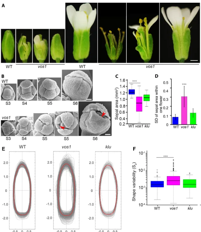
2.3.1 vos1 Mutants Have Increased Variability in Sepal Size and Shape . . . 32
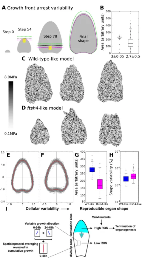
2.3.2 Mechanical Modeling Shows that Spatiotemporal Averaging of Cellular Variability Can Produce Organ Regularity . . . 34
2.3.3 Reduced Local Spatial Variability in the Cell Growth of vos1 Sepals Un-derlies Irregular Sepal Shape . . . 38
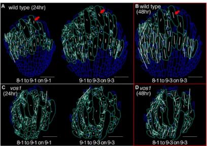
2.3.4 Wild-Type Sepals Undergo Spatiotemporal Averaging of the Principal Di-rection of Growth, Resulting in Regularity, which Is Disrupted in vos1 Mutants . . . 41
2.3.5 vos1 Is a Mutant of the FtsH4 Mitochondrial Protease . . . 42
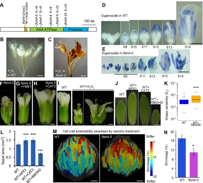
2.3.6 The Increased Irregularity in ftsh4 Sepals Is Caused by Increased ROS Levels . . . 42
2.3.7 ROS Act as a Growth Regulator in Wild-Type Sepals, Promoting Matu-ration and Termination of Growth . . . 44
2.3.8 ftsh4 Sepals Exhibit Cellular Characteristics of Maturation Earlier than Wild-Type . . . 45
2.3.9 ftsh4 Sepals Are Stifer Than Wild-Type . . . 46
2.3.10 Reduced Cellular Variability and Spatiotemporal Averaging Correlate with ROS Accumulation in Maturing Wild-Type Sepal Tips . . . 46
2.3.11 Spatiotemporal Averaging Combined with a Maturation Gradient Regu-lated by ROS Produce Sepal Regularity . . . 46
2.4 Discussion . . . 49
2.4.1 Spatiotemporal Averaging as a General Mechanism to Deal with Stochas-ticity . . . 50
2.4.2 ROS as a Signal that Promotes Cellular Maturation and Growth Arrest . 51 2.5 Experimental procedures . . . 52
2.5.1 Plant Material and Treatment . . . 52
2.5.2 Microscopy and Image Analysis . . . 52
2.5.3 Computational Modeling . . . 53
3 A Mechanical Feedback Restricts Sepal Growth and Shape in Arabidopsis 70
3.1 Summary . . . 71
3.2 Introduction . . . 71
3.3 Results . . . 73
3.3.1 The abaxial sepal exhibits a stereotypical growth pattern . . . 73
3.3.2 The tip of the sepal exhibits a stereotypical cortical microtubule pattern . 73 3.3.3 The sepal growth pattern prescribes a mechanical stress pattern . . . 76
3.3.4 CMTs align along maximal tensile stress in growing sepals . . . 77
3.3.5 A mechanical feedback may channel sepal shape . . . 78
3.4 Discussion . . . 82
3.5 Experimental procedures . . . 83
3.5.1 Plant material and growth conditions . . . 83
3.5.2 Live imaging of the growing abaxial sepal . . . 83
3.5.3 Mechanical perturbations . . . 83
3.5.4 Image analysis . . . 84
3.5.5 Computational Modeling . . . 84
3.6 Supplemental Experimental Procedures . . . 85
4 Discussion 90 4.1 Variability may have beneicial efects during development . . . 91
4.2 Morphogens act in parallel to mechanical signals during morphogenesis . . . 92
4.2.1 Mechanical sensing inluences morphogenesis . . . 92
4.2.2 Cell wall sensing could play a role in compensation . . . 92
4.3 Mechanics and robustness of morphogenesis . . . 93
4.3.1 Cell wall biochemistry and robustness . . . 93
4.3.2 Mechanosensing and robustness . . . 94
4.4 General conclusion . . . 95
5 Appendix 128 5.1 Mechanical isolation of rapidly growing cell bufers growth heterogeneity and con-tributes to organ shape reproducibility . . . 128
Acknowledgments
La in de cette thèse est l’occasion de remercier tous les acteurs qui l’ont rendue possible. En premier lieu, je souhaite exprimer ma gratitude à mon directeur de thèse Arezki. Tu m’as encouragée à revenir à l’issue de mon stage de master 1, en me décrivant un projet super intéressant dans une collaboration - potentielle, à l’époque - de très haut niveau. Le projet était à la hauteur de tes promesses et c’est une réelle chance d’avoir pu en faire partie et de travailler sur des projets aussi chouettes.
De mon point de vue, tu as trouvé le bon compromis entre autonomie et soutien, avec une porte toujours ouverte et du temps à me consacrer, malgré ton emploi du temps de ministre. Tu n’as pas hésité à m’encourager à prendre des vacances et à me reposer quand tu as vu que c’était nécessaire, et ton attention a permis à cette thèse de rester une excellente expérience.
Je voudrais aussi remercier mon co-encadrant, Olivier, pour ta positivité et ton soutien, tes feedbacks et tes nouvelles idées incessantes. La prochaine fois je penserai aux ablations.
I would like to thank the jury of me thesis for having accepted to read my manuscript and for their useful comments. Pareillement, je remercie les membres de mon comité, qui ont su remettre en perspective ce projet lorsqu’il en avait le plus besoin. Leurs conseils ont été précieux pour amener mes projets à maturité.
As I already mentioned, this PhD was part of a collaboration, between labs around the world, with whom the Skypes (and the visits!) really rythmed these years. I am truly aware how lucky I am to have had the chance to interact so often with talented scientists from such diverse backgrounds. I am really proud of the research we have conducted together. In particular, I had the chance to spend a few month in Cornell in one of those labs, and work with Adrienne and Lilan. It was a great human and scientiic experience.
Je souhaite remercier toutes les personnes du RDP qui donnent cette ambiance chaleureuse au labo, et plus particulièrement Nelly, qui a pris beaucoup de photos de sépales avec moi, mais aussi les autres, avec qui on a pris beaucoup de café, Stéphane (gardien attitré du chat), Thomas, Long, Kat, Marion.
Je remercie ma famille, qui, de loin, a su me soutenir quand il y en avait besoin.
Finalement, je remercie surtout Nicolas, toujours présent à mes côtés quand ça ne marche pas, et aussi quand ça marche, bref, tout le temps, je n’y serais pas arrivée sans toi.
1
Introduction
T
wo individuals in the same species or population have similar sizes and shapes, and especially organs such as ears or hands are remarkably similar within a single individual. Even with the tremendous recent work on morphogenesis, how two organs such as hands have such remarkable reproducibility is still a mystery. Indeed, studies usually focus on the mecha-nisms making organs bigger or smaller, but how the organ can sense its shape and regulate it can be studied only by measuring variability of shapes. Arabidopsis thaliana lowers are a suitable tool for this study because each plant has dozens of lowers, allowing a robust estimation of shape variability.Plant cells are surronded by a stif cell wall in tension from the internal pressure. The cell wall mechanical properties are central to cell growth regulation, and in turn play an important role in organ growth and ultimatey shape. Because morphogenesis is at the interplay of intricate actors such as genetic regulation, cell growth and physics, using models is mandatory to get an understanding of the process.
The irst part of the introduction will be focused on the study of variability in biology; I will then describe the cell wall biochemistry and the implication of its mechanical properties on growth. In a third part I will introduce the diferent ways to model plant morphogenesis, and lastly explain why the sepal is a suitable model organ.
1.1 Variability in biology
1.1.1 What can vary in biology?
There are many scales in biology: from populations to subcellular compartments, and every scale displays variability.
Individuals of a population can develop and behave diferently, even in monoclonal populations such as bacteria populations.
Indeed, in a given organism, organs such as hands, ears, or plant leaves, can have varying sizes and shapes from 1 to 10% depending on the organ. At the level of the cell, the concentration of metabolites, transcripts and proteins can vary spatially and temporally.
Diferentiating the importance of each of these possible sources of variability on morphogenesis output has been takled in a study on Drosophila wings, which concluded that the variability at the level of a monoclonal population or at the level of an organism depended on the genetic background of the population: speciic genes had the speciic role to ensure reproducibility of wings sizes at the level of the individual (Debat and Peronnet, 2013).
Similarly, the cells behavior in one organ can vary; and even at the cellular scale, the concen-tration of proteins and the gene expression levels can vary over time and space. The variability of cell behavior is important for some aspects of development. For example, in leaves, stomata and trichomes are distributed along the surface of the leaf, in a speciic pattern. The emergence of such pattern is granted by the variability of behaviors at the cellular scale (Greese et al., 2012). However, studying variability using mutants can be misleading, because the measured variability can be due to incomplete penetrance instead of an actual increase in variability: an allele has an incomplete penetrance when not all individuals carrying the mutation show the mutant phenotype. One way to distinguish between regulation of variability and incomplete penetrance is to also compare mutant and wild-type means: similar means indicate that the gene regulates variability (Raj et al., 2010).
1.1.2 Sources of variability
The observed variability in shape and behavior has diverse origins. For example, the difer-ences observed between individuals of a polyclonal population are likely to be linked to genetic variability. The speciic genes involved in a variable phenotypic trait can be determined using Quantitative Trait Loci (QTLs).
On another hand, the diferences between individuals of the same population can also be due to them growing in diferent environments. However, organs in the same individual develop in the same environment and share the same genotype, for example hands show diferences of about 1% in length (Palmer, 1996). This measured luctuating asymetry is a metric of developmental stability.
The perturbations studied during development usually refer to several processes such as ther-mal noise at the molecular level, random variation of the rates of biological processes impacting
cell-to-cell communication, rates of cell growth or division (Palmer, 1996). Depending on what characteristic of the organims and what source of perturbation is tested, the perturbations can be internal or external, e.g. for the development of a lower, the environmental variability is ex-ternal whereas the cell scale variability is inex-ternal (Masel and Siegal, 2009). One question often addressed in the studies on variability is whether the cellular response is diferent for an internal or an external source or perturbations (Meiklejohn and Hartl, 2002). Moreover, the cellular response can also difer depending on the kind of environmental perturbation: when exposed to temperature or starvation, diferent species of Caenorhabditis nematode reacted diferently, indicating the intricate response of developmental systems to environment (Braendle and Félix, 2008). At an even smaller scale, gene expression itself is noisy, leading to variability in time and between cells in bacteria (McAdams and Arkin, 1999) and eukaryotic cells (Blake et al., 2003).
1.1.3 What should vary but actually does not
Given all the possible aspects of life that can vary, how is it possible that two hands, or two leaves are so similar? Only a few studies focused on this paradox.
During the development of the early embryo, many steps of cell diferentiation rely on the sensing of morphogen gradients. Any stochastic variation of morphogen concentration in this gradient needs to be bufered for a robust development. In particular, in Drosophila, the embryo devel-opment strongly relies on Bicoid gradient. The Bicoid gradient is very variable, it is sensitive to environmental factors such as temperature, the total amount of mRNA in the embryo, and the precise amount of protease. However, the limit of the cells diferentiating, detected by their expression of hunchback along this gradient is strikingly precise (Houchmandzadeh et al., 2002). How the organisms produce such a reproducible limit from a variable input is not resolved. In Drosophila, development is robust against environmental variation (temperature), and protein quantities, but changes in the topology of the regulatory network have large impacts (Houch-mandzadeh et al., 2002, von Dassow et al., 2000).
In plants, phyllotaxis is the regular arrangement of lateral organs in plants. It has been an interest for scientists and scholars since the ancient Greeks, and many properties have been understood as quite robust and reproducible over species. However, how such robustness is achieved is not understood. The phyllotaxis is set up at the shoot apical meristem of the plant, where the positionning of organs is determined by inhibitory ields, which in turn depend on variable processes such as morphogen difusion. Despite much progress in the identiication of molecular regulators of organogensis and possible self-organized properties at the tissue scale (e.g. Reinhardt et al. (2003)), how the temporal and spatial robustness of the inhibitory ields is produced is still a mystery (Mirabet et al., 2012, Vernoux et al., 2011).
Because gene expression is noisy (Blake et al., 2003, McAdams and Arkin, 1999), and because the environment varies, many phenotypic characteristics should vary. Morphogenesis stability is the main example that it does not vary nearly as much as it should.
1.1.4 Bufering mechanisms
Development is robust against environmental and genetic variation. This robustness in the de-velopment is often refered to as canalization (Wagner and Altenberg, 1996), and the mechanisms allowing such robustness have been the focus of many studies.
Gene networks allow the activity of genes to be backed up by other genes, granting a robustness against genetic variation: if the activity of one gene is impaired, another gene or set of genes is likely to be able to ill its function (Hartman et al., 2001). Moreover, comparing Drosophila pop-ulations, the most robust ones against genetic perturbations were also the most robust against environmental perturbations (Stearns et al., 1995).
Topology is an important feature in gene regulatory networks: negative feedback loops can increase robustness of the outcome of the regulatory network, as has been demonstrated in yeast (Howell et al., 2012). In particular, the microRNA miR-7 acts in several feedforward and feedback loops during Drosophila development, bufering the involved genetic networks against environmental variation (Li et al., 2009). MicroRNAs are likely to function in other pathways as well, such as the control of mRNA transcript copy number, and the degradation of impaired mRNA transcripts (Ebert and Sharp, 2012). Moreover, some genes have been demonstrated to speciically increase robustness. The chaperonin Hsp90 is a heat shock protein, and largely stabilizes morphogenetic pathways, degrading misshaped proteins. When the Hsp90 protein is active, it allows other mutations to cryptically accumulate; Hsp90 activity degrading the im-paired proteins.
The identiication of such genes is critical for a full understanding of robustness (Masel and Siegal, 2009). When Hsp90 is compromised, for example by a temperature increase, the cryptic mutations get expressed, leading to alterations of cells behaviors that can be more important than those predicted by only the loss of Hsp90 (Rutherford and Lindquist, 1998). Moreover, Hsp90 is present in all organisms and has been identiied in Arabidopsis thaliana ; where the mutant also presents developmental robustness defects, demonstrating the importance of such bufering mechanisms (Queitsch et al., 2002). Another protein with a large impact on robustness has been described in Arabidopsis thaliana, AtCHR23, which is a chromatin remodeler. This protein has diverse regulates diverse functions of the cell, and when mutated, the plants have a more variable growth in roots, hypocotyls and leaves (Folta et al., 2014).
Most of the time, the proteins involved in robustness have pleiotropic roles in the cell: pro-tein modiication (such as chaperonin), chromatin modeling, DNA integrity, RNA elongation, response to stimuli (Levy and Siegal, 2008), and as we will see in chapter 2, reactive oxygen species regulator. These genes are at the crosses of diferent pathways and are hubs in the genetic network of the cells (Cooper et al., 2006, Levy and Siegal, 2008).
To determine which genes have an impact on robustness, one can also use quantitative trait loci (QTLs). Indeed, QTLs are originally used to map a measured quantity to genetics, but the variability of such quantity can also be mapped to genetics using the same method (Hall et al., 2007, Ordas et al., 2008). Genes have been identiied in Arabidopsis thaliana which
im-pact developmental robustness: for example, the ERECTA gene would be responsible for the robustness of the number of rosette leaves (Hall et al., 2007). However, the robustness of organ size has never been addressed in plants (Boukhibar and Barkoulas, 2015, Lempe et al., 2013).
1.1.5 Spatiotemporal averaging as a bufering mechanism
One way to increase robustness is to average sochastic luctuations in space and time.
Indeed, it has been shown in a theoretical study that averaging protein concentration in space, by difusion, enhanced the precision of gene expression patterns by smoothing out bursts of ex-pression (Erdmann et al., 2009). Similarly, in Schizosaccharomyces pombe the membrane-bond pom1p protein displays a noisy concentration distribution along the cell, but the temporally averaged distributions were much smoother. In this case, it was shown that the smoothening of the protein distribution was due to the clustering of the proteins (Saunders et al., 2012). Temporal averaging is also used by bacteria to sense shallow chemical gradients. Indeed, when a chemical gradient is shallow, the stochastic receptor-ligand interactions can exceed the slight gradient signal. To establish the orientation of the gradient, the bacteria sense the chemical con-centration in several positions and then stabilizes towards the averaged maximum (Dyer et al., 2013). Spatiotemporal averaging has been shown to play an important role in the robustness of morphogen gradient positional precision. Indeed, in Drosophila embryo, the antero-posterior axis is determined by a gradient of Bicoid concentration, which generates cell diferentiation, classically detected by the expression of hunchback. This gradient is highly sensitive to envi-ronmental variability (Houchmandzadeh et al., 2002), however the limit of the cells expressing hunchback is strikingly reproducible. Using a mutant displaying a more lat Bicoid gradient, He et al. (2010) showed that the robustness of the wild-type gradient sensing was due to time and spatial averaging. Another way to smooth protein concentration is intrinsic to the diferent time scales between the biological processes gene activation, transcription, mRNA translation and mRNA and protein degradation, which are suicient to bufer the temporal variability of gene activation and to produce rather smooth concentrations of proteins over time (Paulsson, 2005).
1.1.6 Is variability always detrimental?
At the cellular level, noise of genetic expression can have beneicial efects. Chemical com-pounds increasing expression noise in HIV positive cells but where HIV is latent could, when coupled with reactivating drugs, achieve much more eicient results than the reactivating drugs alone (Dar et al., 2014).
Phenotypic plasticity, or the ability of organisms to produce diferent phenotypes from the same genotype, can be advantageous to a population. Indeed, if the environment changes and a por-tion of the populapor-tion dies, the few that were diferent because of phenotypic stochasticity can prolifer and develop as a new population. Because the survivors have the same genotype as the initial population – if the selection pressure is not too long, the new population has the same
genetical background as the former (Johnston and Desplan, 2010). Similarly, in development and cancer, a cell population can be divided into phenotypic subpopulations. The proportions of each of these subpopulations is determined by stochastic transitions of cells between the states, keeping the proportions constant; even when a subpart of the population is removed (Gupta et al., 2011, Roorda and Williams, 1999, Singh et al., 2013).
Variability at the population level is also useful to interpret signals. For example, the immune NF-κB pathway is activated in response to the extra-cellular TNF signal. If the TNF signal oscillates, in some cells, the NF-κB response can synchronize with the TNF input, but only in a given range of frequencies. However, the cell-to-cell variability allows subpopulations of cells to synchronize with diferent frequencies, allowing the population as a whole to synchronize with a wide range of TNF signal frequencies. In this case, the phenotypic variability of a population of cells is responsible for the ability of the population to respond to an extra-cellular signal (Kel-logg and Tay, 2015). Variability can also be a defense mechanism: it has been shown that insect herbivore performance is reduced when the variability of nutritional content of plants is increased (Wetzel et al., 2016).
Robustness against environmental variations is mandatory for a reproducible development in animals and in plants. Nevertheless, the environment carries information, and organisms have to adapt to environmental changes (Abley et al., 2016); this is especially true for plants since they cannot move. Hence, the organisms need to be able to read environmental cues and ac-cordingly tune their behavior. Here it is worth noticing that the reaction of the plants to a given environmental cue must be robust: they ”produce variability in a reliable manner” (Abley et al., 2016). Strikingly, the strategies that allow robustness in development are often related to the mechanisms that allow environmental plasticity: network topology, feedback and feedforward loops, pleitropic genes (Lachowiec et al., 2016). Thus, if the organism is too robust against environmental variability, it will not adapt to environmental variability, ultimately reducing its itness.
Further than adapting to their environment during their life, robustness and variability have obvious evolutionnary implications. In particular, how the robustness in development, or canal-ization, evolves has been at the center of many studies (Eshel and Matessi, 1998, Gibson and Wagner, 2000, Masel and Bergman, 2003, Pigliucci, 2008, Rice, 1998). These studies demon-strate that the system allows some variability to keep the system evolvable, and that if a system is too robust, it will prevent evolution and ultimately lead to the extinction of the species. It has been theorecized that the bufering mechanisms should be a uniied mechanism for both genetic and environmental robustness (Meiklejohn and Hartl, 2002).
Here, we focus on the robustness of organ shape and size, which has not been investigated so far. In plants, shape is governed by cell wall mechanical properties. Could the intricate and entangled structure of the cell wall contribute to organ shape reproducibility? To investigate this question, we will introduce the cell wall biochemistry.
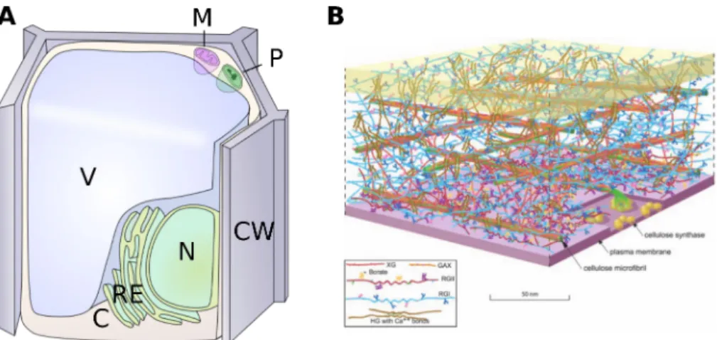
Figure 1.1: The cell is surrounded by the cell wall, which is an intricate structure. (A) Major compartments of a plant cell.
V: vacuole, N: nucleus, ER: endoplasmic reticulum, CW: cell wall, M: mitochondrion, P: plast, C: cytoplasm. (B) The pri-mary cell wall is a complex polysaccharidic structure, composed of cellulose microfibrils, pectin (RGI, RGII, HG) and hemicellulose (XG, GAX), between the cell plasma membrane (at the bottom) and the middle lamella (at the top), sepa-rating cell walls of neighboring cells. (from Somerville et al. (2004))
1.2 The cell wall and its regulators
Plant cells are surrounded by an extracellular matrix called the cell wall (see Figure 1.1). Over a plant’s life, the chemistry and physical properties of the cell wall change, notably in vasculature, to support the increasing weight of the above tissue and maintain the stature of the plant against gravity. The newly synthesized cell wall, or secondary cell wall, is stifer and characterized by the presence of lignin. The cells surrounded by secondary cell wall have stopped growing, and their main function is to support the younger tissue. On the other hand, the cells belonging to the young, growing tissue are surrounded by the much softer primary cell wall.
My project is focused on organ growth, thus I will focus on the primary cell wall only.
1.2.1 The composition and biosynthesis of the cell wall Cell wall composition
In Arabidopsis thaliana leaves, the primary cell wall is composed by pectic polysaccharides (42% of the wall), hemicellulose (24%), cellulose (14%) and proteins (14%) (Zablackis et al., 1995) (see Figure 1.2), but the cell wall also contains other components such as callose, notably involved in the regulation of plasmodesmata opening.
Cellulose is an unbranched homopolysaccharide, composed of β-D-glucopyranose units linked
by 1,4-glycosidic bonds (OSullivan, 1997) (see Figure 1.3). Between 18 and 36 chains assemble via hydrogen bonds into a cellulose microibril, forming the crystalline cellulose I (OSullivan, 1997). The spatial organization of the cellulose microibrils can be studied using X-ray scattering, and several types of crystallized cellulose have been described (Newman et al., 2013).
Figure 1.2: Simplified view of the cell wall. The main components are pectin, hemicellulose and cellulose. Pectin and
hemicellulose are produced in the Golgi and exocytosed in the cell wall, whereas cellulose is synthesised directly at the plasma membrane, following microtubule orientations.
Hemicellulose is a familly of diverse polysaccharides. It contains all the molecules with
equa-torial β-1,4-linkages, but the sugars can be diferent from glucose, such as xylose and mannose, and they often carry side-chains (Scheller and Ulvskov, 2010). The main hemicelluloses are xylan (made of xylose), xyloglucan (made of glucose), mannan (made of mannose) and glucomannan (made of mannose and glucose) (Scheller and Ulvskov, 2010) (see Figure 1.4). Hemicellulose backbone can form hydrogen bonds with the cellulose microibrils (Park and Cosgrove, 2015). Interestingly, mutants of regulators of xyloglucan biochemistry change the growth patterns in plants (Miedes et al., 2013), whereas the mutant depleted of all xyloglucan xxt1/xxt2 shows only a mild phenotype (Park and Cosgrove, 2012).
Pectins also constitute a diverse and compound family of polysaccharides. The main pectin
polysaccharides in Arabidopsis thaliana leaves are homogalacturonan (23% of the cell wall), rhamnogalacturonan I (11%), and rhamnogalacturonan II (8%) (Zablackis et al., 1995). Homo-galacturonan are linear chains of α-1,4-linked D-galacturonic acid; rhamnoHomo-galacturonan I has a backbone composed of repeating disaccharide α-D-galacturonic acid-α-L-rhamnose, linked to various side chains; and rhamnogalacturnan II has a backbone composed of α-1,4-linked D-galacturonic acid, associated with various side chains (Vincken, 2003).
No links between pectins and other components of the cell wall have been documented, however indirect evidence suggest that pectin and cellulose may directly interact (Ralet et al., 2016, Wang and Hussey, 2015). However, Rhamnogalaturonan II molecules can be covalently bound
Figure 1.3: Cellulose molecular composition. (A) Cellulose is composed ofβ-D-glucopyranose units linked by (1→4) glycosidic bonds. (B) A cellulose microfibril is composed of between 18 and 36 chains. Inspired by Park and Cosgrove (2015).
by borate diester bridges on speciic sites (Mazurek and Perlin, 1963, O’Neill et al., 2004) (see Figure 1.5), and homogalacturonan and rhamnogalacturonan II backbones can also be non-covalently linked by calcium bridges (Grant et al., 1973), according to the ”egg-box” model, thus forming a geliied structure, stifer than the free form of pectin (Fraeye et al., 2010) (see Figure 1.5).
Proteins can take up to 14% of the dry mass of the cell wall (Zablackis et al., 1995). The
large variety of proteins in the cell wall adds another layer of complexity and an exhaustive overview of the cell wall proteins has yet to be established (Albenne et al., 2014). Nonetheless, the cell wall proteins can be distinguished between the regulatory enzymes, which modify the other components of the cell wall, and the structural proteins (Showalter, 1993).
The structural proteins of the cell wall are the hydroxyproline-rich glycoproteins (HRGPs) or ex-tensins, the arabinogalactan proteins (AGPs), the glycine-rich proteins (GRPs), the proline-rich proteins (PRPs), and proteins that have a mixture of these domains (Carpita et al., 1996, Cassab, 1998). Extensins are the most abundant and the most studied structural proteins (Cassab, 1998): they are involved in cell recognition pathways, e.g. in the mating system of Chlamydomonas, and in the growth of the pollen tube during pollinization of maize in particular (Cassab, 1998). Extensins are composed of a succession of hydrophobic and hydrophilic motifs of highly modiied amino acids (Fry, 1986), which can be linked together by extensin peroxidase (Fry, 1986, Schn-abelrauch et al., 1996), forming sheets of extensin (Epstein and Lamport, 1984). They can also be linked to other molecules of the cell wall, but neither the mechanisms nor the nature of the links are known (Cassab, 1998, Fry, 1986, Qi et al., 1995). Extensin accumulation is associated with the end of cell growth (Cassab, 1998, Ye and Varner, 1991), in accordance with the increase of cell wall tensile strength due to the formation of an extensin-cellulose network (Cassab, 1998). I will not study this proteic complex during my PhD project. The other cell wall proteins are
Figure 1.4: Molecular composition of the backbones of the four main hemicellulose subtypes. The side-chains are not
represented, and are linked to any carbon of the glycans. After Scheller and Ulvskov (2010).
enzymes modifying the surrounding polysaccharides and proteins. The roles and means of action of these enzymes will be developped in the section 1.2.2.
Lignin is synthesized as a part of the secondary cell wall, so it is not in the focus of this
project. Lignin is formed by phenylalanin altered side-chain modiications such as oxydation, and linked together in the cell wall, forming a macromolecule (Boerjan et al., 2003, Fraser and Chapple, 2011). Lignin also covalently links to other components of the cell wall such as cellulose and hemicellulose (Pérez et al., 2002), thus making the cell wall much stronger.
Callose is composed by glucose residues linked together through β-1,3-linkages. It has many
regulatory roles, but is not present in high proportion in the cell wall. In particular, it regu-lates the opening of plasmodesmata, small channels allowing cell-to-cell symplasmic communi-cation (Levy et al., 2007, Roy et al., 1997, Turner et al., 1994). It is also the most prominent physical defense in fungal infection resistance, forming a plug (Aist, 1976), and is found in speciic structures such as the cell plate in dividing cells, pollen mother cell walls and pollen tubes (Jacobs et al., 2003).
Because the cell wall is so intricate, this presentation of the cell wall components is not exhaus-tive, and decifering exactly which component interacts with which and how is very challenging. The mechanical link between cellulose microibrils, in particular, plays an important role in the growth of the cell wall. The original view of the cell wall as a thethered network, where cel-lulose microibrils are linked by hemicelcel-luloses, has been recently challenged by the ’hot spot’ view, where cellulose microibrils are directly linked in such hot spots via hemicellulose-cellulose
Figure 1.5: Cross-linking of pectin molecules. (A) Homogalacturonan and rhamnogalacturonan II backbones can be
linked by Calcium ions on their non-methylesterified residues. (B) Rhamnogalacturonan II side-chains can be covalently bound through borate diester bridges forming via L-fucose residues. After Vincken (2003)
amalgams (Cosgrove, 2016), but determining the exact relationship between the biochemical components remains a challenge.
Cell wall biosynthesis
Figure 1.6: Schematic representation of cellulose synthesis. Cellulose synthesis takes place at the plasma membrane,
by CESA complexes, which are linked to the microtubules via the Cellulose Synthase Interacting protein CSI1 associated with the Companion of Cellulose Synthase (CC) proteins.
Cellulose is secreted in the membrane via cellulose synthase (CESA) complexes, each one
synthesizing one chain of cellulose (Somerville, 2006). Arabidopsis thaliana genome encodes for at least 9 isoforms of the cellulose synthase. It is known that the CESA complex requires 3 isoforms of CESA proteins in similar amounts, but the exact stoichiometry is not known (Gonneau et al., 2014, Hill et al., 2014). CESA complexes are linked to the cortical microtubule network via
the Cellulose Synthase Interacting (CSI) proteins (Bringmann et al., 2012, Li et al., 2012) and the Companion of Cellulose Synthase (CC) proteins (Endler et al., 2015) (see Figure 1.6). The cortical microtubules array orientation is the result of the auto-organization of the individual microtubules, permitted by a few interaction rules (Wasteneys and Ambrose, 2009), and is also controlled by the cell (Sedbrook and Kaloriti, 2008). The microtubule array and its regulation will be detailed in section 1.2.2.
Most of the other components are synthesized in the cell endoplasmic reticulum and in
the golgi apparatus, where speciic enzymes synthesize and modify the polysaccharides (Geisler et al., 2008, Lerouxel et al., 2006, Mohnen, 2008, Scheller and Ulvskov, 2010) (see Figure 1.2). The exocytosis of the vesicles containing cell wall material is also tightly regulated, as has been shown for the pectin exocytosis (Anderson et al., 2012). Polysaccharide maturation can also take place directly in the cell wall, for example xyloglucan are hydrolyzed in the cell wall (Scheller and Ulvskov, 2010).
1.2.2 The extension of the plant cell wall
The regulation of cell wall biochemistry is central to cell size regulation. The softening of the cell wall has diverse causes, indirect such as the efect of hormones and secondary signals, or direct with the action of speciic molecules such as reactive oxygen species and calcium ions, and regulatory proteins.
Mechanical properties of cell wall components
Both turgor pressure and cell wall mechanical properties inluence cell growth, but the cell wall has been the most studied so far. The role of turgor pressure will be discussed in the section 1.3.1, here the cell wall biochemistry is detailed.
The stifness of the cell wall ranges from 10MPa to 10GPa (Boudaoud, 2003, Keckes et al., 2003, Milani et al., 2011, Mirabet et al., 2011). However, the stifness of the isolated major compo-nents of the cell wall are very diferent: cellulose is the stifest component (the stifness is about 100GPa), whereas hemicellulose (40MPa) and pectin (10-200MPa) are much softer (Mirabet et al., 2011).
Cell growth is oriented by the anisotropy of the mechanical properties. The only structure in the cell wall that is strongly anisotropic geometrically and mechanically is the cellulose mi-croibril: it is the stifest material in the cell wall, and its deposition often leads to the formation of parallel arrays, thus reinforcing the mechanical anisotropy of the wall: the cell wall is stifer in the preferential direction of cellulose microibrils, and softer in the other direction (Kerstens et al., 2001). Because the force driving the growth is isotropic, generated by the internal pres-sure, the resulting deformation is anisotropic and depends on cellulose preferential orientation in the cell wall (Baskin, 2005).
the anisotropy of the cell wall can be regulated by the cell, via the action of hormones, other signaling molecules, mechanical stress, and direct modulators. The roles of these actors are discussed here; the physical aspects of growth are discussed section 1.3.
Phytohormones
Auxin was the irst phytohormone showed to impact cell growth and cell wall mechanical properties, its addition causing cell growth (Kutschera and Schopfer, 1986), by both a decrease of the cell wall pH, and downstream transcriptional efects (Fendrych et al., 2016). Auxin regulates many genes at the transcriptional level, including regulators of the cell wall (Abel and Theologis, 1996), but has also direct efects on the cell wall via the activation of H+-ATPase, decreasing the pH in the cell wall, which has various efects including the activation of many cell wall remodelers (Rayle and Cleland, 1992).
All the other plant hormones have also been shown to impact cell growth: abscissic acid (Kutschera and Schopfer, 1986), gibberellins (de Lucas et al., 2008), brassinosteroids (Sánchez-Rodríguez et al., 2017, Wolf et al., 2012b), and ethylene (Burg, 1973), but the exact pathways are not fully uncovered. For all phytohormones, even if they have an efect on cell wall mechanical properties and ultimately on growth, they also have pleiotropic efects, and distinguishing which efect has which cause can be very tricky.
Signaling molecules
Reactive oxygen species (ROS) are universal secondary signals in plants and animals (Gilroy
et al., 2014). In particular, ROS are important secondary messengers in wound stress signaling, where their production triggers covalent crosslinks between cell wall polysaccharides, ultimately stabilizing the wound (Brisson et al., 1994). ROS also inluence plant development in many ways (Foreman et al., 2003, Gapper and Dolan, 2006, Liszkay et al., 2003) and are involved in tissue ageing. They can cause both stifening and softening of the cell wall depending on the circumstances (Schopfer et al., 2002, Xiong et al., 2015).
Calcium is also a secondary messenger involved in many cellular processes (Gilroy et al., 2014,
Hepler and Wayne, 1985). In the cell wall, one of its major roles is the non-covalent binding to homogalacturonan backbone to form pectate gels.
Reactive oxygen species and calcium have diverse cellular roles apart from their roles in cell wall mechanical properties, making them diicult to use as tools to modify cell wall mechanical properties, since other properties of the cells will change as well.
Mechanical stresses
The cellular response to mechanical stress has been well established in animal (Bischofs and Schwarz, 2003) and in plant cells (Monshausen and Haswell, 2013), and can lead to modiica-tions of cell shape, behavior, and diferentiation. In plants, the origin of mechanical stress is
turgor pressure. No mechanotransduction pathway has been fully described in plants, but pu-tative cell wall sensors have been reported (Ringli, 2010, Wolf et al., 2012a), and the FERONIA receptor-like kinase has been identiied during a screen for mutants not responding to external mechanical stimuli (Shih et al., 2014). Building on analogies from the animal and bacterial ield, stretch-activated channels could also be mechano-sensors; for instance, the mechanosensi-tive channel MSL8 in Arabidopsis is necessary for pollen tube viability during an hypoosmotic shock (Hamilton et al., 2015); the putative stretch-activated calcium channel MCA1 could have a role in sensing mechanical stimuli in roots (Nakagawa et al., 2007); and the OSCA1 channel is required for osmosensing (Yuan et al., 2014).
Downstream of these mechanosensing pathways, cortical microtubules reorient parallel to maxi-mal tensile stress directions in cells and tissues (Hamant et al., 2008). Whether there is a direct link between the identiied mechanosensing pathways and the microtubule reorientation is not known. Moreover, the cellulose deposition follows microtubule orientation, and cellulose is the stifest component of the cell wall (see the section 1.2.1), hence this reaction to stress allows the cell to resist to mechanical stress, and prevents cell damage. At the single-cell scale, cell geometry prescribes a mechanical stress: the shortest width of the cell bears the highest stress. This leads to the accumulation of transversally aligned microtubules along the smallest widths of the cell. The accumulation of microtubules in turn induces the transversal deposition of cellulose, stifening the cell wall and preventing the widening of the cell at this location, which hence remains narrow, recruiting even more microtubules. This positive feedback leads to the formation of the so-called puzzle-shaped cells (Sampathkumar et al., 2014).
However, reactive oxygen species, calcium and mechanical signals all have pleitropic efects, making them diicult tools to investigate the role of cell wall mechanical properties on organ shape robustness.
Wall regulators
Figure 1.7: Non exhaustive summary of cell wall modificators. The proteins modify the components pointed by the
ar-row. A blue arrow means that the presence of this protein is associated with the decrease of growth, a red arrow shows an increase of growth, and a black arrow means that the protein is associated with the control of growth anisotropy. PL: pectate lyase, PG: polygalacturonase, PME: pectin methyl esterase, PMEI: PME inhibitor, XTH: xyloglucan endotransglu-cosylase/hydrolase.
Direct modiications on the cell wall molecules Cellulose can be modiied by cellulases.
Cellulases, also called endo-1-4-β-glucanases, break the covalent bonds between the glucosidic residues of cellulose but also xyloglucan. The KORRIGAN protein is a cellulase linked to the cellulose synthase complex, and is thought to help the cellulose deposition (Vain et al., 2014), but other cellulases are believed to break cellulose at other locations in the cell wall (Glass et al., 2015). Cellulase activity impacts development: several cellulase mutants are smaller than the wild-type (Glass et al., 2015).
Hemicellulose can be modiied by xyloglucan endotransglucosylase/hydrolase or XTH,
xy-losidases, fucosidases and galactosidases.
XTH enzymes can cut and link xyloglucan chains, with the enzymatic activities endohydro-lase and endotranglucosyendohydro-lase, respectively, thus relaxing the stretch in the cell wall (Campbell and Braam, 1999, Rose, 2002). This family of proteins gathers 33 proteins belonging to 3 ma-jor phylogenetic groups. This phylogenetic divergence suggests a specialization of the enzymes: members of groups 1 and 2 are thought to mediate exclusively transglucosylation, whereas mem-bers of the group 3 could mediate xyloglucan endohydrolysis as well. However, this trend has not been tested for all members of the XTH family and exceptions have been reported (Rose, 2002). Such a large family raises the question whether the genes are redundant or complementary in some way. An expression analysis of the XTH genes showed that they were expressed in difer-ent organs at diferdifer-ent times in the developmdifer-ent, and are diferdifer-entially regulated by the plant hormones (Yokoyama and Nishitani, 2001), making the proteins not redundant, but rather each very speciic. Furthermore, the cell wall mechanical properties are directly softened by XTH activity (Miedes et al., 2011), and XTH directly inluence plant growth: the overexpression of XTH increased growth in hypocotyls (Miedes et al., 2013).
Hemicelluloses can also be cleaved by xylosidases. They play a role in plant growth, because the mutants grow smaller silliques whereas the overexpressors silliques are bigger than those of the wild type (Günl and Pauly, 2011). So far, fucosidases and galactosidases have only been studied in the context of fruit ripening, where they modify hemicellulose side-chains (Lazan et al., 1995, Ranwala et al., 1992).
Cellulose microibrils and hemicellulose are linked by non-covalent hydrogen bonds. These can be broken by expansins, loosening the cell wall and inducing cell growth; especially in low pH environments (Cosgrove, 1999, Sampedro and Cosgrove, 2005). It has been shown that expansin expression has direct efects on leaf growth: reducing the amount of expressed expansin induced smaller and less extensible leaves, with smaller petiole, whereas increasing the amount of expansin produced lead to the formation of larger leaves with longer petioles (Cho and Cosgrove, 2000).
The pectin network can be modiied by endo- or exo-polygalacturonases (PG), pectate
lyases (PL), pectin methyl-esterases (PME), pectin acetyl-esterases, and modiications of borate diester and Calcium egg-boxes.
The PGs hydrolyze the homogalacturonan backbone, and the PLs cleave the homogalacturonan 15
backbone via a β-elimination mechanism (Anderson, 2015). Inducing polygalcaturonase activ-ity induced dwarf phenotypes (Capodicasa, 2004), whereas plants with impaired pectate-lyase activity were smaller (Vogel, 2002).
The PMEs cleave the methyl groups from the galactose residues of homogalacturonan and rhamnogalacturonan II (Micheli, 2001). These proteins belong to a large family comprising 66 members in Arabidopsis thaliana (Pelloux et al., 2007). They are regulated by an equally compound gene family of pectin methylesterase inhibitors (PMEI) (Giovane et al., 2004). The methylesteriication of pectins can have two distinct consequences, depending on whether PMEs esterify distant pectin residues, or a large number of neighboring residues (Markovič and Kohn, 1984, Micheli, 2001). When PME activity is sparse, the pectin structure associated with the release of protons in the cell wall due to PME activity leads to an increased activity of PG, severing the network and ultimately softening of the cell wall (Micheli, 2001, Moustacas et al., 1991). When PME activity is clustered, a large number of residues in close vicinity display free carboxyl groups, able to interact with calcium ions and establish the stifer egg-box struc-tures (Goldberg et al., 1996, Micheli, 2001). The interactions between PME and PMEI families are intricate: decifering the respective roles of each of them during development is a challenge. There are many examples of PME or PMEI mutants both inducing and inhibiting growth, re-viewed in Wolf and Greiner (2012) and Peaucelle et al. (2012).
Acetylation can afect both pectin and hemicellulose residues (Naisi et al., 2015). Only one study focused on the impact of acetylation on growth, where the authors show that acute pectin acetylation defects cause dwarism (Manabe et al., 2013).
The boron-deicient plants, presenting defects in the borate-diester structures linking rhamno-galacturonan II polysaccharides, are smaller, more fragile than the wild-type, and some cells do not expand properly (Dell and Huang, 1997, O’Neill et al., 2004). In Arabidopsis mutant mur1, the rhamnogalaturonan II L-Fucose residues are replaced by L-galactose (Reuhs et al., 2004), destabilizing the borate diester bridges; leading to plant dwarism (O’Neill et al., 2001).
Indirect modiications via the microtubule network
The cellulose orientation depends on the orientation of the cortical microtubule network,
itself regulated by microtubule associated proteins (MAPs) (Sedbrook and Kaloriti, 2008). Mi-crotubules have very diferent roles in the dividing cells and the interphase; many MAPs play roles in microtubule regulation in both cases, but I will focus on the MAPs involved in micro-tubule network regulation during the interphase.
The interphasic microtubule network consists in an auto-organizing array, which organization de-pends on MAPs activity and mechanical cues. Moreover, the microtubule array is very dynamic: microtubules grow and shrink continuously. Microtubules also encounter other microtubules at spots called cross-over, and depending on the contact angle, can either depolymerize of zip up to form bundles. Microtubules tend to depolymerize at the edges of the cells if the MAP CLASP is absent (Ambrose et al., 2011, 2007). Ensuing from this behavior, the microtubule array in clasp cells is more sensitive to cell shape (Ambrose et al., 2011). The KATANIN protein cuts microtubules at cross-overs, contributing majorly to the microtubule array self-organization.
Although the exact mechanism remains unknown, the KATANIN protein is antagonized by the SPIRAL2 protein at cross-overs. The katanin plants hence display less dynamic, more disor-ganized microtubule arrays (Lindeboom et al., 2013, Stoppin-Mellet et al., 2006), whereas the spiral2 plants display highly dynamic and aligned arrays (Wightman et al., 2013). The link between microtubule, cellulose deposition, and growth orientation thus explains the mutant phenotypes: katanin have round cells and the plants are dwarf (Burk, 2001, 2002), whereas spi-ral2 have elongated cells and the plants are taller and thinner than the wild-type (Shoji et al., 2004).
Cells regulate their wall through the coordination of all the processes described in this chap-ter. Which modiications are required depend on the circumstances and the state of the cell wall, itself monitored by speciic proteins called integrity sensors. Wall integrity sensors are not well described yet in plants, but understanding their action could help understand better the cell wall regulation process and possible compensatory mechanisms (Cheung and Wu, 2011). All the cell wall components are entangled and interact, forming an excessively complex struc-ture, therefore it starts to be studied with a systemic approach (Somerville et al., 2004). My project focuses on one integrated property of the cell wall: its stifness. To study the efect of cell wall mechanical properties regulation on organ growth, I will use the proteic regulators described in this chapter, which have been shown to have impact on cell wall mechanical properties, but I will not study the precise efect at the cell wall molecular level of these mutations.
1.3 Mechanics and morphogenesis
1.3.1 Turgor pressure and growth
The cell wall biochemistry plays an important role during plant cell growth, since it is an emerging property from the mechanical equilibrium between cell wall and turgor pressure. Only a few studies focused on regulation of turgor pressure during growth, and a full characterization of turgor pressure during growth is yet to be established. Nevertheless, a few examples have shown that turgor pressure can be actively regulated during plant cell growth. Indeed, during lateral root initiation, it is the decrease of turgor pressure in the cortex which allows the emergence of the lateral root. The decrease of turgor pressure in the root cortex is due to the phytohormone auxin which modiies aquaporin activity, in turn impacting turgor pressure (Péret et al., 2012). Furthermore, growing tissues diferentially express genes related to turgor pressure such as a tonoplast aquaporin (Ludevid et al., 1992), and they are more permeable than non-growing tissue (Volkov et al., 2007).
But other examples suggest that turgor pressure is not always correlated with growth patterns:: the pollen tube displays growth oscillations without turgor pressure oscillations, showing that the turgor pressure does not inluence growth in this context (Proseus et al., 1999), or in other contexts (Beauzamy et al., 2014). Nevertheless, turgor pressure is classically correlated to cell growth, in addition to cell wall mechanical properties (COSGROVE, 1993). Both turgor pressure
and cell wall mechancial properties can be estimated by biological experiments.
1.3.2 Measuring turgor pressure
The classical tool used to measure turgor pressure is the pressure probe. The cell is poked by a pressure probe illed with oil, which displacement can be measured to infer the turgor pressure. This technique furthermore allows to modify the internal pressure, by adding luid in the cell, which can be used to study the reaction of the cell to higher pressures (Cosgrove, 1985, Franks et al., 2001). The major drawback of the pressure probe technique is that the measured cell dies in the process, but it has long been the only way to estimate turgor pres-sure in cells, and is nowadays mostly used to assess other less intrusive approaches to meapres-sure turgor pressure. In particular, it has been compared to data obtained by micro-indentation. A micro-indentor measures the force necessary to indent a sample, and it is possible to estimate turgor pressure from such measurement without damaging the cell (Lintilhac et al., 2000). The measurements using such technique were in accordance to pressure probe measurements in the same systems (Beauzamy et al., 2016, Wang et al., 2006). The micro-indentor is also simpler to set up, and allows a higher number of experiments to be performed. The main limitation of indentation is that a model is needed to interpret experiments in terms of turgor pressure.
1.3.3 Measuring cell wall stifness
The cell wall is external, making it reachable to non invasive measurements. Nano- and micro-indentation are the most widely used techniques to measure mechanical properties of the cell wall. The depth of the indentation of a tip into a sample and the force applied to reach this depth are measured, thus allowing the measurement of the sample stifness. The applied forces and depths can range over several orders of magnitude, especially between a nano-indentor, or atomic force microscope (indentations of 10 to 100nm, forces of 10 to 100nN), and a micro-indentor (indentations of 1 to 10µm, forces of 1 to 10µN) (Geitmann, 2006, Milani et al., 2013, Routier-Kierzkowska and Smith, 2013, Vogler et al., 2015). Micro-indentation was successfully used to demonstrate the correlation between pollen tube cell wall biochemistry and stifness (Geitmann and Parre, 2004), and atomic force microscopy was sensitive enough to distinguish cells of diferent identities in the shoot apical meristem of Arabidopsis thaliana (Milani et al., 2011, Peaucelle et al., 2011).
However, a plant tissue is composed of several layers with diferent mechanical identities: each cell is surrounded by a stif cell wall, inlated by turgor pressure, which also resists to the indentation. What portion of the stifness measured by the indentor is due to the turgor pressure, and what portion relects the cell wall stifness? Modeling the behavior of a plant like-tissue material under indentation can greatly help distinguish the diferent stifnesses at play (Malgat et al., 2016): such approach has been successfully used in diferent contexts to measure turgor pressure and cell wall stifness (Felekis et al., 2011, Forouzesh et al., 2013, Hayot et al., 2012, Routier-Kierzkowska et al., 2012). Furthermore, assessing the correct turgor pressure from
indentation data requires taking into account the shape of the cell (Beauzamy et al., 2015, Vogler et al., 2013).
1.3.4 Forces in plants and during morphogenesis Forces shape cells
Plants have to resist gravity to rise above ground. They can do so thanks to the hydrostatic pressure (turgor pressure) typically ranging from 0.1MPa to 1MPa. It has been observed that when peeling a stem, the external tissue shrinks whereas the internal tissue expands, suggesting that the internal layers are in compression and the external layer under tension (Vandiver and Goriely, 2008). Similarly, cutting a plant tissue leads to a deformation; because it relaxes the forces in the tissue, measuring such deformations can inform on stress patterns. In particular, the widening of a gap means that the original tissue was under tension (Dumais and Steele, 2000). To sum up, the force needed to restore the initial coniguration of the cut tissue is equal to the force originally borne by the intact tissue. This approach allows the estimation of the forces at play in a tissue.
At the cellular level, cells are at mechanical equilibrium between the turgor pressure and the stif cell wall under tension. To evaluate the forces at stake at the cellular level, it is possible to remove the turgor pressure by plasmolysis, the displacement of the cell wall will then convey information on the forces in turgid cells. Turgor pressure results from osmotic pressure, hence it is controlled by the diferential of osmolyte concentration between the cell and the outer medium. Hence, it is possible to plasmolyze cells by increasing the outer concentration in osmolytes, reducing the stress due to the turgor pressure and the tension in the cell wall. Comparing plasmolyzed and turgid cells gives information on the tension at play in turgid cell walls (Routier-Kierzkowska et al., 2012). The interplay between turgor pressure and cell wall mechanical properties is at the core of our current understanding of plant cell growth.
Forces and growth
The growth of the plant cell is due to the yielding of the cell wall under the tension generated by turgor pressure. This tension induces deformations of the cell wall, depending on its rheology. In the simplest rheological model, the cell wall is considered purely elastic: it behaves like a spring. In this case, the deformation, or strain ϵ, depends only on the stress σ applied on the string, or the cell wall:
ϵ = σ/E
where E the stifness modulus of the cell wall. The principal limitation of this model is that the spring reverts to its original shape when the force is released: such a cell wall does not grow. A more realistic representation of the cell wall is a visco-elastic material. In this case, it behaves like the association of a spring and a damper (see Figure 1.8):
dϵ
dt = σ/µ + dσ
dt/E 19
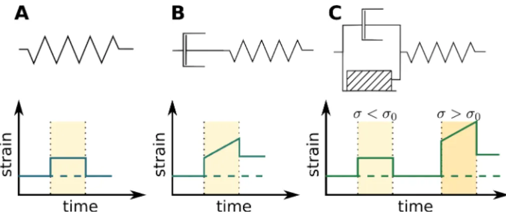
Figure 1.8: Schematics and behaviors of simple rheologies. (A) A pure elastic material behaves like a spring: it returns
to its original state when the stress is released. (B) A visco-elastic material behaves like the association of a spring and a damper, and does not revert to its initial state when the stress is released. (C) The behavior of a visco-elasto-plastic ma-terial depends on the value of the stress applied: if the stress is smaller than the mama-terial-specific thresholdσ0, it behaves
elastically, otherwise it behaves visco-elastically.
where µ is the dynamic viscosity. Here, when the force is released, the material does not revert to its original coniguration: this rheology allows cell wall growth. One of the most elaborate rheological model of the cell wall behavior was introduced by Ortega (1985), and proposes that the cell wall behaves as a visco-elasto-plastic material (see Figure 1.8): the cell wall behaves as an elastic material when the stress is lower than a threshold σ0, but as a visco-elastic material if the stress is larger:
dϵ
dt = (σ − σ0)/µ + dσ
dt/E
Cell wall rheological parameters can be measured, using techniques reviewed in (Milani et al., 2013, Routier-Kierzkowska et al., 2012, Vogler et al., 2015). Depending on the question ad-dressed, one can use one or the other of these rheological model. Note that the rheological parameters of the cell wall can be heterogeneous and/or anisotropic and that these models do not take this into account.
Mechanical forces are omnipresent in plants, and more particularly during morphogenesis; in addition, a growing tissue needs to develop harmoniously integrating all the morphogenetic information over time and space. Models are required to take all actors and their intricate interactions into account.
1.4 Growth of an organ
Despite much progress in developmental biology, we are still far from understanding how organs grow and reach their inal size and shape. Growth is associated with a variety of cellular scale phenomena such as cell expansion, cell proliferation, and cell diferentiation, as well as cell death and cell migration in the case of animals. These processes occur within the thousands of cells that yield a well deined organ. How these various phenomena are coordinated over time and space to shape a consistent and reproducible organ or organism is still an open question. In this