HAL Id: tel-01555654
https://tel.archives-ouvertes.fr/tel-01555654
Submitted on 4 Jul 2017HAL is a multi-disciplinary open access archive for the deposit and dissemination of sci-entific research documents, whether they are pub-lished or not. The documents may come from teaching and research institutions in France or abroad, or from public or private research centers.
L’archive ouverte pluridisciplinaire HAL, est destinée au dépôt et à la diffusion de documents scientifiques de niveau recherche, publiés ou non, émanant des établissements d’enseignement et de recherche français ou étrangers, des laboratoires publics ou privés.
A novel multi-target cancer therapy based on
destabilization of short-lived mRNAs
Felicitas Rataj
To cite this version:
Felicitas Rataj. A novel multi-target cancer therapy based on destabilization of short-lived mRNAs. Molecular biology. Université de Grenoble, 2014. English. �NNT : 2014GRENV040�. �tel-01555654�
THÈSE
Pour obtenir le grade de
DOCTEUR DE L’UNIVERSITÉ DE GRENOBLE
Spécialité : Doctorat CSV/Biologie CellulaireArrêté ministériel : 7 août 2006
Présentée par
Felicitas RATAJ
Thèse dirigée par Dr. Nadia CHERRADI
préparée au sein du Laboratoire Biologie du Cancer et l’Infection dans l'École Doctorale Chimie et Sciences du Vivant
Nouvelle thérapie anti-tumorale
multi-cibles basée sur la dégradation
des ARNms à demi-vie courte
A novel multi-target cancer therapy based on
destabilization of short-lived mRNAs
Thèse soutenue publiquement le 12 Décembre 2014, devant le jury composé de :
Dr. Martin DUTERTRE
UMR 3348 CNRS - Institut Curie Centre de Recherche, Orsay, Rapporteur Dr. Gilles PAGÈS
CNRS UMR 7284 - INSERM U 1081 IRCAN, Nice, Rapporteur Dr. Beatrice EYMIN
INSERM/UJF U823 - Institut Albert Bonniot, Grenoble, Examinateur Pr. Marc POCARD
INSERM U 965 - CHU Paris St-Louis Lariboisière, Paris, Examinateur Dr. Nadia CHERRADI
« Grimpez si vous le voulez, mais n'oubliez jamais que le courage et la force ne sont rien sans
prudence [...]. N'agissez jamais à la hâte, prenez garde au moindre pas. Et dès le début, pensez que
ce pourrait être la fin. » ≈
« Climb if you will, but remember that courage and strength are nought without prudence [...]. Do
nothing in haste; look well to each step; and from the beginning think what may be the end. »
≈
« Klettere, wenn du willst, aber vergiss nicht, dass Mut und Kraft ohne Besonnenheit wertlos sind [...]. Übereile nichts, achte auf jeden Schritt und habe von Anfang an das Ende im Blick. Wir,
die wir die Berge erklettern, wissen, dass jede Höhe durch geduldige und mühsame Anstrengung gewonnen werden muss. Wir wissen
auch, dass ein entschlossener Wille sich den Weg bahnt, und wenn wir zu unseren täglichen Beschäftigungen zurückkehren, so sind wir für
den Kampf des Lebens besser gerüstet und schöpfen aus der Erinnerung neue Kraft und
Lebensfreudigkeit. »
Edward Whymper (1840-1911), Mountaineer
REMERCIEMENTS
Je tiens tout d’abord à remercier les membres de mon jury de thèse :
Martin Dutertre
,Gilles
Pagès
,Marc Pocard
,Béatrice Eymin
d’avoir accepté d’évaluer mon travail.Je souhaite également remercier
Béatrice Eymin
etJean-Luc Coll
d’avoir accepté d’être membres de mon Comité de Suivi de thèse. Merci pour tous vos conseils et suggestions!Je remercie vivement
Jean-Jacques Feige
de m’avoir permis de faire ma thèse au sein du labo BCI et pour toutes ses suggestions scientifiques. Merci pour ton soutien et surtout pour ton effort ces derniers mois afin que je puisse finir mon travail dans de bonnes conditions.Mille mercis à toi
Nadia
, pour avoir eu le courage de partir avec une petite allemande vers cette aventure de trois ans. Ma décision de te rejoindre était la bonne! Tu m’as très bien préparée pour le grand monde de la science. Je me suis sentie toujours très libre et indépendante, mais extrêmement bien encadrée en même temps! Je te remercie vivement pour ton soutien, ton aide, ton enthousiasme, ta rigueur, ta disponibilité, ta patience, ta douceur et surtout pour ta passion pour la recherche!!! J’ai beaucoup apprécié d’avoir eu la chance de travailler avec toi! Je vais faire de mon mieux pour continuer dans cette voie!Je remercie
Séverine Planel
etDelphine Ciais
, qui ont initié ce projet sur TIS11b. Elles ont mis beaucoup de cœur dans ce projet. J’ai eu la chance de vous connaître et d’avoir vos conseils!Odile Filhol-Cochet
etAgnès Castan
,merci pour votre aide et vos suggestions! Vous étiez toujours disponibles pour discuter et prêtes à partager votre savoir-faire et vos réactifs!Vanessa Garnier
, ma chère collègue du bureau. Je te remercie pour ces trois ans ensemble. Nous avons partagé les hauts et les bas. Tu m’as toujours comprise, soutenue et encouragée avec l’élégance d’une parisienne adorable! Je te remercie également pour tout ce temps passé ensemble en dehors du travail – pour ces moments d’amitié. Mille mercis àFrédéric Sergent!
Tu m’as aidé énormément avec les souris, mais plus important encore, tu m’as montré comment gérer des moments de stress avec beaucoup de bonne humeur!Aude Salomon
,Mariela Subileau
,Daniel Vittet
, je vous remercie pour votre aide, vos conseils et les moments de détente pendant les pauses! C’était un grand plaisir pour moi de passer ce temps ensemble!Christine Cogne
etChristine Mallet,
un grand merci pour votre volonté et votre aide!Soumalamaya Bama, Irène Jeannin et Nicolas Chaumontel
, je vous remercie pour votre aide et les moments sympathiques aux côtés des souris!Marie-Pierre Mendez
,Virginie
Lagier
,Sonia Lidy
etNicole
Lefebvre
, je vous remercie vivement pour votre aide administrative!A toutes les autres personnes
du laboratoire avec lesquelles j’ai passé ces trois années. Merci pour votre aide, gentillesse, votre volonté de me faire découvrir la culture française et surtout pour votre patience pour m’expliquer des choses deux, trois fois en français. Merci de m’avoir m’accueillie à bras ouverts!Mille mercis à mes amis en France!
Guillaume
, tu m’as fait découvrir un autre monde – la montagne – avec toutes ses beautés, sa force et ses difficultés. Je te remercie vivement pour ton soutien, pour ton aide et pour toutes nos aventures!Gaëlle et Benoît
, merci beaucoup pour les soirées détendues et rigolotes! Merci pour votre affection! Je me suis beaucoup attachée à vous tous!A ma Maman
etmon Papa, mon frère
etma petite sœur
et toutema famille
, même si c’est dur pour vous de comprendre mon travail, vous étiez toujours derrière moi pendant ces années. Mille mercis pour votre soutien, votre aide, votre confiance! Je suis fière, émue et reconnaissante d’avoir une famille comme vous!Felix
, mille mercis à toi pour ton soutien, ton amour, ta confiance, ton aide pendant ce temps passé en France! J’ai apprécié notre décision de partir ensemble vers cette aventure! C’était une expérience inoubliable de découvrir la culture et la cuisine française, le pays et la montagne avec toi! Je suis fière de toutes nos réussites!L
ist of contentINTRODUCTION ... 17
Chapter 1 Regulation of short-lived mRNA stability ... 19
1.1. The highway to degradation: Deadenylation-dependent mRNA decay ...21
1.2. The highway code: Control of mRNA decay ...23
1.2.1. Cis-acting elements: Focus on AU-rich elements ...23
1.2.2. Trans-acting factors: Focus on ARE-binding proteins ...26
1.2.2.1. Stabilizing ARE-binding proteins ...27
1.2.2.2. Destabilizing ARE-binding proteins ...28
1.3. Regulation of mRNA decay by signalling pathways ...30
In conclusion ...33
Chapter 2 Key player in mRNA decay: The TIS11 protein family ... 35
2.1. Members of the TIS11 protein family ...37
2.1.1. Tristetraprolin (TTP) ...37
2.1.2. TIS11b ...39
2.1.3. TIS11d ...42
2.1.4. Zfp36l3 ...43
2.2. Regulation of TIS11 protein expression ...43
2.3. Protein domains and truncation of TIS11 proteins ...46
2.4. Functions of TIS11 proteins ...49
2.4.1. Key players in mRNA decay ...50
2.4.2. Alternative functions of TIS11 proteins during mRNA lifecycle ...51
2.5. Regulation of TIS11 protein activities ...55
2.5.1. Post-translational modifications – phosphorylation ...56
In conclusion ...59
Chapter 3 The multi-step development of human cancer ... 61
3.1. The hallmarks of cancer ...62
3.1.1. Enabling hallmark: Genome instability and mutation ...62
3.1.2. Enabling hallmark: Tumor-promoting inflammation ...63
3.1.2.1. Cytokines ...65
3.1.2.2. Chemokines ...68
3.1.3. Hallmark: Sustaining proliferative signalling ...70
3.1.4. Hallmark: Evading growth suppressors ...71
3.1.5. Hallmark: Resisting cell death ...72
3.1.7. Hallmark: Inducing angiogenesis ...73
3.1.7.1. Physiological angiogenesis ...73
3.1.7.2. Tumor angiogenesis ...81
3.1.8. Hallmark: Activating invasion and metastasis ...86
3.1.9. Emerging hallmark: Deregulating cellular energetics ...87
3.1.10. Emerging hallmark: Evading immune destruction ...89
3.2. The cancer niche or tumor microenvironment ...90
3.2.1. Cell types of the tumor microenvironment ...91
3.2.1.1. Cancer stem cells ...91
3.2.1.2. Immune inflammatory cells ...92
3.2.1.3. Cancer associated fibroblasts ...92
3.2.2. Heterotypic signalling coordinates cells of the tumor stroma ...93
3.2.3. Hypoxia ...95
In conclusion ...99
Chapter 4 Therapeutic targeting of cancer and its limits ... 101
4.1. Fight against cancer: Focus on anti-angiogenic and anti-inflammatory cancer ...103
therapies ...103
4.1.1. Targeting tumor vasculature: Anti-angiogenic therapies ...103
4.1.1.1. Single-target angiogenesis inhibitors: the first generation ...104
4.1.1.2. Second generation multi-targeted angiogenesis inhibitors: VEGF and related targets ………106
4.1.1.3. Broad-spectrum of angiogenesis inhibitors: the next generation ...106
4.1.1.4. Indirect pharmacological inhibitors of angiogenesis ...107
4.1.2. Targeting the wound that never heals: Anti-inflammatory therapies ...108
4.2. Limits of targeted cancer therapies ...111
4.3. Emerging link between regulation of mRNA stability and cancer ...113
4.3.1. Expression of TIS11 proteins in cancer cell lines ...114
4.3.2. The impact of TIS11 proteins on cancer hallmarks ...115
In conclusion ...121
Chapter 5 Breaking through the other side: Cell penetrating peptides ... 123
5.1. Classes of CPPs ...124
5.2. Different cellular uptake mechanisms of CPPs ...125
5.3. The potential of CPPs in anti-cancer drug delivery ...127
5.4. CTPs ...129
In conclusion ...131
RESULTS ... 135
Article 1 ...137
“A novel phosphorylation-dependent regulation of TIS11b stability and activity by cAMP-dependent protein kinase (PKA) reveals the important role of two conserved N- and C-terminal serines (S54 and S334)” ...137
Introduction ...139
Abstract ...145
Introduction ...147
Material and Methods ...149
Generation of stable Hela Tet-off cells expressing wild type TIS11b and TIS11b mutants ... 152
Results ...154
PKA signalling pathway regulates TIS11b expression and phosphorylation ...154
Identification of PKA target sites within TIS11b sequence: conserved S54 and S334 are phosphorylated in vitro ...155
TIS11b is phosphorylated at S54 and S334 in vivo ...156
S54 and S334 regulate TIS11b mRNA-destabilizing activity ...158
TIS11b protein stability is regulated by S54 and S334 ...159
S54 and S334 modulate the proteasome-dependent degradation of TIS11b ...159
Mimicking a phosphorylation at S54 changes TIS11b subcellular localization ...160
Mimicking a phosphorylation at S54 promotes the binding of TIS11b to endogenous 14-3-3 proteins ...161
TIS11b interacts with endogenous CNOT1 ...161
Discussion ...162
Acknowledgements ...166
References ...167
Legends of the figures ...171
Article 2 ...185
“Targeting AU-rich element-mediated mRNA decay using a mutant version of ZFP36L1/TIS11b zinc finger protein impairs major hallmarks of tumorigenesis” ...185
Introduction ...187
Abstract ...191
Introduction ...193
Material and Methods ...195
Results ...204
The N- and C-terminal domains of TIS11b are active in mRNA decay ...204
R9-ZnC and R9-ZnCS334D fusion proteins reduce luciferase reporter gene activity through VEGF-3’UTR ...205
R9-ZnC and R9-ZnCS334D protein overexpression and purification ...206
Deletion of the N-terminal domain of TIS11b increases the stability of the purified protein under storage ...206
R9-ZnC and R9-ZnCS334D are efficiently internalized into living cells ...207
Purified R9-ZnC and R9-ZnCS334D proteins decrease VEGF mRNA and protein expression ...207
R9-ZnCS334D inhibits proliferation and migration of breast cancer cells in vitro ...208
R9-ZnC and R9-ZnCS334D reduce cancer cell invasion and anchorage-independent cell growth ...208
R9-ZnC and R9-ZnCS334D downregulate the expression of EMT-markers ...209
R9-ZnC and R9-ZnCS334D impair the formation of pseudo capillaries by endothelial cells ...209
Discussion ...210
Acknowledgements ...214
References ...215
Legends of the figures ...218
Supplementary data: In vivo characterization of R9-ZnC and R9-ZnCS334D as novel anti-tumoral agents ...233
Results & Discussion ...235
Intratumoral injection of R9-ZnC or R9-ZnCS334D inhibits 4T1 luc tumor growth ...235
R9-ZnC or R9-ZnCS334D-treatment of 4T1-luc tumors decreases VEGF expression and microvessel density as well as hypoxic areas ...237
Decreased expression of several angiogenic and inflammatory cytokines upon ZnC- or R9-ZnCS334D treatment in vivo ...239
R9-ZnC and R9-ZnCS334D reduces expression of EMT-markers in vivo ...247
Conclusion ...250
Material & Methods ...251
DISCUSSION & CONCLUSIONS ... 255
BIBLIOGRAPHY ... 263
L
ist of figuresFigure 1: Complexity of RNA decay and enzymes involved in eukaryotic cells (Stoecklin & Muhlemann, 2013). ... 18 Figure 2: Mechanisms of deadenylation-dependent mRNA decay in eukaryotes (Planel et al.,
2014). ... 20 Figure 3: Cis-elements located in 3’untranslated region (3’UTR) of mRNAs are recognized
by specific trans-acting factors to modulate the rate of mRNA decay (Thapar & Denmon, 2013). ... 22 Figure 4: Regulation of deadenylation-dependent mRNA degradation through AU-rich
elements (AREs) recognized by ARE-binding proteins (ARE-BPs) in eukaryotes (Planel et al., 2014). ... 26 Figure 5: Phenotype of TTP-knock out (KO-PBS) mice compared to wildtype (WT) and
prevention of this phenotype by injecting anti-TNFα antibodies (KO-Ab) (Taylor et al., 1996). ... 36 Figure 6: Optical projection tomography of vascular abnormalities in TIS11b-/- and TIS11b
+/-embryos at E9.5 (Bell et al., 2006). ... 41 Figure 7: Schematic presentation of TTP gene, mRNA and protein. TTP gene consists of one
intron and two exons (Sanduja et al., 2012). ... 45 Figure 8: Structure similarities between human TIS11 proteins (Ciais et al., 2013). ... 47 Figure 9: Predicted structure of human TTP tandem zinc finger (TZF) domain in a complex
with 5’-UUAUUUAUU-3’-ARE (pink) based on original structure for TIS11d TZF domain (Carrick et al., 2004; Hudson et al., 2004). ... 47 Figure 10: Subcellular localization of TTP, TIS11b (CMG1) and TIS11d (11D) in leptomycin
B-treated HEK 293 cells (Phillips et al., 2002). ... 49 Figure 11: Multiple functions of TIS11 proteins during mRNA life cycle have been shown
(Ciais et al., 2013). ... 52 Figure 12: Subcellular localization of mRNAs is regulated by TIS11 proteins (Franks &
Lykke-Andersen, 2007)... 53 Figure 13: Signalling pathways acting on TTP and TIS11b (Ciais et al., 2013). ... 56 Figure 14: Central role of the p38-MAPKMK2PP2ATTP axis in the regulation of TTP
protein activity, stability and localization (Sandler & Stoecklin, 2008). ... 57 Figure 15: Different phases of tumor development (Thiery, 2002). ... 60 Figure 16: Hallmarks during tumor progression (Hanahan & Weinberg, 2011). ... 60 Figure 17: The intrinsic and extrinsic pathways that describe the link between cancer and
inflammation (Mantovani et al., 2008). ... 64 Figure 18: The complex role of cytokines and chemokines during cancer initiation and
progression (Landskron et al., 2014). ... 67 Figure 19: Involvement of chemokines in angiogenesis of breast cancer (Palacios-Arreola et
al., 2014). ... 69 Figure 20: Interaction of CXCR4 with its ligand CXCL12 in primary tumor and metastasis of
Figure 21: A balance between pro- and anti-angiogenic factors controls angiogenesis. ... 74
Figure 22: Regulation of VEGF-A expression by alternative splicing (Eymin et al., 2014)... 75
Figure 23: The binding specificities of VEGF proteins and VEGFR signalling complexes. .... 78
Figure 24: The multi-step process of angiogenesis including the activation phase, the sprouting and the maturation phase (David et al., 2009). ... 80
Figure 25: The angiogenic switch during tumor development (Bergers & Benjamin, 2003). .. 82
Figure 26: Schematic representation of tumor angiogenesis including sprouting angiogenesis, vasculogenesis (bone marrow-derived endothelial progenitor cells are recruited to the tumor by different factors such as the stromal-derived factor 1 (SDF-1), the basic fibroblast growth factor (bFGF), IL-6 and G-CSF), intussusception and vessel co-option (Zhu et al., 2011). ... 84
Figure 27: Tumor vasculature is abnormal in their structure and function (Carmeliet & Jain, 2011). ... 85
Figure 28: Abnormalities of tumor microvasculature (Jain, 2005; Goel et al., 2011). ... 85
Figure 29: The invasion-metastasis cascade (Thompson & Haviv, 2011). ... 87
Figure 30: Complexity of the interactions between tumor cells and cells of the tumor microenvironment (Hanahan & Weinberg, 2011). ... 90
Figure 31: Signalling network of cancer cells and the tumor microenvironment during tumorigenesis (Hanahan & Weinberg, 2011). ... 94
Figure 32: Oxygen-dependent regulation of HIF-1α stability and HIF-1 signalling (Semenza, 2013). ... 96
Figure 33: Therapeutic targeting of cancer hallmarks (Hanahan & Weinberg, 2011). ... 100
Figure 34: The basic concept of anti-angiogenic cancer therapies (Zetter, 2008). ... 102
Figure 35: The hypothesis of vessel normalization (Jain, 2005; Goel et al., 2011). ... 104
Figure 36: Adaptive (evasive) resistance and intrinsic non-responsiveness are the two modes of tumor resistance to anti-angiogenic cancer therapies (Bergers & Hanahan, 2008). ... 110
Figure 37: Mechanisms of adaptive resistance (Bergers & Hanahan, 2008). ... 110
Figure 38: Network of TTP-mediated post-transcriptional control of mRNAs encoding oncogenic and tumor-promoting factors (Ross et al., 2012). ... 115
Figure 39: Cellular uptake mechanisms and possible intracellular trafficking of CPPs (Chou et al., 2011). ... 126
Figure 40: Activatable CPP, which is fused to an anionic inhibitory sequence via MMP-cleavable linker (Jiang et al., 2004). ... 128
Figure 41: Biodegradable pH-sensitive micelle delivery system (Sethuraman et al., 2008).. . 128
Figure 42: Multi-target effect of truncated/mutated TIS11b fusion proteins on cancer hallmarks. ... 258
L
ist of tablesTable 1: Classification of AU-rich elements after Chen et al. (Chen et al., 1995). ... 25 Table 2: Classification of AU-rich elements after Bakheet et al. (Bakheet et al., 2003). ... 25 Table 3: Overview of important ARE-binding proteins in eukaryotes (Planel et al., 2014). ... 27 Table 4: Identified targets of TTP (Brooks & Blackshear, 2013). ... 38 Table 5: Identified targets of TIS11b (Baou et al., 2009). ... 40 Table 6: TTP interaction partners (Brooks & Blackshear, 2013). ... 50 Table 7: Anti-angiogenic agents, their molecular targets and current indications for cancer
therapy (Limaverde-Sousa et al., 2014). ... 102 Table 8: Examples of CPPs, their origin, structure and proposed mechanism of cellular uptake
L
ist of abbreviations5ʼ/3ʼUTR 5ʼ/3ʼ untranslated region ACTH Adrenocorticotropic hormone AGO2 Argonaute 2
AMF Autocrine motility factor Ang1 Angiopoietin 1
Ang2 Angiopoietin 2 ARE AU-rich elements ARE-BP ARE-binding proteins
ARED AU-rich elements (AREs) containing mRNA database Arg (R) Arginin
Asp (D) Aspartate
ASTRC AUF1-and-Signal Transduction-Regulated Complex BAC Bovine adrenocortical cells
Berg36 B-cell early response gene encoding a 36-kDa protein BM Basement membrane
BMDC Bone-marrow derived cells BRF1/2 Butyrate Response Factor 1/2 BSA Bovine serum albumin
C, CC(R) C/CC-motif chemokine (receptor) CA Carbonic anhydrase
CAF Cancer-associated fibroblast CDKN2A Cyclin dependent kinase inhibitor 2A CHX Cycloheximide
COX-2 Cyclooxygenase 2 CPP Cell-penetrating peptide CSC Cancer stem cell
CSF-1 Colony-stimulating factor 1 CTD C-terminal domain CTP Cell-targeting peptide
CXC, CX3C(R) CXC/CXC3-motif chemokine (receptor) Cul4B Cullin 4B
Da Dalton
Dll4 Delta-like 4
DRB 5,6-Dichloro-β-D-ribofuranosyl benzimidazol EC Endothelial cells
ECM Extracellular matrix
EDTA Ethylenediaminetetraacetic acid EGF Epidermal growth factor EGFR EGF receptor
ELAV Embryonic-lethal abnormal visual in Drosophila melanogaster EMT Epithelial-mesenchymal transition
ENDO Endonucleolytic mRNA decay eNOS Endothelial nitric oxide synthase
EPR Enhanced Permeability and Retention effect ERF-1/2 Epidermal Growth Factor-response factor 1/2 ERK Extracellular-signal-regulated kinase EXO Exonucleolytic mRNA decay FDA Food and Drug Administration FFF Formalin-free fixation solution FGF Fibroblast growth factor (=bFGF) Flk-1 Fetal liver kinase 1
Flt-1/4 Fms-like tyrosine kinase 1/4 GFP Green fluorescent protein G0S24 G0/G1 switch regulatory gene 24
GM-CSF Granulocyte macrophage-colony stimulating factor GLUT1-4 Glucose transporter 1 to 4
GSK3β Glycogen synthase kinase-3 beta GST Glutathion-S-transferase HEK Human embryonic kidney
HER Human epidermal growth factor receptor 2 HNF1α Hepatocyte nuclear transcription factor 1α HIF-1 Hypoxia-inducible factor-1
HNF1α Hepatocyte nuclear transcription factor 1α HPRT Hypoxantin phosphoribosyl transferase HRE Hypoxia responsive element
HUVEC Human umbilical vein endothelial cells ICAM Intercellular adhesion molecule IFP Interstitial fluid pressure IFNγ Interferon γ
IL Interleukin
IP Immunoprecipitation
IP-10 Interferon γ-induced protein 10
IPTG Isopropyl-beta-D-thiogalacto-pyranoside IRE Iron response element
IREBP IRE-binding protein IRES Internal ribosome entry sites JAK Janus-activated kinase JNK C-jun-N-terminal kinase αKG α-ketoglutarate
KA in vitro kinase assay
KH-domains K homology-domains KO Knock out
KSRP K homology splicing regulatory protein LDHA Lactate dehydrogenase
LH Luteinizing hormone LHR LH receptor
LLC Lewis lung carcinoma LOX Lysyl-oxydase LPS Lipopolysaccharide
MCP-1 Monocyte-chemoattractant protein 1 MCT4 H+/monocarboxylate transporter 4 MEK1/2 Mitogen-activated protein kinase kinase1/2 MET Mesenchymal-epithelial transition MIP-1 Macrophage inflammatory protein 1 miRNA micro RNA
MK2 p38MAPK-activated protein kinase 2 MMP Matrix-metalloprotease
MR Mineralocorticoid receptor MSC Mesenchymal stem cell mTOR
NES Nuclear export sequence
NF-κB Nuclear factor kappa-light-chain-enhancer of activated B cells NGD No-go mRNA decay
NLS Nuclear localization sequence NMD Nonsense-mediated mRNA decay NOV Nephroblastoma overexpressed gene NRP1/2 Neuropilin 1/2
NTD N-Terminal domain
NSAIDs Non-steroidal anti-inflammatory drugs OE Overexpression
P-body (PB) Processing body
p38 MAPK p38 mitogen activated protein kinase PABP Poly(A)-binding protein
PARN Poly(A)-specific ribonuclease PBS Phosphate buffer saline PDGF Platelet-derived growth factor PFA Paraformaldehyde
PHD Prolyl-hydroxylases
PI3K Phosphatidylinositol 3ʼ–kinase PIMO Pimonidazol
PKA cAMP-protein kinase A PKB Protein kinase B (=AKT) PKC Protein kinase C PLGF Placental growth factor PMA Phorbol myristyl acetate PP2A Protein phosphatase 2A PTD Protein transduction domain
PTEN Phosphatase and tensin homologue deleted on chromosome 10 PTX3 Pentraxin 3
PVDF Polyvinylidene fluoride RB Retinoblastoma proteins RBP RNA-binding protein RGD Arginine-glycine-aspartate
RISC RNA-induced silencing complex RONS Reactive oxygen/nitrogen species RPL13A Ribosomal protein 13a
RRM RNA Recognition Motifs SDF-1 Stromal-derived factor 1 SDS Sodium dodecyl sulfate Ser (S) Serine
SG Stress granule siRNA Small interfering RNA SLBP Stem-loop-binding protein SSC Saline sodium citrate
TAM Tumor-associated macrophage TAN Tumor-associated neutrophils
TAT Transcription-transactivating protein of HIV-1 TBS Tris buffer saline
TCA Tricarboxylic acid cycle (Krebs cycle) TGFβ Transforming growth factor beta Th T-helper cell
Thr threonine
TIL Tumor-infiltrating leukocyte TIMP Tissue Inhibitor of metalloprotease TIS11 TPA-inducible sequence 11 TKI Tyrosine kinase inhibitor TNFα Tumor necrosis factor alpha
TPA 12-O-tetradecanoylphorbol-13-acetate TSP1/2 Thrombospondin 1/2
TTP Tristetraprolin
TTR-BPs Turnover and translation regulatory RNA-binding proteins TZF Tandem zinc finger
Ub Ubiquitin
uPA(R) Urokinase-type plasminogen activator (receptor) Y2H Yeast two hybrid
VECAM Vascular cell adhesion protein VEGF Vascular endothelial growth factor VEGFR VEGF receptor
VHL Von Hippel Lindau factor VPF Vascular permeability factor
WST-1 4-[3-(4-iodophenyl)-2-(4-nitrophenyl)-2H-5-tetrazolio]-1, 3-benzene disulfonate WT Wildtype
ZF Zinc finger
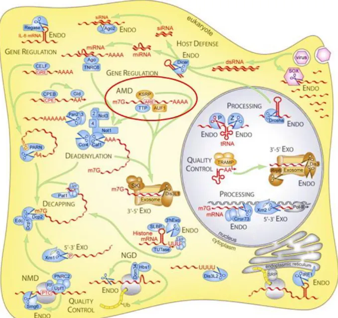
Figure 1: Complexity of RNA decay and enzymes involved in eukaryotic cells (Stoecklin &
Muhlemann, 2013). Among other mechanisms, ARE-mediated mRNA decay (AMD) is
an important regulatory process of mRNA stability and is further described in this chapter. ENDO, endonucleolytic mRNA decay; 5ʼ-3ʼ EXO, 5ʼ-3ʼ exonucleolytic mRNA decay; 3ʼ-5ʼ EXO, 3ʼ-5ʼexonucleolytic mRNA decay; NGD, no-go mRNA decay; NMD, nonsense-mediated mRNA decay.
C
hapter 1
Regulation of short-lived mRNA stabilityGene expression is a tightly controlled mechanism. Beside transcription, post-transcriptional mechanisms are also important for controlling gene expression. Multiple regulatory systems ensure the balance between gene products (RNA or protein) and their need for cellular growth, function and fate. Especially during development, the presence of certain transcripts during specific time windows is crucial. Protein concentration is determined by the cytoplasmic concentration of the corresponding mRNA, which depends on mRNA synthesis and decay rates, and also on post-translational modifications regulating protein stability. Post-transcriptional regulatory mechanisms, sub-divisible into processes determining translatability of mRNAs and processes defining equilibrium between transcription rate and degradation of the transcript, gained a lot of interest during the last years. Pre-mature mRNA processing, nuclear mRNA export, RNA interference, mRNA sequestration, codon usage, translational repression by microRNAs (miRNAs) or proteins, and the control of mRNA turnover are limiting mechanisms for mRNA synthetic rate. Figure 1 illustrates the fascinating biology of eukaryotic ribonucleases and how they are involved in the mRNA life cycle (Stoecklin & Muhlemann, 2013). Although most mRNAs are degraded through the deadenylation-dependent decay pathway triggered either by RNA-binding proteins or microRNAs, deadenylation-independent mechanisms exist (Fabian et al., 2010; Garneauet al., 2007). For certain mRNAs, degradation is initiated by endonuclease cleavage within the nucleotide sequence, followed by further degradation via either the exosome (3’→5’ decay) or the exoribonuclease Xrn1 (5’→3’ decay). Mammalian cells have developed several mRNA surveillance mechanisms to clear defective transcripts and to avoid the synthesis of abnormal proteins. Nonsense-mediated mRNA decay (NMD) seems to be restricted to newly synthesized mRNAs that contain premature termination codons. The translation of these transcripts would lead to truncated proteins with aberrant function. No-go mRNA decay (NGD) induces the degradation of faulty transcripts associated with stalled ribosomes, to release sequestered components of the translation machinery. Transcripts lacking a stop codon are degraded by the non-stop mRNA decay pathway, thus preventing translation to proceed along the poly(A)tail of the transcript and to result in aberrant proteins.
However, this chapter will focus on the post-transcriptional regulation of mRNA stability, in particular on AU-rich element-mediated control of mRNA half-life, which enables cells to quickly adjust transcript levels and their translational potential in response to various stimuli (Guhaniyogi & Brewer, 2001).
Figure 2: Mechanisms of deadenylation-dependent mRNA decay in eukaryotes (Planel et al.,
1.1. The highway to degradation: Deadenylation-dependent mRNA decay
Eukaryotic mRNAs contain two stabilizing structures, the 5’-end 7-methylguanosine cap (m7G-cap) and the poly(A) tail located at the 3’-end, which are added to the pre-mature mRNA after transcription. These two structure elements are recognized by the cytoplasmic proteins eIF-4F and the poly(A)-binding protein (PABP), to enhance translation initiation and to protect mRNAs from unspecific exonuclease activity. Translocation of the mature mRNA occurs at the level of cytoplasmic polysomes. In response to a still unknown signal, mRNAs are deadenylated and transported to processing bodies (P-bodies). In P-bodies, mRNAs can be either stored for return to translation or recruited to the mRNA decay machinery for degradation. P-bodies are dynamic, cytoplasmic foci, which harbour non-translatable mRNAs. In addition, translation repressors and mRNA decay factors are present in these cellular structures (Kulkarni et al., 2010).
The decay of the majority of eukaryotic mRNAs is initiated by a rate-limiting process called deadenylation, which leads to the shortening of the poly(A) tail (Figure 2). Several deadenylases, located in the cytoplasm of eukaryotic cells as components of P-bodies, are well characterized. Pan2-Pan3 is a PABP-dependent deadenylase unit, which trims the nascent poly(A) tail (Brown et al., 1996). Ccr4-Not is a large protein complex, which consist of nine subunits including the conserved canonical subunits Ccr4, three Caf proteins (Caf1/Pop2, Caf40/Not9, Caf130/Not10) and five Not proteins (Not1, Not2, Not3, Not4 and Not5) (Wahle & Winkler, 2013; Inada & Makino, 2014). In this complex, Ccr4 and Caf1 are the catalytic subunits, whereas Not1 functions as a central scaffold. The role of the other subunits remains unknown. Deadenylation in eukaryotic cells seems to be a two-phase process, where Pan2-Pan3 is performing a first, incomplete shortening of the poly(A) tail, followed by the entire poly(A) tail degradation through Ccr4-Not1 deadenylase activity (Zheng et al., 2008). A third mammalian deadenylase is the poly(A)-specific ribonuclease (PARN), which plays an important role during embryogenesis (Godwin et al., 2013). PARN activity is cap-dependent and is increased by interacting with the 5’-end m7G-cap. The naked 3’-end is further attacked by the exosome, a macromolecular complex of 10-11 subunits. Nine subunits form the central core of the exosome. Two other subunits of multi-domain polypeptides, which carry the catalytical activity of the eukaryotic exosome, associate with the central core depending on cellular localization, leading to mRNA degradation (Figure 2) (Chlebowski et al., 2013). The 5’-cap on the remaining nucleotide sequence is metabolized by the scavenger decapping enzyme DcpS.
Figure 3: Cis-elements located in 3’untranslated region (3’UTR) of mRNAs are recognized by
specific trans-acting factors to modulate the rate of mRNA decay (Thapar & Denmon,
2013). (A) Poly(A) tail and PABP. (B) AU-rich elements (AREs) and ARE-binding
proteins (ARE-BP). (C) Histone stem-loop and the stem-loop-binding protein (SLBP). (D) Iron response elements (IREs) and the IRE-binding protein (IRE-BP). (E) MiRNAs targeting mRNA sequences and recruiting the RNA-induced silencing complex (RISC) complex.
Alternatively to the exosome-dependent pathway, deadenylation can be followed by removing the 5’-cap through the major activity of decapping enzymes Dcp1 and Dcp2 (Figure 2) (Arribas-Layton et al., 2013). Within this complex, Dcp2 has the intrinsic decapping activity. Xrn1, a 5’ 3’exoribonuclease, recognizes the unprotected 5’-end leading to 5’ 3’decay of the mRNA (Nagarajan et al., 2013).
1.2. The highway code: Control of mRNA decay
Traditionally, transcriptional regulation has been the major focus of gene expression studies. However, recently the important contribution of mRNA decay mechanisms has emerged and numerous pathways linked to this aspect were reported. Most regulatory sequences (cis-acting elements) involved in these control mechanisms are located in the 5’ and 3’untranslated regions (UTR) of mRNAs, which function as platforms for association of multi-subunit complexes initiated by RNA-binding proteins (RBPs) and other trans-acting factors (miRNAs, long non-coding RNAs). Whereas the 5’UTR is more important in translational control including cap-dependent and internal ribosomal entry site (IRES)-mediated processes, determinants of mRNA stability, cellular location and translatability are predominately found in the 3’UTR (Moore, 2005; Pickering & Willis, 2005). Interestingly, the length of 3’UTR could act as determining factor of mRNA stability. Experiments have shown a positive correlation between length of 3’UTR and number of cis-acting elements, respectively, and mRNA half-life (Akashi et al., 1994). Importantly, these mechanisms are often altered during cancer development and other life-threatening diseases, suggesting that they might contribute to human pathologies (Vislovukh et al., 2014).
The following paragraphs will describe how mRNA fate is controlled by a balance between stabilizing and destabilizing RNA-binding factors which recognize regulatory cis-acting sequences located in the 3’UTR.
1.2.1. Cis-acting elements: Focus on AU-rich elements
Several cis-acting sequences, such as the poly(A) tail, iron-responsive elements (IRE), Jun-kinase response elements, histone stem-loop and AU-rich elements (AREs) are located in the 3’UTR of the mRNA to determine its stability (Figure 3) (Knapinska et al., 2005; Matoulkova et al., 2012). Stem-loop destabilizing elements are present in mRNAs encoding for Interleukin-2/6 or the granulocyte-stimulating factor (G-CSF). This sequence motif requires at least one stem-loop structure to be functional. Furthermore, CU-rich regulatory elements confer instability to mRNAs. The poly(A)-tail promotes longer stability of transcripts and efficient translation. AREs
are the landmark cis-acting elements of short-lived mRNAs (Figure 3) (Matoulkova et al., 2012). AREs were originally identified as instability determinants (Shaw & Kamen, 1986). Shaw & Kamen gave the initial evidence of the destabilizing function of AREs as they demonstrated that the half-life of the otherwise stable β-globin mRNA was decreased when fused to the 3’UTR of the granulocyte macrophage-colony stimulating factor (GM-CSF) which is known to harbours AREs. AREs are generally conserved between species (Halees et al., 2008). Initially, it was
estimated that 8-10 % of total mammalian mRNAs contain AREs
(http://brp.kfshrc.edu.sa/ARED/) (Halees et al., 2008). More recently, a database elaborated for a comprehensive investigation of AREs reported 3 275 protein encoding genes which harbour at least one ARE in their 3ʼUTR, underscoring the importance of these sequences (Halees et al., 2008; Gruber et al., 2011). By searching AREs also in the introns of human genes, 9 114 additional genes were found, meaning that around 50 % of human genes are part of the ARE-regulome (Halees et al., 2008). ARE appearance correlates with rapid patterns of mRNA decay for genes encoding for factors involved in proliferation, inflammation, transcription, immune response, development and signalling or proto-oncogenes (Shaw & Kamen, 1986; Shyu & Wilkinson, 2000). ARE-bearing mRNAs are intrinsically labile short-lived mRNAs with half-lives of a few hours.
AREs are 40- to 150-nt long adenylate uridylate-rich sequences with various copies of an AUUUA motif. UUAUUUAWW (W=A/U) is considered as the minimally functional ARE. Historically, AREs were categorized into three groups based on the number of the AUUUA pentamer (ARE length) and functional characteristics (Table 1) (Chen & Shyu, 1994). Class I presents several isolated AUUUA motifs flanked by U-rich sequences found in early-response-gene mRNAs like transcription factors. Genes categorized in class II harbour clustered or tandem, overlapping pentamers. Representatives of this group are cytokines like the GM-CSF, the tumor necrosis factor α (TNFα), the Vascular endothelial growth factor (VEGF) or Interleukin-3 (IL-3). Class III represents U-rich sequences in absence of the AUUUA pentanucleotide. All three ARE-categories induce mRNA decay, but via different mechanisms and kinetics. Class I, such as c-fos ARE, and class III, like c-jun ARE, mediate mRNA degradation through a synchronous shortening of the poly(A) tail, followed by the rapid digest of the mRNA core molecule. In contrast, mRNAs containing class II AREs, such as GM-CSF or IL-3 are eliminated asynchronously – poly(A) tail is removed simultaneously to endo-/exonuclease activity (Chen et al., 1995). Recently, an alternative classification, which takes also the context of the AUUUA pentamer into account, was proposed and is the basis of the ARE-mRNA database “ARED” (Table 2) (Bakheet et al., 2003; Bakheet et al., 2006). The
computationally predicted 13 bp sequence WWUUAUUUAUUWW is the most consensus motif in labile mRNAs. Sequence variations around this core motif are the basis of ARED classification into five groups. Cluster I-IV contains two to five AUUUA pentanucleotides. Cluster V harbours only one 13 bp consensus motif. Interestingly, AREScore, a recent study using an algorithm to identify mRNAs containing AUUUA motifs described further potential ARE-regulated mRNAs, which are not listed in ARED (Spasic et al., 2012).
Table 1: Classification of AU-rich elements according to Chen et al. (Chen et al., 1995). GM-CSF, Granulocyte macrophage-colony stimulating factor ; TNF, Tumor necrosis factor ; VEGF, Vascular endothelial growth factor; IL-3, Interleukin-3.
Table 2: Classification of AU-rich elements according to Bakheet et al. (Bakheet et al., 2003). INF, Interferon ; COX-2, Cyclooxygenase 2; FGF2, Fibroblast growth factor 2; uPAR, Urokinase-type plasminogen activator receptor
1.2.2. Trans-acting factors: Focus on ARE-binding proteins
AU-rich elements are specifically recognized by trans-acting factors, called ARE-binding proteins (ARE-BPs), which belong to a group of proteins, called turnover and translation regulatory RNA-binding proteins (TTR-BPs) (Pullmann et al., 2007). Therefore, ARE-BPs are involved in processing, nuclear export, cellular localization, degradation and translation of mRNAs. The final effect on mRNA strongly depends on the primary mRNA regulatory function of the bound ARE-BP. mRNA stability is determined by a balance between counteracting factors which compete for their binding to AREs or co-activate each other due to direct interactions (Cherradi et al., 2006; Hinman & Lou, 2008; Kedar et al., 2012). mRNAs are either stabilized and their translation is enhanced or they are tagged for degradation and translationally repressed. A common structure characteristic of ARE-BPs is the RNA-binding domain, which could be of different nature, including RNA Recognition Motifs (RRMs), K homology (KH)-domains and CCCH tandem zinc fingers. The N- and C-terminal part of these factors is less conserved and contains numerous interaction sites with other proteins. Therefore ARE-BPs are considered as linkers between mRNAs and the mRNA decay machinery by recruiting components of these protein complexes implicated in every step of mRNA degradation (Figure 4).
Figure 4: Regulation of deadenylation-dependent mRNA degradation through AU-rich elements (AREs) recognized by ARE-binding proteins (ARE-BPs) in eukaryotes (Planel et al.,
To date, around twenty ARE-BPs are known, the most important are listed in Table 3.
Table 3: Overview of important ARE-binding proteins in eukaryotes (Planel et al., 2014).
1.2.2.1. Stabilizing ARE-binding proteins
The best studied stabilizing ARE-BP is HuR, a ubiquitously expressed protein (Table 3). This protein is a member of the ELAV (embryonic-lethal abnormal visual in Drosophila
melanogaster) protein family, comprising the nervous-system specific members HuB, HuC and Hu-D (Ma et al., 1996). HuR harbours three RNA recognition motifs through which it interacts with ARE-containing mRNAs. HuR targets a variety of mRNAs including cytokines, proto-oncogenes, growth factors and cell cycle regulators, leading to transcript stabilization and/or enhanced translation (Hinman & Lou, 2008; Lebedeva et al., 2011). Upon cell stimulation, nuclear HuR translocates reversibly into the cytoplasm (Fan & Steitz, 1998). The mechanism of mRNA stabilization remains unknown. Based on experimental observations, one could assume a competition between HuR and destabilizing ARE-BPs for the ARE (Cherradi et al., 2006; Hinman & Lou, 2008). Alternatively, HuR could interact directly or indirectly with components of the mRNA decay machinery, avoiding the assembly of these protein complexes, or strengthening the interaction of PABP to the poly(A) tail. In addition to its role in regulation of mRNA stability, HuR can also affect the expression of target proteins at the level of translation. HuR may serve as enhancer or repressor of translation. Furthermore, Hu proteins have been shown to influence alternative splicing and polyadenylation of the pre-mature transcript (Hinman & Lou, 2008).
It is worth mentioning that HuR-triggered mRNA functions are regulated by post-translational modifications of HuR involving several signalling cascades (Doller et al., 2008). HuR is target of various kinases such as MAPKs, MAPK-activated protein kinase-2 (MK2), AMP-activated kinase (AMPK) or the cell-cycle checkpoint kinase 2 (Chk2) leading to the phosphorylation of the protein. Furthermore, methylation and acetylation as well as ubiquitination are described for HuR. Altogether these post-translational modification regulate HuR subcellular localization, impact the sequestration of HuR and HuR binding to ligand proteins (e.g. 14-3-3). However, detailed mechanisms are poorly understood.
1.2.2.2. Destabilizing ARE-binding proteins
The antagonists of stabilizing ARE-BPs are factors promoting mRNA decay by binding to AREs. Overexpression of such factors is associated with rapid degradation of ARE-containing mRNAs. Once bound to the ARE, destabilizing ARE-BPs function as platforms for assembly of protein complexes implicated in mRNA decay by interacting directly or indirectly with mRNA-degrading proteins. In addition, ARE-BPs have been found in cellular micro-organelles, such as P-bodies, where mRNA decay takes place (Garneau et al., 2007). It needs to be emphasized that these trans-acting factors do not harbour an intrinsic nuclease activity.
The most important and best-studied destabilizing ARE-BPs, including AUF1, K homology splicing regulatory protein (KSRP), RHAU and TIS11 proteins are listed in Table 3.
Due to alternative splicing, four isoforms (p37AUF1, p40AUF1, p42AUF1, p45AUF1) of the trans-acting ARE-BP AUF1 with unique functions are present in mammals (White et al., 2013). AUF1 binds with high affinity via its RRM domain to ARE-containing mRNAs of Myc, Fos and GM-CSF. A current model suggests that p37AUF1 recognizes the ARE-containing mRNA and orchestrates the formation of a large multi-subunit complex, the AUF1-and-Signal Transduction-Regulated Complex (ASTRC), composed of translation initiation factors and molecular chaperones. Furthermore, the ASTRC seems to recruit specific deadenylases and exosome components accelerating deadenylation and consequent degradation of targeted mRNAs. The interaction of AUF1 with either translation initiation factors or components of the mRNA decay machinery of the ASTRC supports the double-edged function of AUF1 as either stabilizing or destabilizing ARE-BP.
The ubiquitously expressed destabilizing protein KSRP is a more general post-transcriptional regulator of gene expression (Briata et al., 2013). Despite other functions of KSRP during mRNA life-cycle, this protein can bind AREs via its KH-domains to form complexes with the deadenylase PARN, the exosome components and the decapping enzyme Dcp2, favouring rapid mRNA decay. mRNAs of c-fos, TNFα, IL-8 as well as other cytokine and growth factor mRNAs are part of the KSRP target-repertoire. In addition to its role in mRNA decay, KSRP serves as a component of the complexes Dicer and Drosha thus promoting the maturation of miRNAs (Trabucchi et al., 2009).
Like KSRP, RHAU interacts with the deadenylase PARN and exosome components, thus inducing deadenylation and destabilization of ARE-bearing mRNAs (Tran et al., 2004).
Tristetraprolin (TTP), TIS11b and TIS11d, classified as TIS11 protein family, are well known as potent mediators of ARE-mediated mRNA decay. All three proteins will be introduced in more detail in Chapter 2 as they are the main subject of the present work. TIS11 proteins bind through a tandem CCCH type zinc finger mainly to AUUUA motifs flanked by additional uridylate residues (Brewer et al., 2004). Mounting evidence indicates that the main function of TIS11 proteins is to recruit the deadenylation machinery on their target mRNA thus enhancing the shortening of the poly(A) tail and promoting mRNA destabilization. Co-Immunoprecipitation experiments have demonstrated the interaction between TTP and different deadenylases (Lai et al., 2003; Marchese et al., 2010). Very interestingly, a recent study demonstrated the direct interaction of TTP and Not1, the scaffold protein of the Ccr4-Not1 deadenylation complex (Sandler et al., 2011; Fabian et al., 2013). In vitro, TIS11 proteins could also induce PARN activity, while having no impact on ARE-lacking transcripts (Lai et al., 2003). In addition, TIS11 proteins seem to be also implicated in later steps of mRNA decay, as co-immunoprecipitation
experiments have identified the decapping enzyme Dcp2, the cytoplasmic 5’ 3’exoribonuclease Xrn1 and components of the exosome as TIS11 protein interaction partners (Fenger-Gron et al., 2005; Lykke-Andersen & Wagner, 2005; Hau et al., 2007). Based on the phenotype of TTP knock out mice, which exhibit a syndrome of systemic inflammation due to TNFα overexpression, TNFα was identified as one of the first TTP target mRNAs in macrophages (Taylor et al., 1996; Carballo et al., 1998). Other important factors destabilized by TIS11 proteins are GM-CSF, VEGF, cyclooxygenase 2 (COX-2), IL-1, IL-8, IL-3 and hypoxia-inducible factor-1 (HIF-1) (Stoecklin et al., 2002; Boutaud et al., 2003; Ciais et al., 2004; Chen et al., 2006; Marderosian et al., 2006; Essafi-Benkhadir et al., 2007; Suswam et al., 2008; Kim et al., 2010; Bourcier et al., 2011; Chamboredon et al., 2011). Even though mRNA destabilization remains their best-characterized role, TIS11 proteins seem to have alternative functions throughout the life of an mRNA, which will be discussed in Chapter 2.
1.3. Regulation of mRNAdecay by signalling pathways
Even though mRNA decay is currently intensively investigated, the question of how extracellular stimuli or cell-cycle checkpoint signals are interacting with cytoplasmic mRNA turnover and how key players within this game are regulated remains unclear. During the last years, an additional level of complexity regarding the sophisticated regulation of mRNA turnover has appeared. Almost all ARE-BPs are targets of post-translational modifications, which alter their RNA-binding affinity or protein-protein interactions. Regulatory post-translational modifications, including phosphorylation, ubiquitination and methylation have been described for ARE-BPs. However, the impact of these chemical modifications on ARE-BP functions is not well understood. Phosphorylation is one of the best studied post-translational modifications of ARE-BPs. Several signal transduction pathways are implicated and the most important ones will be briefly introduced (Thapar & Denmon, 2013). Most data derived from disease conditions, like cancer and inflammatory syndromes, where altered mRNA turnover due to unbalanced activity of ARE-BPs was observed. In most cases, phosphorylation was shown to repress the mRNA destabilizing activity while enhancing ARE-BP protein stability. These reversible post-translational modifications ensure the immediate availability of ARE-BP to allow cells rapid adaptation to environmental changes.
The p38 mitogen activated protein kinase (p38 MAPK) pathway is one of the most important signalling cascades regulating ARE-mediated mRNA decay. Activation of p38 MAPK by environmental stress leads to the stabilization of class II ARE-containing mRNAs, such as TNFα, VEGF, Macrophage inflammatory protein 1 (MIP-1α), GM-CSF, COX-2,
matrix-metalloprotease 1 (MMP-1) and MMP-3 (Wang et al., 1999; Dean et al., 2001; Reunanen et al., 2002; Tebo et al., 2003; Wilson et al., 2003; Cha et al., 2011). Therefore, it is not surprising, that stimulation of this pathway is associated with inflammatory diseases and cancer. Among other ARE-BPs, TTP is the best studied target of the p38 pathway. Upon activation, the protein kinase 2 (MK2), a downstream target of p38, phosphorylates TTP at two Serines (Ser). TTP thus loses its mRNA binding affinity and interacts with 14-3-3 proteins. This leads to an altered localization of TTP within the cell and prevents its destabilizing activity (Chrestensen et al., 2004). The inhibition of KSRP-induced myogenin mRNA decay via its p38 phosphorylation has been described (Briata et al., 2012).
The Extracellular-signal-regulated kinase (ERK or MAPK) pathway plays a central role in cell growth and cell cycle progression under healthy conditions as well as in uncontrolled cell proliferation in cancer and in inflammatory response of macrophages and eosinophils. For example, the ARE-BP AUF-1 induces exosome-mediated decay of GM-CSF mRNA in unstimulated eosinophils (Shen et al., 2005). Stimulation of the ERK signalling cascade in these cells ends in phosphorylated AUF-1, which dissociates from the ARE, leading to GM-CSF mRNA stabilization. The constitutive activity of ERK in Ras-transformed fibroblasts inhibited TTP-mediated destabilization of VEGF mRNA (Essafi-Benkhadir et al., 2007). Interestingly, the RNA-binding capacity of TTP to AREs in the VEGF 3’UTR was not changed by ERK activity. Furthermore, inducible TTP overexpression in melanoma cell lines with constitutive ERK activity revealed that ERK triggers the proteasome-dependent degradation of TTP (Bourcier et al., 2011).
As another important signalling pathway, the phosphatidylinositol 3-kinase (PI3K) and its most important target the Ser/Thr kinase AKT (or protein kinase B) need to be mentioned. Downstream effectors of AKT are implicated in cell survival, metabolism, cell cycle and protein synthesis. Transcriptome studies of human glioblastoma cells upon PI3K inhibition revealed an increased mRNA degradation of around 20 genes (Graham et al., 2010). SiRNA experiments identified TIS11b and KSRP as ARE-BPs responsible for this effect. In addition, TIS11b has been described as target of AKT (Schmidlin et al., 2004). Phosphorylation by AKT facilitates the interaction between TIS11b and 14-3-3 proteins in the cytoplasm leading to a decrease of its ARE binding-affinity and a consequent mRNA stabilization. Similar observations were reported for KSRP (Briata et al., 2012). Phosphorylation of KSRP and consequent inhibition of this ARE-BP increased the half-life of β-catenin and myogenin mRNAs in myoblasts.
I
n conclusion, gene expression, from transcriptional initiation to translation of a mature protein, is a highly controlled mechanism. In between this start- and end-point, a mRNA, as a part of a messenger ribonucleoprotein complex, needs to pass several key steps during its life-cycle, including processing of the pre-mature mRNA, nuclear export, quality assessment as well as translational repression and de-repression. Among these processes, the regulation of mRNA stability is an additional key control point. This post-transcriptional regulation allows for rapid changes in mRNA levels during cell adaptation to extracellular stimuli.The majority of eukaryotic mRNAs is degraded via the deadenylation-dependent mRNA decay. The steady-state level of an mRNA is determined by cis-acting sequences, mainly located in the 3’UTR, which are recognized by trans-acting factors, which are directly or indirectly bound to components of the translational or mRNA decay machinery. mRNA stability is determined by the balance between stabilizing factors, like HuR, and destabilizing factors, such as TTP, AUF1 or KSRP, which bind to cis-acting AU-rich elements pre-dominantly located in the 3`UTR of mRNAs. ARE-BPs are implicated in each step of the deadenylation-dependent mRNA decay. ARE-BPs themselves undergo post-translational modifications, mainly phosphorylation, upon activation of signalling cascades, including p38 MAPK, ERK and PI3K/AKT pathway. Phosphorylation of ARE-BPs impairs their interaction with the mRNA decay machinery and increases their protein stability.
Importantly, AREs and ARE-BPs are associated with a rapid decay of growth factor, inflammatory cytokine and proto-oncogene mRNAs. In addition to their phosphorylation by overactive signalling pathways, the altered abundance of ARE-BPs could lead to impaired half-lives of short-lived mRNAs and has pathological consequences. It is therefore not surprising that several diseases, such as cancer and inflammation, are correlated with deregulated mRNA stability. Before discussing this emerging link, TIS11 proteins as major regulators of ARE-mediated mRNA decay are introduced in the next chapter.
C
hapter 2
Key player in mRNA decay: The TIS11 protein familyDuring a genetic screening of murine fibroblasts (3T3 cells) treated with the tumor promoting phorbol ester 12-O-tetradecanoylphorbol-13-acetate (TPA), Varnum et al. identified in the late 1980s an early response gene that they called “TPA-Inducible Sequence 11” (TIS11). At the same time, several other groups confirmed Varnum et al.’s observations, which led to the definition of the TIS11 family (Varnum et al., 1989; DuBois et al., 1990; Gomperts et al., 1990; Lai et al., 1990; Varnum et al., 1991; Nie et al., 1995). These proteins are nearly undetectable under quiescent conditions, but show a rapid, transient induction of their mRNA in response to external stimuli.
TIS11 proteins are tandem CCCH zinc finger-containing RNA-binding proteins which are ubiquitously expressed and play a crucial role in embryonic development and mRNA decay of short-lived mRNAs. By targeting specific response elements (AU-rich elements) located in the 3’UTR of mRNAs, TIS11 proteins promote mRNA degradation. In addition to this key function, TIS11 proteins seem to be also implicated in mRNA transcription, splicing, polyadenylation as well as translation of mRNAs. Even if the different members of this protein family share common structural characteristics and functions in vitro, each TIS11 protein holds a unique role in vivo. TIS11 proteins are post-translationally modified, mainly due to kinase-mediated phosphorylations. These modifications regulate their localization, activity and stability. Depletion of TIS11 proteins causes the abnormal stabilization of target short-lived mRNAs and is associated with cancer and systemic inflammatory diseases.
This chapter aims at introducing the TIS11 protein family by highlighting the different members, their function and regulation.
Figure 5: Phenotype of TTP-knock out (KO-PBS) mice compared to wildtype (WT) and prevention of this phenotype by injecting anti-TNFα antibodies (KO-Ab) (Taylor et al., 1996).
2.1. Members of the TIS11 protein family
Three members of the TIS11 protein family are known in mammals: TTP, TIS11b and TIS11d (Lai et al., 1990). In 2005, Blackshear et al. discovered a fourth member, ZFP36L3, which is exclusively expressed in placenta of rodents (Blackshear et al., 2005). TIS11 proteins are encoded by different chromosomes and are therefore products of distinct genes. Nevertheless, TIS11 proteins have several common structural characteristics. All members are able to bind to ARE-containing mRNAs and to induce their deadenylation and degradation in vitro (Baou et al., 2009). By contrast, each member of the TIS11 family possesses a unique role in vivo as demonstrated by gene knock out-studies in mice which will be deciphered in the following paragraphs.
2.1.1. Tristetraprolin (TTP)
Tristetraprolin (TTP), the prototype of the tandem CCCH zinc finger TIS11 protein family, is also known as 12-O-tetradecanoylphorbol-13-acetate-inducible sequence (TIS11) or Zinc finger protein 36 (ZFP36) as well as Nup475 or G0/G1 switch regulatory gene 24 (G0S24). Full-length TTP was initially cloned from either insulin-, phorbolester- or serum-induced murine fibroblasts and further described for humans (DuBois et al., 1990; Lai et al., 1990). In addition, TTP expression was shown to be induced by lipopolysaccharide (LPS), cinnamon polyphenols, and green tea extract (Cao et al., 2004; Cao et al., 2007a; Cao et al., 2007b). TTP is localized in the nucleus of quiescent fibroblasts, then is phosphorylated rapidly after induction due to external stimuli and translocates into the cytoplasm (Taylor et al., 1995; Taylor et al., 1996). By contrast, TTP is exclusively localized in the cytoplasm in macrophages (Carballo et al., 1998). To characterize the in vivo function of TTP, a murine knock out model was generated (Taylor et al., 1996). These mice are viable, but lose weight and body fat several weeks after birth (Figure 5). The TTP-KO mice exhibit furthermore growth retardation, cachexia, arthritis, inflammation and autoimmunity. The high similarity between the phenotype of TTP-KO mice and TNFα overexpressing mice led the authors to hypothesize that TNFα could be the major cause of the systemic inflammatory syndrome seen in TTP-deficient mice (Keffer et al., 1991). Indeed, this phenotype could be prevented by frequently treating TTP-KO mice with anti-TNFα antibodies early after birth (Taylor et al., 1996). Thus, this KO model described for the first time, a link between TTP and the pro-inflammatory cytokine TNFα and revealed the function of TTP in mRNA decay. To identify the origin of TNFα overproduction in TTP-KO mice Carballo et al. transplanted bone marrow of TTP-KO or TTP-WT mice in irradiated immunodeficient mice (Carballo et al., 1997).
After several months, mice which received TTP-KO bone marrow developed TTP-KO phenotype, suggesting that hematopoietic progenitors are responsible for the described syndrome. Additional in vitro studies showed an increase in TNFα secretion by LPS-stimulated TTP-KO macrophages that was accompanied by an increase in TNFα mRNA levels. Finally, Carballo et al. demonstrated the physical interaction between TTP and the ARE located in the 3’UTR of TNFα (Carballo et al., 1998). They concluded that TTP-KO phenotype is caused by an accumulation of TNFα mRNA in macrophages due to increased TNFα mRNA stability in the absence of TTP. These results could be further strengthened by two additional in vivo models. Interbreeding of TTP-KO mice with TNFα receptor-deficient mice protected the animals from the systemic inflammatory syndrome (Carballo & Blackshear, 2001). The same effect was observed in the murine TNFα-ΔARE model (Kontoyiannis et al., 1999).
Another well-known target of TTP is the Granulocyte-macrophage colony-stimulating factor (GM-CSF), a growth factor for myeloid cells. GM-CSF 3ʼUTR contains AU-rich elements for TTP binding (Shaw & Kamen, 1986). Carballo et al. observed increased GM-CSF secretion by LPS-induced TTP-KO-bone marrow-derived stromal cells compared to the wildtype due to increased GM-CSF mRNA half-life (Carballo et al., 2000). Our laboratory identified HIF-1α mRNA as target of a TTP in endothelial cells submitted to hypoxia (Chamboredon et al., 2011). TTP directly interacts with AREs located in the 3ʼUTR of HIF-1α mRNA and induces destabilization of the transcript. Interleukin 2 (IL-2) (Ogilvie et al., 2005), IL-3 (Carballo et al., 1998), IL-6 (Stoecklin et al., 2001), IL-8 (Winzen et al., 2007; Bourcier et al., 2011), IL-23 (Lee et al., 2013), c-fos (Raghavan et al., 2001), cyclooxygenase 2 (COX-2) (Sawaoka et al., 2003), c-myc (Marderosian et al., 2006), Vascular endothelial growth factor (VEGF) (Essafi-Benkhadir et al., 2007) and many other transcripts were also identified as TTP targets (Table 4). It is worth mentioning that no correlation between these potential targets and the TTP-KO model has been reported.
2.1.2. TIS11b
The second member of the TIS11 protein family, TIS11b or Zinc finger protein 36-like 1 (ZFP36L1), cMG1, Butyrate Response Factor 1 (BRF1), Epidermal Growth Factor-response factor 1 (ERF-1) or B-cell early response gene encoding a 36-kDa protein (Berg36) was described by Gomperts et al. in 1990 (Gomperts et al., 1990). This study reported that growth factor-stimulated epithelial cells of rat intestine displayed an induced expression of an early response gene showing high sequence homology with TIS11. ZFP36L1 mRNA expression was induced by several mitogens and growth factors like EGF, Insulin, LPS, TPA, Phorbol myristyl
Table 5: Identified targets of TIS11b (Baou et al., 2009).
acetate (PMA) or Angiotensin II, as already shown for TTP (Gomperts et al., 1990; Corps et al., 1995; Cao et al., 2004; Cao & Lin, 2008). Interestingly, the kinetic of gene expression induction differs between TIS11b and TTP. By contrast, redundancy of TIS11b and TTP was observed in cell-free deadenylation assays and cell-free RNA-binding as well as transfection experiments (Lai et al., 2000; Lai et al., 2003).
Less is known about the physiological role of TIS11b compared to TTP, so far. Storch et al. described a regulation of cardiac and hepatic TIS11b expression by the circadian rhythm proposing a potential role of ZFP36L1 in the circadian variability of cytokine levels in the blood stream (Storch et al., 2002). Stabilization of IL-3 in cells expressing a TIS11b mutant was observed by Stoecklin et al. (Stoecklin et al., 2002). In this study, an elegant functional approach based on the idea of translating mRNA stability changes into a fluorescent signal to identify regulators of mRNA turnover in mammalian cells was used. A reporter-gene (Green fluorescent protein (GFP) or β-globin) was fused to the ARE-containing 3’UTR of IL-3 and transfected in vitro. Cells underwent several rounds of mutagenesis, followed by subsequent analysis of the reporter mRNA stability. Clones showing increased GFP half-life were supposed to have a loss of function mutation of an ARE-binding protein and were re-transfected with a retroviral cDNA library. By using this strategy, TIS11b was identified as regulator of GM-CSF, TNFα, IL-2, IL-3 and IL-6 mRNA stability (Table 5) (Stoecklin et al., 2001). Ectopic expression of TIS11b in this context could restore the rapid decay of these mRNAs.
Our lab was first to demonstrate that TIS11b induction was concomitant with the decrease of the mRNA of the angiogenic cytokine VEGF in adrenocorticotropic hormone (ACTH)-stimulated adrenocortical cells (Chinn et al., 2002). A functional interaction between TIS11b and the 3'UTR of VEGF mRNA was observed in NIH-3T3 cells. Indeed, co-transfection of TIS11b and