Dopamine treatment for severe ovarian hyperstimulation syndrome
4
0
0
Texte intégral
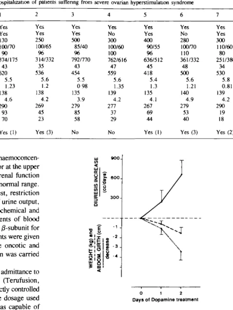
Figure

Documents relatifs