Drug Deposition and Distribution in Healthy and Atherosclerotic
Arteries and in Models of Atherosclerosis Following Bulk or
Stent-Based Drug Delivery
byNeda Vulakmirovic
M.S., Chemical Engineering, MIT, 2000
B.S., Chemical Engineering, Oregon State University, 1997
SUBMITTED TO THE HARVARD-MIT DIVISION OF HEALTH SCIENCES AND TECHNOLOGY IN PARTIAL FULFILLMENT OF THE REQUIREMENTS FOR THE
DEGREE OF
DOCTOR OF PHILOSOPHY in the subject of
MEDICAL ENGINEERING AND MEDICAL PHYSICS AT THE
MASSACHUSETTS INSTITUTE OF TECHNOLOGY and
HARVARD UNIVERSITY MAY 2007
C 2007 Neda Vukmirovic. All rights reserved.
The author hereby grants permission to MIT to reproduce and to distribute publicly paper and electronic copies of this thesis document in whole or in part.
Signature of Author: • ..
Division of Health Sciences and Technology May 10, 2007 Certified by:
Elazer R. Edelman, M.D., Ph.D.
Thomas D. and Virginia W. Cabot Professor of Health Sciences and Technology Thesis Supervisor
Accepted by: . ,
I
Martha L. Gray, Ph.D.Edward Hood Taplin Professdr of Medical and Electrical Engineering MASSACHUSETTS INSTITUTE Director, Harvard-M.I.T. Division of Health Sciences and Technology
OF TECHNOLOGY
JUL 1 3 2007
al,,•
Drug Deposition and Distribution in Healthy and Atherosclerotic
Arteries and in Models of Atherosclerosis Following Bulk or
Stent-Based Drug Delivery
by Neda Vukmirovic
Submitted to the Harvard-MIT Division of Health Sciences and Technology on May 10, 2007, in partial fulfillment of the requirements for the degree of Doctor of Philosophy in the subject of Medical
Engineering and Medical Physics.
Abstract
Drug eluting stents have revolutionized the practice of medicine and the landscape of medical devices. Yet, more than four years after introduction clinical trial data and clinical use have still not fully clarified what drives the safety and efficacy of these devices. The goal of this thesis was to help fill this void by describing the mechanisms by which stent-eluted drugs are distributed within healthy and atherosclerotic vascular models.
In the first part of the thesis we investigated the effect of drug physicochemical properties on drug deposition, retention, and distribution in a healthy vascular model. We found that hydrophobic drugs are deposited to a far greater degree than hydrophilic drugs, with longer retention times, and distribution patterns that likely track with specific and general binding sites. The second part of the thesis investigated how arterial ultrastructure in health and disease modulates the arterial deposition and distribution of hydrophobic antiproliferative drugs used with drug-eluting stents. We tracked the distribution of radiolabeled and FITC-labeled
compounds and demonstrated that macrostructural changes in arterial architecture led to profound changes in drug deposition. Paclitaxel in particular was sensitive to tissue state. This drug binds specifically to tubulin and to lesser extent in a general manner to elastic. Drug levels
fell as paclitaxel, tubulin and elastin were displaced by lipid and collagen. These observations might well explain how drugs may partition within different arterial lesions as determined by lesion composition.
Finally, we demonstrated that association with these binding sites was governed by association kinetics that reflects the different components of the arterial wall compartments. Slower release kinetics yielded up to 64% higher deposition of a drug from stents implanted in rabbit iliac arteries over a 28-day period. Mathematical modeling illustrates that the dependence of drug deposition on stent release kinetics is contingent on drug retention. Further model development is implicated for predicting drug deposition profiles for different types of drugs, arterial states, and stent release kinetics.
Thesis Supervisor:
Elazer R. Edelman
Thomas D. and Virginia W. Cabot Professor of Health Sciences and Technology Thesis Committee: Alan T. Hatton, Robert S. Langer, Frederick Welt
Table of Contents
Abstract ... ... 2
Table of Contents ... 3
List of Figures ... 5
Acknowledgements ... 7
CHAPTER 1: Background and Research Significance ... 9
1.1 Cardiovascular Disease and Atherosclerosis ... 9
1.2 History of treatments... 11
1.3 The Challenge of Restenosis met by Drug Eluting Stents ... 12
1.4 Motivation, Research Significance, and Hypothesis ... ... 12
1.5 References ... ... 19
CHAPTER 2: Pharmoacokinetics in the Healthy Arterial Wall - In Vitro Study... 21
2.1 Background, Hypothesis, and Specific Aims ... ... ... 21
2.2 Materials & Methods ... 22
2.3 Results... ... 23
2.3.1 Equilibrium Drug Deposition as a Function of Bulk Concentration. ... 23
2.3.2 Tissue-U ptake K inetics ... 23
2.3.3 T issue-E lution K inetics... ... 23
2.3.4 Total U ptake Capacity. ... ... 23
2.3.5 Transmural Drug Distribution... ... 24
2 .4 D iscu ssion ... ... ... ... 24
2.5 R eferences ... ... ... ... ... ... ... 32
CHAPTER 3: Pharmacokinetics in the Atherosclerotic Arterial Wall - In Vitro Study ... 33
3.1 Background, Hypothesis, and Specific Aims ... ... 33
3.2 M aterials & M ethods ... 33
3.2 .1 M odel D rugs ... 33
3.2.2 Arterial Models of Atheorsclerosis ... 34
3.2.3 Drug Detection Tecniques ... ... 34
3.2.4 Ultrastructure Determination Techniques... ... ... 35
3.3 R esults ... ... ... . .. 37
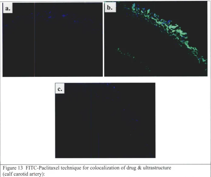
3.3.1 FITC-Paclitaxel Tracking Tecniques ... ... ... 37
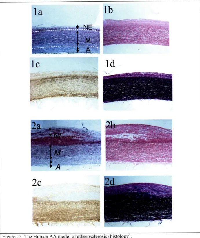
3.3.2 H um an tissue... ... 37
3.3.3 Rabbit Abdominal Aorta Tissue: 1 month of high fat diet ... 38
3.3.4 The Rabbit Iliac Artery: 1 month of high fat diet ... ... 38
3.3.5 Rabbit Abdominal Aorta: 5-month high fat diet... ... 39
3.4 Discussion ... ... ... 41
3.4.2 Changes in Drug Deposition and Distribution with Disease ... 3.4.3 Experimental Limitations and Suggestions ... ... 3.4.4 C onclusions... ... 3.5 R eferences... ... ... CHAPTER 4: Drug Release Kinetics from Stents in Vivo... ...
4.1 Background, Hypothesis, and Specific Aims ... ... 4.2 M aterials & M ethods ... ...
4.2.1 D evice D escription... ... 4.2.2 A nim al M odel ... ... 4.2.3 Tissue Analysis for Drug Content... 4.2.4 Statistical A nalysis... ... 4.2.5 Radioactive Material Disposal... 4.2.6 Mathematical Description of Drug Deposition Profiles ... 4.3 R esults... ...
4.3.1 Observations at Different Time Points ... 4.3.2 Drug Release from Stents ... 4.3.3 Arterial deposition. ... 4.3.4 O ther deposition... ... 4.3.5 Mathematical Description of Drug Deposition... ... 4.4 D iscussion ... ... 4.5 R eferences... ...
CHAPTER 5: Summary and Future Work ... 88
5.1 Thesis Sum m ary ... 88
5.2 Future W ork ... ... 89
5.2.1 Drug Transport in Healthy Arteries ... ... 89
5.2.2 Drug Transport in Diseased Arteries ... ... 90
5.2.3 Three-compartment modeling of in vivo data ... ... 92
5.3 R eferences ... ... 98
APPENDICES ... 99
Appendix 1: Quantifying a Radiolabeled Compound in the Bulk Arterial Tissue ... . 99
Appendix 2: Cryosectioning and Quantifying a Radiolabeled Compound ... 100
Appendix 3: Preparation of FITC-labeled PXL Solution for Tissue Incubation ... 102
Appendix 4: Tissue Incubation if FITC-labeled PXL, Freezing, and Cutting... 104
Appendix 5: Sample preparation for Histology ... 106
Appendix 6: Oil-Red-O staining... 107
Appendix 7: IHC (Immunohistochemistry) Staining Preparation ... 108
Appendix 8: IHC (Immunohistochemistry) Staining Protocol ... 109
Appendix 9: IHC Staining of Frozen Sections by CRP... 114
Appendix 11: Data Tables for Chapter 4 ... 118
Appendix 12: Radiolabeled drug quatification in Rabbit tissues... 123
List of Figures
Figure 1 A diagram of the wall of human aorta... ... ... 15
Figure 2 Pathways in evolution and progression of human atherosclerotic lesions ... 16
Figure 3 Proposed role of LDL oxidation in the initiation of fatty streak lesions... 17
Figure 4 Rationale for stent implantation and management of in-stent restenosis ... 18
Figure 5 Equilibrium tissue uptake capacity of paclitaxel as a function of bulk concentration.. 27
Figure 6 Tissue uptake profiles ofradiolabeled dextran (*), paclitaxel (o), and rapamycin (0) 28 Figure 7 Tissue elution profiles of radiolabeled dextran (*), paclitaxel (o), and rapamycin (0) 28 Figure 8 Tissue uptake capacity of radiolabeled dextran, rapamycin, and paclitaxel ... 29
Figure 9 Fractional tissue uptake capacity of labeled paclitaxel (m) and rapamycin () ... 29
Figure 10 Transmural equilibrium distribution... ... 30
Figure 11 Summary of drug-tissue interactions ... ... 31
Figure 12 Chemical structure of FITC-paclitaxel conjugate ... 36
Figure 13 FITC-PXL technique for colocalization of drug & ultrastructure... ... 46
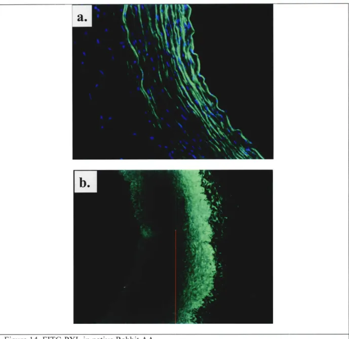
Figure 14 FITC-PXL in native rabbit AA... 47
Figure 15 The human AA model of atherosclerosis (histology)... ... 48
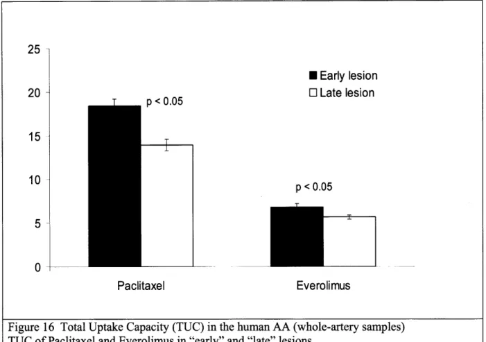
Figure 16 Total uptake capacity (TUC) in the human AA (whole-artery samples) ... 49
Figure 17 The human abdominal aorta model of atherosclerosis (I) ... 50
Figure 18 The human abdominal aorta model of atherosclerosis (II)... . 51
Figure 19 Total uptake capacity in the human AA - compartmentalized. ... 52
Figure 20 The Rabbit 1-month Abdominal Aorta model of atherosclerosis ... 52
Figure 21 Total uptake capacity (TUC) in the rabbit abdominal aorta ... 53
Figure 22 Transmural distribution of paclitaxel in the rabbit abdominal aorta ... 54
Figure 23 Transmural distribution of everolimus in the rabbit abdominal aorta... 55
Figure 24 The Rabbit 1-month Iliac Artery model of atherosclerosis: Tubulin & FKBP-12... 56
Figure 25 The Rabbit 1-month iliac artery model of atherosclerosis: Lipid... 57
Figure 26 Variability in lipid concentration around the perimeter of diseased arteries... 57
Figure 27 Variability in Tubulin and FKBP around the perimeter in diseased arteries ... 58
Figure 28 Variability in ultrastructure around the perimeter in diseased arteries... 59
Figure 29 The Rabbit 5-month abdominal aorta model of atherosclerosis - injured artery... 60
Figure 30 The Rabbit 5-month abdominal aorta model of atherosclerosis - diseased artery... 61
Figure 31 FITC-PXL (green) and lipid (inset, red)... 62
Figure 32 FITC-PXL deposition relative to lipid content... 63
Figure 33 Loss of paclitaxel content with disease relative to injured arteries ... 63
Figure 34 FITC-PXL deposition relative to collagen content ... 64
Figure 35 FITC-PXL deposition relative to "elastin content" ... 65
Figure 36 FITC-PXL deposition relative to b-tubulin content ... 66
Figure 37 Tw o stent platform s... 83
Figure 38 Surgical procedure of stent implantation into a rabbit iliac artery ... 83
Figure 39 The analysis segment of the iliac artery ... ... 84
Figure 40 Two-compartmental model for drug release from stents and its deposition into the surrounding arterial tissue... ... 84
Figure 41 Drug release from stents... ... 85
Figure 43 Model solution for C (i.e., tissue drug concentration)... ... 86
Figure 44 Two-compartment model of drug delivery from stents and uptake by the tissue ... 86
Figure 45 Model of drug distribution through tissues at equilibrium (from Lovich et al.) ... 94
Figure 46 Adobe Photoshop histogram of a histology image ... 94
Figure 47 Luminosity calculated from MetaMorph RGB values ... .95
Figure 48 Rabbit AA containing Tubulin Tracker and immunostained for b-tubulin... 95
Figure 49 A 3-compartmental model proposed to describe deposition of a drug released from a stent and into a neighboring arterial tissue. ... 96
Figure 50 A b-tubulin image of a diseased artery, and two linescanes of pixel intensities along the line across the artery. ... 97
Acknowledgements
During my doctoral thesis, I have been challenged in many ways and to the point where I questioned whether I had chosen well the place or the subject matter. Yet all decisions I had made would make sense if done again. The experience of having tested my limits and a fundamentally different way of thinking have been the gifts.
This work would not have been completed without a generous support of a number of people. Elazer, I thank you for the opportunity to work in your laboratory, where I've benefited from interacting with you and with outstanding clinicians, researchers, and colleagues, and where I've been able to work in a uniquely multifaceted area of a deep interest to me. Thank you for supporting me through the long graduate experience, for not doubting my at times ambitious goals, and for your kindness.
I am thankful to my thesis committee for their input and guidance. Professor Alan Hatton, you have contributed valuable suggestions on modeling drug transport, and I've greatly appreciated your humor and encouragement since my first year in Chemical Engineering. Professor Robert Langer, I thank you for introducing me to Elazer, and for reminding me along the way that the best use of science is in its applications. Dr. Frederick Welt, thank you for your invaluable guidance into animal models and your support with the early in vivo experiments.
Dr. Rami Tzafriri, I owe you a great deal of gratitude for many hours of discussions on modeling and transport, and for focusing my thinking on important questions. Your excitement for
research has made many long days hopeful, and I am grateful I've had a chance to work with and learn from you..
Professor Collin Stultz, I am deeply thankful for your support in clarifying my scientific and academic questions and for helping me put my graduate experience in perspective. Your guidance during this last year has been invaluable and has helped turn confusion and concern I felt at times into motivation and resolve.
Dr. David Ettenson, I am indebted to you for many hours of teaching and explanations of vascular and molecular biology. Being able to speak with you many times has been a great source of support. Adam Groothuis and Anna Spognardi, without your help with animal
surgeries this thesis would not have been possible. I thank you and also Katie Madden, Dr. Bob Marini, and Chris Autieri for help with the animal work. Bill McCarthy and the Radiation Protection Program staff have been instrumental in my learning about radiation, and Michele Miele in numerous lab matters.
Philip Seifert and Gee Wong, your assistance with histology has been invaluable. Thank you for your patience and for letting me learn from you. Discussions with Dr. David Wu, Dr. Mark Lovich, Dr. Michael Jonas, and Kha Lee have been helpful in defining the models of disease and transport. Dr. David Wu and Dr. Kumaran Kolandaivelu, I fondly remember and appreciate your guidance and support at the early times of my work in the laboratory. I am thankful for technical expertise and kindness of Dr. Vijaya Kolachalama towards the end of this work. The UROP
students, Pamela Lee and Jacqueline Brazin, have contributed in early and late stages, respectively.
Dr. Campbell Rogers and Dr. Sahil Parikh, I cannot forget how encouraged I was receiving your emails related to my clinical questions during late nights before my committee meetings.
Professor Roger Mark, thank you for many discussions during this past year and for a warm welcome into your and Dottie's home.
Dr. Aaron Baker, I've admired your ability to effortlessly blend humor with work, and have greatly appreciated your support with data analysis and instrumentation on a number of occasions.
Peter Wu, I am thankful for your friendship and confidence, and many late night conversations. At times when my mind was tired or confused and heart indecisive, hearing your insight brought relief and joy.
Dr. Roy Beigel and Brinda Balakrishnan, thank you for your kindness and cheer, and for giving me a perspective on life over work during many days, when that perspective was hardly
appreciated on its own.
Everyone else in the Edelman Laboratory, including the Spanish armada, I thank each one of you for contributing to the rich community that I've been a part of and that I've learned from. Finally, this work would not have been possible without the support of family and friends. I am indebted to Dilan Seneviratne, Waleed Farahat, Mayssam Ali, Erich Caulfield, Victoria Tai, Uttam Kumbhat, Rob Jagnow, Ron Dror, and Xiaomin Mou, among others, and to the
Sidney&Pacific and Ashdown graduate communities.
To my sister Rada for continuous challenge to discover who I am and who I could be, and for providing a turning point in my graduate work that made a goal become a reality.
To Vuk and Mila, my parents, for letting me go far into unknown at age 17, and for supporting me in my endeavors.
Neda Vukmirovic May 10, 2007
CHAPTER 1: Background and Research Significance
1.1
Cardiovascular Disease and Atherosclerosis
Statistics
In 2004, about 79 million Americans had one or more forms of cardiovascular disease (CVD), which included high blood pressure, coronary heart disease (CHD), and stroke [1] [24]. Cardiovascular disease claims more lives each year than the next 4 leading causes of death combined, which are cancer, chronic lower respiratory diseases, accidents, and diabetes mellitus [25]. In 2004, CVD claimed 871,500 lives. CHD claimed 452,300 lives and is the single leading cause of death in America today [24]. The estimated direct and indirect cost of CVD for 2006 was $403.1 billion, and for CHD $142.5 billion [25].
Biological Basis
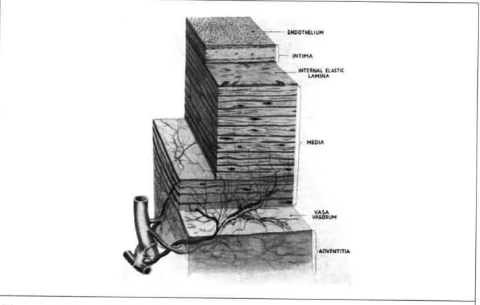
To understand the basis of CVD it is necessary to consider the structure of the blood vessel wall, as shown in Figure 1 [7].
The blood vessel wall consists of three layers: the intima, media, and adventitia. The intima is the innermost layer and contains the endothelial cells. Its dividing line with the media is a layer of elastin (internal elastic lamina). The media is made up of smooth muscle cells, a varied number of elastic sheets, bundles of collagenous fibrils, and a network of elastic fibrils. Its dividing line with the adventitial is a layer of elastin (external elastic lamina). The adventitial layer contains collagen fibers, ground substances, and some fibroblasts, macrophages, blood vessels (vasa vasorum), myelinated and nonmyelinated nerves.
Pathology
Well-recognized cardiovascular risk factors (hypertension, dyslipidemia , diabetes, smoking, prior or family history of CVD, age, sex, obesity and physical inactivity) are associated with endothelial injury and changes in the vessel wall that lead to cardiovascular disease and clinical events. One of the earliest changes associated with these risk factors is an increase in oxidative stress in the vessel wall. This causes endothelial cells to decrease production of some compounds and increase production of others [8]. For example, production of nitric oxide (NO), a powerful vasodilator, is decreased. Production of vasoconstrictors, notably endothelin and angiotensin II, is increased. Greater production of local mediators (e.g., vascular cell adhesion molecule, VCAM, and plasminogen activator inhibitor 1, PAI-1) promotes inflammation, disrupts fibrinolysis, and at later stages of disease increases vascular remodeling and plaque rupture. The vascular endothelium secretes numerous other substances that modulate blood vessel tone and participate in the development and progression of atherosclerosis.
Atherosclerosis
Coronary heart disease (CHD) is caused by atherosclerosis, which is a multifactorial disease that affects the intima of elastic arteries and manifests itself in progressive narrowing and hardening of the arteries over time. The disease is characterized by intramural deposits of lipids, proliferation of vascular SMCs (smooth muscle cells) and fibroblasts, and accumulation of macrophages. These changes are accompanied by a calcification of arterial wall, loss of elasticity of vessel wall, and narrowing or dilatation of the vascular lumen (Figure 2, [11]).
Atherosclerosis is responsible for more disability and death than any other disease (95% of deaths from coronary artery disease, about 50% of deaths from diabetes and about 50% of those from cerebrovascular disease [9]). The disease is commonly seen in the aorta, but also frequently found in the smaller arteries such as the coronaries and the cerebrals and to a lesser extent in the medium-sized vessels.
Coronary atherosclerosis is clinically the most important aspect of atherosclerosis. Coronary arteries are relatively narrow, and atherosclerosis could seriously reduce the blood flow through them. Slow, progressive narrowing of the arterial lumen that occurs over an extended period causes myocardial hypoxia and presents clinically either as angina pectoris (prolonged substernal, thoracic pain) or congestive heart failure (a condition where the heart is failing to pump efficiently, which often results in congestion in the lungs). Sudden occlusion of a coronary artery by a thrombus formed over an ulcerated atheromas causes myocardial infarct. Pathogenesis of Atherosclerosis
The pathological effects of atherosclerosis occur over decades, through several stages of disease progression, from early fatty streaks to advanced lesions (found beginning in the fourth decade of life) and clinical events. According to the response to injury hypothesis [12], Figure 3, the pathogenesis of atherosclerosis begins with an infiltration of lipid into the vessel wall. A subtle injury to the endothelium initiates the process [10] by injuring the impermeable endothelial layer. This allows small pools of extracellular lipid to form within smooth muscle cell layers, disrupting the intimal lining of the vessel. This is accompanied by attachment of monocytes and platelets to the internal surface of the artery and local thrombus formation. These changes alter the permeability of the arterial wall, facilitating even more influx of lipids.
The principal lipids, which accumulate in the intima are cholesterol and its esters. In the free state these substances are insoluble in the body fluids, and when introduced into connective tissue excite a "foreign-body" type of tissue reaction, or an inflammatory reaction, where scavenger monocytes infiltrate the intima, transforming into macrophages that phagocytose lipid-laden fragments of damaged cells and stroma. Macrophages that have accumulated lipid in their cytoplasm appear histologically as foam cells, or microscopically as a fatty streak.
Macrophages secrete growth factors and cytokines, which recruit additional monocytes, macrophages, and other cells. Cytokines and growth factors also stimulate the proliferation of smooth muscle cells and their ingrowth into the intima from the tunica media. Lipid accumulates not only in macrophages but also in smooth muscle cells. From dead and dying cells, cholesterol is released into interstitial spaces. These events lead to the formation of fibro-fatty plaques and atheromas. Atheromas consist of amorphous, lipid-rich material (lipid, cell debris, smooth muscle cells collagen, and calcium in older persons) and are soft. On the luminal side, atheromas are typically covered with an intimal fibrous cap, consisting of fibroblasts surrounded by collagen, which replaces the normal intimal cells. Many atheromas undergo fibrosis
(overgrowth of' surrounding fibrous tissue/fibroblasts) and become hard plaques.
Once the plaque becomes fibrous, the danger of the plaque rupture increases. A rupture of the protective fibrous cap permits contact between the blood and the thrombogenic material in the lipid core of the lesion, and leads to a clinical complication that involves thrombosis. Sizable atheromas may remain silent for decades or produce stable angina. The time of rupture will be determined by the balance between collagen formation and its degradation by matrix metalloproteinases (MMPs) as well as the presence of the SMCs in the fibrous cap.
Besides fibrous cap rupture, atheromas are complicated by other secondary changes, which include calcification and ulceration, which overlie endothelium with secondary thrombosis.
1.2
History of treatments
CABG (coronary artery bypass graft) is a clinical procedure, which is done to reroute or "bypass" the blood around a clogged coronary artery and thereby improve the supply of blood to the heart.
PTCA (percutaneous transluminal coronary angioplasty) has been used as a less invasive and a less expensive alternative to CABG. It is a procedure used to dilate the constricted coronary arteries. A catheter with a deflated balloon at its tip is inserted into the narrowed part of the artery (it is introduced into the body through an incision in the femoral artery and guided radiographically to the coronary artery stenosis). The balloon is inflated, compressing the plaque and enlarging the inner diameter of the blood vessel so that the blood can flow more easily. Then the balloon is deflated and the catheter withdrawn.
Immediately after the intervention, however, there is pathobiologic responses to intervention, which includes deposition of proteins and platelets. This is a natural response and intended to "passivate" (i.e., render inert) the injured surface. Platelet aggregation and release of platelet-derived growth factor (PDGF) lead to formation of a white, platelet-rich thrombus [16]. If passivation is successful, the extent of thrombus formation is limited, leaving an adequate lumen. However, while thrombus formation may not be significant in terms of lumen encroachment, it serves as a locus for the second phase of repair.
The second phase is an inflammatory reaction involving mononuclear and polymorphonuclear leukocytes, which adhere to the injury site and secrete many mediators including interleukin-1 (IL-1), interleukin-6 (IL-6), tumor necrosis factor alpha (TNF ), and basic fibroblast growth factor (basic FGF, [17]).
The inflammatory response stimulates smooth muscle cell migration from the media to the intima. These cells proliferate, secrete matrix material, and become intermixed with the monocytes. This third phase peaks approximately one week after angiography and continues for some weeks after [17].
The final phase involves collagen deposition and remodeling of the arterial wall. Expansion of the matrix causes progressive narrowing and (within 6 to 12 months) restenosis. The arterial wall may expand and grow (remodel), thus facilitating maintenance of the lumen, or it may shrink and contribute to restenosis.
Prognosis in some patients is suboptimal, as the dilated segment of the artery renarrows within six months after the procedure, and they may require another PTCA, a stent placement, or a coronary artery bypass surgery.
Stenting provides scaffolding for the vessel wall and was originally developed to prevent or treat two major complications of PTCA (abrupt vessel closure and restenosis), but soon it came to be
employed in over 80% of PTCA procedures. Stents are either balloon-delivered or self-expanding and are commonly composed of stainless steel, tantalum, or nitinol (a shape-memory alloy of nickel and titanium). These metals are not ferromagnetic and thus do not interfere with MRI (magnetic resonance imaging). Common stent types include coil, slotted tube, mesh and ring, and are either made from a single strand of wire formed into a repeating pattern (coil), cut from a single piece of metal (tube), or from overlapping wire (mesh). Each type has its own advantages in terms of flexibility, uniform expansion, possibility of recoil, etc.
Development of new stents (Palmaz-Schatz, Crown, Multi-link, Duett, Wiktor, etc.) is evolving rapidly. Custom-designed stents allow access to lesions that are difficult to treat. Researchers are currently exploring the use of stents as a method of local and sustained drug delivery via stent coatings employing biodegradable polymers, which appears to be a promising technique to prevent restenosis (explained in the next section).
Figure 4 summarizes the rationale for stent implantation and the management of its complications [15]. Immediately after balloon angioplasty (Figure 4a), the plaque is cracked and irregular, and the intima is damaged. Within hours or days, the artery wall recoils (Figure 4b), significantly narrowing the lumen. SMC (smooth muscle cells) proliferate. Later (Figure 4c), blood cells and platelets collect at the site of intimal damage, and the SMCs migrate in, proliferate, and form the neointima (Figure 4d). Stenting (Figure 4e) may result in a larger vessel diameter compared to balloon angioplasty alone and may prevent early elastic recoil and late remodeling. But it also causes vessel wall injury, which in turn leads to a proliferation of the neointima at the stenting site. An improved revascularization strategy (Figure 4f) using a drug coated stent reduces in-stent restenosis.
1.3 The Challenge of Restenosis met by Drug
Eluting
Stents
During PTCA, intraluminal balloon inflation causes dissection of the vessel wall. This leads to restenosis in 30% to 50% of patients, likely as a result of elastic recoil of the vessel wall, platelet-mediated thrombus formation, SMC (smooth muscle cell) proliferation, and late remodeling. Abrupt vessel closure occurs in 4% to 10% of patients [15].
Implantation of stents (or bare metal stents) reduces abrupt closure and restenosis, leading to improved short-term and long-term vessel patency. However, it does not limit and likely stimulates SMC proliferation, leading to restenosis within the stent and vessel renarrowing with the risk in the range of 15% to 20%. Subsequently, drug-eluting stents (DES) were developed. They were designed to release pharmacological agents after deployment to inhibit the response to injury that is mainly responsible for restenosis after BMS implantation. As a result, restenosis and target-vessel revascularization have been reduced to rates below 10% after DES implantation.
1.4
Motivation, Research Significance, and Hypothesis
MotivationMany pharmacological agents considered for DES have been tested in animal models and have been effective. For example, animal studies have shown that drugs like paclitaxel inhibit cell proliferation and intimal hyperplasia following balloon angioplasty and stent implantation [19,
20]. However, the mechanism by which drugs distribute within atherosclerotic lesions and act to inhibit restenosis are not precisely known. Moreover, many of the agents, which have been successful in animal trials have been ineffective in human clinical trials [21]. This disparity in results between animal-model research and clinical trials has led to skepticism about the validity of animal models in restenosis research.
However, whether this skepticism is founded is uncertain as virtually all studies on drug distribution and transport kinetics have been done on previously intact blood vessels in healthy animals [22, 23]. Since drug administration builds its efficacy on the response of the disease state the potential inferences from examination of drug transport in healthy and not diseased tissues might be limited. For example, the variations in the lipid content of atherosclerotic plaque may alter the drug distribution pattern after directed vascular delivery and change the efficacy [20]. At the very least, the increased tissue beneath endovascular stents placed in anatomically large or hyperplastic arteries increases the time and mass through which drugs must diffuse, potentially leading to delayed and diluted pharmacological response [23]. The mechanism of drug transport, the relative importance between the diffusive and convective forces, may also change as the diffusive resistance increases with vessel thickness (convection may come to play a more significant role). Therefore, studies of drug interaction with healthy tissues can only be useful to a certain extent, and need to be followed by studies in diseased tissues.
This view is further supported by disparity between early and late results of DES that could not have been anticipated from animal models. The early Sirius[26] clinical trial showed that 93.4% of patients were free from target lesion revascularization (TLR) 2yrs following the initial procedure. This was contrasted to 80.4% of patients who had received bare metal stents. However, the Basket Late Trial (presented at the ACC 2006, [27]) investigated the incidence of late clinical events with respect to restenosis. The results showed a non-statistically significant increase of thrombotic events in DES when compared to BMS (2.6 vs. 1.3% respectively), and a statistically significant increase in combined fatal and non-fatal MI in DES when compared to BMS (4.9 vs. 1.3%, respectively). Further, Aoki et al. [28] showed in 2005 that plaque volume and hypoechogenicity decrease (indicating normal tissue formation) only 2-4yrs following the DES placement. This implies that DES are potent modulators of vascular repair, and delay vessel healing beyond the time line previously anticipated. This delayed healing have not been observed in preclinical studies, where healthy models are used, emphasizing the impact of atherosclerosis on the healing process and the importance of using diseased models for device design and evaluation.
This body of work was designed to investigate mechanisms of drug deposition, distribution and transport kinetics across healthy and atherosclerotic arteries. The results will enable us to understand more fully the potential of local cardiovascular drug delivery within the system in which it is to act. In addition, they will further guide establishing valuable and meaningful disease models for design and evaluation of endovascular devices.
Research Significance
This work examines the transport of several compounds (dextran, paclitaxel, rapamycin, everolimus) with different physicochemical properties (hydrophobicity, binding properties)
across atherosclerotic-like arteries (hyperplastic, inflammatory, and lipid enriched), where the disease state was defined in terms of arterial ultrastructure (lipid, binding protein, collagen, and elastin content). I showed that:
1. uptake and elution kinetics, as well as equilibrium deposition and distribution, are guided by drug physicochemical and binding properties,
2. altered stent release kinetics affected drug deposition,
3. disease changes the arterial ultrastructure, which in turn affects drug deposition and distribution across the arterial wall.
Hypothesis
The underlying hypothesis that drove my work was that pathology changes the state of an artery. That change alters tissue properties, which in turn affect drug transport (deposition, distribution, binding, and kinetics) across an arterial wall and atherosclerotic lesions. The resulting drug deposition and distribution within the target arterial wall will determine the efficacy and safety of drugs eluted from endovascular implants.
ELASIC A
A
RUM
WIMMU
istated maeIsphs ge bern caes
ype I fa1ly atrseak) lskion
ma'tly litice lpar
pe
ll.
(nem
la)
sion
Type It changes & sxelieotrac r Ik id pdools
Type I[ clanges & core oi
Typ V (IAbrutetrona) lesion
TIid core Ibrtic & 0ty,.
Sormupl tpd cores & ftbrotk
l•ers~ or mainly clclc.i
Iromm 'ttst accetieratotalsn e a •scle lnically and s !ent riybuon 'or
NOmOncatue and
MaEn
Clinicl
Sequences in P~prression a
M ; Pmecanim , ,la ...
or ma nly fjotiC r --- - .u ov ,
decade
Type VI (cwlplIcaeid) limalus ulac
d1crt,
tr/f
wmbomis,he: ram-emoehrage.
VIernajma
Wironka~s
Figure 2 Pathways in Evolution and Progression of Human Atherosclerotic Lesions
From type I to type IV, changes in lesion morphology occur primarily because of increasing accumulation of lipid. The loop between types V and VI illustrates how lesions increase in thickness when thrombotic deposits form on the surfaces.
Figure 3 Proposed Role of LDL Oxidation in the Initiation of Fatty Streak Lesions
LDL crosses the endothelium in a concentration-dependent manner and can become trapped in the extracellular matrix (1). The subendothelium is an oxidizing environment, and if the LDL remains trapped for a sufficiently long period of time, it undergoes oxidative changes (2). Mildly oxidized forms of LDL contain biologically active phospholipid oxidation products that affect the pattern of gene expression in endothelial cells (ECs), leading to, among other things, changes in the expression of monocyte binding molecules (designated X-CAM), monocyte
chemoattractant protein (MCP-1), and macrophage colony stimulating factors (CSFs) (3). These factors in turn promote the recruitment of monocytes (4) and drive their phenotypic
differentiation to macrophages (5). Further oxidation leads to alterations in apolipoprotein B such that LDL particles are recognized and internalized by macrophages (6), progenitors of the lipid-laden foamn cells. Marked increases in lipid and cholesterol oxidation products render the LDL particles cytotoxic, leading to further endothelial injury (7) and favoring further entry of LDL and circulating monocytes and thus a continuation of the disease process [60].
drwCng monoWoto
G
XAM>(I
blood
Or
1
::1
1.5 References
1. 2001 heart and stroke statistical update. Dallas: American Heart Association, 2000.
2. Morbidity and mortality 1998 chartbook on cardiovascular, lung, and blood diseases. National Institute of Health: National Heart, Lung, and Blood Institute, 1998.
3. Mokdad, A.H., et al. Diabetes trends in the U.S.: 1990-1998. Diabetes Care, 2000. 23(9): p. 1278-83.
4. The hidden epidemic of cardiovascular disease. Lancet, 1998. 352(9143): p. 1795.
5. Murray, C.J. and A.D. Lopez. Alternative projections of mortality and disability by cause 1990-2020: Global Burden of Disease Study. Lancet, 1997. 349(9064): p. 1498-504.
6. Fung, Y.C. Biomechanics : mechanical properties of living tissues. New York: Springer-Verlag, 1993.
7. Florey, H. General pathology. Philadelphia: W. B. Saunders Co., 1970.
8. Gibbons, G.H. and V.J. Dzau. The emerging concept of vascular remodeling. N Engl J Med, 1994.
330(20): p. 1431-8.
9. Boyd, W. Pathology for the Physician. Philadelphia: Lea & Febiger, 1958.
10. Pepine, C.J. The effects of angiotensin-converting enzyme inhibition on endothelial dysfunction: potential role in myocardial ischemia. Am J Cardiol, 1998. 82(10A): p. 23S-27S.
11. Stary, H.C., et al. A definition of advanced types of atherosclerotic lesions and a histological classification of atherosclerosis. A report from the Committee on Vascular Lesions of the Council on Arteriosclerosis, American Heart Association. Circulation, 1995. 92(5): p. 1355-74.
12. Damjanov, I. Hitopathology: a color atlas & textbook. Baltimore: Williams & Wilkins, 1996. 13. Eagle, K.A., et al. ACC/AHA guidelines for coronary artery bypass graft surgery: executive
summary and recommendations : A report of the American College of Cardiology/American Heart Association Task Force on Practice Guidelines (Committee to revise the 1991 guidelines for coronary artery bypass graft surgery). Circulation, 1999. 100(13): p. 1464-80.
14. Pentecost, M.J., et al. Guidelines for peripheral percutaneous transluminal angioplasty of the abdominal aorta and lower extremity vessels. A statement for health professionals from a special writing group of the Councils on Cardiovascular Radiology, Arteriosclerosis, Cardio-Thoracic and
Vascular Surgery, Clinical Cardiology, and Epidemiology and Prevention, the American Heart Association. Circulation, 1994. 89(1): p. 511-31.
15. Marso, S.P., S.G. Ellis, and R. Raymond. Intracoronary stenting: an overview for the clinician. Cleve Clin JMed, 1999. 66(7): p. 434-42.
16. Topol, E.J. and P.W. Serruys. Frontiers in interventional cardiology. Circulation, 1998. 98(17): p. 1802-20.
17. Edelman, E.R. and C. Rogers. Pathobiologic responses to stenting. Am J Cardiol, 1998. 81(7A): p. 4E-6E.
18. Rogers, C., et al. Endogenous cell seeding. Circulation, 1996. 94(11): p. 2909-14. 19. Drachman, D.E., et al. Neointimal thickening after stent delivery of paclitaxel: change in
composition and arrest of growth over six months. JAm Coll Cardiol, 2000. 36(7): p. 2325-32. 20. Heldman, A.W., et al. Paclitaxel stent coating inhibits neointimal hyperplasia at 4 weeks in a
porcine model of coronary restenosis. Circulation, 2001. 103(18): p. 2289-95.
21. Simon, D.I. and C. Rogers. Vascular Disease and Injury. Totowa: Humana Press, 2001.
22. Creel, C.J., M.A. Lovich, and E.R. Edelman. Arterial paclitaxel distribution and deposition. Circ Res, 2000. 86(8): p. 879-84.
23. Lovich, M.A. and E.R. Edelman. Mechanisms of transmural heparin transport in the rat abdominal aorta after local vascular delivery. Circ Res, 1995. 77(6): p. 1143-50.
24. http://www.americanheart.org Cardiovascular Disease Statistics, April 23, 2007
http://circ.ahajoumals.org/cgi/reprint/ 113/6/e85
26. Weisz G, Leon MB, Holmes DR Jr, Kereiakes DJ, Clark MR, Cohen BM, Ellis SG, Coleman P, Hill C, Shi C, Cutlip DE, Kuntz RE, Moses JW. Two-year outcomes after sirolimus-eluting stent implantation: results from the Sirolimus-Eluting Stent in de Novo Native Coronary Lesions (SIRIUS) trial. J Am Coll Cardiol. 2006 Apr 4;47(7):1350-5. Epub 2006 Mar 20.
27. http://www.medscape.com/viewarticle/529648
28. Aoki et al. Evaluation of Four-Year Coronary Artery Response After Sirolimus-Eluting Stent Implantation Using Serial Quantitative Intravascular Ultrasound and Computer-Assisted Grayscale Value Analysis for Plaque Composition in Event-Free Patients. JACC 2005; 46:1670
CHAPTER 2: Pharmacokinetics in the Healthy Arterial Wall
-
In
Vitro Study
2.1
Background, Hypothesis, and Specific Aims
Background
The success of drug-eluting stents is predicated to as great a degree upon the extent of drug deposition and distribution through the arterial wall as virtually any other factor (1-5). The biological effects of a candidate drug are essential, but, ultimately, tissue residence time will determine effect and toxicity (6, 7). Fueled by clinical relevance (8-11), a number of studies have been carried out to detect, model, and predict the distribution of drugs within arterial segments beneath, adjacent to, and distant from stents (12-15). Drugs that are retained within the blood vessel are far more effective than those that are not. Heparin is a perfect example of a compound whose ubiquitous biological potential is lost by virtue of its physicochemical properties. Heparin regulates virtually every aspect of the vascular response to injury (16). Yet heparin is so soluble and diffusible that it simply cannot stay in the artery for more than a few minutes after release and has no effect on intimal hyperplasia when eluted from a stent (4, 17,
18).
Paclitaxel, in contradistinction, is a far smaller compound, with fewer effects specific to vascular biology, but paclitaxel is so hydrophobic and insoluble that it binds tenaciously to tissue protein elements and remains beneath stent struts long after release (13). The clinical efficacy of paclitaxel at reducing restenosis rates following elution from stents appears incontrovertible (8, 11). In addition to its hydrophobic, nonspecific binding behavior, paclitaxel also binds to its protein target, polymerized microtubules (19), with nanomolar specificity. Analogously, rapamycin, also successful after local vascular delivery (10), acts on a specific target through a series of events that requires binding to the specific binding proteins FKBP12 (FK506 binding protein 12) and FRAP (FKBP12-rapamycin-associated protein) (20, 21). The distribution of tissue-binding proteins is not uniform in space or time. For example, FKBP12 is found most abundantly in vascular smooth muscle cells at a concentration of -10-5 M (22). FKBP12 is up-regulated in neuronal systems with injury (23) and likely after arterial injury as well. Microtubules have a similar cellular concentration of =10-5 M (24). The nonuniform distribution of paclitaxel previously observed in the arterial wall may reflect an inhomogeneous distribution (13) of polymerized microtubules or carrier proteins (25).
Although regulated at a coarse level by transport forces and lipid avidity, the distribution of paclitaxel and rapamycin is also regulated at a fine level by the distribution and availability of their protein targets, a level of control not present for drugs such as heparin that are rapidly cleared from arterial tissue. With a more complete understanding of the role of specific binding in arterial drug distribution, stent design, drug composition, and release formulation can be better optimized.
Hypothesis
We hypothesized that drug physicochemical properties will affect their tissue pharmacokinetics. More specifically, hydrophobic drugs, as opposed to hydrophilic drugs, will have increased deposition and retention levels, and longer equilibration times. Further, the arterial wall distribution and local pharmacological effects of compounds such as paclitaxel and rapamycin
will be influenced by tissue-specific binding proteins, above and beyond the influence of general hydrophobic interactions alone.
Specific Aims
To test the above hypothesis we examined tissue uptake and elution kinetics, equilibrium deposition levels, and arterial wall distribution profiles for a model hydrophilic drug, dextran, and two model hydrophobic drugs, paclitaxel and rapamycin.
2.2 Materials & Methods
Equilibrium Drug Deposition as a Function of Bulk Concentration.
Samples of calf carotid artery were placed in drug solution (radiolabeled paclitaxel in PBS, or a mixture of radiolabeled and unlabeled paclitaxel) at room temperature for 48hrs. Following incubation, tissue samples were digested, liquid scintillation cocktail was added, and drug quantity determined by LSC.
Arterial Loading and Elution. We defined the tissue pharmacokinetic profiles of dextran (10,000 Da), paclitaxel (853.9 Da), and rapamycin (914.2 Da) in calf carotid arterial wall segments. Calf internal carotid arteries were harvested and transported in PBS with physiological calcium and magnesium [PBS++/0.01 mol/liter CaC12/0.01 mol/liter MgC12 (Sigma)] at 40C. Arteries were cleaned of excess fascia, opened longitudinally, cut into segments (400-600 mg), and placed in centrifuge tubes with 1.0 ml of [3H]dextran (10-6 M), [3H]paclitaxel (10-6 M), or [14C]rapamycin (10-6 M). All bulk solutions were made fresh immediately before experimentation, and the same initial bulk concentrations were used for both loading and elution experiments.
For loading experiments, segments were removed in triplicate at indicated time points, briefly washed in PBS++, and blot-dried before being dissolved in Solvable (Packard-Canberra). Liquid scintillation mixture (6 ml) was added to dissolved samples before counting with liquid scintillation spectroscopy (2500 TR Liquid Scintillation Analyzer, Packard-Canberra). For elution experiments, segments were allowed to equilibrate for 60 h and were then placed in 50 ml of PBS++ for the indicated time periods before being processed in triplicate as indicated with the loading experiments.
Drug Tissue Uptake Capacity (TUC) and Distribution.
We defined the bulk differential capacities of the arterial wall for dextran, paclitaxel, and rapamycin at equilibrium. Arterial segments were weighed before being placed in drug bath solutions. Segments were allowed to equilibrate for 60 h and were then processed for liquid scintillation counting. The drug concentration of each tissue sample was normalized by tissue mass and then by drug concentration in the bulk fluid during equilibrium incubation to determine the uptake capacity. As indicated, tissue was incubated with (i) labeled drug alone to determine TBC; (ii)drug and supersaturated levels of the same unlabeled drug to demonstrate that specific and nonspecific tissue-binding sites can be displaced; and (iii) supersaturated levels of the other nonlabeled hydrophobic drug (i.e., labeled rapamycin was mixed with cold paclitaxel, and vice versa) to displace only nonspecific binding.
Equilibrated transmural drug distribution in the artery was measured through en face cryosectioning. Arterial segments were incubated in the drug bath for 60 h, and then laid flat and snap-frozen in a plastic encasement with Tissue-Tek OCT compound (Sakura Finetechnical, Tokyo). Samples were stored in a -800C freezer until they were sectioned parallel to the intima
with a refrigerated microtome, the Cryotome SME (Shandon, Pittsburgh) (12, 26, 27). Upon sectioning, the segment length and width were measured with a caliper. Sections 0.040 mm thick were cut parallel to the intima, and the drug content of each sample was determined by liquid scintillation spectroscopy. Tissue drug concentration (cT) at each transmural location was calculated as the mass of drug normalized by the measured tissue area and slice thickness. Tissue drug concentration was then normalized by the bulk fluid drug concentration during equilibrium incubation (cbulk) to determine the uptake capacity (k) at each transmural location (x).
2.3 Results
2.3.1 Equilibrium Drug Deposition as a Function of Bulk Concentration.
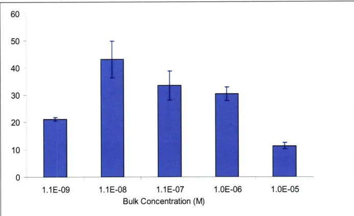
In preliminary experiments, it was determined that equilibrium drug deposition in calf carotid or rabbit abdominal aorta samples is reached in 24-36 hours. Next, we were interested to know if this deposition is dependant upon the drug concentration in the bulk or surrounding solution. The total tissue uptake capacity (TUC) of paclitaxel increased twofold as the bulk concentration increased from 10-9M to 10-8M. As the bulk concentration was increased further to 10-5M paclitaxel uptake capacity continually decreased (Figure 5).
2.3.2 Tissue-Uptake Kinetics.
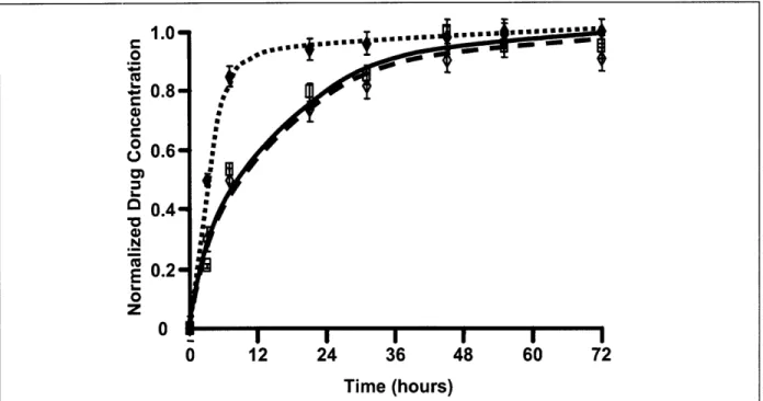
Arterial samples were incubated in [3H]dextran (10-6 M), [3H]paclitaxel (10-6 M), or [14C]rapamycin (10-6 M) and harvested in triplicate over a period of 72 h. Loading data were normalized to an average peak value at 72 h (Figure 6). Whereas the hydrophilic dextran reached 80% of equilibrium value within several hours, paclitaxel and rapamycin took nearly 1 day to achieve the same level. The loading profiles for these two hydrophobic compounds are indistinguishable and approach steady state only after 60 h.
2.3.3 Tissue-Elution Kinetics.
Arterial samples were pre-equilibrated in [3H]dextran (10-6 M), [3H]paclitaxel (10-6 M), or [14C]rapamycin (10-6 M) for 60 h and then placed in an elution sink of 50 ml of PBS++. Samples were processed in triplicate over the following 60 h, and data were plotted as percentages of the pre-elution load value (Figure 7). Dextran elutes most rapidly, losing 90% of its equilibrated load within 2 h and reaching a steady state of =10% of original material in <5 h. The hydrophobic drugs take =1 day to reach an elution steady-state value. At 48 h, arterial segments loaded with rapamycin retain =60% of their initial load, and those loaded with paclitaxel, =35%.
2.3.4 Total Uptake Capacity.
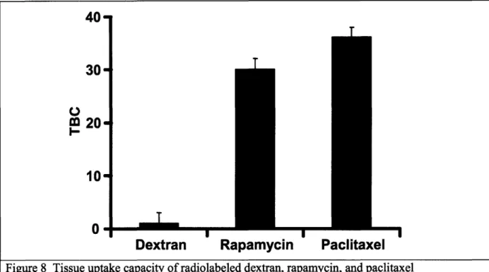
TUC was defined as the tissue concentration (cT) at equilibrium normalized by the bulk concentration at equilibrium (cbulk). Arterial segments were incubated in the drug for 60 h to allow for equilibration. Upon equilibration, bulk solutions were sampled, and tissue samples were processed for liquid scintillation counting. Variation in initial bulk concentrations over an order of magnitude did not affect TBC. Dextran has little uptake capacity in arterial tissue with a
coefficient of =0.60. Because this value is similar to its physically accessible volume fraction in arterial tissue (17), dextran may not leave the extracellular space. The paclitaxel and rapamycin binding coefficients are not statistically different (P > 0.05) by the two-tailed Student t test and are significantly greater than 1, indicating that nonspecific and/or specific binding interactions are sequestering these drugs in the tissue (Figure 8).
To assay for the binding specificity, experiments were conducted with mixtures of labeled and nonlabeled drug (Figure 9). Molar excess of unlabeled paclitaxel or rapamycin displaced the binding of the corresponding labeled drug to <5% of control value. At this level the TBC value fell to nearly 1, indicating displacement of specific and nonspecific binding. When the cold drug was switched, the TBC decreased to =35% of control for paclitaxel and =50% for rapamycin, suggesting that these compounds were now displacing the unrelated labeled drug, although displacement occurred only from nonspecific sites, leaving specific binding intact. By the two-tailed Student t test, the paclitaxel and rapamycin specific retention fractions are statistically different (P < 0.05).
2.3.5 Transmural Drug Distribution.
Arterial samples were incubated in [3H]dextran, [3H]paclitaxel, or [14C]rapamycin for 60 h and then snap-frozen for en face sectioning (Figure 10). Previous work with paclitaxel in BSA showed that TBC was maximal in the intima and declined precipitously within the most intimal regions of the arterial media, to less than half the intimal level. At the outer edge of the media, the paclitaxel uptake capacity increased gradually and peaked within the adventitia before falling off to unity (13). These data have been recapitulated, but without BSA, for direct comparison with dextran and rapamycin. In this case, paclitaxel shows a very similar medial profile to the BSA case, but rises sharply in the adventitia, suggesting that BSA provides a nonspecific binding sink for adventitial binding. Rapamycin shows a uniform transmural distribution, in stark contrast to the nonuniform distribution of paclitaxel. Dextran again shows little uptake capacity throughout the artery.
2.4 Discussion
Local drug delivery has great theoretical and practical appeal for vascular diseases (28, 29). One important surprise is that biological potency is not the sole determinant of biological effect. Drug-specific physicochemical properties determine, to a great degree, whether concentrations sufficient for therapeutic activity can be sustained. Drugs that bind to tissue elements, for example, are retained within tissue and have dramatic clinical effects; nonbinding hydrophilic drugs are rapidly cleared and ineffective against restenosis. Our data suggest that specific binding also plays a critical role in determining drug distribution. Whereas paclitaxel distributes heterogeneously through arterial tissue, rapamycin distributes more uniformly through the media and adventitia. In addition, the tissue binding and diffusion results suggest that binding site availability and distribution regulate the fine structure of drug deposition beyond the coarse structure imposed by transport forces and lipid avidity. Ultimately, local tissue ultrastructure and the concentrations they enforce on the artery at a microscopic scale, together with specific and nonspecific binding site distribution, become critical considerations in the optimization of vascular drug delivery.
Specific and Nonspecific Binding Determine TBC. Compounds must bind to proteins, intracellular or extracellular, to have a biological effect. This binding can take two forms: nonspecific interactions, such as those influenced by charge or water affinity, and specific binding idiosyncratic to the individual drug. Paclitaxel and rapamycin both can bind nonspecifically to serum proteins and hydrophobic tissue microenvironments. Paclitaxel demonstrates nanomolar specificity to polymerized microtubules, whereas rapamycin shows similar specific binding properties to FKBP12, a ryanodine-receptor-associated protein. Dextran, by virtue of its extreme hydrophilicity, exhibited neither type of binding and, accordingly, its TBC was <1; its potential tissue level can never exceed the concentration in the bathing solution. In contrast, paclitaxel and rapamycin were deposited in the blood vessel at concentrations 30- to 40-fold higher than that in surrounding bulk solutions. Thus, tissue concentrations of paclitaxel and rapamycin can exceed the applied concentration several fold, establishing an effective volume of distribution within arteries larger than anticipated from surrounding solution concentration.
Because both microtubules and FKBP, which specifically bind paclitaxel and rapamycin, respectively, reside in calf internal carotid segments at -10-5 M (24), offering a large specific drug sink, we investigated the specificity and potential of tissue binding by using a competitive binding tissue assay. Both specific and nonspecific binding contributions were observed. When cold rapamycin was substituted for a high concentration molar excess of paclitaxel in the labeled paclitaxel system, the fractional TBC was nearly 35%, indicating that approximately one-third of the TBC was from specific binding. Comparably, when the same procedures were carried out with labeled rapamycin and excess cold paclitaxel, the fractional TBC was 50%, with near equal contributions from specific and nonspecific binding events. Taking into account specific, nonspecific, and general binding a summary diagram can be made to demonstrate types of
interactions that hydrophobic drugs can be involved in (Figure 11).
Tissue Pharmacokinetics of Hydrophilic and Hydrophobic Compounds. It is expected that paclitaxel and rapamycin will have similar transport properties, given that both compounds have solubilities of =6 gLg/ml, molecular masses of <1 kDa, and nanomolar binding constants to their specific protein targets. Indeed, the compounds act quite similar when compared with dextran. Whereas only several hours are required for dextran to reach a steady-state tissue concentration that never exceeds the surrounding media levels, paclitaxel and rapamycin take four to six times longer to reach a steady state. Kinetics, not concentration, accounts for this effect, as the time to reach steady state was independent of the concentration of the same drug applied over a broad range. Although the net binding of paclitaxel and rapamycin were nearly identical, their distribution and tissue elution after binding and uptake were not. At steady state the artery retained only 10% of the applied hydrophilic dextran. Paclitaxel retention was 3.5-fold that of dextran, and rapamycin levels were almost twice those of paclitaxel. These differences in tissue retention closely parallel the percentages attributed to specific binding (Figure 9), and it is clear that both hydrophobic compounds are retained to a much greater extent than dextran. Moreover, at steady-state loading, whereas paclitaxel remained in the subintimal space and partitioned significantly in the adventitia, rapamycin was evenly distributed throughout the arterial wall (Figure 10).
Movement of a molecule through a composite structure, such as a blood vessel wall, is driven by a range of forces and phenomena. The influence of effective molecular radius can dominate when all other :factors are equal but may recede in importance when other factors are present. For instance, despite being nearly 20 times smaller than dextran, paclitaxel and rapamycin diffuse more slowly in both the transmural and planar directions. This difference may be attributed to the hydrophobicity of the compounds or possibly to the role of binding. Whereas dextran has few binding sites, paclitaxel and rapamycin will repeatedly bind to and dissociate from their respective specific and nonspecific targets as they diffuse through tissue, in effect slowing the leading edge of the diffusion front. We have previously shown that albumin and dextrans diffuse at least an order of magnitude faster in the planar direction than they do in the transmural direction (3). For paclitaxel and rapamycin, the planar diffusivity exceeded transmural diffusivity by at least two orders of magnitude, although both drugs' diffusivities were two orders of magnitude smaller than those of dextran. These phenomena are likely governed by similar forces for all three drugs, despite vastly different lipid avidities. The transport of hydrophilic compounds is enhanced in aqueous regimes of the vessel wall but retarded by hydrophobic elastin layers. For hydrophobic compounds, these layers act in a reverse fashion; the movement of paclitaxel and rapamycin is likely impeded by the more water-rich regions of the blood vessel wall and aided by lipid pools or even the protein-studded elastin lamina. In both cases, however, whereas individual layers might be isotropic, the greater composite of alternating layers of the arterial wall provide for planar diffusion that far exceeds transmural flux (3).
As the number of available drug-eluting devices increases, distinction and choice may reside not only in ease of use but also in the physicochemical functionality of the drug-stent unit. Local drug distribution is modulated by transport and lipid avidity at a coarse level, but for clinically relevant compounds such as rapamycin and paclitaxel, it is also modulated by the distribution of the compounds' specific binding sites. Arterial ultrastructure also influences transport, because alternating tissue layers of varying permeability result in anisotropic transport for both hydrophobic and hydrophilic drugs. Design and evaluation of a drug-eluting device thus requires a unified understanding of the drug, its physicochemical characteristics, and its specific and nonspecific interactions with arterial structures.
60 50 -40 30
-T
T 20-10 01.1E-09 1.1E-08 1.1E-07 1.OE-06 1.OE-05
Bulk Concentration (M)
Figure 5 Equilibrium tissue uptake capacity of Paclitaxel as a function of bulk concentration (in PBS and room temperature, calf internal carotid artery samples; mixtures of radiolabeled and non-labeled drugs were used). Contrary to expectations, the profile is not linear nor does it reach saturation.
C O C O rU N E L0
O
ZTime (hours)
Figure 6 Tissue uptake profiles ofradiolabeled dextran (*), paclitaxel (0), and rapamycin (0)
in calf internal carotid artery samples. Hydrophobic paclitaxel and rapamycin reach steady state later than the hydrophilic dextran.
1
0.8
o 0.6
'E
0.4
0.2
0
B
Time (hours)
Figure 7 Tissue elution profiles of radiolabeled dextran (*), paclitaxel (o), and rapamycin (0)
from calf internal carotid artery samples. Hydrophobic paclitaxel and rapamycin reach an elution steady state later than the hydrophilic dextran, and also demonstrate significant residual concentrations.
40-r T
30-
20-
10-o-
U . U.Dextran
Rapamycin
Paclitaxel
Figure 8 Tissue uptake capacity of radiolabeled dextran, rapamycin, and paclitaxel in calf internal carotid samples at equilibrium (after 60 hours of incubation).
DU"
S50-O
40-3 40-
30-Lno020-0
0*
.0a
Rx
Rx'
Figure 9 Fractional tissue uptake capacity of labeled paclitaxel (m) and rapamycin (0) when in the presence of excess unlabeled drug, relative to the capacity of labeled drug alone, after 60 hours of incubation. (1) Rx: 10-6M labeled drug with 10-3M of the same unlabeled drug. (2) Rx': 10-6M labeled paclitaxel with 10-3M unlabeled rapamycin, and 10-6M labeled rapamycin
with 103M unlabeled paclitaxel (courtesy of Andrew Levin).
I M
Media
50-
40-
30-
20-
1
0-I
I I
IIIII1
I0
0.1
0.2
I0.3
IAdventitia
i
Ii I I I0.4
0.5
0.6
0.7
Depth from Lumen (mm)
Figure 10 Transmural equilibrium distribution
of labeled dextran (,), paclitaxel (o), and rapamycin (0) in 0.040 mm thick calf internal carotid samples. Compared to hydrophilic dextran, the profiles ofhydrophobic drugs demonstrate nonuniform distribution across the arterial wall as well as higher deposition levels.
ii ii I"~
Rx
Hydrophilic
Rx Hydrophobic
Extracellular
I
Extracellular
compartment
Non-Specific
Specific
Other Rx can displace - ns only Rx can displace - sp
competition competition
knp Iksp Cellular
NI
\
compartment
General
Specific
binding site
binding site
(protein,
lipid)
or
Receptor
/7
4
/
no effect
Effect
-
Cell
activation
Figure 11 Summary of drug-tissue interactions:
Hydrophilic drugs deposit into and clear from the tissue too fast to exert a pharmacologic effect. Hydrophobic drugs bind long enough for an effect to take place. Within the extracellular
compartment, a given hydrophobic drug (ex, paclitaxel) will bind to non-specific sites that are also accessible to other drugs. It will also bind to specific sites that are accessible to that drug alone (ex, a specific site on elastin). Intracellularly, a drug can bind to a general site from which it can be displaced by other drugs (ex, a lipid droplet) or to a specific site from which it can't be displaced by other drugs (ex, microtubules).