HAL Id: inserm-01142383
https://www.hal.inserm.fr/inserm-01142383
Submitted on 15 Apr 2015
HAL is a multi-disciplinary open access
archive for the deposit and dissemination of sci-entific research documents, whether they are pub-lished or not. The documents may come from teaching and research institutions in France or abroad, or from public or private research centers.
L’archive ouverte pluridisciplinaire HAL, est destinée au dépôt et à la diffusion de documents scientifiques de niveau recherche, publiés ou non, émanant des établissements d’enseignement et de recherche français ou étrangers, des laboratoires publics ou privés.
Protein S inherited qualitative deficiency: novel
mutations and phenotypic influence.
Martine Alhenc-Gelas, Marianne Canonico, Pierre-Emmanuel Morange,
Joseph Emmerich
To cite this version:
Martine Alhenc-Gelas, Marianne Canonico, Pierre-Emmanuel Morange, Joseph Emmerich. Protein S inherited qualitative deficiency: novel mutations and phenotypic influence.. Journal of Thrombosis and Haemostasis, Wiley, 2010, 8 (12), pp.2718-26. �10.1111/j.1538-7836.2010.04073.x�. �inserm-01142383�
Accepted for publication in the Journal of Thrombosis and Haemostasis doi: 10.1111/j.1538-7836.2010.04073.x
Received 5 May 2010, accepted 31 August 2010 Original Article
PROTEIN S INHERITED QUALITATIVE DEFICIENCY: NOVEL MUTATIONS AND PHENOTYPIC INFLUENCE
M Alhenc Gelas*, $ M Canonico** PE Morange*** , $$ J Emmerich****, $
for the GEHT genetic thrombophilia group
* Hématologie biologique, AP-HP, Hôpital Européen Georges Pompidou, Paris, France
** INSERM Unit 780, Cardiovascular Epidemiology section, Villejuif, France *** INSERM, Unit 626, Marseille, France
$$ Laboratoire d’Hématologie, CHU Timone, Marseille, France
**** Médecine Vasculaire-HTA, AP-HP, Hôpital Européen Georges Pompidou, Paris, France
$ INSERM Unit 765, Paris, France
Correspondence to :
Dr M Alhenc-Gelas, Service d’hématologie biologique, Hôpital Européen Georges Pompidou, 20-40 rue Leblanc, 75908 Paris cedex 15, France, phone: 33(1)56093938, fax : 33(1)56093393, martine.alhenc-gelas@egp.aphp.fr
Short title: PROS1 type II novel mutations
Keywords: endogenous thrombin potential, PROS1, protein S deficiency,
ABSTRACT
Background: Only few mutations associated with qualitative protein S deficiency
have been already described. Sensitivity and specificity for type II PROS1 mutations of commercially available reagents for measuring PS activity are not well established . Whether these mutations are significant risk factors for thrombosis remains an unresolved question. Methods: In order to address the first point, we present and discuss the results of PROS1 analysis performed in the 30 probands with type II PS inherited deficiency suspicion and 35 relatives, studied in our laboratory between 2000 and 2008. In order to investigate the influence of type II mutations on the coagulability level, thrombin generation tests were performed on plasma from 102 PROS1 type II, type I/III, or PS Herleen mutation heterozygous carriers and controls.
Results: Mutations (12 novel, 6 already described) which likely explain the qualitative
phenotype, were found in 27 (90%) among the 30 probands studied. In relatives, 78% of heterozygotes presented with a type II phenotype. An APC resistance phenotype was documented in type II and type I/III defects heterozygous carriers; however, the effect of type II was milder than the effect of type I/III PS mutations.
Conclusions: PS functional assay (Staclot PS, Stago) was efficient in screening for
PROS1 type II defects, particularly in probands. A significant positive influence of type II mutations on ex vivo thrombin generation was demonstrated. However, whether these mutations increase the risk of venous thromboembolism requires further investigation.
INTRODUCTION
Protein S (PS) is a cofactor of activated protein C (APC) in the proteolytic inactivation of blood coagulation factors Va (FVa) and VIIIa (FVIIIa), thus providing a negative feedback on thrombin generation and making the APC/PS pathway essential for normal haemostasis. The anticoagulant activity of PS might also be direct, i.e independent of APC, as PS is also a nonenzymatic cofactor for tissue factor pathway inhibitor (TFPI) in the inhibition of factor Xa (FXa), and in addition, directly limits thrombin generation by binding to and inhibiting FXa and FVa in the prothrombinase complex [1].
PS deficiency (MIM:#176880) is a recognized genetic risk factor for venous thromboembolism (VTE) [2]. It is an autosomal dominant trait with incomplete penetrance and variable clinical and biochemical expression, which affects 2% to 10% of patients with thrombosis in Caucasian populations [3]. It could be more frequent in Asian populations [4]. According to plasma levels of total PS (TPS), free PS (FPS) antigen and PS activity (PSA), three types of PS deficiency are defined: type I and III are quantitative deficiencies; type II or qualitative PS deficiency is characterized by normal FPS level and reduced PSA, and therefore a decreased PSA/FPS ratio due to the presence of a dysfunctional variant in plasma. Type II deficiency seems rather uncommon ; to the best of our knowledge only sixteen mutations able to explain impaired PS APC cofactor activity have been found in patients with a type II phenotype, and no case of PS-deficient subject with impaired direct anticoagulant activity has been reported so far; most type II mutations are unsurprisingly located at positions known to be important for the protein function, i.e:
in the propeptide cleavage site (p.Arg40Leu, p.Arg41His) [5] [amino acid numbering
p.Thr78Met) [5, 6], in the thrombin sensitive loop (TSR) (p.Arg101Cys) [7] and in the
epidermal-growth factor (EGF) like domains, [p.Thr144Asn in EGF1, in EGF2
p.Asp157Gly, p.Cys175Phe, and p.Lys196Glu in EGF2, p.Cys217Tyr, p.Tyr225Cys in EGF3, p.
Ile244-Asp245 del in EGF4] [5, 8, 9, 10]. A substitution in the donor splice site of intron 5
(c.469+5G>A) leading to exon 5 skipping induces full loss of EGF1 [5]. In addition, two
natural missense mutations at position 95, p.Gly95Glu and p.Gly95Arg were reported in
patients with mild decrease in the PSA/FPS ratio [5]. Type II PS inherited deficiency could be in fact more frequent than generally considered since PS functional assay is performed by a minority of laboratories in the screening for PS deficiency; the reason for measuring FPS level rather than PSA is “historical” due to insufficient specificity of the first generation of commercial PS activity assays. Moreover, no commercial reagent allowing direct PS activity measurement are available. However, it would be important to include searching for type II variants in thrombophilia screening if these variants do increase risk of thrombosis.
The two objectives of the present study were: 1) to assess the usefulness of Staclot PS (Stago, Asnières, France) for screening for type II inherited deficiency; this assay is based on modified activated partial thromboplastin time (APTT). Diluted patient plasma is added to PS-depleted plasma in the presence of purified APC and factor Va, therefore the assay measures the APC dependent activity of PS; and 2) to gain insight on phenotypical consequences of PROS1 type II mutations. We report the results of PROS1 analyses performed in the whole group of probands with a suspicion of inherited qualitative PS abnormality studied in our laboratory between 2000 and 2008 and in relatives. We also report the results of thrombin generation tests (TGTs) performed by calibrated automated thrombinography (CAT) [11] on plasma from 76 carriers of distinct PROS1 mutations, including 26 with type II
defects, and we compare thrombosis history in relatives with type II or type I/III defects.
SUBJECTS AND METHODS Subjects
Subjects studied were initially referred to specialized French clinical haematology laboratories for thrombophilia screening in the context of unexplained personal or family history of thrombosis. Recording of clinical characteristics of the subjects including objectively confirmed episodes of venous thrombosis (VT) or pulmonary embolism (PE), and screening for biological risk factors for VT (antiphospholipid antibodies, antithrombin, protein C, protein S deficiency, the FV Leiden and the F2 G20210A mutations) were performed in each centre. Family history of VTE was defined by the occurrence of VT in at least one first degree relative. When an inherited PS deficiency was suspected, DNA and plasma samples, when available, were sent to our laboratory for PROS1 genotyping. An informed consent authorizing genetic analyses was obtained from every patient, according to the Helsinki declaration.
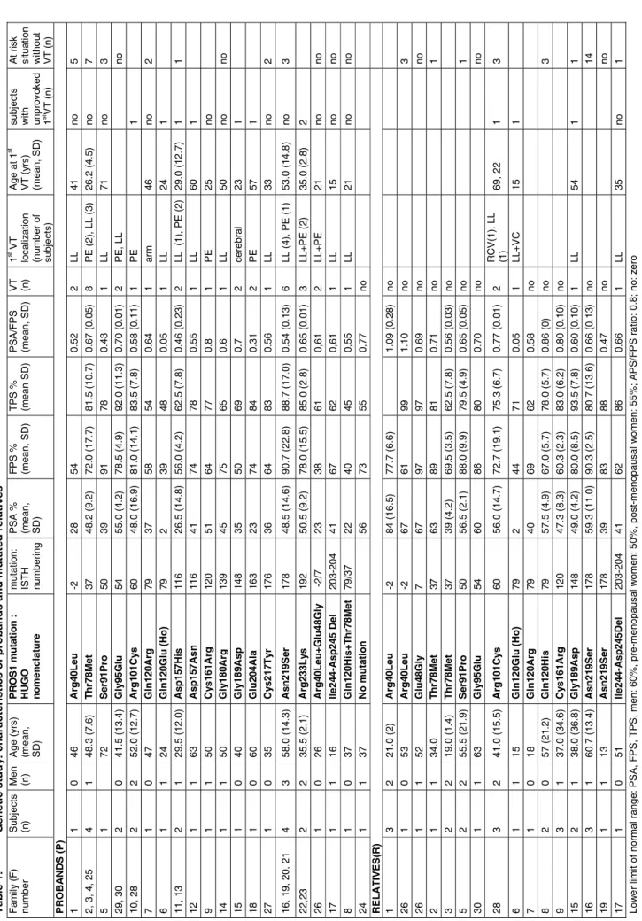
Genetic study: In the period 2000-2008, we analyzed PROS1 in 30 probands
(14 men, 16 women), mean age 44.7 (extreme values 16-73), in whom a PS inherited qualitative deficiency was suspected and 35 relatives from 15 of these kindreds. Plasma PS activity (PSA) and TPS levels were measured using Staclot PS and Asserachrom TPS reagents (Stago). FPS level was measured using the FPS reagent from Instrumentation Laboratory (Milano, Italy) in two patients and using the Asserachrom FPS kit (Stago) in the others. PS type II inherited deficiency was suspected in probands based on the observation of a persistent decreased or
borderline PSA and abnormal PSA/FPS ratio (< or =0.80) in the absence of factors susceptible to induce falsely low Staclot PS level (FVIII>250% [12], FV Leiden [13], antiphosholipid antibodies..).
TGTs: TGTs were performed on plasma from: 26 subjects heterozygous for
type II PROS1 mutations and 34 subjects with type I/III mutations previously identified by sequencing, one individual with a Multiplex Ligation-dependent Probe Amplification (MLPA) profile suggesting heterozygosity for a complete deletion of the gene, 15 PS Herleen (Ser501Pro) heterozygous carriers, 7 relatives from these families who did not carry the mutation present in their pedigree. They did not receive hormonal treatment and had no other biological risk for thrombosis. The subjects were selected on their genotype and on availability of plasma samples for TGTs. A last group consisted of 19 “healthy subjects” with no personal history of thrombosis and no biological risk factor for VT, studied in our laboratory during the same period, Symptomatic subjects were studied at distance from thrombosis events, at least 1 month after stopping oral anticoagulant treatment.
All samples for PS assessment and thrombin generation work were taken in the absence of anticoagulant treatment.
PROS1 defects carrying and thrombosis history: We compared thrombosis
history in adult (> 15years old) relatives from all families with PROS1 mutations associated with type II (36 relatives from 9 families) or quantitative PS deficiency (47 relatives from 11 families) and no other risk factor for thrombosis studied in our laboratory between 2000 and 2010, for whom full clinical information was available. Mutations responsible for quantitative deficiency were premature stop-codons or missense detrimental mutations already described.
PROS1 analysis
In the probands, the exons, the intron-exon boundaries and 700bp of the proximal promoter of the functional gene for PS, PROS1 (GeneID: 5627, MIM#176880) were studied by direct sequencing as previously described [14, 15]. In family members, gene analysis was restricted to the region found abnormal in the proband. Mutations are numbered according to the HUGO recommendations for mutation nomenclature (http://www.hgvs.org) based on the start codon (ATG) of the first translated methionine. Amino-acid numbering according to the ISTH database [5], based on the mature protein, is obtained by substracting 41 for residues of the mature protein or 42 for residues that belong to signal peptide and propeptide.
Thrombin generation tests
Venous blood was collected on 0.109M sodium citrate in the recruiting centres. Platelet poor plasma was prepared by double-centrifugation for 15mn at 2000-2500g, at temperature between 15 and 22°C, within 2hours from blood sampling, then stored frozen in small aliquots at temperature <-30°C. For every patient, all clotting tests were performed on a unique plasma sample, within 2 hours after thawing. Thrombin generation in the presence or absence of activated protein C (APC) was measured according to the method described by Hemker et al. [11] using the reagents PPP (-a mixture of phospholipids (PL) and tissue factor (TF)-), thrombin calibrator and the fluorogenic substrate FluCa from Thrombinoscope (Maastricht, Netherlands), following the manufacturer’s instructions except for the following modification: thrombin generation was triggered by the addition of 20ul PPP reagent to 80ul plasma in the presence of 5ul APC (10ug/ml) (Hyphen Biomed,
Neuville s/Oise, France) or 5ul 0.15MNaCl. Fluorescence measurements were performed after addition of 20ul of FluCa. Final concentrations of PL and TF in the mixture were 4.8pM TF and 3.9uM PL. The parameters studied were the area under the curve (endogenous thrombin potential: ETP), the lag time, time to peak (PT), peak height and the start tail (ST), time at which thrombin generation has come to an end. Inter-assay coefficients of variation (CV) calculated on the results obtained for a normal plasma pool in 2 series of respectively 7 and 6 independent runs were <10% for ETP-APC, peak height-APC, ST-APC, PT +/-APC, and 10-20% for the other parameters studied. CV of the ETP -APC/+APC ratio was 15%.
PSA, factor II (FII), FV, FVIII, F-TFPI, TPS, FPS, and fibrinogen level when not previously determined on fresh plasma, were also measured on this plasma sample, using reagents from Stago (Asnières, France). FV level was checked in order to verify that storage did not alter the quality of plasma samples
Statistical analysis of TGT results
Distribution of each parameter was tested for normality and transformed variables were used for non-parametric distributions. Mean levels were given in arithmetic form. Analysis of variance and Fisher test were used to compare the baseline characteristics of subjects. Multiple comparison procedures including the Bonferroni correction were used to assess the differences in thrombin generation parameters between subgroups. Stepwise multiple linear regressions were used to assess the relative contribution of haemostatic variables to the difference in each thrombin generation parameter between subgroups. Statistical analysis used procedures available in the Statistical Analysis System (SAS) software (SAS Institute, Inc., Cary, NC, USA. Annual incidences of VTE were calculated by dividing the
number of symptomatic relatives by the total number of observation years. Observation time was defined as the period from the age of 15 years until the end of the observation period, or until the first episode of VTE. Freedom of VTE was analysed by the Kaplan-Meier method.
RESULTS Genetic study
Information concerning thrombosis history, and plasma PS levels in the probands and mutated family members are shown in Table 1. A personal history of VT was present in 28 probands; P10, asymptomatic, was explored because of a family history of VT; P24 had a history of distal arterial thrombosis in association with a systemic lupus. Other biological risk factors for thrombosis were present in 3 probands (heterozygosity for a detrimental PROC mutation in P22 and for the F2 nt 20210G>A substitution inP24 and 29), but in no relative. Thrombosis antecedents
were present in one 1st degree relative in 9 families, and in 4 relatives in another
kindred.
Nineteen different mutations were found in 29 probands and in 28 relatives. Fifty-three subjects were heterozygotes. Two individuals were homozygotes, and two
others were compound heterozygotes. We found 12 mutations [c.143A>G (p.Glu48Gly),
c.271T>C (p.Ser91Pro), c.359A>G (p.Gln120Arg), c.360G>C (p.Gln120His), c.358C>G (p.Gln120Glu), c.469G>C (p.Asp157His), c.469G>A (p.Asp157Asn), c.481T>C (p.Cys161Arg), c.538G>A (p.Gly180Arg), c.566G>A (p.Gly189Asp), c.611A>C (p.Glu204Ala), c.656A>G
(p.Asn219Ser) ] which, to the best of our knowledge, were not previously reported. We
also found 6 previously described detrimental changes [c.119G>T (p.Arg40Leu), c.233C>T
c.728A>T which induces the loss of Ile244 and Asp245] and a substitution previously reported
as a neutral polymorphism [c.698G>A (p.Arg233Lys)].
No mutation was found in 7 asymptomatic relatives which presented with normal PS levels.
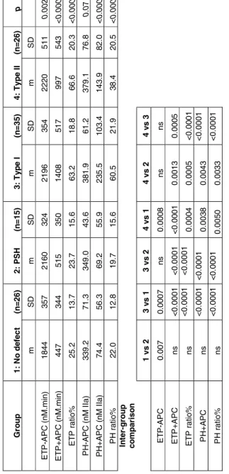
Thrombin generation tests
Relatives non-carrier of PROS1 mutations and healthy subjects were combined in the analyses since no significant differences in descriptive characteristics or TG parameters were found among the subjects.
General characteristics are reported in Table 2. Age, factor II, FV, FVIII, fibrinogen mean levels were similar among the groups. F-TFPI level was higher in type II than in type I/III deficient patients. As expected, PSA and FPS levels were clearly low in PROS1 type I/III mutation carriers and mildly decreased in PS Herleen carriers. In type II mutation carriers, mean PSA was borderline. PSA/FPS ratio was significantly decreased in patients with type II mutations. Sixty-six among these 102 subjects were asymptomatic.
Multiple regression analysis revealed significant positive influence of FII (r=0.44, <0.001, and r=0.29, p=0.003) and fibrinogen (r=0.39, p=0.002 and r=0.25, p=0.01) on ETP and peak height in the absence of APC; PS (PSA r=-0.23, p=0.02, FPS r=-0.18, p=0.06) and FV (r=0.29, p=0.02) had a mild significant influence on ETP only and FVIII on peak height only (r=0.21, p=0.03). In the presence of APC, ETP and peak height were highly dependent on PS (PSSTA: r= -0.69 p<0.0001, FPS: r=-0.64, p<0.0001) and F-TFPI (r=-0.43, p<0.0001), and a mild influence of FVIII on both parameters was observed (r=0.21, p=0.03).
Overall comparison demonstrated significant inter-group differences for ETP and no significant difference in lag time or start tail. Table 3 shows the influence of PROS1 defects on ETP and peak height. Both the statistical significance and the means were adjusted for age, sex, personal history of VTE, and for FII, fibrinogen, FV and FVIII, which had significant influence on these parameters. ETP in the absence of APC was 20% higher in carriers of PROS1 mutations than in non-mutated subjects, whatever the type of defect. Highly significant differences between type I/III or type II mutation carriers and non-mutated subjects for both ETP and peak height in the presence of APC and for ETP and peak ratios (demonstrating an APC resistance) were observed (p<0.0001). Thrombin generation in the presence of APC was indeed 3 fold higher in patients with PS type I/III deficiency than in PS Herleen carriers or subjects without mutations and 2 fold higher in patients with type II mutations. Significance was lost after adjustment on PSA which explained more than 80% of inter-group differences in the presence of APC, demonstrating that in this condition the effects on TGT are mainly due to PS defects, and 30-60% of inter-group differences, showing a lower contribution, in the absence of APC. Further adjustment on F-TFPI did not change the results. No significant differences between PS Herleen carriers and non mutated subjects were observed. There was no significant difference in TGT parameters between individuals who had suffered VT and those who had not.
PROS1 mutations and thromboembolic history
Characteristics of the relatives studied and incidences of VT events (deep or superficial VT or PE) are given in Table 4, and event free survival curves for VT are shown in Figure 1. The 15 non-mutated relatives from the 30 families studied were
asymptomatic. Eight among 37 relatives with type I/III mutation and 2 among 21 relatives with type II mutation displayed a thrombosis history. Annual incidence of VT in relatives with type I/III mutation was 0.56 (0.17-0.95). Heterozygosity for a PROS1 type I/III mutation conferred an odds ratio for VT of at least 4.3 (95% CI 0.3-69.0) as compared with the non-mutated relatives from the type I/III group. However, these estimations are not fully reliable due to the absence of VT event among non-carriers. Incidence was 2 fold lower in relatives with type II mutation [0.24 (0-0.58)]. However, the difference was non significant (p=0.22).
DISCUSSION
The 12 novel mutations, which affect residues 48, 91, 120, 157, 161, 180, 189, 204, or 219 could explain a qualitative deficiency for the following reasons: Firstly, the affected residues, except Ser 91, are highly conserved in PS among mammalian species.
Secondly, most novel mutations (-except those at positions 204 and 219-) are located in regions of PS (GLA, TSR, EGF1 and 2) known to be crucial for APC
cofactor activity [16-19]. Among these mutations, Glu 48, Ser 91 and Gln 120 are
located “at strategic positions”. Glu 48 is located in a crucial ω loop of the GLA region
involved in interaction of PS with anionic PL surfaces [16], and Ser 91 is located at a site where cleavage by thrombin leads to PS inactivation [17]. Gln 120 together with Asp 119 and Asp 136 in EGF1 could be part of the principal functional interaction site for APC [20]. A p. Gln 120 Ala variant expressed in vitro and analysed in a TG assay was recently reported to have severely reduced APC cofactor activity [20]. Residues 204 and 219 are located in EGF3 which has less understood functions. However, these residues belong to typical Ca2+ binding consensus sequences
possibly important for the structural integrity of PS as specific cooperative effect of EGF2 and EGF3 for calcium binding have been demonstrated [21]. Moreover, a p.Glu204Gly PS variant expressed in vitro was previously reported to have decreased anticoagulant function [10]. In addition, functional importance of the EGF domains is supported by previous description of natural mutations associated with type II PS deficiency at positions 144, 157, 175, 196 and 217 [5].
Thirdly, p.Cys161Arg, p.Ser91Pro and p.Gly180Arg may induce significant modifications of the protein structure due to disruption of a disulfide bridge for the first one or differences in physicochemical characteristics for the others.
Lastly, cosegregation observed in families with mutations at position 91, 189 and 219 and observation of type II deficiency in unrelated carriers of distinct mutations at position 120 support their detrimental character.
Interestingly, among carriers of p.Gln120Glu, two consanguineous patients were homozygotes and another subject was a compound heterozygote, carrier of both p.Gln120Glu and p.Thr78Met mutations. Low PS antigen level in these patients suggests a significant effect of p.Gln120Glu on both function and expression or metabolism of PS. The consequences of p.Cys161Arg in the family reported here are not clear, with borderline FPS levels and normal PSA/FPS ratios observed in 3 among 4 heterozygous carriers. However, a patient previously studied in our laboratory heterozygous for another mutation at the same position (p.Cys161Gly) (- not initially reported as type II-) displayed in fact also a qualitative phenotype (PSA/FPS: 0.5-0.75 on 2 different occasions) [5].
Five mutations (p.Arg40Leu, p.Gly95Glu p.Thr78Met, p.Arg101Cys, p.Ile244-Asp245 del) had been previously described, and found associated with type II deficiency [5, 7, 22].
p.Arg233Lys was the unique mutation found in probands 22 and 23. However, as Arg233Lys was previously reported to be a neutral polymorphism [5], its presence probably does not explain plasma levels.
Absence of mutation was observed in only one proband (P24) in whom an acquired PS abnormality cannot be excluded accounting for the clinical context (systemic lupus), even if no antiphospholipid or anti PS IgG (Zymuphen anti PS IgG, Hyphen BioMed, Neuville s/Oise, France) could be detected.
Finally, mutations possibly explaining decreased PSA/FPS ratios were found in 27 among the 30 probands (90%) studied documenting the usefulness of Staclot PS for type II mutation screening, when applied to probands with no cause of ‘false positive” results (i.e FV Leiden, FVIII>250%) [12, 13]. In relatives, the assay was not 100% sensitive as illustrated by the observation of fully normal PSA in 6 among the 27 heterozygotes studied (carriers of Arg40Leu (3), Glu48Gly (1), Arg101Cys (1) or Asn219Ser (1) respectively). However, PSA/FPS ratio was decreased in 3 among them. Possible explanations for “false-negative” results are the following: firstly, the nature of the assay, based on a modified APTT, therefore possibly sensitive to negative as well as positive interferences; secondly, a global increase in PS level due to acquired conditions; thirdly, a genetic origin: phenotypic variability in Thr78Met carriers was previously observed, and explained by the “mild” influence of the mutation on both secretion and function of PS [6]. A mild influence of the mutation could similarly explain phenotypic variability associated with heterozygosity for Arg40Leu or Lys50Glu [5], or other mutations reported here.
We used thrombinography to gain insight into the influence of type II mutations carrying on procoagulant potential. It was previously reported that CAT monitoring performed at low TF concentration (1pM) in the absence of APC and at
high concentration (10-15pM) in the presence of APC are conditions with an optimal sensitivity to the activity of the TFPI/PS system [23, 24]. In such conditions, pooled plasma from heterozygous type I PS deficient individuals generated 3-5 fold more thrombin than normal plasma [25]. In accordance with this finding, in a large group of heterozygous carriers of type I/III mutations, we found a significant APC resistance in TGTs performed at 4.8pM TF, which can be fully explained by PS defects.
Interestingly, in the study by Castoldi et al., full length TFPI and PS low levels were simultaneously observed in PS type I deficient patients, and TFPI was shown to contribute significantly to the increased TG observed in PS deficient plasma. Full normalization of TG was only obtained through simultaneous normalization of both PS and TFPI levels [25]. More recently, Castoldi et al. reported that TFPI levels are reduced in type I but normal in type III PS deficiency [26]. In the present study, mean F-TFPI level was indeed lower in the 12 PS type I deficient subjects [mean (SD): 7.4 (3.5) ng/ml] than in the 23 type III deficient subjects [8.4 (3.8)]; however, the difference was non significant and F-TFPI contribution to inter-group ETP and peak height variations was mild.
At 4.8pM TF in the absence of APC, sensitivity of the assay to the activity of the TFPI/PS system is milder. Seré et al, reported that, at 3.5pM TF, ETP increase was only 20%after full neutralization of PS [23]. This may explain why we observed relatively mild effects at 4.8pM and a weak contribution of PS defects to inter-group ETP differences.
PS Herleen contains a S501P mutation that results in loss of N-linked glycosylation at position 499. PS Herleen has a reduced half-life in vivo [27] which may explain the association of PS Herleen heterozygosity with mildly reduced levels of plasma FPS and the observation of clearly low FPS level in homozygotes [28].
Whether the risk of VT is increased in PS Herleen heterozygous subjects remains doubtful; the prevalence of the S501P mutation is indeed rather high in healthy populations (around 0.5%) and no significant association was found between PS Herleen carrying and VT [29]. PS Herleen allele may be in fact significantly involved in the occurrence of VT only in individuals who carry other genetic traits [28, 30]. We did not observe any influence of PS Herleen on TGT parameters except on ETP-APC, supporting no more than a mild influence on procoagulant potential.
To the best of our knowledge, the influence of type II mutations on CAT parameters has not been studied yet. Our finding of a significant 2-fold increased ETP+APC and ETP ratio in carriers of type II mutations compared to subjects with no defect or PS Herleen carriers is consistent with a procoagulant phenotype and suggests that type II inherited deficiency may be a risk factor for VT.
Recent studies on risk of a first VT event in carriers of PS type I/III inherited deficiency reported annual incidences ranging from 0.27 to 1.5% in line with our own result [31-35]. To the best of our knowledge, the risk of VT associated with type II defects has not yet been studied. In the present study, no significant influence on the risk of VT could be demonstrated. However, the number of relatives investigated was small and should be increased for obtaining more reliable estimate of the risk.
APPENDIX
We thank the following colleagues from the GEHT thrombophilia group for giving us the opportunity to study their patients: Drs N Ajzenberg (AP-HP, Hôpital Bichat), C Biron-Andreani (CHU Montpellier), C Boinot (CHU Poitiers), L Darnige (AP-HP, Hôpital Européen G Pompidou), M Dreyfus (AP-HP, Hôpital Bicêtre), M Hanss (CHU Lyon), MH Horellou (AP-HP, Hôtel-Dieu), B Jude (CHRU Lille), Y Laurian (AP-HP, Hôpital J Verdier), F Lellouche (CH Quimper), C Leroy-Matheron (AP-HP, Hôpital H Mondor), E Mazoyer HP, Hôpital Avicenne), L Rugeri (CHU Lyon), A Robert (AP-HP, Hôpital St Antoine), N Schlegel (AP-(AP-HP, Hôpital R Debré), B Tardy (St Etienne), C Ternisien (CHU Nantes), C Trichet (CH Argenteuil), N Trillot (CHRU Lille), P Sié (CHU Toulouse).
ACKNOWLEDGMENTS
We thank Dr B Villoutreix (INSERM UMRS 973-MTi) for helpful discussion, ML Aubry and N Ochat for excellent technical assistance.
This study was in part supported by the DHOS (Direction de l’Hospitalisation et de l’Organisation des Soins) program "Soutien financier en faveur des laboratoires pratiquant le diagnostic par génétique moléculaire des maladies rares".
DISCLOSURE OF CONFLICTS OF INTEREST
REFERENCES
1. Castoldi E, Hackeng TM Regulation of coagulation by protein S. Curr Opin Hematol ; 2008: 15: 529-36.
2. Garcia de Frutos P, Fuentes-Prior P, Hurtado B, Sala N. Molecular basis of protein S deficiency. Thromb Haemost 2007; 98: 543-45.
3. Seligsohn U, Lubetsky A. Genetic susceptibility to venous thrombosis. New Engl J Med 2001; 344: 1222-31.
4. Sakata T, Okamoto A, Mannami T, Tomoike H, Miyata T. Prevalence of protein S deficiency in the Japanese general population: the Suita Study. J Thromb Haemost 2004 ; 2: 1012-3.
5. Gandrille S, Borgel D, Sala N, Espinosa-Parrilla Y, Simmonds R, Rezende S, Lind B, Mannhalter C, Pabinger I, Reitsma PH, Formstone C, Cooper DN, Saito H, Suzuki K. Scientific and standardization committee communication. Protein S deficiency: a database of mutations. First update. Thromb Haemost 2000; 84: 918-18
6. Rezende SM, Lane DA, Mille-Baker B, Samama MM, Conard J, Simmonds RE. Protein S Gla-domain mutations causing impaired Ca2+ induced phospholipid binding and severe protein S deficiency. Blood 2002; 100: 2812-19.
7. Boinot C, Borgel D, Kitzis A, Guicheteau M, Aiach M, Alhenc-Gelas M. Familial thrombophilia is an oligogenetic disease: involvement of the prothrombin G20210A, PROC and PROS gene mutations. Blood Coag Fibrinol 2003; 14: 191-96.
8. Hermida J, Faioni EM, Mannucci PM. Poor relationship between phenotypes of protein S deficiency and mutations in the protein S alpha gene. Thromb Haemost 1999; 82: 1634-38.
9. Labrouche S, Reboul MP, Guerin V, Vergnes G, Freyburger G. Protein C and protein S assessment in hospital laboratories: which strategy and what role for DNA sequencing? Blood Coag Fibrinol 2003; 14: 531-38.
10. Biguzzi E, Razzari C, Lane DA, Castaman G, Cappellari A, Bucciarelli P, Fontana G, Margaglione M, D’Andrea G, Simmonds R, Rezende SM, Preston R, Prisco D, Faioni E, and protein S Italian Team (PROSIT). Molecular diversity and thrombotic risk on protein S deficiency: the PROSIT Study. Hum Mutat 2005; 25: 259-269.
11. Hemker HC, Al Dieri R, De Smedt E, Beguin S. Thrombin generation, a function of the haemostatic-thrombotic system. Thromb Haemost 2006; 96: 553-61.
12. Wolf M, Boyer-Neumann C, Leroy-Matheron C, Martinoli JL, Contant G, Amiral J, Meyer D, Mannucci PM. Functional assay of protein S in 70 patients with congenital and acquired disorders. Blood Coag Fibrinolysis 1991; 2: 705-1213.
13. Faioni EM, Boyer-Neumann C, Franchi F, Wolf M, Meyer D, Mannucci PM. Another protein S functional assay is sensitive to resistance to activated protein C. Thromb Haemost 1994 ; 72 : 648.
14. Gandrille S, Borgel D, Eschwege-Gufflet V, Aillaud M, Dreyfus M, Matheron C, Gaussem P, Abgrall JF, Jude B, Sie P. Identification of 15 different candidate causal point mutations and three polymorphisms in 19 patients with protein S deficiency using a scanning method for the analysis of the protein S active gene. Blood 1995; 85: 130-38.
15. Hall AJ, Peake IR, Winship PR. Regulation of the human protein S gene promoter by liver enriched transcription factors. Brit J Haematol 2006; 135: 538-46.
16. Huang M, Rigby AC, Morelli X, Grant MA, Huang G, Furie B, Seaton B, Furie BC Structural basis of membrane binding by Gla domains of Vitamin K-dependent proteins. Nature Structural Biol 2003; 10: 751-56.
17. Chang GT, Aaldering L, Hackeng TM, Reitsma PH, Bertina RM, Bouma BN. Construction and characterization of thrombin-resistant variants of recombinant human protein S. Thromb Haemost 1994; 72: 693-97.
18. Hackeng TM, Fernandez JA, Dawson PE, Kent SBH, Griffin JH. Chemical synthesis and spontaneous folding of a multidomain protein: anticoagulant microprotein S. PNAS 2000; 97: 14074-78.
19. Mille-Baker B, Rezende SM, Simmonds RE, Mason PJ, Lane DA, Laffan MA. Deletion or replacement of the second EGF-like domain of protein S results in loss of APC cofactor activity. Blood 2003; 101: 1416-18.
20. Andersson HM, Arantes MJ, Crawley JTB, Luken BM, Tran S, Dahlbäck B, Lane DA, Rezende SM. Activated protein C cofactor function of protein S: a critical role for Asp 95 in the EGF1-like domain. Blood 2010; 115: 4878-85.
21. Stenberg Y, Linse S, Drakenberg T, Stenflo J. The high affinity calcium-binding sites in the epidermal growth factor module region of vitamin K-dependent protein S. J Biol Chem 1997; 272: 23255-60.
22. Franchi F, Viscardi Y, Razzari C, Faioni EM, Bonara P, Biguzzi E, Mannucci PM c.301C>T(p.Arg101Cys): a novel mutation in the thrombin-sensitive region of protein S associated with a dysfunctional protein. Thromb Haemost 2006; 96: 381-83.
23. Seré KM, Rosing J, Hackeng TM. Inhibition of thrombin generation by protein S at low procoagulant stimuli: implications for maintenance of the hemostatic balance. Blood 2004; 104: 3624-30.
24. Hackeng TM, Maurissen LF, Castoldi E, Rosing J. Regulation of TFPI function by protein S. J Thromb Haemost 2009; 7 (Suppl.1): 165-68.
25. Castoldi E, Simioni P, Tormene D, Rosing J, Hackeng TM. Hereditary and acquired protein S deficiencies are associated with low TFPI levels in plasma. J Thromb Haemost 2009; 8: 294-300.
26. Castoldi E, Maurissen LF, Tormene D, Spiezia L, Gavasso S, Radu C, Hackeng TM, Rosing J, Simioni P. Similar hypercoagulable state and thrombosis risk in type I and type III protein S deficient individuals from mixed type I/III families. Haematologica 2010, in press
27. Denis C, Robert SJ, Hackeng TM, Lenting PJ. In vivo clearance of human protein S in a mouse model. Influence of C4b-binding protein and the Herleen polymorphism. Arterioscler Thromb Vasc Biol 2005; 25: 2209-15.22.
28. Espinosa-Parrilla Y, Navarro G, Morell M, Abella E, Estivill X, Sala N. Homozygosity for the protein S Herleen allele is associated with type I PS deficiency in a thrombophilic pedigree with multiple risk factors. Thromb Haemost 2000; 83: 102-6.
29. Bertina RM, Ploos Van Amstel HK, Van Wunjgaarden A, Coenen J, Leemhuis MP, Deuz-Terlouw PP, Van der Linden IK, Reitsma PH. Herleen polymorphism of protein S, an immunologic polymorphism due to dimorphism of residue 406. Blood 1990; 75: 538-48.
30. Giri TK, Yamazaki T, Sala N, Dahlback B, Garcia de Frutos P. Deficient APC-cofactor activity of protein S Herleen in degradation of factor Va Leiden: a possible mechanism of synergism between thrombophilic risk factors. Blood 2000; 96: 523-31 31. Martinelli I, Mannucci PM, De Stefano V, Taioli E, Rossi V, Crosti F, Paciaroni K, Leone G, Faioni EM. Different risks of thrombosis in four coagulation defects associated with inherited thrombophilia: a study of 15 families. Blood 1998; 92: 2353-58.
32. Sanson BJ, Simioni P, Tormene M, Moia M, Friederich PW, Huisman MV, Prandoni P, Bura A, Rejto L, Wells P, Mannucci PM, Girolami A, Büller HR, Prins MH. The incidence of venous thromboembolism in asymptomatic carriers of a deficiency of antithrombin, protein C, or protein S: a prospective cohort study. Blood 1999;94: 3702-06.
33. Brouwer JLP, Veeger NJGM, Van der Schaaf W, Kluin-Nelemans C, Van der Meer J. Difference in absolute risk of venous and arterial thrombosis between familial protein S deficiency type I and type III. Results from a family cohort study to assess the clinical impact of a laboratory test-based classification. Brit J Haematol 2005;128: 703-10.
34. Vossen CY, Conard J, Fontcuberta J, Makris M, Van der Meer JM, Pabinger I, Palareti G, Preston FE, Scharrer I, Souto JC, Svensson P, Walker ID, Rosendaal FR. Risk of a first venous thrombotic event in carriers of a familial thrombophilic defect. The European Prospective Cohort on Thrombophilia (EPCOT). J Thromb Haemost 2005;3: 459-64.
35. Mahmoodi BK, Brouwer JLP, Ten Kate MK, Lijfering WM, Veeger NJGM, Mulder AB, Kluin-Nelemans HC, Van der Meer J. A prospective cohort study on the absolute risks of venous thromboembolism and predictive value of screening asymptomatic relatives of patients with hereditary deficiencies of protein S, protein C, or antithrombin. J Thromb Haemost 2010;8: 1193-200.
T a bl e 1: Genet ic st ud y : char act er is ti c s o f pr o b a n ds and m u ta te d r e la ti ve s Fa mi ly ( F ) num ber Sub jec ts (n ) Me n (n) Age ( y rs ) (m ean , SD ) P R O S 1 m u ta ti o n : H U G O n o m e n cl at u re m u ta ti o n : IS T H nu m b er in g PSA % (m ean, SD ) FP S % (m e an, SD ) T PS % (m ean SD ) PS A/ F PS (m ea n, SD ) VT (n) 1 st VT lo c a liz at ion (num ber o f su b je c ts ) Age at 1 st VT (y rs ) (m ea n, SD ) su b je c ts wi th unpr ov o k ed 1 stVT ( n ) At ri s k s it u at ion w it hout VT ( n ) P R O BANDS ( P ) 1 1 0 46 A rg 4 0L eu -2 28 54 0 .52 2 L L 4 1 no 5 2, 3, 4, 25 4 1 48 .3 (7 .6 ) T h r78M et 3 7 48. 2 (9. 2 ) 72. 0 (17. 7 ) 81. 5 (10 .7) 0 .67 ( 0 .0 5 ) 8 PE (2) , LL (3) 2 6 .2 ( 4 .5 ) no 7 5 1 72 S e r9 1 P ro 5 0 39 91 78 0 .43 1 L L 7 1 no 3 29, 3 0 2 0 41 .5 (1 3. 4) Gl y 95G lu 5 4 55. 0 (4. 2 ) 78. 5 (4. 9 ) 92. 0 (11 .3) 0 .70 ( 0 .0 1 ) 2 PE , LL no 10, 2 8 2 2 52 .0 (1 2. 7) A rg 1 01C y s 6 0 48. 0 (16 .9) 81. 0 (14. 1 ) 83. 5 (7. 8 ) 0 .58 ( 0 .1 1 ) 1 PE 1 7 1 0 47 Gl n 1 2 0 A rg 7 9 37 58 54 0 .64 1 a rm 4 6 no 2 6 1 1 24 Gl n 1 2 0 Gl u (H o ) 7 9 2 39 48 0 .05 1 L L 2 4 1 11, 1 3 2 1 29 .5 (1 2. 0) As p 1 5 7 H is 1 16 26. 5 (14 .8) 56. 0 (4. 2 ) 62. 5 (7. 8 ) 0 .46 ( 0 .2 3 ) 2 L L ( 1 ), PE (2) 2 9. 0 ( 12. 7) 1 1 12 1 1 63 A s p1 5 7 As n 1 16 41 74 78 0 .55 1 L L 6 0 1 9 1 1 50 C y s16 1 A rg 1 20 51 64 77 0 .8 1 PE 2 5 no 14 1 1 50 Gl y 180 A rg 1 39 45 75 65 0 .6 1 L L 5 0 no no 15 1 0 40 Gl y 189 A s p 1 48 35 50 69 0 .7 2 c e rebr al 2 3 1 18 1 0 60 Gl u 2 0 4 A la 1 63 23 74 84 0 .31 2 PE 5 7 1 27 1 0 35 C y s 2 1 7T yr 1 76 36 64 83 0 .56 1 L L 3 3 no 2 16, 1 9 , 20 , 21 4 3 58 .0 (1 4. 3) As n 2 1 9 S e r 1 78 48. 5 (14 .6) 90. 7 (22. 8 ) 88. 7 (17 .0) 0 .54 ( 0 .1 3 ) 6 L L (4 ), P E (1) 5 3 .0 ( 14. 8) no 3 22, 2 3 2 2 35 .5 (2 .1 ) A rg 2 33L y s 1 92 50. 5 (9. 2 ) 78. 0 (15. 5) 85. 0 (2. 8 ) 0 .65 ( 0 .0 1 ) 3 L L +PE (2) 3 5 .0 ( 2 .8 ) 2 26 1 0 26 A rg 4 0L eu +Gl u 48G ly -2/ 7 23 38 61 0 ,61 2 L L +PE 2 1 no no 17 1 1 16 Il e24 4-A s p 245 D e l 2 03-2 04 41 67 62 0 ,61 1 L L 1 5 no no 8 1 0 37 Gl n 1 2 0 H is+T h r78M et 7 9 /37 22 40 45 0 ,55 1 L L 2 1 no no 24 1 1 37 N o mu ta ti o n 56 73 55 0 ,77 no R E L A T IV E S (R ) 1 3 2 21 .0 (2 ) A rg 4 0L eu -2 84 ( 16. 5 ) 77. 7 (6. 6 ) 1 .09 ( 0 .2 8 ) no 26 1 0 53 A rg 4 0L eu -2 67 61 99 1 .10 no 3 26 1 1 52 Gl u 4 8 G ly 7 67 97 97 0 .69 no no 2 1 1 34 .0 T h r78M et 3 7 63 89 81 0 .71 no 1 3 2 2 19 .0 (1 .4 ) T h r78M et 3 7 39 ( 4 .2 ) 69. 5 (3. 5 ) 62. 5 (7. 8 ) 0 .56 ( 0 .0 3 ) no 5 2 2 55 .5 (2 1. 9) S e r9 1 P ro 5 0 56. 5 (2. 1 ) 88. 0 (9. 9 ) 79. 5 (4. 9 ) 0 .65 ( 0 .0 5 ) no 1 30 1 1 63 Gl y 95G lu 5 4 60 86 80 0 .70 no no 28 3 2 41 .0 (1 5. 5) A rg 1 01C y s 6 0 56. 0 (14 .7) 72. 7 (19. 1 ) 75. 3 (6. 7 ) 0 .77 ( 0 .0 1 ) 2 R C V(1) , LL (1 ) 6 9 , 22 1 3 6 1 1 15 Gl n 1 2 0 Gl u (H o ) 7 9 2 44 71 0 .05 1 L L +VC 1 5 1 7 1 0 18 Gl n 1 2 0 A rg 7 9 40 69 62 0 .58 no 8 2 0 57 (21 .2) Gl n 1 2 0 H is 7 9 57. 5 (4. 9 ) 67. 0 (5. 7 ) 78. 0 (5. 7 ) 0 .86 ( 0 ) no 3 9 3 1 37 .0 (3 4. 6) C y s16 1 A rg 1 20 47. 3 (8. 3 ) 60. 3 (2. 3 ) 83. 0 (6. 2 ) 0 .80 ( 0 .1 0 ) no 15 2 1 38 .0 (3 6. 8) Gl y 189 A s p 1 48 49. 0 (4. 2 ) 80. 0 (8. 5 ) 93. 5 (7. 8 ) 0 .60 ( 0 .1 0 ) 1 L L 5 4 1 1 16 3 1 60 .7 (1 3. 4) As n 2 1 9 S e r 1 78 59. 3 (11 .0) 90. 3 (2. 5 ) 80. 7 (13 .6) 0 .66 ( 0 .1 3 ) no 14 19 1 1 13 As n 2 1 9 S e r 1 78 39 83 88 0 .47 no no 17 1 0 51 Il e24 4-A s p 245D el 2 03-2 04 41 62 86 0 .66 1 L L 3 5 no 1 Lo w e r lim it of n o rm al ran ge: P SA, F P S , T P S , m en: 6 0 %, pre-m e n o p aus a l w o m en: 50%, po s t-m en opau s a l w o m e n : 55%; A PS/ F P S r a ti o: 0. 8 ; n o : z e ro LL: l o w e r lim b , R C V : r e ti nal c e nt ra l v e in , PE: pul m o nary e m boli s m , V C : v ena c a v a
T a b le 2 : T h ro m b in gener a ti o n t est s: c h ar ac te ri st ic s of t h e st u d y po pul a ti o n G rou p 1 : No de fe c t 2 : PS H e rl een 3 : T y p e I/II I 4 : T y p e II P* n 26 1 5 35 2 6 A g e (y rs ) 37. 5 (15. 9 ) 42. 3 (16. 1) 39. 9 (14. 3) 45. 8 (15 .4) 0. 23 W o m e n 2 3 (88 .5) 1 3 (8 6. 7) 2 0 (5 7. 1) 12 (4 6. 1) 0 .002 Sy m p to m a ti c 1 (3. 9 ) 4 (2 6. 7) 1 8 (5 1. 4) 13 ( 50. 0) < 0 .000 1 PR OS 1 m u ta ti o n s (n pat ien ts ** ) p. Glu67 Ala (3 ), p. T y r85St op (1 ), p .T y r182St op (4), p. C y s 186 T y r (2), p. Val30 9 Phe (1 ), p .Glu319S to p (2 ), p .As p 333Glu (1) , p .Le u446Phe (1) , p. Gly 4 4 8 Glu (1 ), p .C y s 4 34S top ( 1 ) p. C y s 475 A rg (1) , p. C y s 4 75S top ( 2 ), p. Val510 St o p (2 ), p. T y r56 0 S top ( 3 ), p. Glu5 93St o p ( 1 ), c .235-1G> A (1 ), c .236 _241 delins AA ( 1 ), c .76+T A >G (2) , c .46 9+4C > T (1 ), c .1 155-1G>A (1 ), c .1 492 -3 T > G (1) , c .43 3_434 ins T T ( 1) , ab norm a l M L P A (1 ) p .A rg40L eu ( 7 ) p. A rg41Le u (2 ) p. T h r78 M et ( 4 ) p .Glu48G ly (1) p .A rg101 C y s (4) p. Gly 180 A rg (1 ) p. C y s 2 17T y r (1 ) p. As n2 19Ser ( 6 ) F II % 94. 0 (6. 5 ) 89. 6 (15. 3) 95. 1 (10. 8 9 ) 92. 0 (12 .0) 0. 39 F V % 94. 2 (15. 4 ) 95. 3 (15. 0) 100. 5 (20 .9) 98. 5 (21 .0) 0. 59 F ib rinoge n g/ L 2. 9 (0. 5 ) 2. 9 (0. 6 ) 2. 9 (0. 7 ) 3. 0 (0. 7 ) 0. 94 F V II I% 108. 8 (44. 9) 110. 5 (27 .9) 111. 8 (49 .6) 133 .8 (5 3. 6) 0. 20 PSA% 87. 3 (14. 9 ) 61. 2 (16. 2) 32. 2 (14. 4) 53. 9 (14 .6) < 0 .000 1 F P S% 78. 9 (11. 3 ) 52. 5 (14. 9) 29. 1 (12. 3) 70. 1( 19. 9) < 0 .000 1 PS A /F P S 1. 1 (0. 1 ) 1. 2 (0. 1 ) 1. 1 (0. 2 ) 0. 8 (0. 2 ) < 0 .000 1 F -T F P I ng/ m l 9. 0 (3. 1 ) 9. 5 (3. 6 ) 8. 0 (3. 6 ) 11 .4 (4 .3 ) 0. 01 Al l pa ti e n ts s tu d ied a re het e ro z y g ot es Va lu e s a re m eans ( S D ) o r n(% ); * p ANOVA f o r c o n ti n u o us v a ri ab le s or p Fi s h e r t e st f o r c a te g o ri c a l var iab le s In te r-gr ou p c o m p a ri s o n 2 v s 1 3 v s 1 4 vs 1 4 vs 3 4 vs 2 3 vs 2 P S A < 0 .000 1 < 0 .000 1 < 0 .0 0 01 <0 .0 00 1 ns <0 .0001 F PS < 0 .000 1 < 0 .000 1 ns <0 .0 00 1 0. 0 004 <0 .0001 PS A /F P S n s ns < 0 .0 0 01 <0 .0 00 1 <0 .0001 ns F-T F P I n s n s ns 0. 000 9 ns ns
T a b le 3 : Res u lt s of t h ro m b in ge n e ra ti on te st s Gr o u p 1: N o d e fec t (n = 26) 2: PSH (n =15) 3: T y p e I (n =35) 4: T y p e I I (n = 26) p m SD m SD m SD m S D ET P -A P C (n M .m in) 184 4 3 57 2160 3 24 2196 3 54 222 0 51 1 0 .002 ET P+AP C ( n M .m in) 447 3 44 515 3 50 1408 5 17 997 54 3 <0 .0001 ET P r a ti o % 2 5 .2 13. 7 2 3 .7 15. 6 63 .2 18. 8 6 6 .6 20. 3 <0 .0001 P H -APC (nM I Ia ) 3 39. 2 71. 3 34 9. 0 43. 6 38 1. 9 61. 2 3 79. 1 76. 8 0 .07 PH +A P C ( n M I Ia) 7 4 .4 56. 3 6 9 .2 55. 9 23 5. 5 103. 4 1 43. 9 82. 0 <0 .0001 P H r a ti o % 2 2 .0 12. 8 1 9 .7 15. 6 60 .5 21. 9 3 8 .4 20. 5 <0 .0001 In te r-gr oup c o m p a ris o n 1 vs 2 3 v s 1 3 vs 2 4 vs 1 4 vs 2 4 v s 3 ET P-AP C 0 .007 0 .0007 ns 0. 000 8 ns ns ET P + A P C ns < 0 .000 1 <0. 0001 <0. 0 0 0 1 0 .0 0 13 0. 000 5 ET P r a ti o % ns < 0 .000 1 <0. 0001 0. 000 4 0. 0 005 < 0 .0 0 0 1 PH + A P C n s < 0 .000 1 < 0 .0 0 01 0. 003 8 0. 00 43 <0 .000 1 P H r a ti o % n s < 0 .000 1 < 0 .0 0 01 0. 005 0 0. 00 33 <0 .000 1 IIa: thro m b in
T a b le 4 : T h ro m b o e mb o lic h is to ry in a d u lt re la tiv e s w ith o r w ith o u t P R O S 1 ty p e I/I II o r ty p e II mu ta ti o n s PROS1 mu ta ti o n T y p e I /III (2 1 fa m ilie s ) T y p e II (9 fa milie s ) p N o n m u ta te d M u ta te d N o n m u ta te d M u ta te d N u m b er of r e la ti v e s 10 3 7 1 5 2 1 W o m e n ( n , % ) 8 (8 0) 2 3 ( 64) 8 ( 5 3 ) 9 ( 4 3 ) NS A g e a t e n ro lm e n t (y rs ) (m ea n, S D ) 42 .7 ( 14. 8) 4 0 .8 ( 1 7 .1 ) 49. 5 ( 1 7. 4 ) 40. 0 ( 1 8. 6 ) NS S y m p to m a ti c s u b jec ts 0 8 0 2 NS A g e at 1s t V T ( y rs ) (m ea n, S D ) 34 .8 ( 1 5. 4) 4 7 .0 ( 3 1 .1) NS O b s e rv at io n p e ri o d (p er s o n .yr s) 4 2 7 1 4 17 742 826 An n ual I n ci d e nc e o f VT (% ) (9 5 % C I) 0 .56 ( 0 .1 7 -0 .9 5 ) 0. 24 ( 0 -0 .5 8 ) NS