HAL Id: hal-01581980
https://hal.sorbonne-universite.fr/hal-01581980
Submitted on 5 Sep 2017
HAL is a multi-disciplinary open access
archive for the deposit and dissemination of
sci-entific research documents, whether they are
pub-lished or not. The documents may come from
teaching and research institutions in France or
abroad, or from public or private research centers.
L’archive ouverte pluridisciplinaire HAL, est
destinée au dépôt et à la diffusion de documents
scientifiques de niveau recherche, publiés ou non,
émanant des établissements d’enseignement et de
recherche français ou étrangers, des laboratoires
publics ou privés.
Distributed under a Creative Commons Attribution| 4.0 International License
To cite this version:
Dario de Vecchis, Laetitia Cavellini, Marc Baaden, Jérôme Hénin, Mickaël M. Cohen, et al.. A
membrane-inserted structural model of the yeast mitofusin Fzo1. Scientific Reports, Nature Publishing
Group, 2017, 7 (1), pp.10217. �10.1038/s41598-017-10687-2�. �hal-01581980�
A membrane-inserted structural
model of the yeast mitofusin Fzo1
Dario De Vecchis
1, Laetitia Cavellini
2, Marc Baaden
1, Jérôme Hénin
1, Mickaël M. Cohen
2&
Antoine Taly
1Mitofusins are large transmembrane GTPases of the dynamin-related protein family, and are required for the tethering and fusion of mitochondrial outer membranes. Their full-length structures remain unknown, which is a limiting factor in the study of outer membrane fusion. We investigated the structure and dynamics of the yeast mitofusin Fzo1 through a hybrid computational and experimental approach, combining molecular modelling and all-atom molecular dynamics simulations in a lipid bilayer with site-directed mutagenesis and in vivo functional assays. The predicted architecture of Fzo1 improves upon the current domain annotation, with a precise description of the helical spans linked by flexible hinges, which are likely of functional significance. In vivo site-directed mutagenesis validates salient aspects of this model, notably, the long-distance contacts and residues participating in hinges. GDP is predicted to interact with Fzo1 through the G1 and G4 motifs of the GTPase domain. The model reveals structural determinants critical for protein function, including regions that may be involved in GTPase domain-dependent rearrangements.
The mitochondrial network is in constant motion, and its morphology is determined by the fusion and fission of mitochondrial membranes. This process, known as mitochondrial dynamics, is essential for the maintenance, function, distribution and inheritance of mitochondria, allowing the cell to respond to its ever-changing
physio-logical conditions1, 2. Defects in mitochondrial dynamics are associated with neurological disorders and plasticity;
thus, investigations into this subject are physiologically relevant3.
Mitochondria have evolved a set of large GTPases in the dynamin-related protein (DRP) family that consti-tute the fusion/fission apparatus. Among these DRPs, mitofusins (Mfn1 and Mfn2 in mammals) are dedicated
to the homotypic tethering and fusion of mitochondrial outer membranes (OMs)4, 5. In addition, Mfn2 is also
localized on endoplasmic reticulum (ER) membranes to regulate contacts between the ER and mitochondria6–8.
Fzo1p (hereafter called Fzo1) is the sole mitofusin homologue in Saccharomyces cerevisiae9. Fzo1 is embedded
in the mitochondrial OM with a transmembrane (TM) region that spans the membrane twice, exposing the
N- and C-terminal portions to the cytosol and a loop to the intermembrane space10. Fzo1 is characterized by an
N-terminal GTPase domain flanked by two coiled-coil heptad repeats (HRs) (HRN and HR1, respectively)9, 11.
The C-terminal region contains an additional HR (HR2).
Although the factors promoting the fusion of mitochondrial membranes were identified more than a decade ago, our understanding of the precise sequence of events that lead to the fusion of OMs has long been chal-lenged by the lack of information regarding mitofusin structures. GTP hydrolysis was recently shown to be
required to bring the OMs together prior to fusion12, and the integrity of the Fzo1 GTPase domain is essential
for binding the Mdm30 ubiquitin ligase to the mitofusin13. These observations led to the hypothesis that GTPase
domain-dependent rearrangements of Fzo1 presumably occur concomitantly with mitochondrial tethering12–15.
These conformational changes would be comparable to the changes observed in the bacterial dynamin-like
pro-tein (BDLP), a propro-tein related to mitofusins16. The BDLP structure exists in two conformational states: a “closed”
compact structure observed upon GDP binding16 and an “opened” extended structure observed in the presence
of a non-hydrolysable GTP analogue17. A similar two-state model may be hypothesized for mitofusins.
The structure of a human Mfn1 fragment (residues 1–364) containing the GTPase domain (residues 75–336) linked to the second half of the HR2 domain (residues 694–741) was recently solved and found to exhibit a fold
that shares a striking similarity to the corresponding regions in BDLP18, 19. This long-awaited observation is not
1Institut de Biologie Physico-Chimique, Laboratoire de Biochimie Théorique, UPR 9080, Centre National de la
Recherche Scientifique, Paris, France. 2Institut de Biologie Physico-Chimique, Laboratoire de Biologie Cellulaire et
Moléculaire des Eucaryotes, UMR 8226, Centre National de la Recherche Scientifique, Sorbonne Universités, UPMC University of Paris 06, Paris, France. Correspondence and requests for materials should be addressed to M.M.C. (email: cohen@ibpc.fr) or A.T. (email: taly@ibpc.fr)
Received: 10 April 2017 Accepted: 14 August 2017 Published: xx xx xxxx
only consistent with the established homology between mitofusins and BDLP but also indicates that molecular modelling based on BDLP structures is an effective approach to investigate the architecture of full-length
mito-fusins15, 20, 21. However, previous models of mitofusins were not dynamically assessed in a membrane environment
and lacked experimental validation.
Here, we present a full-atom homology model of Fzo1 in complex with GDP, which is based on the solved
crystal structure of BDLP in the closed conformation16. As the proteins are distantly related, the modelling
strat-egy integrated information from several template structures and published experimental data. The proposed Fzo1 model was simulated in three extended molecular dynamics (MD) simulations in a fully hydrated lipid membrane environment. Model consistency was validated by in vivo experiments using controlled site-directed mutagenesis targeting predicted interactions and comparison to partial crystal structures of human Mfn1.
The presented model provides structural insights at the residue level and will be instrumental in shedding light on the structural determinants of mitochondrial membrane fusion.
Results and Discussion
A near full-length and consistent mitofusin model based on BDLP.
The template search identi-fied BDLP from Nostoc punctiforme as the most suitable template for homology modelling of S. cerevisiae Fzo1(structure 2J68, 3.1 Å16), as it shared 20% sequence identity and 43% similarity with Fzo1 (Supplementary
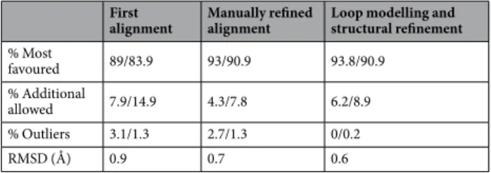
Table 1). Several iterations were crucial for improving the stereochemical quality of the model, as shown by steady
improvements in the stereochemical measures (Table 1).
Percentage of residues within the different Ramachandran plot regions are indicated. The values were
deter-mined using MolProbity22/ProCheck23. The RMSD was computed using UCSF Chimera24 after the superposition
of the BDLP crystal structure and Fzo1 model for each of the iterations indicated.
Although homologous regions were detected throughout the amino acid sequences of the target and template, the N-terminal region showed greater differences between the two species. This divergence is not surprising because, unlike other mitofusin family members, Fzo1 possesses a unique heptad repeat (HRN) upstream of the
GTPase domain2. Furthermore, our analysis using different alignment algorithms showed that the Fzo1 sequence
aligns well to the template starting at approximately residue 100 (Supplementary Figs 1–4). Secondary
struc-ture predictors (Supplementary Fig. 5) described the region upstream of HRN as unstructured, and the first 86
N-terminal residues were predicted to be disordered by the DISOPRED3 algorithm25 (Supplementary Fig. 6).
We took advantage of the requirement for Fzo1 during respiration9, 26 to obtain insights into the role of the
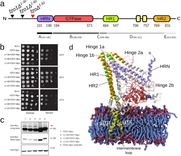
first 86 N-terminal residues upstream of the Fzo1 HRN domain. Thus, we assessed the fermentative (glucose) and respiratory (glycerol) growth capacities of three distinct N-terminal deletion mutant strains of FZO1, namely fzo1Δ1–30, fzo1Δ1–60 and fzo1Δ1–91 as compared to positive (wild-type) and negative (fzo1Δ) control cells (Fig. 1b).
Although the fzo1Δ1–30 and fzo1Δ1–60 mutants displayed normal growth on both glucose and glycerol-containing
media, the respiratory growth of the fzo1Δ1–91 mutant was nearly completely abolished (Fig. 1b). Interestingly, the
steady-state level of the fzo1Δ1–91 protein was significantly increased compared with that in the wild-type Fzo1 or
the Fzo1Δ1–30 and Fzo1Δ1–60 mutants (Fig. 1c), suggesting that the deletion of residues 61 to 91 induces
stabiliza-tion of the mitofusin, possibly by inhibiting Mdm30-mediated degradastabiliza-tion13. Consistent with this observation,
Mdm30-mediated ubiquitylation of Fzo1Δ1–91 was undetectable (Fig. 1c). These results indicate that residues 61
to 91 of Fzo1 are essential for Fzo1 function and its ubiquitin-dependent regulation by Mdm30. These phenotypes
mimic the previously observed phenotypes following inactivation of the GTPase domain13, suggesting that
resi-dues upstream of the HRN may participate in regulating GTP binding or hydrolysis by the mitofusin.
Despite the previously unappreciated functional importance of residues 61 to 91, the lack of a template for the region upstream of the HRN domain and the resulting uncertainty in its structure assignment imposed omitting the first 100 amino acids of Fzo1 from the final model. At this stage, we are tempted to speculate that together with the N-terminal helix observed in the model presented below (Helix α1 in the HRN), residues 61 to 100 may fall back on the GTPase domain to regulate its activity, although further investigation is required.
The Fzo1 model and its essential domains are illustrated in Fig. 1d. Most of the protein is exposed to the
cyto-plasmic side of the lipid bilayer and presents a similar architecture to BDLP, in which the stalk domain is
com-posed of two four-helix bundles connected by hinges16 (Fig. 1d). This organization is consistent with the recently
solved minimal GTPase domain structures from human mitofusin Mfn1, in which the N- and C-terminal region
of the GTPase domain form a four-helix bundle together with the HR2 helix that follows hinge 1b18, 19. Since our
Fzo1 model was built before the partial Mfn1 structures were published, this similarity validates the homologous regions in our model and provides substantial confidence in domains of the molecule that are not present in the Mfn1 fragment (e.g., the TM region, the trunk and hinges 1a/1b).
RMSD (Å) 0.9 0.7 0.6
The Fzo1 model has a stable core when simulated in a membrane environment.
The conforma-tional stability of the Fzo1 model was assessed in three 500-ns MD trajectories in a mixed lipid bilayer environ-ment (referred to as Fzo1.I, Fzo1.II and Fzo1.III).The predominant secondary structure of the Fzo1 model confirms a coherent and stable architecture between
the three simulations (Fig. 2), with a clear pattern of long α-helices retained during the time course of the
simula-tion, notably for the predicted coiled-coil domains HRN, HR1 and HR2, as well as for the TM dimer.
Supplementary Fig. 7 and Supplementary Table 2 show the drift of the protein structure from the
energy-minimized initial model as time series of the RMSD. The high RMSD values observed for the whole
protein in the last frame (5.7–7.3 Å) account for significant local structural deviations (Supplementary Fig. 7,
top panels, black lines). The decomposition into protein segments (Supplementary Fig. 7, top panels) reveals
that these high values are mostly due to i) unstructured regions, ii) portions of the structure that possess high
flexibility, such as the N-terminal helices, as measured by the RMSF (Supplementary Fig. 7, top panels, red lines),
and iii) regions that are unresolved in the template crystal structure, i.e., residues 502–510 (residues 631–653 in the Fzo1 model). When the contribution of these components is removed, a lower RMSD with a plateau between
4–5 Å is observed (Supplementary Fig. 7, top panels). The analysis of the ab initio modelled TM domain shows
the same average RMSD value of 2.4 Å (Supplementary Table 3), indicating stability and consistency among the
three trajectories.
Figure 1. Architecture and domain organization of the Fzo1 model. (a) Scheme showing the domains of Fzo1 from S. cerevisiae. Residue numbers for each domain and the deletion mutants are indicated. The red arrow highlights the deletion mutant that causes a defect in respiration. HRN, N-terminal truncated heptad repeat (violet, residues 101–190); GTPase, GTPase domain (red, residues 194–373); HR1, heptad repeat 1 (green, residues 484–547); transmembrane segments (yellow, residues 706–757), HR2, heptad repeat 2 (orange, residues 769–831). The fragments between the hinges are indicated below the alignment and are designated A to E. (b) Dextrose and glycerol growth spot assay for the 30-, 60- and 91-amino acid N-terminal FZO1 deletion
mutant strains, namely fzo1Δ1–30, fzo1Δ1–60 and fzo1Δ1–91, respectively. The fzo1Δ and FZO1-Myc strains were
used as negative and positive controls, respectively. (c) Anti-Myc and Anti-Pgk1 immunoblots of whole-cell extracts prepared from the strains used in (b). The FZO1-Myc mdm30Δ strain was used as a control for lack of Fzo1 ubiquitylation. Molecular weight markers are indicated on the right of long or short exposures of the immunoblots. (d) Fzo1 model after the equilibration phase (as described in the Methods section). The blue
surface represents POPE and POPC lipids from a portion of the bilayer. The GDP nucleotide and the Mg2+ ion
Together, these results indicate consistency among the trajectories that show a similar fluctuation profile
(Supplementary Fig. 6) with the model that underwent the equilibration and production stages, maintaining the
proposed fold.
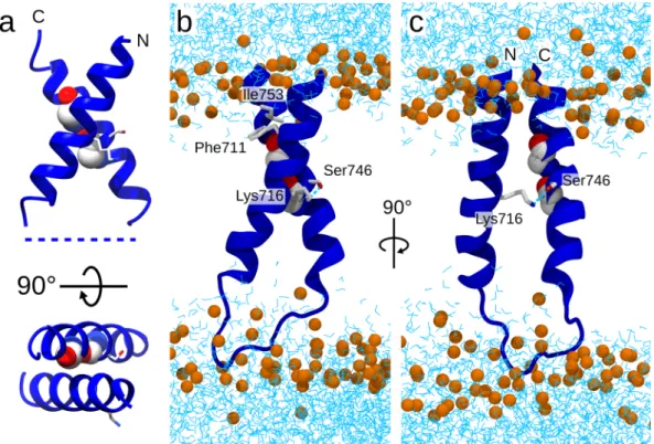
Fzo1 transmembrane domain. The highest-scoring model constructed using PREDDIMER27 shows a compact
structure characterized by a crossing angle (χ) of 119.7° (other models are shown in Supplementary Fig. 8).
The second TM helix (TM2) harbours a GxxxG motif (with x representing any amino acid) that was previously
reported to play a role in helix dimerization28, 29. We based our choice on the observation that interactions with
(small)xxx(small) motifs are often associated with the formation of right-handed dimers characterized by
neg-ative helix-helix crossing-angles in parallel dimers30–32. Here we have a right-handed antiparallel dimer, with
χ = 120° (Supplementary Fig. 8). This is currently the most plausible arrangement. The second-highest
scor-ing model has a similar score and involves the same dimerization motif, although with a straighter geometry (χ = 175°, nearly antiparallel). While the latter arrangement is less common it cannot be excluded and the reader should be aware that alternative transmembrane arrangements are possible.
According to the RMSD analysis for each MD simulation trajectory (Supplementary Fig. 7), the TM domain
is well anchored and does not undergo notable structural changes, as confirmed by the analysis of the dominant
secondary structure (Fig. 2). The observed fluctuation values are similar in the three replicates (Supplementary
Fig. 6). These features suggest that the structure of the TM domain is realistic.
To characterize the interaction between the two TMDs in more details than just pure geometrical contacts, we analysed specific hydrogens bonds as described in methods. All simulations indicated a hydrogen bond between the two TM helices that involved the side-chains of Lys716 and Ser746 (38%, 19% and 18% persistence for Fzo1.I, Fzo1.II and Fzo1.III, respectively). Notably, Ser746 resides on the TM2 helix and belongs to the aforementioned GxxxG motif.
Analysis of the contact residues in the TM dimer revealed an abundance of promiscuous stabilizing
inter-actions (Supplementary Table 4). The only unambiguous interaction seemed to be Phe711-Ile753, whereas the
remainder of the interactions varied among the trajectories. After manually adjusting the side-chain rotamer (see Methods), the Ile753 residue on helix TM1 directly faced residue Phe711 on TM2, increasing the packing of the
TM helices (Fig. 3b). The analysis of protein-lipid hydrogen-bonding indicated that Fzo1 had more interactions
with POPE compared to POPC (Supplementary Table 5). Given that the bilayer contains both lipids in the same
proportion, this observation suggests a preference for POPE.
Experimental validation of the Fzo1 model.
The yeast mitofusin Fzo1 has previously beencharacter-ized by introducing many mutations across its functional domains (Supplementary Fig. 9). We aimed to evaluate
the relationship between our structural model and the wealth of published data available, as well as the additional
targeted mutations summarized in Supplementary Table 6 and discussed hereafter. This analysis was performed
on the most representative structure (i.e., a centroid) identified after a cluster analysis of the Fzo1.I trajectory (see the Methods).
Figure 4 shows the secondary structure elements. The organization of the HR domains in our model reveals a
more complex organization than initially hypothesized, as these repeats are not merely assembled as continuous helices. In particular, HRN, HR1 and HR2 are characterized by a complex structure and are composed of several tandem helices. This observation may be key to interpreting the functional outcomes of several Fzo1 mutations. For instance, according to our model, previously uncharacterized regions, such as the α14 and α27 helices, may contribute to protein function. Although these helices are not defined by any heptad periodicity, they represent
a structural continuation of the HR1 and HR2 repeats, separated by the hinges 1a and 1b, respectively (Fig. 4,
blue arrows). Consequently, helices α14 and α27 are located parallel to the HRN in our 3D model (Fig. 1d). This
configuration is strikingly similar to the four-helix bundle observed in the recent crystal structure of an Mfn1
fragment (Supplementary Fig. 10), in which the C-terminal helix has been proposed to stabilize the identified
bundle18. Here, we propose that Fzo1 helix α27 (Fig. 4) plays a similar role (Supplementary Fig. 10). Consistent
with this observation, deletion of the last 24 residues of Fzo1 (Fzo1 ∆826–855) was previously shown to abolish
mitochondrial fusion34. Although the reconstitution of helix α27 in our model (res 815–845) results from the
manual modification of the target-template alignment (see Methods), the considerations described above provide a significant validation of our modelling approach.
Based on these observations, we refined our analysis of the model using a series of site-directed mutagenesis experiments across the predicted interactions revealed during the MD analysis.
Figure 2. Stable secondary structure motifs in simulations. Comparison of the Fzo1 domain organization
shown in Fig. 1a with the stable secondary structures (persistence greater than 90%) in the three replicate
simulations. The colour code is blue, α-helix; red, β-sheet; yellow, turn; green, bend; black, β-bridge; and violet, π-helix.
Interactions between the N- and C-terminal halves of the protein. We searched for salt bridges to test experi-mentally the putative interactions and validate the proposed model for Fzo1. We focused our attention on the
predicted salt bridge D335-K464 (Fig. 5) because the interaction was identified in the model after the
minimiza-tion phase and is conserved in BDLP, where both charged residues are inverted (His202 and Glu331, respectively, Figure 3. Insights into the Fzo1 transmembrane domain. (a) PREDDIMER27 ab initio prediction of the helical
dimer (FSCOR 3.113, crossing angle χ 119.7°). (b,c) Snapshots from the Fzo1.I trajectory showing the most representative structure (i.e., the centroid). Glycine residues within the GxxxG motif are presented as a space-filled representation, whereas residues involved in interactions are depicted in stick form. Phosphate atoms from POPC and POPE (orange) and water molecules (cyan) are indicated, whereas the rest of the protein is omitted for clarity.
Figure 4. Cartoon representation of the Fzo1 model and its functional domains. (top) Residue numbers delimiting the domains, (bottom) secondary structure elements are annotated with the Fzo1 mutations performed in this study. Mutants considered for the charge swap strategy are connected by a bar. The colour code is cyan, loss of function (LOF) and maroon, wild-type phenotypes. Putative hinge regions are indicated by blue arrows. Previously reported mutations across Fzo1 functional domains are shown in Supplementary Fig. 9. Green and pink horizontal bars above the secondary structure elements depict the N- and C-terminal halves,
Supplementary Fig. 11). We maintained the focus on D335-K464, even though the interaction was weakened in subsequent simulations (1.5%, 2% and 27% for Fzo1.I, Fzo1.II and Fzo1.III, respectively). This choice was moti-vated by the following observations: a preliminary MD study suggested that the salt bridge was present in 61% of structures, and the solved fragment from Mfn1 shows an interaction between the homologous pair of charged
residues, Asp200 and Lys33618, 19 (Supplementary Fig. 10). In addition, these mutants provide us with the
oppor-tunity to investigate the importance of the Lys464 residue, which is a target for post-translational modification by ubiquitin in Fzo115.
We employed a charge swap strategy to assess this predicted interaction, which relies on the assumption that a double charge reversal mutation across a predicted salt bridge should restore a wild-type phenotype, whereas Figure 5. Swap mutations across the predicted salt bridge D335-K464. (a) Relative positions of D335 (red) and K464 (blue) in the Fzo1 model. The green and pink regions correspond to the N- and C-terminal halves,
respectively (Supplementary Fig. 9). (b) Dextrose and glycerol growth spot assay. (c) Anti-Fzo1 and anti-Pgk1
immunoblots of whole-cell extracts prepared from the strains analysed in (b). Molecular weight markers are indicated on the right. (d) Anti-Myc and Anti-Pgk1 immunoblots of whole-cell extracts prepared from the indicated strains. Molecular weight markers are indicated on the right of long or short exposures of the immunoblots.
single point mutations may abolish the salt bridge, therefore affecting protein function. Although the single point mutations D335K and K464D induced a total inhibition of respiratory growth, this strong defect was partially but
significantly corrected by the swap mutation D335K-K464D (Fig. 5b), thus confirming the proximity between
residues at position 335 and 464. Consistent with the role of K464 in the ubiquitin-mediated degradation of
Fzo115, the level of the Fzo1 K464D mutant protein increased compared with that of the wild-type control. Both
the Fzo1 D335K single mutant and, most surprisingly, the D335K-K464D double mutant (charge swap) displayed levels comparable to the wild-type protein. This observation led to compare the ubiquitylation status of single (K464D) or double swap (D335K-K464D) Fzo1-13Myc mutants with that of K398R that is established to alter
ubiquitylation of the mitofusin15, 35. As expected, detection of the Mdm30-dependent ubiquitylation doublet
was strongly impaired in K464D and K398R mutants (Fig. 5d). Strikingly, however, the double swap mutation
D335K-K464D restored partial ubiquitylation of Fzo1-13Myc (Fig. 5d). As aspartate cannot be ubiquitylated, the swap mutations may thus also allow position 335 to be ubiquitylated by restoring the proximity of residues 335 and 464. Alternatively, this restored proximity may contribute to maintain a correct fold of Fzo1 that would favor ubiquitylation on K398. The latter possibility is more likely as K398 is the main target for Mdm30-dependent
ubiquitylation of Fzo135. In aggregate, these results provide experimental confirmation of a physical interaction
between the two residues located in the N- and C-terminal halves of Fzo1 and contribute to validating the pre-dicted proximity in our model.
In order to further cross-validate our model, we have constructed a new partial Fzo1 model based on human
Mfn1 crystal19 (Supplementary Fig. 10). We compared key interactions predicted by our BDLP-based model with
the alternative partial model. Interestingly, among the numerous interactions conserved (Supplementary Fig. 10),
the predicted interaction between K464 and D355 (Fig. 5), is also observed in the Mfn1-based model.
Electrostatic interactions are involved in organizing the HR domain. The HR regions in Fzo1 are required for mitofusin architecture, showing LOF phenotypes upon disruption of their presumed helical folds imposed by
proline mutations11. In our model, the coiled-coil HR1 and HR2 domains form an antiparallel bundle (Fig. 1d).
Yet surprisingly, we did not observe the typical HR interaction pattern in the HR1/HR2 coiled-coil structure
(Supplementary Table 7 and Supplementary Fig. 12). The hydrophobic spine of HR2 is solvent-exposed, and the
HR1 hydrophobic spine actively interacts with the nearby α18 helix (residues 584–630, Fig. 4 and Supplementary
Fig. 12), which interestingly does not exhibit heptad periodicity. Consequently, the HR2 repeat may be available
for putative interactions between Fzo1 molecules similarly to human Mfn1, in which the HR2 domain dimerize
through anti-parallel binding36. Whether these interactions take place in trans (between Fzo1 molecules from
opposing membranes) as previously proposed20, 36 or in cis (between Fzo1 molecules from the same membranes)
prior to mitochondrial tethering37 is an important question that will need future clarification.
In contrast to the lack of a classical pattern of hydrophobic interactions, we observed a network of intra- and interhelical salt bridges in the simulations. According the convention of labelling residues in a heptad repeat
from a to g, with a and d usually indicating hydrophobic residues38, intrahelical interactions were observed in
all three domains, with the majority of the b-e type, similar to the pattern previously observed for the neuronal
SNARE complex39. Almost all interhelical salt bridges connected HR1 and HR2, with one exception connecting
HRN and HR2 (observed only in Fzo1.II). The 8 salt bridges that persisted in all three trajectories are listed
in Supplementary Table 8. We used site-directed mutagenesis to investigate a possible interhelical salt bridge
between the HR1 and HR2 domains, the D523-H780 salt bridge, which was identified early on as having high persistence in a preliminary simulation. Indeed, although the distance rapidly rises above the adopted interaction cut-off of 6.5 Angstrom, the distance between its charged groups remains constant below 10 Å during the sim-ulation. The charge swap strategy applied to these two residues revealed that the D523H point mutation caused a temperature-dependent respiratory growth defect. However, the H780D mutant yields a wild-type phenotype
(Fig. 6b). Although this result may seem difficult to rationalize, an interpretation based on common observations
with charge reversal mutations is that D523 and/or D780 may interact with more than one partner, allowing for a form of compensation when H780 is mutated. Strikingly, the swap mutant D523H-H780D induced com-plete rescue of the respiration defect caused by the D523H mutation. This result is consistent with the proximity between HR1 and HR2 in the Fzo1 model and suggests that this proximity is a requirement for mitofusin
activ-ity. Although the predicted salt bridge D523-H780 (HR1 and HR2, respectively) shown in Fig. 6 is not directly
conserved, it is located near a salt bridge identified in the template structure of BDLP (D523 and H780 in Fzo1 correspond to I390 and F624 in BDLP, next to bridging residues R393 and D625; see the sequence alignment in
Supplementary Fig. 11).
A recent study hypothesized that the HR2 domain may dissociate from the HR1 domain in active forms of Mfn2 and unfold itself from the rest of the protein to become available for trans interactions with exposed HR2
domains of Mfn2 molecules from opposing mitochondria20. However, a dissociation of one of the repeats seems
unlikely because our model features tight interactions between the coiled-coil HR1 and HR2 domains, which
appear to be required for maintaining the mitofusin fold (Fig. 1d).
Functional importance of the regions surrounding hinges 1a and 1b in the Fzo1 model. A tempting alternative hypothesis to the mitofusin conformational switch mediated by HR2 extension is the possibility that mitofusins
perform extensive GTPase domain-dependent rearrangements through hinges 1a and 1b (Fig. 4), similar to
BDLP17. In this regard, Mdm30 was previously shown to bind the N-terminal half of Fzo1 (HRN/GTPase domain)
in a manner that required the GTPase domain-dependent displacement of the C-terminal half (HR1/TM/HR2
domain)13. Mutations within the HR2 domain that disrupt the interaction between the N- and C-terminal halves
of Fzo111 were thus expected to promote constitutive Mdm30 binding. Consistent with this hypothesis, the L819P
mutation induced an increase in Mdm30-dependent ubiquitylation of Fzo1 and markedly accelerated the degra-dation of the mitofusin13.
The Leu819 residue is located on helix α27 of the Fzo1 model, with its side chain facing the helix α14 (Fig. 7a). Consistent with this arrangement, introduction of a negative charge at position 819 (L819E) impaired respiratory
growth at 30 and 37 °C whereas replacement with an uncharged residue (L819A) did not have any effect (Fig. 7b).
As previously shown, the L819P mutation abolished respiration at all temperatures (Fig. 7b) and enhanced
degra-dation of Fzo1 (Fig. 7f). The location of Leu819 in hinge 1b suggests that the L819P mutation may induce a kink
in this region that would in turn favor a conformational rearrangement of the mitofusin. To explore this possibil-ity, we reasoned that perturbing the backbone conformation around Leu819 should also promote conformational remodelling of Fzo1 and mimic the L819P mutation effect. In this regard, Glu818 is predicted to lie in hinge 1b, with a water-exposed side-chain. Removing the side-chain charge (E818A) or even reversing it (E818R) did not affect respiratory growth, indicating that this position is not sensitive to modifications of the side-chain. However, we confirmed that this position is sensitive to backbone effects by introducing a proline residue (E818P) that
completely abolished yeast growth on glycerol media (Fig. 7c). Moreover, while the E818P mutation induced an
Fzo1 decrease that does not depend on Mdm30 (Fig. 7d; compare lanes 2 and 4), this mutant was also degraded
by the Mdm30-mediated pathway twice more than wild-type Fzo1 (graph Fig. 7d). This observation is consistent
with the effects of L819P and the potential conformational switch it exerts on Fzo1.
Tyr490 is located in hinge 1a in our model and is predicted to interact with Leu819 in Fzo1.I and Fzo1.II
(75% and 61%, respectively) (Fig. 7a). Similar to L819A but in contrast to L819E, mutation of Tyr490 in Alanine
(Y490A) or Lysine (Y490K) did not affect respiratory growth (Fig. 7e). However, combining Y490K with L819E
Figure 6. Swap mutations across the predicted salt bridge D523-H780. (a) Location of D523 (red) and H780 (blue) in the Fzo1 model. The green and pink regions correspond to the N- and C-terminal halves, respectively
(Supplementary Fig. 9). The positions of HR1 and HR2 are indicated. (b) Dextrose and glycerol growth spot
assay. (c) Immunoblots of whole-cell extracts prepared from the strains analysed in (b). The Pgk1 immunoblot was used as a loading control. Molecular weight markers are indicated on the right.
(Y490K-L819E) induced an enhanced defect in respiration as compared to L819E (Fig. 7e) without affecting Fzo1
levels (Fig. 7f). This confirms the predicted proximity between Tyr490 and Leu819. Moreover, the Y490P
muta-tion was previously reported to cause total inhibimuta-tion of respiratory growth and induce the formamuta-tion of loose
mitochondrial aggregates that were hypothesized to result from a tethered trapping state11. Similar to hinge 1b, a
mutation of Tyr490 to proline within hinge 1a may thus affect mitochondrial fusion by inducing conformational rearrangements in Fzo1.
Single point mutations, L501A and L504A, of residues located on the other side of the hinge were previously
shown to be functionally silent but abolished mitochondrial fusion in combination11. Thus, these mutations were
hypothesized to affect hydrophobic interactions, as both leucines were predicted to occupy the a and d positions
in the HR1 domain11. The analysis of the contacts within our model revealed that these two leucine residues may
in fact belong to a leucine cluster between residues Leu501, Leu504 and Leu802, which is located in close vicinity
to hinge 1b (Fig. 7a). This cluster may explain the differential phenotypes obtained with the L501A or L504A
mutants, as Leu802 may compensate for the single mutation but not the double point mutations.
Figure 7. Critical residues in the Fzo1 model hinge region. (a) Detailed structure of the putative hinge region. The colour code is: cyan: Leu819, yellow: Tyr490, purple: Leu802, red: Glu818, grey: Leu501, and light grey: Leu504. The domains are: violet: HRN, green: HR1, and orange: HR2. The location of this hinge region within the model is presented in the right panel. (b), (c) and (e) Dextrose and glycerol growth spot assay with indicated strains. (d) and (f) Anti-Fzo1 and anti-Pgk1 immunoblots of whole-cell extracts prepared from the strains used in (b), (c) and (e). Molecular weight markers are indicated on the right of immunoblots. The graph in (d) represents the quantified ratio of Mdm30-dependent degradation for wild-type Fzo1 or the Fzo1 E818P mutant.
Thus, these results highlight the critical importance of regions surrounding hinges 1a and 1b in Fzo1 function and suggest that they may be involved in mitofusin rearrangements.
Atomistic insights into the GTPase domain.
Binding site architecture and protein-GDP interac-tions. Protein-GDP interactions were modelled based on the homologous interacting residues in the BDLPtemplate. Ten H-bonds were identified in the BDLP structure (Supplementary Table 9). The 10 corresponding
donor-acceptor distances were restrained during the modelling procedure and the equilibration phase (Fig. 8
and methods for details). The time series of these distances in the three simulations were monitored during the
equilibration phase and for each trajectory (Fig. 8 right and Supplementary Fig. 13).
The residues forming persistent, tight contacts were Asn197, Ser201 and Ala202. Contacts showing more variation after the restrained equilibration phase were formed by Thr198, Lys370, Lys371 and Asp373. Contacts Figure 8. Analysis of the key protein-GDP interactions in the Fzo1 nucleotide-binding site. The colour code is grey during the equilibration phase, cyan for simulation Fzo1.I, green for Fzo1.II and purple for Fzo1.III. Distance restraints and persistent H-bonds over 50% of the simulation time are indicated with orange and green dotted lines, respectively. The G-boxes from G1 to G4 are highlighted. The evolution of the donor-acceptor distance network for the residues contacting the ligand is presented on the right. The last three blocks show the new interactions observed in Fzo1 with respect to BDLP, and the red line delimits the end of the equilibration phase. Donor-acceptor distances are depicted using a colour gradient from the darkest (below or equal to 3.5 Å) to lightest colours (up to 4.0 Å).
involving Asp428 and Gly427 were predicted based on the template but were not retained in the unrestrained trajectories. An equivalent network involving the homologous residues N237 (Fzo1-K370), D240 (Fzo1-D373)
and others (Supplementary Table 10) was found in the recent crystal structures from human Mfn118, 19, and is
conserved in our Mfn1-based model (Supplementary Fig. 10). Our approach thus provides insight into the
resi-dues that promote the stabilization of the nucleotide within the Fzo1 GTPase domain.
MD simulations further revealed new interactions in the Fzo1 model. In particular, His426 contacted the
GDP.N1 in Fzo1.I after the displacement of the Asp428 residue (Fig. 8 right). Furthermore, the Gly199 residue
contacted the bridging oxygen between the two phosphates (GDP.O3A), and the Glu215 residue was involved in an interaction, characterized by a 46% persistence in Fzo1.III, with the ribose in position O2′ oriented towards
the backbone (Fig. 8 left). Our model also includes a magnesium ion in the nucleotide binding site, which
partic-ipates in coordinating GDP with the Ser201 residue (see Supplementary Fig. 14), similar to human dynamin or
atlastin40, 41. This feature is not only in agreement with the structure of the human Mfn1 fragment in which the
Ser89 residue directly coordinates a bound magnesium ion19 but is also consistent with the requirement for this
ion in the tethering of proteoliposomes by the Mfn1 construct18.
GDP primarily interacts with the G1 and G4 motifs. In particular, G1 (the P-loop) anchors both phosphate
groups (Fig. 8 left). The G2 (switch I) and G3 motifs point away from the active site, suggesting a more structural
role. This observation is consistent with the orientation of Thr103 in the BDLP crystal structure away from the active site16 and the critical role of the corresponding conserved Thr221 (i.e., G2 motif) in Fzo1 function9, 11, 13.
These properties are typical of GTPase domains41, 42.
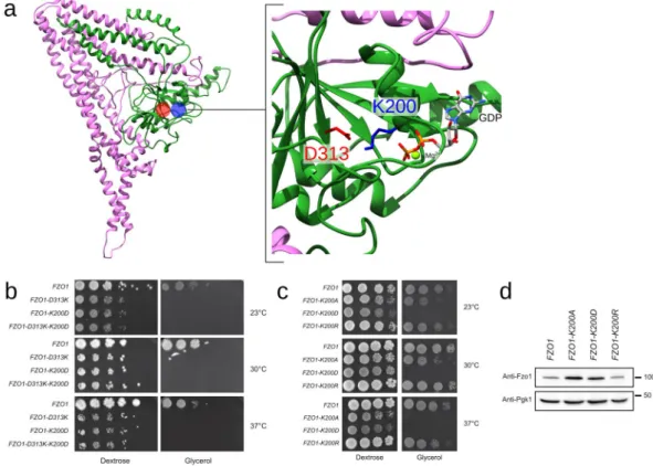
A positive charge at position 200 is essential for GTPase domain integrity. The vicinity between the residues of the GTPase binding site in the model features a high confidence salt bridge that connects the G1 (Lys200) and G3 motifs (Asp313). This salt bridge is not only conserved in BDLP (Lys82 and Asp180, respectively) but also detected in all three MD replicates, Fzo1.I, Fzo1.II and Fzo1.III, with a persistence greater than 90%. Surprisingly, the charge swap strategy applied to this predicted salt bridge revealed that although the single mutations K200D and D313K induced an expected and strong inhibition of respiratory growth, the double K200D-D313K mutation (charge swap) did not rescue this defect (Fig. 9b).
Figure 9. Swap mutations across the predicted salt bridge K200-D313. (a) Location of D313 (red) and K200 (blue) in the Fzo1 model. The most representative structure after the cluster analysis on the Fzo1.I trajectory is shown (i.e., the centroid). The green and pink regions correspond to the N- and C-terminal halves, respectively
(Supplementary Fig. 9). The GDP atoms and the bound magnesium are indicated. (b) Dextrose and glycerol
growth spot assay. Single mutations across the predicted interaction cause a severe respiration defect, and the double mutant (charge swap) does not rescue these defects. (c) Dextrose and glycerol growth spot assay. The K200A and K200D point mutations affect respiration, whereas the K200R mutation does not impact growth on glycerol media. (d) Anti-Fzo1 immunoblot of whole-cell extracts prepared from the strains used in (c). The Pgk1 immunoblot was used as a loading control. Molecular weight markers are indicated on the right.
provided by the lysine at position 200 may be essential for GTP hydrolysis, which could explain the failure of the K200D-D313K swap mutation to restore functional mitochondrial fusion. Furthermore, a negative charge at position 200 may weaken (if not abolish) the interaction with the nearby negatively charged nucleotide.
We introduced a conservative charged mutation to arginine at position 200 (K200R) and compared the effects of the K200A, K200D and K200R mutations on respiratory growth to test this hypothesis. Remarkably, although K200D abolished growth on glycerol media and K200A induced strong thermosensitive defects, K200R
main-tained full respiratory growth capacity at all tested temperatures (Fig. 9c). Consistent with this observation, the
Mdm30-mediated degradation, and thus the GTPase domain integrity13, were affected in Fzo1 K200A and Fzo1
K200D but not Fzo1 K200R (Fig. 9d). Therefore, a positive charge in position 200 is required for Fzo1 function. This finding explains the failure of the D313K-K200D charge swap mutant to rescue respiratory growth but also suggests that the positive charge at position 200 participates in GTP hydrolysis.
Conclusions
Using the homology modelling approach based on the structure of the bacterial dynamin BDLP, we have mod-elled the structure of the Fzo1 mitofusin in complex with the nucleotide GDP. The structural model is stable under simulation, experimentally validated in vivo and consistent with published data. The model provides insights at the residue level and describes Fzo1 as being partitioned into distinct structural regions: the GTPase domain, a four-helix bundle, a hinge region, and a three-helix trunk connected to the TM domain. Little is known about the precise sequence of events that may couple GTP hydrolysis to potential conformational rearrangements within
mitofusins. In this regard, the conserved four-helix bundle observed in the Mfn1 partial structures18, 19 and in
our full-length Fzo1 model is reminiscent of the characteristic bundle signalling element (BSE) of dynamins41, 42.
The BSE domain has been proposed to transfer conformational information from the GTPase domain to the
trunk region of dynamin during membrane fission catalysis41. Considering the continuity between the four-helix
bundle and the putative hinge region in Fzo1 (Supplementary Fig. 10), this structural motif may play a similar
role during membrane fusion. When comparing the Mfn1- and the BDLP-based models we have constructed, the
bundles are arranged differently (Supplementary Fig. 10). While this difference may involve the artificial linker
that allowed obtaining the Mfn1 crystals18, 19, our BDLP-based model suggests that the bundle likely adopts a
distinct conformation that could provide optimal flexibility in the hinge 1a/1b regions, which warrants further insights on the structure of full-length mitofusins. Mitofusins do not function as monomers but oligomerize in both cis (on the same lipid bilayer) and trans (from opposing lipid bilayers) to mediate membrane attachment and fusion4, 11, 19, 36, 37, 44. As the precise principles for these oligomerization properties emerge, future studies should
aim to model these complexes in presumably “active” and “inactive” conformations and thereby further elucidate the precise events that lead to mitofusin-mediated membrane fusion.
Materials and Methods
Bioinformatic analysis.
Sequences identification and alignments. The S. cerevisiae Fzo1 target sequencewas obtained from the UniProtKB database (Universal Protein Resource Knowledgebase45), entry: P38297. A
putative template was identified using the BLASTP tool (Basic Local Alignment Search Tool for proteins46) against
the PDB (Protein Data Bank47, with default settings. Homologues for the template in the phylum Cyanobacteria
were then selected using the BLAST method (with default settings), from which a set of 43 sequences annotated as dynamin-like proteins was selected. The identity ranged from 22% to 87%, and the sequence coverage was greater
than 65%. This set was then aligned with Expresso48 using structural information from the template structure with
PDB-id 2J6816. After removing the first 100 N-terminal residues of Fzo1, we merged the multiple sequence
align-ment (MSA) described above with the target sequence using M-Coffee49. We used NCBI HomoloGene (https://
www.ncbi.nlm.nih.gov/homologene, accessed 10-05-2016) to add a restricted set of 11 homologous sequences belonging to the families of fuzzy onions (FZO1), FZO-like (FZL), mitofusin (MFN1 and MFN2) (hgid: 31469,
95893, 11481 and 8915, respectively), which are listed in Supplementary Table 1.
Supplemental alignments were constructed to identify the target-template correlation in the N-terminal
region. Two different alignment algorithms, Clustal Omega50 and T-Coffee51, were chosen to combine the set of
43 homologous template sequences. Then, the resulting MSA was merged with the target Fzo1 sequence with and
without its first 100 N-terminal residues using M-Coffee (Supplementary Figs 1–4).
The T-Coffee method was used to align protein sequences from all the mitofusins belonging to the FZO1
family listed in Supplementary Table 1 that were used in this study.
Structure prediction and model assessment. The secondary structure of Fzo1 was predicted from its sequence
using the following algorithms: CONCORD52; PSIPRED (v 3.3)53; PSSpred54; and PORTER55. The LigPlot+
pro-gram56 was used to inspect the homologous residues in the target and template that contact the GDP substrate
using a donor-acceptor distance of 3.5 Å. The PCOILS v1.0.1 algorithm57 was used to predict heptad periodicity in
Fzo1 and BDLP. Disordered residues in the target sequence were investigated using the predictor DISOPRED325.
Transmembrane domain. The BDLP template used for modelling Fzo1 possesses a hydrophobic region (res
TM helices of Fzo1 (Supplementary Table 11) do not match in the final target-template alignment after
refine-ment (Supplerefine-mentary Fig. 11). For this reason, the ab initio prediction of the helix dimer structure was crucial in
the modelling process.
The location of the TM segments was identified using the predictors MEMSAT-SVM58, OCTOPUS59
and TMpred60 (Supplementary Table 11). Ab initio modelling of the TM segments was performed using the
PREDDIMER server27, and the best model was selected according to the proposed ranking score (F
SCOR 3.113).
Strategy used to model the Fzo1-GDP complex.
We manually refined the target-template alignment described above to avoid fragmenting the secondary structure elements. In particular, we modified the alignment to open a gap of 21 positions within the predicted TM helices of Fzo1. This modification enabled us to recon-stitute the predicted fold for the C-terminal fragment (res 826–855) that was previously shown to be requiredfor mitochondrial respiration34, but was unstructured and located outside the template in the initial alignment.
Consistently, this modification positioned the residue L819, which induces a severe loss of function (LOF) when
mutated to proline11, from a nearby unstructured region precisely within the putative hinge thought to enable
the GTPase domain-dependent rearrangements. Moreover, consistent with the crystal structure of human Mfn1, this C-terminal fragment may participate in stabilizing the identified four-helix bundle18, 19. Finally, guided by the
secondary structure predictors, small adjustments were made to restore helices that were disrupted by insertions and deletions (indels).
We combined three different templates to model the Fzo1 structure. The TM segments (res 706–726 and 737–757) were based on the ab initio model, the pentapeptide 216–220, which was unresolved in structure 2J68,
was modelled on residues 98–102 from structure 2J69 (3.0 Å16), and the remainder of the protein followed
struc-ture 2J68. One hundred models were generated using the MODELLER program (v 9.15)61. The GDP nucleotide
coordinates from structure 2J68 were included during the modelling procedure. During modelling, harmonic restraints were imposed on donor-acceptor distances, based on the LigPlot+ analysis of protein-nucleotide inter-actions in template 2J68. Moreover, we imposed canonical α-helix conformations for residues 101–123, 299–304, 426–433, 548–553, 700–726, 737–757 and 847–855, as well as β-strand conformations for residues 234–241 and 281–283.
We performed loop refinement of the intermembrane loop (res 727–736) and we applied the loop modelling
routine of MODELLER62 resulting in fewer Ramachandran outliers. We ranked solutions according to the
dis-crete optimized protein energy (DOPE) method63.
The Dunbrack rotamer library64 implemented in University of California, San Francisco (UCSF) Chimera24
was used to change the rotamers of the Lys727, Lys735 and Lys736 residues, which are located within the inter-membrane loop, by orienting the charged side-chains towards water to stabilize the TM domain during the MD simulation. Similarly, the rotamer of residue Ile753 was changed to increase TM helix packing. The PROPKA
method65 (v 3.00) implemented in the automated PDB2PQR pipeline66 (v 2.0.0) was used to assign protonation
states at pH 7. A double protonation was predicted for the His240 residue. The lipid-buried Lys716 residue is
conserved in half of the genes belonging to the FZO1 family (Supplementary Table 1 and Supplementary Fig. 15).
According to recent data, lysine residues are titrated in a membrane environment, suggesting that changes in the
ionization state may regulate membrane-protein function67. Therefore, the protonation state of the lipid-buried
Lys716 was manually set to a neutral amine. The stereochemical quality was assessed using the MolProbity
server22 and PROCHECK23. The model obtained was then oriented with respect to the lipid bilayer using the PPM
server68. These coordinates were subsequently manually adjusted by a 6° rotation around the x axis and a
transla-tion of −1.5 Å along the z axis to improve the lipid-packing around the Fzo1 TM domain, and empty spaces in the bilayer were avoided during model building using the CHARMM-GUI pipeline (see below).
Simulation setup and equilibration.
The GROMACS 5.0.4 program was used to perform the MDsimulations69. The simulation system was assembled using the CHARMM-GUI Membrane Builder70 with the
CHARMM36 force field71. In order to mimic the mitochondrial outer membrane composition, the protein model
was embedded in a 1:1 mixed lipid bilayer composed of 156 molecules of palmitoyl-oleoyl-phosphatidylcholine (POPC) and 156 molecules of palmitoyl-oleoyl-phosphatidylethanolamine (POPE). The solvent is composed
of a 150 mM KCl solution with 36,919 TIP3P water molecules72. One magnesium ion was manually positioned
between the α and β GDP phosphates, consistent with crystal structures 2X2E, 3T34, 3W6P and 5CA9 for human
dynamin 141, dynamin-related protein 1 A from Arabidopsis thaliana73, human dynamin-1-like protein74 and
Sey1p from Candida albicans75, respectively. Two K+ counterions were subsequently added to neutralize the
sys-tem. Supplementary Table 12 reports the composition of the simulation system. Before running the simulations,
the system was subjected to 8000 steps of energy minimization. Afterwards, the structure was thermalized at
310 K for 200 ps using the NVT ensemble, with a time step of 2 fs and position restraints of 1000 kJ·mol−1·nm−2
on all protein heavy atoms. Subsequently, the same restraint settings were applied in 10 ns simulations using the NPT ensemble to allow the lipids to relax. The optimized and relaxed system was further equilibrated for a total of 250 ns using the NPT ensemble by combining position restraints on side-chains and backbone atoms, which
were gradually relaxed from 1000 to 5 kJ·mol−1·nm−2 in seven successive steps. Simultaneously, we maintained
distance restraints of 1000 kJ·mol−1·nm−2 for the previously identified GDP homologous hydrogen bond network
(see above). After 250 ns of equilibration, three replicas of the same system (Fzo1.I, Fzo1.II and Fzo1.III) were simulated for a total of 500 ns with an integration time step of 2 fs in the NPT ensemble under periodic boundary
conditions. Long-range electrostatics were managed using the particle-mesh Ewald (PME) method76. All bond
lengths were constrained using the LINCS algorithm77. Global translational and rotational motions were removed
the DSSP method78. Hydrogen bonds and salt bridges were identified using g_hbond from GROMACS and VMD
1.9.279 (http://www.ks.uiuc.edu/Research/vmd/), respectively. Contacts within a distance cut-off of 3.5 Å (and up
to 30 degree off-axis angle) and 6.5 Å were considered for H-bonds and salt bridges, respectively, to assess these interactions. Other analyses were conducted using in-house programs. When not otherwise specified, the persis-tence means how much the considered event occurs during the simulation time, expressed as a percentage over the number of frames. The cluster analysis was performed using g_cluster from GROMACS with the GROMOS
method80, with a cut-off of 0.25 Å. Molecular graphics were generated with the VMD 1.9.279 (http://www.ks.uiuc.
edu/Research/vmd/) and UCSF Chimera 1.924 packages. Data were plotted using Grace (
http://plasma-gate.weiz-mann.ac.il/Grace/).
Plasmids, yeast strains and growth conditions.
FZO1 mutant plasmids are listed in SupplementaryTable 13. Plasmids MC377 to MC382, MC396 and MC397 were synthesized by GeneCust (Ellange, Luxembourg).
A QuikChange Lightning Site-Directed Mutagenesis Kit (Agilent Technologies, Santa Clara, California, USA) was used to generate plasmids MC400 and MC406 to MC408. Mutagenic primers were designed based on the
MC250 plasmid sequence with the web-based QuikChange Primer Design Program available online at www.
agilent.com/genomics/qcpd.
Yeast strains are listed in Supplementary Table 14. Standard methods were used for growth, transformation
and genetic manipulation of S. cerevisiae. Minimal synthetic media [Difco yeast nitrogen base (Voigt Global Distribution, Inc., Lawrence, Kansas, USA), and drop-out solution] supplemented with 2% dextrose (SD) or 2%
glycerol (SG) were prepared as previously described81.
Strains lacking FZO1 lose their mitochondrial DNA because of decreased mitochondrial fusion efficiency9, 26.
Consequently, a plasmid-shuffle strategy was used as a reliable analysis of fzo1 mutants in vivo. The FZO1 shuffle strain MCY571 (fzo1Δ strain covered by an FZO1 shuffling plasmid) was employed. MDM30 was
chromosom-ally deleted in the FZO1 shuffle strain using a previously described method82 to generate the mdm30Δ strain
(MCY585).
Shuffle strains were transformed with the plasmids shown in Supplementary Table 13 and plated on SD
selec-tive media lacking uracil and tryptophan. Ten colonies were systematically isolated on SD selecselec-tive media and replica-plated on 5′-fluoroorotic acid (5′-FOA) plates. Strains grown on 5′-FOA plates and cured from the FZO1 shuffling plasmid were in turn replica-plated on SD and SG selective plates lacking tryptophan. The glycerol growth phenotypes of strains cured from the shuffling plasmids were reproducibly observed in 100% of the clones tested after 1 to 3 days of growth at 30 °C. Representative clones were subsequently used for spot assays and the preparation of protein extracts.
Spot assays.
Cultures grown overnight in SD selective media lacking tryptophan were pelleted, resuspendedat an optical density at 600 nm (OD600) of 1, and serially diluted (1:10) five times in water. Three microliters of
the dilutions were spotted onto SD- and SG-selective plates and grown for 2 to 4 days (dextrose) or 3 to 6 days (glycerol) at 23 °C, 30 °C or 37 °C.
Protein extracts and immunoblotting.
Cells grown in SD-selective media lacking tryptophan werecollected during the exponential growth phase (OD600 = 0.5–1). Total protein extracts were prepared using the
NaOH/trichloroacetic acid (TCA) lysis technique83. Proteins were separated on 8% SDS-PAGE gels and
trans-ferred to nitrocellulose membranes (AmershamTM HybondTM-ECL; GE Healthcare, Little Chalfont, United
Kingdom). The primary antibodies used for immunoblotting were monoclonal anti-Pgk1 (Abcam, Cambridge, United Kingdom), monoclonal anti-myc (9E10), and polyclonal anti-Fzo1 (generated by GeneCust). Primary antibodies were detected using horseradish peroxidase-conjugated secondary mouse or rabbit
anti-bodies (HRP, Sigma-Aldrich, Saint-Louis, Missouri, USA), followed by incubation with a ClarityTM Western
Kit (Bio-Rad, Hercules, California, USA). Images of the immunoblots were acquired using a Gel DocTM XR+
(Bio-Rad) and analysed using the Image Lab 3.0.1 software (Bio-Rad). The anti-Fzo1 is not sensitive enough to detect ubiquitylated species of the mitofusin. For this reason, the ubiquitylation status of Fzo1 was analyzed using FZO1-13MYC strains and immunoblotting with anti-myc.
References
1. Labbé, K., Murley, A. & Nunnari, J. Determinants and functions of mitochondrial behavior. Annu. Rev. Cell Dev. Biol. 30, 357–391 (2014).
2. Westermann, B. Mitochondrial fusion and fission in cell life and death. Nat. Rev. Mol. Cell Biol. 11, 872–884 (2010).
3. Bertholet, A. M. et al. Mitochondrial fusion/fission dynamics in neurodegeneration and neuronal plasticity. Neurobiol. Dis. 90, 3–19 (2016).
4. Ishihara, N., Eura, Y. & Mihara, K. Mitofusin 1 and 2 play distinct roles in mitochondrial fusion reactions via GTPase activity. J. Cell.
Sci. 117, 6535–6546 (2004).
5. Rojo, M., Legros, F., Chateau, D. & Lombès, A. Membrane topology and mitochondrial targeting of mitofusins, ubiquitous mammalian homologs of the transmembrane GTPase Fzo. J. Cell. Sci. 115, 1663–1674 (2002).
6. de Brito, O. M. & Scorrano, L. Mitofusin 2 tethers endoplasmic reticulum to mitochondria. Nature 456, 605–610 (2008). 7. Filadi, R. et al. Mitofusin 2 ablation increases endoplasmic reticulum–mitochondria coupling. Proc Natl Acad Sci USA 112,
8. Naon, D. et al. Critical reappraisal confirms that Mitofusin 2 is an endoplasmic reticulum–mitochondria tether. PNAS 113, 11249–11254 (2016).
9. Hermann, G. J. et al. Mitochondrial fusion in yeast requires the transmembrane GTPase Fzo1p. J. Cell Biol. 143, 359–373 (1998). 10. Fritz, S., Rapaport, D., Klanner, E., Neupert, W. & Westermann, B. Connection of the mitochondrial outer and inner membranes by
Fzo1 is critical for organellar fusion. J. Cell Biol. 152, 683–692 (2001).
11. Griffin, E. E. & Chan, D. C. Domain interactions within Fzo1 oligomers are essential for mitochondrial fusion. J. Biol. Chem. 281, 16599–16606 (2006).
12. Brandt, T., Cavellini, L., Kühlbrandt, W. & Cohen, M. M. A mitofusin-dependent docking ring complex triggers mitochondrial fusion in vitro. Elife 5 (2016).
13. Cohen, M. M. et al. Sequential requirements for the GTPase domain of the mitofusin Fzo1 and the ubiquitin ligase SCFMdm30 in mitochondrial outer membrane fusion. J. Cell. Sci. 124, 1403–1410 (2011).
14. Escobar-Henriques, M. & Anton, F. Mechanistic perspective of mitochondrial fusion: tubulation vs. fragmentation. Biochim.
Biophys. Acta 1833, 162–175 (2013).
15. Anton, F., Dittmar, G., Langer, T. & Escobar-Henriques, M. Two deubiquitylases act on mitofusin and regulate mitochondrial fusion along independent pathways. Mol. Cell 49, 487–498 (2013).
16. Low, H. H. & Löwe, J. A bacterial dynamin-like protein. Nature 444, 766–769 (2006).
17. Low, H. H., Sachse, C., Amos, L. A. & Löwe, J. Structure of a bacterial dynamin-like protein lipid tube provides a mechanism for assembly and membrane curving. Cell 139, 1342–1352 (2009).
18. Qi, Y. et al. Structures of human mitofusin 1 provide insight into mitochondrial tethering. J. Cell Biol. 215, 621–629 (2016). 19. Cao, Y.-L. et al. MFN1 structures reveal nucleotide-triggered dimerization critical for mitochondrial fusion. Nature 542, 372–376
(2017).
20. Franco, A. et al. Correcting mitochondrial fusion by manipulating mitofusin conformations. Nature 540, 74–79 (2016).
21. Knott, A. B., Perkins, G., Schwarzenbacher, R. & Bossy-Wetzel, E. Mitochondrial fragmentation in neurodegeneration. Nat. Rev.
Neurosci. 9, 505–518 (2008).
22. Chen, V. B. et al. MolProbity: all-atom structure validation for macromolecular crystallography. Acta Crystallogr. D Biol. Crystallogr.
66, 12–21 (2010).
23. Laskowski, R. A., MacArthur, M. W., Moss, D. S. & Thornton, J. M. PROCHECK: a program to check the stereochemical quality of protein structures. Journal of Applied Crystallography 26, 283–291 (1993).
24. Pettersen, E. F. et al. UCSF Chimera–a visualization system for exploratory research and analysis. J Comput Chem 25, 1605–1612 (2004).
25. Jones, D. T. & Cozzetto, D. DISOPRED3: precise disordered region predictions with annotated protein-binding activity.
Bioinformatics 31, 857–863 (2015).
26. Rapaport, D., Brunner, M., Neupert, W. & Westermann, B. Fzo1p is a mitochondrial outer membrane protein essential for the biogenesis of functional mitochondria in Saccharomyces cerevisiae. J. Biol. Chem. 273, 20150–20155 (1998).
27. Polyansky, A. A. et al. PREDDIMER: a web server for prediction of transmembrane helical dimers. Bioinformatics 30, 889–890 (2014).
28. Brosig, B. & Langosch, D. The dimerization motif of the glycophorin A transmembrane segment in membranes: importance of glycine residues. Protein Sci. 7, 1052–1056 (1998).
29. Lemmon, M. A., Treutlein, H. R., Adams, P. D., Brünger, A. T. & Engelman, D. M. A dimerization motif for transmembrane alpha-helices. Nat. Struct. Biol. 1, 157–163 (1994).
30. Hubert, P. et al. Single-spanning transmembrane domains in cell growth and cell-cell interactions: More than meets the eye? Cell
Adh Migr 4, 313–324 (2010).
31. Walters, R. F. S. & DeGrado, W. F. Helix-packing motifs in membrane proteins. Proc. Natl. Acad. Sci. USA 103, 13658–13663 (2006). 32. Zhang, S.-Q. et al. The membrane- and soluble-protein helix-helix interactome: similar geometry via different interactions. Structure
23, 527–541 (2015).
33. Hutchinson, E. G. & Thornton, J. M. HERA–a program to draw schematic diagrams of protein secondary structures. Proteins 8, 203–212 (1990).
34. Sesaki, H. & Jensen, R. E. Ugo1p links the Fzo1p and Mgm1p GTPases for mitochondrial fusion. J. Biol. Chem. 279, 28298–28303 (2004).
35. Cavellini, L. et al. An ubiquitin-dependent balance between mitofusin turnover and fatty acids desaturation regulates mitochondrial fusion. Nat Commun 8, 15832 (2017).
36. Koshiba, T. et al. Structural basis of mitochondrial tethering by mitofusin complexes. Science 305, 858–862 (2004).
37. Anton, F. et al. Ugo1 and Mdm30 act sequentially during Fzo1-mediated mitochondrial outer membrane fusion. J. Cell. Sci. 124, 1126–1135 (2011).
38. Mason, J. M. & Arndt, K. M. Coiled coil domains: stability, specificity, and biological implications. Chembiochem 5, 170–176 (2004).
39. Durrieu, M.-P., Lavery, R. & Baaden, M. Interactions between neuronal fusion proteins explored by molecular dynamics. Biophys. J.
94, 3436–3446 (2008).
40. Bian, X. et al. Structures of the atlastin GTPase provide insight into homotypic fusion of endoplasmic reticulum membranes. Proc.
Natl. Acad. Sci. USA 108, 3976–3981 (2011).
41. Chappie, J. S., Acharya, S., Leonard, M., Schmid, S. L. & Dyda, F. G domain dimerization controls dynamin’s assembly-stimulated GTPase activity. Nature 465, 435–440 (2010).
42. Daumke, O. & Praefcke, G. J. K. Invited review: Mechanisms of GTP hydrolysis and conformational transitions in the dynamin superfamily. Biopolymers 105, 580–593 (2016).
43. Santel, A. & Fuller, M. T. Control of mitochondrial morphology by a human mitofusin. J. Cell. Sci. 114, 867–874 (2001).
44. Shutt, T., Geoffrion, M., Milne, R. & McBride, H. M. The intracellular redox state is a core determinant of mitochondrial fusion.
EMBO Rep 13, 909–915 (2012).
45. Boutet, E. et al. UniProtKB/Swiss-Prot, the Manually Annotated Section of the UniProt KnowledgeBase: How to Use the Entry View.
Methods Mol. Biol. 1374, 23–54 (2016).
46. Altschul, S. F., Gish, W., Miller, W., Myers, E. W. & Lipman, D. J. Basic local alignment search tool. J. Mol. Biol. 215, 403–410 (1990).
47. Berman, H. M. et al. The Protein Data Bank. Nucl. Acids Res. 28, 235–242 (2000).
48. Armougom, F. et al. Expresso: automatic incorporation of structural information in multiple sequence alignments using 3D-Coffee.
Nucleic Acids Res. 34, W604–608 (2006).
49. Wallace, I. M., O’Sullivan, O., Higgins, D. G. & Notredame, C. M-Coffee: combining multiple sequence alignment methods with T-Coffee. Nucleic Acids Res. 34, 1692–1699 (2006).
50. Sievers, F. et al. Fast, scalable generation of high-quality protein multiple sequence alignments using Clustal Omega. Mol. Syst. Biol.
7, 539 (2011).
51. Notredame, C., Higgins, D. G. & Heringa, J. T-Coffee: A novel method for fast and accurate multiple sequence alignment. J. Mol.


