HAL Id: tel-02864832
https://tel.archives-ouvertes.fr/tel-02864832
Submitted on 11 Jun 2020
HAL is a multi-disciplinary open access
archive for the deposit and dissemination of sci-entific research documents, whether they are pub-lished or not. The documents may come from teaching and research institutions in France or abroad, or from public or private research centers.
L’archive ouverte pluridisciplinaire HAL, est destinée au dépôt et à la diffusion de documents scientifiques de niveau recherche, publiés ou non, émanant des établissements d’enseignement et de recherche français ou étrangers, des laboratoires publics ou privés.
Neurogenesis regulation and homeostasis : role of Pax6
signalling and mathematical modelling
Yi Cui
To cite this version:
Yi Cui. Neurogenesis regulation and homeostasis : role of Pax6 signalling and mathematical modelling. Neurons and Cognition [q-bio.NC]. Sorbonne Université, 2018. English. �NNT : 2018SORUS309�. �tel-02864832�
1
THÈSE DE DOCTORAT DE L’UNIVERSITÉ PARIS VI
ÉCOLE DOCTORALE CERVEAU-COGNITION-COMPORTEMENT
Neurogenesis regulation and homeostasis:
role of Pax6 signalling and mathematical modelling
Présentée par CUI Yi
Soutenance le 21 Décembre 2018
Composition du jury
Dr. Hugues Berry
Rapporteur
Dr. Frédéric Causeret
Rapporteur
Dr. Sylvie Schneider-Maunoury
Examinatrice
Dr. Fiona Francis
Examinatrice
Pr. Philip Kumar Maini
Examinateur
Dr. Alessandra Pierani
Examinatrice
Pr. Alain Prochiantz
Directeur de thèse
3
Summary
The cerebral cortex is the largest region of the cerebrum in the mammalian brain. It controls higher-order brain functions such as sensory perception, cognition, motor commands and language. Thus, a tight control of the organization of the cerebral cortex is vital for most species. Understanding the regulatory mechanisms supporting these processes is an important endeavor in developmental biology. Here, we focused on the processes taking place during neurogenesis of the cerebral cortex, and took a multi-disciplinary approach combining biological experiments and mathematical models.
For the mathematical modelling of neurogenesis, we start with a model describing the probability of progenitor divisions sequence as a function of time during the neurogenesis. It is parsimonious, but sufficient to explain and draw predictions on the phenotype observed in Lhx2 conditional knock-out mutant that precocious neurogenesis affected cortical surface and thickness. Moving one step further, we relaxed the constraints prescribing the timing of division probability in the model, and designed a model with intrinsic clocks whereby the progenitors undergo a sequence of divisions as they progress along a fixed profile, much like the movement of a particle descending a tilted potential with wells, in response to two parameters: ‘force’ and ‘noise’. Thus, the switches from different type of divisions are modeled as an intrinsic property, so that the timing is generated by the cells themselves. The new model not only can explain the microcephaly, but also explain the ‘force’ and ‘noise’ together influence cortex thickness and neuron layer proportion in different cortical areas, especially in detail the timing of piriform cortex and entorhinal cortex generation. The first part of the thesis presents our works in this domain, and is organized as follows. In Chapter I, we introduce the basic notions of the cerebral cortex organization and the characteristics of neocortex (and neocortical different functional areas), piriform cortex and entorhinal cortex. We discuss in more detail dorsal telencephalon neural progenitor proliferation and differentiation to produce neurons of the pallium. In Chapter II, the first model and the application of explain Lhx2 conditional knock-out mice microcephaly phenotype is explained in the publication I. Then we describe the second model and its application.
4
The second topic we studied is how the brain compensates cell death to maintain homeostasis during development. In two mutant mouse models with neuronal death between embryonic days 11 to 14, different compensation phenotypes are observed: in one mutant, around 30% reduced volume is observed but deep layer and upper layer neuron proportions remain untouched. In the other mutant, normal neuron number per unit was kept with abnormal neuron layer proportions, 30% reduction of deep layer neurons and 20% increase of upper layer neurons. Here, we develop a unified mathematical model that reconciles those two opposite observations. This model is based on two fundamental compensation mechanisms, each supported by biological evidence, and that can, alone, explain both phenotypes: 1) an increase in the probability and maximal number of intermediate progenitor proliferative divisions; 2) a delay in the switching time between upper- and deep-layer neurons generation by a maximum of 24h. In the last section of Chapter I, we introduce the different progenitor types, types of division of progenitors and their cell cycle duration. In the introduction part of Chapter III, we discuss a selection of papers on neuronal death during development and the possible biological regulatory mechanism of neuronal homeostasis after cell death. In the publication II, we applied the compensation model to one of the cell death mutant mice model showing that increased intermediate proliferation is a powerful compensation mechanism. Then we adapted the model and added the second mechanism for a comprehensive understanding of homeostasis and compensation of neuron death during brain development.
The third topic we studied is the role of extracellular Pax6 on neurogenesis. Pax6, as classical transcriptional factor, is one of the master regulators of neuronal progenitor proliferative division and differentiation. Meanwhile, with its homeodomain, it can transfer between cells and exert non-cell autonomous activities. We showed that an overexpression of extracellular Pax6 at Cajal-Retzius neurons source inhibits their generation. In contrast, blocking extracellular Pax6 by electroporation switches pyramidal neuron progenitors generating Cajal-Retzius neurons ectopically in the dorsal region. Similarly blocking extracellular Pax6 by genetic approach at cortical hem induces Cajal-Retzius neurons generation in ventricular zone of neighboring region. This ectopic induction of Cajal-Retzius neurons is timing- and region-specific. Basic mechanisms reported in the literature on the generation and migration of
5
Cajal-Retzius neurons and their role in cortical development is summarized in Chapter I. The role of Pax6 in dorsal telencephalic development is described in the neocortical regionalization section of Chapter I and detailed with extracellular function of homeoproteins in the introduction part in Chapter IV. The result section in Chapter IV is the topic of a publication in preparation on the role of extracellular Pax6 on neurogenesis.
6
Acknowledgements
I would like to thank the honored members of my thesis committee for having accepted to be part of this board of examiners, especially Dr. Frédéric Causeret and Dr. Hugues Berry, for taking time to read and evaluate my thesis.
To Pr. Alain Prochiantz and Dr. Jonathan Touboul for starting this collaboration and having given me the opportunity to work on these exciting projects. Also for their patient supervision, and for encouraging me during the research. Their keen and vigorous academic observations have helped me throughout last past four years and have provided a thread for my future.
To Dr. Alessandra Pierani for hosting me to perform the experiments and having all the fruitful scientific discussions and encourage.
To Dr. Shen-Ju Chou for sharing the experiment results and ideas to start the modelling project.
To all the people in the lab I have closely worked with: Dr. Hadhemi Kaddour for working together on Pax6 project. Dr. Betty Freret-Hodara for working together on compensation of neuron loss. Dr. Yoko Arai for tutoring and help with experiments. Dr. Iffat Sumia for being my best lunch friend.
To all current and former members of the Prochiantz, Touboul and Pierani team, for making work in the lab a real pleasure.
7
Chapter I: Introduction ... 1
1. Cerebral cortex organization ... 1
1.1 Neocortex ... 2
1.2 Piriform cortex ... 4
1.3 Entorhinal cortex ... 5
2. Neocortical arealization ... 5
3. Cajal-‐Retzius cell generation and migration and role in cortical development ... 8
3.1 Cajal-‐Retzius cell generation and migration ... 8
3.2 Role of Cajal-‐Retzius cell ... 10
4. Development of the pallium ... 13
4.1 Origin of excitatory neurons ... 13
4.2 Cortical progenitors cell cycle duration ... 16
5. Mathematical modelling of brain development ... 20
Chapter II: Modeling the neurogenesis ... 22
1. Results/Publications ... 22
1.1 Mathematical of neurogenesis based on progenitor divisions ... 22
1.2 Publication I ... 23
1.3 Theoretical calculation of the model ... 29
1.4 Mathematical of neurogenesis based on progenitor potential drop 49 2. Discussion ... 57
Chapter III: Modeling the cell death and neurogenesis homeostasis 60
1. Introduction ... 601.1 Cell death during development ... 60
1.2 Regulation of neuronal homeostasis after cell death by feedback from post-‐mitotic neurons ... 62
1.3 Several predictions of mathematical model of compensation ... 63
2. Methods ... 64
3. Results ... 68
3.1 Publication II ... 68
3.2 Basic response mechanisms to cell death ... 86
3.3 Compensation breakdown in mutant models ... 88 3.4 Homeostasis and compensation of mild to severe neuronal death . 90
8
3.5 Programmed and abnormal cell death, homeostasis and its
breakdown ... 93
4. Discussion ... 96
4.1 Possible biological compensation mechanisms ... 96
4.2 A universal mathematical model of compensation ... 97
4.3 Compensation capacity of the model ... 97
4.4 Prenatal ethanol exposure induced neuron loss triggers both of the compensation mechanisms ... 98
Chapter IV: Role of homeoprotein Pax6 diffusion in cortical
development ... 100
1. Introduction ... 100
1.1 Role of homeoproteins during development ... 100
1.2 Homeoprotein transfer ... 102
1.3 Role of Pax6 during cortical development ... 103
2. Materials & Methods ... 105
3. Results ... 108
3.1 Neutralizing extracellular Pax6 induces ectopic generation of neurons 108 3.2 Induction of CR neurons generation by blocking ePax6 is time and region specific. ... 111
4. Discussion ... 115
1
Chapter I: Introduction
The cerebral cortex is the largest region of the cerebrum in the mammalian brain. It controls higher-order brain functions such as sensory perception, cognition, motor commands and language. The size of cerebral cortex has undergone a strong expansion during evolution. The mammalian cerebral cortex comprises the neocortex, the hippocampus and the piriform cortex. Both the hippocampus and the piriform cortex conserve common characteristics with the three-layered general cortex of reptiles, while the neocortex is organized in six layers. The neocortex is composed of with highly connected excitatory (~80%) and inhibitory (~20%) neurons.
Neurogenesis is the process during development by which neural stem cells generate neurons of the central nervous system, followed by migration of neurons, formation of dendrites and axons and synaptogenesis. After neurogenesis, the stem cells that generated the neurons also participate in the generation of glial cells (astrocytes and oligodendrocytes) during a phase called gliogenesis.
The first neurons to be generated during cortical development are the Cajal-Retzius cells (CRs) and the subplate cells that form the preplate (Allendoerfer & Shatz, 1994; Luskin & Shatz, 1985). The preplate is split into superficial marginal zone and the deeper subplate by the excitatory pyramidal neurons generated from pallium progenitors forming the cortical plate (CP) in between. The CP of the neocortex is composed of six layers of neurons that migrate in an inside-out fashion, the first neurons generated are positioned in the deepest layer of CP and the last in the most superficial (Berry, Rogers, & Eayrs, 1964; Rakic, 1974). Inhibitory GABAergic neurons are generated by the sub-pallium progenitors and by tangential migration inserted into the pallium (Anderson, Eisenstat, Shi, & Rubenstein, 1997; Parnavelas, Barfield, Franke, & Luskin, 1991; Rakic, 1988).
1. Cerebral cortex organization
The structure of the cerebral cortex is not uniform throughout. Actually, an almost monotonic variation of its phenotype is immediately apparent when considering brain
2
atlases. In particular, substantial variation of thickness can be observed. According to cytoarchitectural differences in layer thickness and cell density, at the beginning of the 20th century, the pioneer histological work of Brodmann identified in the cortex 52 cortical areas (figure 1A) (Brodmann, 1909). Up to a two-fold expansion was observed between the thinnest of cortical regions Brodmann’s area 3 on the posterior bank of the central sulcus (with an average thickness of less than 2 mm) and the thickest regions Brodmann’s area 4 on the anterior bank (4mm) (Fischl & Dale, 2000). The relationship between cortical areas and cortical function was deduced from patients who lost a certain cognitive ability after a brain injury. Broca discovered that patients lost speaking ability due to single cortical region damage, demonstrating that this region was responsible for language processing (Broca, 1861). This theory was then confirmed by Penfield's electrical stimulation and ablation work. Each area is involved in the processing of specific information: motor, sensory or cognitive (Penfield, 1961).
1.1 Neocortex
The neocortex is composed of neurons and glial cells. It is organized in six layers segregated principally by cell type and neuronal connections. On several aspects the overall structure of the neocortex can be found relatively uniform. However, finer investigation reveals many exceptions to this uniformity, both globally and locally. Indeed, there is an important variation in thickness of neuron layers, along a gradient from a thick rostro-lateral region to a thinner caudo-medial region. At a more local scale, one can find also rapid variation in brain’s organization; in primate for example, there is a sharp transition of cytoarchitecture between the area 17 and area 18 (primary and secondary visual areas): the layer 4 in area 17 is much thicker and complex compared to the same layer in area 18 (figure 1B) (O'Leary & Nakagawa, 2002).
Neurons in the neocortex are connected in an intricate network with subcortical structures (Mayhew, 1991). Connections established by pyramidal neurons can be divided into two groups: intracortical (commissural and associative) projections and corticofugal (subcortical and subcerebral) projections neurons (figure 1C). Neurons located in the superficial layers (2,3 and 4) establish mainly intracortical connections.
3
They can extend their axons to the opposite hemisphere, thus forming the contralateral connections crucial for synchronization and integration of bilateral connections. They also form ipsilateral connections with other neurons in the same hemisphere. Ipsilateral connections are also observable within the same hemisphere. This is the case of the pyramidal neurons of the layer 4 which project their axons over short distances (Migliore & Shepherd, 2005; Molyneaux, Arlotta, Menezes, & Macklis, 2007). The neurons of the deep layers (5 and 6) send their axons to non-cortical brain areas, thus forming corticofugal connections. Layer 6 neurons will establish corticothalamic connections while those in the layer 5 will mainly send their axons long distances out of the cortex to different targets such as the brainstem and spinal cord creating subcerebral connections (S. Lodato, Shetty, & Arlotta, 2015b) (figure 1C). The cytoarchitecture of the different cortical areas serves specific functions. For example, layer 4, which is the primary target for thalamic sensory afferents, is much more developed in primary sensory areas than in motor areas. The layer 5, composed of pyramidal neurons that send their axons to the level of subcortical structures, is considerably developed in the motor region. This specificity is established at early stages of cortical development but will be refined with individual experience during the postnatal period via afferents joining the cortex (Alfano, Magrinelli, Harb, Hevner, & Studer, 2014).
Figure 1. Regionalization in cortex and diversity of cortical projection neurons and A
intracortical projection
corticofugal projection
C
4
their connections. (A) Map of the 52 cortical areas in humans according to Brodmann. (B) The sharp transition of cytoarchitecture between the area 17 and area 18 primary and secondary visual areas in primate. The layer 4 in area 17 is much thicker and complex than in area18. (C) The neurons of the superficial layers (2, 3 and 4) establish intracortical connections. Neurons can project on the contralateral hemisphere via commissural connections or in the same hemisphere via associative connections. The neurons of the deep layers (5 and 6) establish corticofugal connections which can be towards the thalamus: corticothalamic connections or towards the brainstem and the spinal cord in particular: subcerebral connections. Adapted from M. A. Lodato et al., 2015a; O'Leary & Nakagawa, 2002.
1.2 Piriform cortex
The piriform cortex is located in the ventrolateral part of the telencephalon. Its function is to contribute to odor coding and representation. Anatomically, it is thinner than the neocortex (Srinivasan & Stevens, 2017), and presents a simpler layer structure: it is composed of three layers, a characteristic feature of allocortical structures. The most superficial layer of the piriform cortex is the lateral olfactory tract (LOT) formed by the axons receiving the input from the olfactory bulb. Underneath the LOT cells layer, layers 1a and 1b are both primarily neuropil with axodendritic synapses from the LOT and cortico-cortical association axons respectively (Haberly, Hansen, Feig, & Presto, 1987). Neurons in layer 2 are divided into 2 subtypes: semilunar and superficial pyramidal cell forming a compact band of projection neurons. The deepest layer III includes a low density of so-called deep pyramidal cells. Inhibitory neurons are distributed throughout all three layers (Aboitiz, Montiel, Morales, & Concha, 2002; Shipley & Ennis, 1996).
Moreover, unlike the “inside-out” pyramidal neuron generation order in the neocortex, the relation between order of excitatory neuron generation and layer distribution is more complicated in piriform cortex. The neurogenesis duration of the piriform cortex is premature and abbreviated compared with the neocortex, starting from E10 and ending around E15. It has been demonstrated that the deeper layer 3 neurons are generated before layer 2, showing a temporal canonical “inside-out” pattern. However, within the piriform layer 2, neurons in the superficial layer 2a are formed earlier than
5
deep layer 2b, exhibiting a inverse “outside-in” temporal neurogenic pattern. Due to the shorter duration of neurogenesis time window in the piriform cortex, neurons born at the same time are less well separated between layers in the piriform cortex compared with neocortex (Martin-Lopez, Ishiguro, & Greer, 2017). Neuron layer markers expressed in the neocortex, including Cux1, Barhl1, Tle4, Foxp2 and Fezf2, are also expressed in piriform cortex, but in different layer organization (Diodato et al., 2016).
1.3 Entorhinal cortex
The entorhinal cortex is an area of the brain located at the caudal end of the temporal lobe in rodents. It functions as a hub in a widespread network for memory, navigation and the perception of time (Hafting, Fyhn, Molden, Moser, & Moser, 2005; Tsao et al., 2018). Entorhinal cortex principal cells divide into pyramidal neurons (mostly calbindin positive) and dentate gyrus-projecting stellate cells (Tang et al., 2014). Stellate cells are generated first starting before E11 and pyramidal cells start appearing from E13. At E16, more than 90% of new born neurons are pyramidal cells. Stellate cells exhibit an orderly birthdate-dependent distribution along the dorso-ventral axis. Early born stellate cells are prevailingly on the dorsal regions, whereas later born neurons were found at progressively more ventral positions. Similar to the case of the piriform cortex, the canonical inside-out generation of neuron is not observed in the piriform cortex: the pyramidal cells are distributed randomly in the layers irrespective of their birthdate (Donato, Jacobsen, Moser, & Moser, 2017).
2. Neocortical arealization
The neocortex has four primary areas. Three of those are sensory: the primary visual (V1), somatosensory (S1), and auditory (A1) cortices, respectively processing information received from the retina, body, and cochlea. The fourth primary area of the neocortex is the motor (M1) area, which controls voluntary movement of body parts (figure 2). In the adult, the transition from one neocortical area to another is typically abrupt, with borders that can be sharply defined by area differences in
6
architecture, and in some instances by the distributions of projection neurons, input projections, or gene expression patterns. For example, SVZ progenitors proliferate more in V1 resulting a major increase in the numbers of superficial layer neurons compared to adjacent higher order visual areas (Dehay & Kennedy, 2007). In the primate brain, between these primary areas, there appears an increasing number of higher level areas (O'Leary & Nakagawa, 2002; Sur & Rubenstein, 2005).
The specification and differentiation of neocortical areas are controlled by an interplay between intrinsic mechanisms and extrinsic mechanisms (figure 2). Intrinsic mechanisms correspond to the specific combination and concentration of transcription factors expressed in the associated cortical progenitors. Important evidence demonstrated the significance of the intrinsic genetic mechanisms regulating arealization, chiefly based on the finding that many of the differential transcription factor expression patterns appear before the thalamocortical axons input reach the cortex. Cell fate is indeed determined at very early stages in progenitors along the dorsoventral and anteroposterior axis (Campbell, 2003; Jessell, 2000; Miyashita-Lin, Hevner, Wassarman, Martinez, & Rubenstein, 1999; Schuurmans & Guillemot, 2002). Patterning centers, cortical hem (between cortical and choroidal fields), septum (at the rostromedial pole of the telencephalon) and antihem (at the pallial–subpallial boundary) lie at the borders of the telencephalon and participate in the arealization of the cortex. The morphogens and signaling molecules secreted from patterning centers and diffused on the developing cortex induced the graded expression of these transcription factors in cortical progenitors, such as Emx2, Pax6, COUP-TFI, and Sp8 (figure 2). These transcription factors were shown to have direct and significant functions in arealization. The combination of the expression level of these different transcription factors provide positional information for the cortical progenitors to form different cortical areas (Arai & Pierani, 2014; Borello & Pierani, 2010; O'Leary & Sahara, 2008).
7
Figure 2. Patterning centers and graded transcription factors drive arealization of the neocortex. The initial, tangential gradients of transcription factors in the VZ are established by morphogens secreted from telencephalic patterning centers. The graded expression of certain TFs, such as Pax6, Emx2, COUP-TFI, and Sp8, imparts positional or area identities to cortical progenitors, which is transmitted to their neuronal progeny that form the CP. The CP also initially exhibits gradients of gene expression that are gradually converted to distinct patterns with sharp borders. Coincident with this process, distinct cortical layers (2–6), and the anatomically and functionally distinct areas seen in the adult, differentiate from the CP. Genes that are differentially expressed across the cortex are often expressed in different patterns in different layers, suggesting that area-specific regulation of such genes is modulated by layer-specific properties, and questions the definition of area identity. Adapted from O'Leary & Nakagawa, 2002.
Extrinsic mechanisms are just as crucial as the intrinsic. Arealization happens when signals from subcortical structures thalamocortical axons reach the cortex. The study of how extrinsic mechanisms influence cortical arealization dates back to the discovery of the emergence of the barrel field in the somatosensory cortex by in 1973. This indeed led the authors to later evidence that thalamocortical axons control the formation of the barrel field, and that the loss of a vibrissa results in the reorganization of the barrel field (Van der Loos & Woolsey, 1973; Woolsey & Wann, 1976).
Signaling molecules (patterning centers) Fgf8, Fgf17 (ANR, CoP) Wnts, Bmps (hem) Shh
Anti-hem Graded gene expression in VZ (dTel)
Intrinsic mechanisms Anatomically and functionally distinct areas P M A L A M L P Discontinuous, sharply bordered gene expression Graded gene expression in VZ and CP 2/3 4 5 6 P A CP VZ Pax6 Sp8 COUP-TFIEmx2 F/M S1 A1 V1 ? Extrinsic mechanisms
8
3. Cajal-Retzius cell generation and migration and role in
cortical development
3.1 Cajal-Retzius cell generation and migration
CRs are the first post-mitotic neurons migrating into the developing cortex between E10.5 and E12.5 in mice. They participate in the formation of the pre-plate and are located in the marginal zone during cortical development. It was first suggested that these neurons come from the pallial VZ, since they express pallial markers such as Tbr2 (Hevner, Neogi, Englund, Daza, & Fink, 2003). However, various laboratories have shown that CRs are generated from multiple sources at signaling centers and migrate into the developing cortex (figure 3B). Using tracing and electroporation methods, Takiguchi-Hayashi and colleagues have showed that the reelin positive CRs originated at the cortical hem and joined the developing cortex by tangential migration in order to cover its entirety (Takiguchi-Hayashi et al., 2004). Since then, it has been reported that between 60% and 70% of CRs come from this region. One year after the publication of these results, Bielle in Pierani’s team identified two other sources at the suptum and pallial-subpallial border (PSB) as sources of CRs from Dbx1-expressing progenitors, using genetic tracing and ablation experiments in mice (figure 3A) (Bielle et al., 2005). Various proteins have been identified expressing in CRs and the generation of several transgenic lines gave the possibility of permanent tracing of the CRs migration. The transcription factor p73 was found in particular at the level of the hem in the mouse at E12.5. Tissir and colleagues generated the knock-in line DeltaNp73Cre to follow the CRs permanently until postnatal stage. This mouse lines allowed observing a distribution of CRs of the hem in the whole cortex: neocortex, median cortex and also in the hippocampus (Tissir et al., 2009).
9
Figure 3. Sources of CRs in mice. (A) Schematic representation of the three CRs sources at coronal slices on the rostro-caudal axis (L1, L2, L3) in E 11.5 mice. The septum (in green) is found at the pallial-subpallial rostro-medial border (L1 section), the PSB (red) is found at the pallial-subpallial rostro-lateral border (L1 section), the hem (blue) is found at the pallial-subpalial medio-caudal border (L3 section). The color points correspond to the CRs of the different sources distributed in the dorsal cortex. DM: dorsomedian, D: dorsal, DL: dorso-lateral and L: lateral. (B) Side view of an E12.5 mouse brain with the three CRs sources represented by the same color code as in (A). Adapted from Griveau et al., 2010.
The migration of a CR cell depends on various elements: its origin, environment and its interactions with other CR cells. The meninges, especially the pia, are in contact with the CRs during migration. The pia mater is composed of blood vessels, meningeal cells and fibroblasts with a high secretory activity. A secreted factor in the extracellular matrix of meninges plays an important role in the correct positioning of CRs. In the study of chemokine CXC12, a protein highly secreted by the meninges, and its receptors expressed by CRs: CXCR4 and CXCR7, researchers found that disturbing the expression of both the ligand and receptors causes ectopic distribution of CRs in CP, SVZ of the developing cortex. This ectopic distribution is found preferentially in the dorsolateral cortex (Trousse et al., 2014). Using time-lapse video microscopy technique, Villar-Cerviño and colleagues found that movement of CRs is regulated by repulsive interactions between themselves; CR cells perform repelled random walks and are eventually found throughout the whole cortical surface. They also have shown that this contact repulsion between CRs is mediated by Eph/ephrin interactions(Villar-Cerviño et al., 2013). Brain-derived neurotrophic factor and neurotrophin 4 are also involved in the control of CR cell migration. Ectopic over-expression of brain-derived neurotrophic factor prior to the onset of its endogenous expression down-regulates reelin and produces a polymicrogyric cortex with disorganized CRs and aberrant cortical lamination (Alcántara, Pozas, Ibañez, & Soriano, 2005).
10
the early development of the nervous system. Implants of GFP-positive CR cells into Sey mutant mice at E11 and E12, compared with WT embryos, shows more cells that arise from the cortical hem and seem to migrate by unclear and expanded routes. A large number of cells lose their subpial position and appear at different levels of the neuroepithelial thickness, demonstrating that the absence of Pax6 has an effect on CR cells migration(Ceci, López-Mascaraque, & de Carlos, 2010).
Barber and colleagues have also shown that the VAMP1-3 protein family was necessary for the migration and positioning of CRs. One of the members of this family, VAMP3, is strongly expressed in septum derived CRs and hem derived CRs in contrast to VAMP1 and 2 expressed slightly in CRs. It is possible to invalidate this protein with the transgenic line Ibot, which allows the expression of the light chain of botulinum neurotoxin B to cleave and inactivate the VAMP1-3 proteins. In order to target only the septum derived CRs and hem derived CRs, the Ibot line was crossed with the DeltaNp73Cre line causing an increase in the migration speed of these two subpopulations as well as the ectopic distribution of septum CRs in the caudo-dorsal cortex and hem CRs in the rostro-dorsal cortex. In a non-cell autonomous manner, a dorsolateral expansion of the PSB CRs was also found, the total number of CRs remaining unchanged in this mouse model (Barber et al., 2015).
3.2 Role of Cajal-Retzius cell
Among the multiple function of CRs in the development of the cortex, the most known is their role in the establishment of cortical lamination through the secretion of reelin. The absence of reelin is found in the Reeler mutant mice first described by Falconer in 1951. This spontaneous mutant exhibits abnormal behaviors such as ataxia and tremors. The histological study of the mutant mouse brain reported aberrant lamination of the cortex, cerebellum and hippocampus due to lack of projection neuron migration (Falconer, 1951).
Only in 1995 the gene responsible for this phenotype has been identified and the protein named reelin. Using the in situ hybridization technique to detect reelin mRNA in mice, the expression of reelin has been identified in multiple brain areas such as cortex, olfactory bulbs, striatum and cerebellum. In mice developing cortex, only the CRs in layer 1 expressed reelin (D'arcangelo et al., 1995). This protein has therefore
11
become a marker of choice for studying CRs during embryonic development.
In order to study the involvement of CRs in cortical lamination, different teams performed ablations of CRs subpopulations using fragment A of diphtheria toxin leading to cell death. In each case, it was impossible to eliminate all populations of the CRs. Tissir et al by disabling the cortical hem CRs and septum CRs using the DeltaNp73Cre line; Wnt3aCre: RosaDTA; Reelin +/- showed that suppression of the majority (up to 75%) of CRs caused no cortical lamination defects (Tissir et al, 2009). These observations were also made during the suppression of only cortical hem CRs with the Wnt3aDTA line (Yoshida et al, 2005) and the suppression of septum CRs and PSB CRs with the NesCre line; Dbx1DTA (Bielle et al, 2005). In all the cases, the remaining CRs alone are enough to cover the whole cortex and modulate pyramidal neuron radial migration.
CRs also regulate cortical arealization. In 2010, Griveau and colleagues performed a specific ablation of septum CRs. Based on the Reelin staining, there were less CRs in the rostro-medial part of the cortex around E11. This depletion was accompanied by a decrease in the proliferation of underlying progenitors in VZ. At E12.5, a redistribution of hem CRs and PSB CRs filled this vacant space on the surface of the cortex. At this stage, no proliferation defects were observed in the underlying VZ. The ablation of septum CRs also influenced the early transcriptional factor gradients of the cortex such as Pax6, Emx2 and Sp8, further, changing the arealization of cortex at postnatal stages (Griveau et al., 2010). In 2015, Barber and colleagues observed a small but significant modification of the primary area subregions and the establishment of secondary and associative areas in the mutant mice when CRs migration speed and distribution were disturbed (Barber et al., 2015).
12
Figure 4. CRs control the regionalization of the cerebral cortex by their signaling activity. (A) Diagram showing dorsal views of mouse brains in the case of redistribution of PSB-CRs (red population) and CH-CRs (blue population) during ablation of S-CRs (green population) (scheme) top left) and when invalidating Vamp1-3 in the S-CRs and CH-CRs with the line Np73Cre; Ibot (Diagram at the top right). These redistributions are accompanied by a deformation of the cortical areas observed at P8. (B) Diagram illustrating the concept of CR as a mobile signaling center. The CR is represented in green and secretes various factors necessary for the early regionalization of the cortex away from its place of origin, the cell in pink represents a fixed source that will diffuse its factors in gradient from its position. Adapted from Causeret & Pierani, 2016.
A
ablation WT iBotmigration of CR neurons
morphogen carried by cell migration
13
4. Development of the pallium
4.1 Origin of excitatory neurons
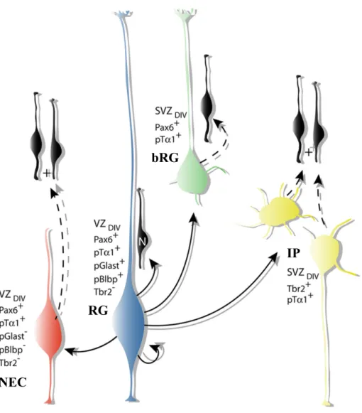
In the beginning of mouse embryo development, the neuroepithelial cell (NECs) perform proliferative symmetric divisions, allowing the exponential growth of progenitor pool (Noctor, Martínez-Cerdeño, Ivic, & Kriegstein, 2004). Then NECs transform into radial glia progenitors (RGs) (figure 5) by losing tight junctions but maintaining adherents junctions and initiating the expression of astroglial cell markers (Hatakeyama et al., 2004). RGs are bipolar cells whose cell body is located in the ventricular zone (VZ) and fibers span the width of the cortex (Rakic, 1972). From E10.5, the first population of post-mitotic neurons, namely Cajal–Retzius (CR) cells, are generated from the border of neocortex, migrate through the marginal zone and give rise the layer I of the cortex (Bielle et al., 2005; Takiguchi-Hayashi et al., 2004; Valverde, De Carlos, & López-Mascaraque, 1995). Very soon after, around E11, the RGs start asymmetric divisions, keeping one progenitor in the VZ and generating one intermediate progenitor (IP) or one neuron (Noctor et al., 2004). IPs delaminate into the subventricular zone (SVZ) and divide at basal positions. IPs have limited proliferative potential. After a small number of proliferative divisions, IPs divide symmetrically and generate two neurons (figure5). In ferrets and primates, the vast majority of IPs perform proliferative divisions and the they undergo multiple rounds of proliferative divisions before generating neurons (Betizeau et al., 2013; Fietz et al., 2010).
RGs through the indirect IPs pathway, transiently amplify the capacity of projection neurons production (Noctor et al., 2004; Wu et al., 2005). During evolution, the emergence of another type of progenitor, basal radial glial cells (bRGs) who keep one basal process and that they divide in the SVZ, contribute to the expansion of the neocortex in mammals. Unlike IPs, these basal progenitors with glial characteristics are capable of self-renewal, performing asymmetric divisions to maintain one bRGs and add one neuron or IP (Franco & Müller, 2013).
New born neurons either directly from RGs or indirectly through IPs or bRGs migrate radially along RGs fibers into the cortical plate (CP) (Noctor, Flint, Weissman, Dammerman, & Kriegstein, 2001; Rakic, 1972). At the end stage of neurogenesis,
14
once neuron radial migration is complete, RGs lose neurogenesis capacity and start producing oligodendrocytes or astrocytes (figure5) (Misson, Takahashi, & Caviness, 1991).
Figure 5. Progenitors and their divisions in cortical neurogenesis. The main types of neuronal progenitors: neuroepithelial cell, intermediate progenitors and basal radial glial cell are represented as well as differentiated cells from their division over time. Progenitors mainly found in primates are represented in the dashed rectangle. Adapted from Paridaen & Huttner, 2014.
The development starts from a monolayer of NECs in VZ that expand tangentially in surface and in thickness (Williams & Price, 1995). From E12 to E14 NECs differentiate into RGs initiating the expression of astroglial cell markers: Brain lipid-binding protein (BLBP) and Glast promoters (Hatakeyama et al., 2004). Within the cell cycle the nuclei of NECs and RGs undergo stereotyped movement called interkinetic nuclear migration. Their nuclei migrate radial away from the ventricular
Symmetric
proliferative NEC – RGtransition
NEUROGENESIS Asymmetric
neurogenic Additional NPC types (e.g., mammalian neocortex)
Up p er la ye rs Deepe r la ye rs Inte r-mediat e zo n e (O u te r) Su bv en tr ic u la r zo n e Ve n tr ic u la r zo n e Neurona l l ay er s/ C or tica l plat e LATE NEUROGENESIS PRE-NEUROGENESIS Symmetric self-consuming NEC 2 NEC 2 Macroglia Time Progenitor cell types
NEC Neuroepithelial cell
RG Radial glial cell
IP Intermediate progenitor
bRG Basal radial glial cell Differentiated cell types
DL Deep layer neuron
UL Upper layer neuron
m Macroglia (oligodendrocyte/astrocyte) RG RG 1 bRG 1 bRG bRG 1 RG 1 Neuron + RG RG RG 1 RG 1 IP 1 IP 1 IP + + + 2 Neurons 2 Neurons IP 1
15
surface during G1 phase, reach maximum movement at S phase and then move back to the apical side during G2 to divide at the ventricular surface (figure 8A) (Sauer, 1935)
There are two main types of progenitors in the SVZ: bRGs and IPs. The bRGs had first been identified in ferret and human (Fietz et al., 2010; D. V. Hansen, Lui, Parker, & Kriegstein, 2010). Soon after, several research groups also found them in rodents’ developing cortices but with a much lower frequency (Shitamukai, Konno, & Matsuzaki, 2011; Xiaoqun Wang, Tsai, LaMonica, & Kriegstein, 2011). bRGs are generated from RGs losing the apical process, but keeping the basal process that reaches the pia membrane and retaining epithelial features characteristic. NECs, RGs and bRGs are self-renewable progenitors that express markers Pax6, Sox2 and Nestin (Fietz et al., 2010; D. V. Hansen et al., 2010). IPs show either multipolar shape in SVZ or ‘short radial’ in VZ (Kowalczyk et al., 2009). IPs divide symmetrically either into 2 IPs or 2 neurons. All types of IPs express marker Tbr2, while neurogenic IPs lose the expression of Pax6 (Figure6) (Noctor et al., 2004; Wu et al., 2005).
16
Figure 6. Molecular profiles and morphologies of different types of mouse cortical precursors and their lineage relationships. Adapted from Tyler & Haydar, 2013.
4.2 Cortical progenitors cell cycle duration
The cell cycle has long been considered identical in all different types of cortical progenitor cells (Cai, Hayes, & Nowakowski, 1997), however, more detailed studies of the cell cycle have identified that the duration of the cell cycle varies between the different progenitor types, but also, within a a single progenitor population, as a function of time in development . In particular, the rapid division rate observed in early progenitors is attributed to a significantly short cell cycle caused by short G1 phase. This regulation of the cell cycle is limited in pluripotency progenitors and prevent differentiation (Lange & Calegari, 2010; Orford & Scadden, 2008; Singh &
J Neurosci. Author manuscript; available in PMC 2013 September 27. RG
bRG
IP
17
Dalton, 2009; White & Dalton, 2005). Studies have shown that the cell cycle duration of RGs increases through the development (Calegari, Haubensak, Haffner, & Huttner, 2005; T. Takahashi, Nowakowski, & Caviness, 1995). Cell cycle duration enlarged after the switching from proliferative division to neurogenic division (Dehay & Kennedy, 2007; Salomoni & Calegari, 2010). Several studies have shown that the main contribution of this enlargement is longer G1 phase duration in a neurogenic division compared with in a proliferative division (Arai et al., 2011; Calegari et al., 2005; Lukaszewicz, Savatier, Cortay, Kennedy, & Dehay, 2002). Manipulations the duration of G1 phase in mouse influence the progenitors division preference: an increase in the G1 phase correlates with an increase in neurogenic divisions and, conversely, a reduction in the duration of the G1 phase, with an increase of proliferative divisions (Calegari & Huttner, 2003; Lange, Huttner, & Calegari, 2009; Pilaz et al., 2009).
Thanks to advances in experimental techniques of BrdU incorporation, the duration of each cycle phase of the main progenitor cells that populate VZ and SVZ in the mouse on E14.5 days has been established (figure 7) (Arai et al., 2011). This study has shown that APs have a shorter cell cycle time than BPs, in particular by reducing the length of the G1 phase. In addition, the duration of the S phase is greatly increased in progenitor cells that begin proliferative rather than neurogenic division.
Figure 7. Parameters of the cell cycle of progenitor cells at the peak of neurogenesis. The duration of the cell cycle and especially the G1 phase of apical progenitor cells is shorter than the basal progenitor cell cycle time. Within these populations, proliferating progenitor cells (Tis21-GFP-) have a longer S-phaseduration than neurogenic progenitor cells (Tis21-GFP +). Adapted from Arai et al., 2011.
18
Pyramidal neurons are generated sequentially from RGs in VZ or indirectly through IPs or bRGs in SVZ and migrate radially into CP successive waves. The earlier-born neurons will be placed at the level of the basal surface of the CP. The later-born neurons will cross this first layer of post-mitotic neuron to place themselves at the surface. This "inside-out" process evidenced by Angevine et al, in 1961 performing pulses of tritiated thymidine at different stages of mouse corticogenesis and by Rakic et al, in 1974 in rhesus macaque. Radioactive tritiated thymidine is incorporated by progenitors in S phase, will be inherited by their progeny and only remain undiluted in cells that have not undergone subsequent division thus labeling their birthdate (Jun & Sidman, 1961; Rakic, 1974).
Two mechanisms are involved in pyramidal neuron radial migration: somal translocation and glia-guided locomotion. Translocation alone is preferred during the early stages of cortical development when the migration distance is short and does not appear to require support. Neurons that use this mechanism have a long extension to the marginal zone. Their nuclei are translocated slowly and continuously to their final position. Neurons that move by locomotion have a very short extension and migrate freely along the basal processes of the radial glia (figure 8B) (Nadarajah & Parnavelas, 2002; Nadarajah, Brunstrom, Grutzendler, Wong, & Pearlman, 2001). This migration entails complex mechanisms. It is described into 4 steps: first, the neurons generated at the VZ move radially in the SVZ. Second, they become multipolar in SVZ. At this stage, the neurons do not seem to be attached to the guidance extension of the RGs and are still able to migrate tangentially. Then, neurons stretch and attach to the extension of the adjacent RGs to migrate toward the CP, using locomotion. Finally, the neurons get off the RGs and switch to soma translocation (figure 8C) (Azzarelli, Guillemot, & Pacary, 2015).
19
Figure 8. Modes of migration in the cortex. (A) Interkinetic nuclear migration. The nuclei of neuroepithelial cells or radial glia cells occupy different positions along the apical-basal axis depending on the phase of the cell cycle. (B) Somal translocation of early-born cortical neurons. Newborn neurons lose their apical attachment and reach the PP by translocation of the soma and progressive shortening of the basal process. (C) Glia-guided radial migration of cortical neurons. Four phases of radial migration can be distinguished. Newborn neurons leave the proliferative areas (I) and reach the SVZ/IZ, where they acquire a multipolar morphology (II). After pausing in the SVZ/IZ, cells migrate toward the CP, using locomotion (III). At the end of their migration, cortical neurons switch to soma translocation (IV). MZ, marginal zone; CP, cortical plate; PP, preplate; IZ, intermediate zone; SVZ, subventricular zone; VZ, ventricular zone. Adapted from Azzarelli et al., 2015.
20
5. Mathematical modelling of brain development
The history of mathematical models in morphogenesis, from shapes of organisms (D’Arcy Thompson, 1942) to the spots on the leopard skin (Murray, 1988) is remarkably rich and fruitful for both mathematics and biology. In the narrower domain of brain development also, a number of models have been very useful for a better understanding of the mechanisms supporting the development of brain morphology.
A large part of the mathematical modeling in brain development is concerned with the organization of the brain phenotype at a macroscopic scale (full brain or brain areas). In this domain, an important landmark in mathematical models of morphogenesis Alan Turing’s 1952 celebrated article The Chemical Basis of Morphogenesis (Turing, 1952) describing how a uniform state could evolve into a non-uniform pattern during development. The latter reaction–diffusion theory of morphogenesis, has served as a basic model in theoretical biology, but also in brain macroscopic and functional phenotype (Lefèvre & Mangin, 2010; Striegel & Hurdal, 2009).
In the 1960s, Lewis Wolpert offered another conceptual definition of a morphogen and devised a model to describe basic pattern formation in development, the French Flag Model. In the French flag model, a morphogen diffuses between a source and a sink and cells decide on their "states" depending on the local morphogen concentration. Due to the existence of thresholds in the cell response, the states are discrete and in the simple model proposed by Wolpert, there are only three states represented by the different colors of the French flag (Wolpert, 1969). Homeoprotein diffusion was also studied using theoretical models, first using discrete-space models to derive mechanisms for gradient and boundary formation (Holcman, Kasatkin, & Prochiantz, 2007; Kasatkin, Prochiantz, & Holcman, 2008), and, few years after, combining Turing’s models with spatial cues, Quininao, Prochiantz and Touboul (Perthame, Quiñinao, & Touboul, 2015; Quiñinao, Prochiantz, & Touboul, 2015) showed that slow diffusion of homeoproteins can stabilize and regularize boundaries between brain areas.
In this thesis, we are interested in models of brain development at a smaller scale, in terms of number of cells generated and of the layers they belong to. A number of
21
models have also considered this question in recent years. For instance, Slater and colleagues have developed a stochastic model of neurogenesis based cell lineage tree; their model emulates a single population of multipotent progenitors and their stochastic escape from symmetric divisions (Slater, Landman, Hughes, Shen, & Temple, 2009). Barbara Finlay and collaborators have built a model consider progenitor cell-cycle duration, cell death rate, and the probability to exit symmetric division, to explain how different parameters influence neuron number and the expansion through an evolutionary perspective (Cahalane, Charvet, & Finlay, 2014; Workman, Charvet, Clancy, Darlington, & Finlay, 2013).
The model we will present in this thesis was developed with the purpose of being simple enough to be easily implementable and parameterized, yet precise enough to be fitted to actual data. Compared to existing models in the literature, the model presented in Chapters II of this thesis emulates multiple cell populations and their divisions as a function of time, either within a prescribed program or through an intrinsic clock. With this model, we were able to draw quantitative predictions on the role of the switching time between proliferative and neurogenic divisions in microcephalies. Also, contrasting with previous studies, our model was directly applied to mutant mice models of microcephalies to better understand the regulation mechanisms at play in brain development.
Very recently, new developments in mathematical modeling of neurogenesis at cell populations scales were developed. In particular, Picco and colleagues proposed a deterministic model reproducing a similar sequence of divisions of progenitor cells, and concluded that this switch from proliferative and neurogenic divisions could be used to describe the diversity of cortical phenotypes in mammalian species (Picco, García-Moreno, Maini, Woolley, & Molnár, 2018). Delving at finer temporal scales, Postel and collaborators have developed a mathematical model considering both RGs and IPs divisions depending on the precise phase within the cell cycle of each cell. This model, fitted to experimental data of mouse embryos (cell numbers at different cortical development stages), was applied to a mutant mouse model with a shortening of the neurogenic period and an increased of number IPs (Postel et al., 2018).
22
Chapter II: Modeling the neurogenesis
1. Results/Publications
1.1 Mathematical of neurogenesis based on progenitor divisions
Mathematical models of neurogenesis can provide a new tool to understand the determinants of brain architecture and size. In collaboration with Shen-Ju Chou (Taipei), we developed a simple model of the sequence of divisions and differentiations of cells during neurogenesis, to explain her experimental data on microcephaly in mice.
In detail, Nestin-cre; Lhx2 conditional knockout mice shows a significantly smaller and thinner cortex, which is associated with a precocious initiation of cortical neurogenesis. To demonstrate that this early neurogenesis can indeed account for the phenotype we observed in Lhx2 cKO, we developed a parsimonious mathematical model that simulates the sequence of divisions of progenitors during cortical neurogenesis.
The model reproduces the four main types of divisions observed during corticogenesis; namely
(i) symmetric division into two progenitors (proliferative phase);
(ii) asymmetric division into a progenitor and an intermediate progenitor, itself generating two neurons;
(iii) asymmetric division into a progenitor and a neuron (ii and iii are neurogenesis phase);
(iv) loss of capacity to generate neurons (gliogenesis phase).
Moreover, neurons generated are considered to belong to a specific layer depending on the time at which the division of progenitors occurred, according to the classical radial inside-out migration of neurons to the distinct layers. All divisions and differentiations thus occur at rates (probabilities of occurrence) that vary in time. Using that model, we first reproduced the smaller and thinner phenotype of the
23
Nestin-cre; Lhx2 conditional knockout mice. Also we analyzed the consequence of
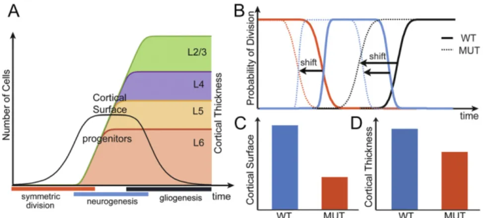
several possible timing shifts. The mathematical model provided us with further information that the duration of symmetric division (i) into two progenitors controls the number of progenitors (the cortical surface); the duration of neurogenic phase divisions (ii) and (iii) controls the number of neurons generated by each progenitor (the cortical thickness). Early initiation of gliogenesis division (iv) during the neurogenesis phase would lead to a more dramatic reduction of neuronal numbers in superficial layers than in deep layers. Together with the transition time point from deep to upper layer neurons, (iv) controls the proportion of deep and upper layer neurons numbers.
24
Lhx2 regulates the timing of β-catenin-dependent
cortical neurogenesis
Lea Chia-Ling Hsua,b,c,1, Sean Nama,1, Yi Cuid, Ching-Pu Changa, Chia-Fang Wanga, Hung-Chih Kuoa,
Jonathan D. Toubould,e, and Shen-Ju Choua,b,c,2
aInstitute of Cellular and Organismic Biology, Academia Sinica, Taipei, 115, Taiwan;bMolecular Cell Biology, Taiwan International Graduate Program,
Academia Sinica, Taipei, 115, Taiwan;cGraduate Institute of Life Sciences, National Defense Medical Center, Taipei, 114, Taiwan;dCollège de France, Centre
for Interdisciplinary Research in Biology, CNRS UMR 7241, INSERM U1050, MemoLife Paris Science Lettres, 75231 Paris, France; andeInria Paris-Rocquencourt,
Mycenae Team, 78153 Le Chesnay, France
Edited by John L. R. Rubenstein, University of California, San Francisco, San Francisco, CA, and accepted by the Editorial Board August 18, 2015 (received for review April 12, 2015)
The timing of cortical neurogenesis has a major effect on the size and organization of the mature cortex. The deletion of the LIM-homeodomain transcription factor Lhx2 in cortical progenitors by Nestin-cre leads to a dramatically smaller cortex. Here we report that Lhx2 regulates the cortex size by maintaining the cortical progenitor proliferation and delaying the initiation of neurogenesis. The loss of Lhx2 in cortical progenitors results in precocious radial glia differen-tiation and a temporal shift of cortical neurogenesis. We further investigated the underlying mechanisms at play and demonstrated that in the absence of Lhx2, the Wnt/β-catenin pathway failed to maintain progenitor proliferation. We developed and applied a mathematical model that reveals how precocious neurogenesis af-fected cortical surface and thickness. Thus, we concluded that Lhx2 is
required for β-catenin function in maintaining cortical progenitor
proliferation and controls the timing of cortical neurogenesis.
cortical neurogenesis|Lhx2|β-catenin
U
nderstanding how genetic mechanisms interact to set up a precise developmental timing is a fundamental issue in bi-ology. In the cerebral cortex, excitatory neurons are generated by progenitor cells in the dorsal telencephalon (dTel) lining the lateral ventricle. During the early developmental stages, cortical pro-genitors undergo symmetric divisions, resulting in the proliferation of progenitors and thereby allowing expansion of the developing cortex. Soon after, cortical progenitors start generating distinct types of neurons through asymmetric differentiative divisions (1–5). The precise timing of the switch from proliferative division to differentiative division is crucial to determining the number of cortical neurons, and thus the cortical size.The switch from proliferation to differentiation is reportedly regulated by the canonical Wnt signaling pathway, in which β-catenin (β-Cat) is the major downstream effector. In the absence of Wnt signaling, β-Cat is phosphorylated by glycogen synthase kinase 3 and targeted for proteosome degradation. Once Wnt ligands bind to the Frizzled-Lrp5/6 receptors, the activity of glycogen synthase kinase 3-Axin-APC (adenomatous polyposis coli) destruction complex is inhibited. As a consequence, β-Cat accumulates in the cytoplasm, translocates to the nucleus, and activates downstream gene tran-scription together with the lymphoid enhancer-binding factor (LEF)/ T-cell factor (TCF) transcription factors (6). Overexpression of the stabilized, N-terminally truncated form of β-Cat in cortical pro-genitors during early neurogenesis promotes their overproliferation (7, 8), whereas inactivation of β-Cat in the cortex promotes neuro-genesis (9, 10). However, stabilized β-Cat was also shown to pro-mote cortical progenitor differentiation (11). Thus, it has been proposed that Wnt/β-Cat signaling promotes proliferation and differentiation of cortical progenitors at early and late de-velopmental stages, respectively (12). This raises the essential and largely open question of how Wnt/β-Cat regulates cortical progenitor proliferation and differentiation.
The LIM-homeodomain transcription factor Lhx2 plays an important role in cortical development. In the neocortex, Lhx2 is
expressed by neocortical progenitors within the ventricular zone (VZ) of the dTel throughout cortical neurogenesis. Lhx2 was shown to play stage-specific roles determining the fate of cortical pro-genitors during early stages of corticogenesis (13–15). Further, the mutant mice with Lhx2 deleted in the neural progenitors at embry-onic day 11.5 (E11.5) by Nestin-cre exhibited a significantly smaller neocortex than WT mice (16). Overall, these previous studies showed that Lhx2 is important for the determination and mainte-nance of neocortical progenitors, although how Lhx2 regulates the proliferation and differentiation of cortical progenitors is unclear.
In this study, we identified a role for Lhx2 in regulating the function of the Wnt/β-Cat signaling pathway in maintaining progenitor proliferation. The deletion of Lhx2 in cortical pro-genitors leads to a temporal shift of neurogenesis. We found that by regulating how cortical progenitors respond to Wnt/β-Cat signaling, Lhx2 regulates cortex size. By delaying neuronal dif-ferentiation and maintaining progenitor symmetric division, Lhx2 allows a suitable increase of the numbers of cortical progenitors needed to develop a proper cortex.
Results
Lhx2 Regulates the Timing of Sequential Cortical Neurogenesis.The
deletion of Lhx2 in the cortical progenitors at E11.5 by Nestin-cre in Lhx2 conditional knockout (cKO, Lhx2f/f:Nestin-cre) leads
to a significantly smaller and thinner cortex, although all six
Significance
The cerebral cortex is the most highly evolved structure in the human brain. Generating the correct number and types of neurons is crucial for brain function. We show a central role of the Lhx2 homeoprotein in this task: deleting Lhx2 in cortical progenitors leads to a temporal shift of neurogenesis initiation, resulting in a much smaller cortex with decreased numbers of neurons in all cortical layers. Further, we found that Lhx2 is required for the Wnt/β-catenin pathway to maintain progenitor proliferation. Using a parsimonious mathematical model, we demonstrated that such disruptions of neurogenesis timing are enough to explain the cortical size and thickness modifications observed. Our findings enlighten how neurogenesis timing is regulated molecularly and how it affects cortical size and organization.
Author contributions: J.D.T. and S.-J.C. designed research; L.C.-L.H., S.N., Y.C., C.-P.C., C.-F.W., J.D.T., and S.-J.C. performed research; H.-C.K. contributed new reagents/analytic tools; L.C.-L.H., S.N., Y.C., and S.-J.C. analyzed data; and J.D.T. and S.-J.C. wrote the paper.
The authors declare no conflict of interest.
This article is a PNAS Direct Submission. J.L.R.R. is a guest editor invited by the Editorial Board.
1L.C.-L.H. and S.N. contributed equally to this work.
2To whom correspondence should be addressed. Email: schou@gate.sinica.edu.tw.
This article contains supporting information online atwww.pnas.org/lookup/suppl/doi:10.
1073/pnas.1507145112/-/DCSupplemental.
www.pnas.org/cgi/doi/10.1073/pnas.1507145112 PNAS | September 29, 2015 | vol. 112 | no. 39 | 12199–12204
NEUR
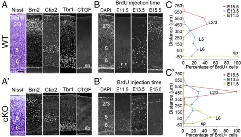
25 cortical layers were generated in the cKO, and the relative posi-tions of cortical neurons in each were generally normal (16). To investigate the mechanisms for Lhx2 to regulate cortical neuro-genesis, we first examined the timing of neurogenesis by injecting BrdU into pregnant mothers when embryos were at different de-velopmental stages and analyzing the distribution of BrdU-labeled neurons at postnatal day 7 (P7), when the six cortical layers are easily distinguishable. To define each cortical layer in WT (Lhx2f/+
or Lhx2f/f) and Lhx2 cKO cortex, we analyzed the expression of
cortical layer markers in P7 brains (17, 18). We found that in both WT and cKO cortex, the CTGF (connective tissue growth factor)-expressing subplate neurons were distributed in a single layer. We used Brn2, Ctip2, and the Tbr1 expression domain to determine layers (L) 2/3, 5, and 6, respectively (Fig. 1 A and A′). We found that the number of cells in all layers all significantly decreased in cKO cortex (SI Appendix, Fig. S1). In WT cortices, almost all neurons generated at E11.5 contributed to the subplate, whereas most neurons generated at E13.5 and E15.5 contributed to the deep (L6, L5) and superficial (L2/3) layers, respectively (Fig. 1 B and C) (17). In cKO cortices, although the inside-out organization was maintained, we found that neuronal birth dates shifted to earlier points. For example, in the cKO brain, most neurons born at E11.5 contributed to layer 6, and most neurons born at E13.5 contributed to superficial layers. In cKO cortices, the number of neurons generated at E15.5 dramatically decreased relative to WT, and these neurons were located superficially in layer 2/3 (Fig. 1 B′ and C′). BrdU birth dating analyses suggested that Lhx2 deletion in cortical progenitors altered the timing of neurogenesis. To confirm this, we examined expression of neuronal markers at E13.5. We observed comparable numbers of Reelin-positive Cajal-Retzius cells in WT and cKO cortices, whereas the cortical plate, which is labeled by Tbr1, Ctip2, and Satb2, was relatively thicker in the cKO cortex (SI Appendix, Fig. S2). Overall, these findings confirmed that cortical neurons are produced earlier in Lhx2 cKO.
Neurogenesis and Radial Glia Differentiation Initiate Earlier in Lhx2 cKO.To demonstrate that the loss of Lhx2 leads to increased neurogenesis during early cortical development, we analyzed the
production of divisions by cortical progenitors derived from E13.5 WT and cKO dTel. We cultured progenitors at clonal density for 24 h to allow them to divide once to form two-cell pairs. We ob-served significantly fewer two-cell pairs formed from cells from the cKO compared with WT dTel (Fig. 2A). We also stained the two-cell pairs with antibodies against TuJ1 (neuron-specific class III b-tubulin) to label neurons and Sox2 (sex-determining region Y-related HMG box 2) to label progenitors. Pairs were identified as symmetric proliferative (P–P) divisions to form two Sox2-positive progenitors, symmetric neurogenic (N–N) divisions to form two TuJ1-positive neuronal cells, or asymmetric (P–N) divisions with one Sox2-positive and one TuJ1-positive cell. In WT and cKO cortices, the percentage of N–N pairs was comparable. In WT cortex, the majority of the two-cell pairs were P–P, indicating pro-liferative division of most progenitors at this stage. We found that in cKO cortex, the percentage of P–P and P–N pairs was signifi-cantly decreased and increased, respectively (Fig. 2B). This finding agreed with our previous report that Lhx2 deletion by Nestin-cre enhances neurogenesis, with an increased number of cortical pro-genitors exciting cell cycle at early developmental stages (16).
To provide additional evidence to support that the deletion of Lhx2 leads to increased neurogenic progenitors, we examined the expression of neurogenic progenitor markers, such as Tis21 (Btg2) and Hes6 (19–21), in E12.5 WT and cKO cortices. We found significantly increased expression of Tis21 and Hes6 in the dTel VZ in Lhx2 cKO relative to WT (Fig. 2 C, C′, D, and D′ and
SI Appendix, Fig. S3), an effect consistent with increased neu-rogenesis in the cKO.
Our results suggested that neurogenesis initiates earlier in Lhx2 cKO, and thus we further examined whether the differentiation of radial glia, the neurogenic progenitors, occurs earlier in cKO. In the developing dTel, we defined radial glial cells (RGCs) with RGC markers Blbp (brain lipid-binding protein; or Fabp7, fatty acid binding protein 7), Glast (glial high-affinity glutamate trans-porter; or Slc1a3, solute carrier family 1 member 3), and TnC (Tenasin-C) (22–27). We found that all these RGC markers are precociously up-regulated in the VZ of cKO dTel. (Fig. 2 E, E′, F, and F′ andSI Appendix, Figs. S3 and S4). The precocious
Fig. 1. Lhx2 regulates the timing of sequential neurogenesis. (A and A′) Immunostaining for markers of specific cortical layers on coronal sections of P7 WT (A) and cKO (A′) cortices. In both WT and cKO samples, six neuronal layers are present, including the Brn2-expressing L2/3 (2/3), Ctip2-expressing L5 (5), Tbr1-expressing L6 (6), and CTGF-expressing subplate (sp). (B and B′) Immunostaining for BrdU on coronal sections of P7 WT (B) and cKO (B′) cortices. BrdU was injected into pregnant mothers at E11.5, E13.5, or E15.5. BrdU injected at E11.5 labeled neurons distributed in the subplate in WT mice (arrowheads), but BrdU-labeled cells were detected in layer 6 in cKO mice. BrdU injected at E13.5 labeled neurons concentrated in layer 6 in WT mice, but E13.5 BrdU-labeled cells were spread to L2/3 in cKO mice. BrdU injected at E15.5 labeled many neurons in L2/3 in WT, but only a few superficially in L2/3 in cKO. (C and C′) Quantification of results from B and B′ indicating the location of BrdU-positive neurons in P7 WT and cKO cortices labeled at indicated times (n = 3). In a 100-μm-wide radial column, BrdU-labeled neurons were counted at 100-μm intervals from the subplate (defined as 0) to the pial surface. The percentage of BrdU-positive cells was calculated by determining the number of BrdU-positive cells in a 100 × 100 μm box divided by the total number of BrdU-positive cells in the entire radial column. (Scale bar, 100 μm.)