Brain plasticity related to the consolidation of motor
sequence learning and motor adaptation
Karen Debasa,b, Julie Carriera,b,c, Pierre Orbana, Marc Barakata,b, Ovidiu Lungua, Gilles Vandewallea,c,
Abdallah Hadj Tahara, Pierre Belleca, Avi Karnid, Leslie G. Ungerleidere,1, Habib Benalif, and Julien Doyona,b,f,1 aFunctional Neuroimaging Unit, Centre de Recherche de l’Institut Gériatrique, Université de Montréal, Montreal, QC, Canada H3W 1W5;bCentre de Recherche en Neuropsychologie et en Cognition, Department of Psychology, University of Montreal, Montreal, QC, Canada H3C 3J7;cCentre d’Étude du Sommeil et des Rythmes Biologiques, Hôpital du Sacré-Cœur de Montréal, Montreal, QC, Canada H4J 1C5;dLaboratory for Functional Brain Imaging and Learning Research, The Brain-Behavior Center, University of Haifa, Haifa 31905, Israel;eLaboratory of Brain and Cognition, National Institute of Mental Health, National Institutes of Health, Bethesda, MD 20892; andfUnité Mixte de Recherche-S 678, Institut National de la Santé et de la Recherche Médicale/ University of Paris 6, Centre Hospitalier Universitaire Pitié-Salpêtrière, 75634 Paris, France
Contributed by Leslie G. Ungerleider, September 7, 2010 (sent for review May 20, 2010)
This study aimed to investigate, through functional MRI (fMRI), the neuronal substrates associated with the consolidation process of two motor skills: motor sequence learning (MSL) and motor adapta-tion (MA). Four groups of young healthy individuals were assigned to either (i) a night/sleep condition, in which they were scanned while practicing afinger sequence learning task or an eight-target adaptation pointing task in the evening (test) and were scanned again 12 h later in the morning (retest) or (ii) a day/awake condi-tion, in which they were scanned on the MSL or the MA tasks in the morning and were rescanned 12 h later in the evening. As expected and consistent with the behavioral results, the functional data revealed increased test–retest changes of activity in the stria-tum for the night/sleep group compared with the day/awake group in the MSL task. By contrast, the results of the MA task did not show any difference in test–retest activity between the night/sleep and day/awake groups. When the two MA task groups were combined, however, increased test–retest activity was found in lobule VI of the cerebellar cortex. Together, thesefindings high-light the presence of both functional and structural dissociations
reflecting the off-line consolidation processes of MSL and MA.
They suggest that MSL consolidation is sleep dependent and reflected by a differential increase of neural activity within the corticostriatal system, whereas MA consolidation necessitates ei-ther a period of daytime or sleep and is associated with increased neuronal activity within the corticocerebellar system.
functional MRI
|
memory consolidation|
motor learning|
sleep|
wakefulnessM
otor memory consolidation refers to the“off-line” process by which a memory trace initially labile becomes more robust and fixed. Accumulated evidence has shown that sleep contributes to this physiological process, but that its effect depends on the nature of the motor learning demands (see ref. 1 for a review). For example, several researchers have demon-strated that the consolidation of a newly learned sequence of movements (motor sequence learning, MSL) acquired through explicit mechanisms is sleep dependent, as performance gains have been observed after nocturnal sleep, but not after the simple passage of time (2–5). By contrast, the role of sleep in the consolidation of skills, in which subjects have to adapt to sen-sorimotor perturbations (motor adaptation, MA), has been more controversial. Whereas Huber and colleagues (6) have reported that better performance on such a task was only observed in subjects who slept following training, Doyon et al. (2) have re-cently demonstrated that similar performance gains could be seen after a night of sleep or an equivalent period of daytime.Numerous studies have previously demonstrated that the stri-atum, cerebellum, and motor-related cortical regions play a crit-ical role in the acquisition of MSL and MA skill behaviors (7–9). Investigations of the brain regions mediating the consolidation process of these motor abilities, and how they relate to sleep,
however, have revealed inconsistent findings. Whereas sleep-dependent posttraining improvement in performance on MSL has been associated with a reduction in brain activation in pre-frontal, premotor, and primary motor cortex (M1) areas, as well as increased activity in parietal regions (10), a different pattern of results characterized by increases of activation in M1, medial prefrontal, hippocampus, and cerebellum, along with a decrease of activity in parietal cortices, have also been reported (11). Furthermore, the neural correlates associated with the consoli-dation of MA skill during sleep have never been fully studied. Using electroencephalography (EEG), it was suggested that slow wave activity localized in the parietal cortex contributes to the consolidation of a rotation adaptation task during sleep (6). However, EEG recordings do not allow the measurement of subcortical activity changes in purportedly critical structures like the cerebellum.
The goal of this study was to determine, using functional MRI (fMRI), the cerebral structures affected by the off-line consoli-dation of both MSL and MA skills, and how they relate to sleep vs. passage of time. To do so, we compared activation maps between night/sleep and day/awake groups of young healthy subjects that were asked to execute motor tasks designed to measure one of these two forms of motor skills. The night/sleep groups were thus trained and scanned in a posttraining session after having reached asymptotic performance on a version of a five-item finger sequence learning task (MSL) or an eight-target reaching task (MA) in the evening. They were then retested in the morning after a 12-h delay encompassing a night of sleep in the laboratory. The day/awake groups followed the same procedure, except that they were trained and scanned on either of these two tasks in the morning and retested 12 h later in the evening (Fig. 1). On the basis of previous behavioral (3, 4) and imaging data (9), we hypothesized that MSL would elicit sleep-dependent changes in the corticostriatal system, whereas MA would produce sleep-independent changes in the corti-cocerebellar network.
Results
Behavioral Data: Motor Sequence Learning. Performance on this
task was measured using the execution time per sequence (TpS) for each block of trials. All subjects improved during the training session and reached asymptotic performance. (For additional information regarding this initial session, see SI Text as only
Author contributions: J.C., A.H.T., A.K., L.G.U., H.B., and J.D. designed research; K.D. performed research; K.D., P.O., and O.L. analyzed data; and K.D., M.B., G.V., P.B., and J.D. wrote the paper.
The authors declare no conflict of interest.
1To whom correspondence may be addressed. E-mail: [email protected] or [email protected].
This article contains supporting information online atwww.pnas.org/lookup/suppl/doi:10.
1073/pnas.1013176107/-/DCSupplemental. PSYCHOL OGICAL AND COGN ITIVE SC IENCES
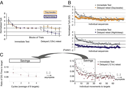
results collected while scanning are discussed below.) To test for the amount of gains in performance following consolidation, at test comparing normalized data of thefirst block in the delayed retest session between the night/sleep and day/awake groups was used. This approach allowed us to minimize the possible in-fluence of testing and retesting subjects at different times of day. As expected, the night/sleep group at retest was significantly faster than the day/awake group t(38) = 12.4, P < 0.0001 (Fig. 2A). Compared with its baseline performance in the evening, the night/sleep group started off in the morning with a decreased TpS of 83 ms (0.47 SD) in the delayed retest session, whereas the day/awake group started off with a nonsignificant increase of 24 ms TpS in the evening. In addition, to make sure this pattern of results was not driven by averaging across trials within thefirst block of the retest session (12), the subjects’ individual times to execute each sequence were analyzed (Fig. 2B). The TpSs were normalized on the lastfive blocks of the testing session, and the subject’s performance on the first five sequences of the retest session was compared between the night/sleep and day/awake groups. Results of the t test revealed again a significant differ-ence between the night/sleep group (−0.23 SD below their baseline) and the day/awake group (0.70 SD above their base-line) [t(22) = 2.14, P = 0.04], hence demonstrating that subjects that slept experienced spontaneous gains in performance (con-solidation) compared with the group that did not.
Behavioral Data: Motor Adaptation.The main measure of
perfor-mance on the MA task consisted of a ratio between the accuracy and the time taken to reach a target. Accuracy was calculated in pixel units with the difference in surface (DS) between the subject’s actual trajectory and the ideal one that had to be fol-lowed. Both the night/sleep and day/awake groups demonstrated
learning across the training session and reached asymptotic performance (SI Results). The savings, a behavioral reflection of the consolidation process in motor adaptation learning (13, 14), were assessed by comparing the amount of execution that sub-jects required before reaching asymptotic performance in both test and delayed retest sessions. We found that for both night/ sleep and day/awake groups, there were significant savings from the immediate test to the delayed retest session, as evidenced by a significant interaction between cycles (i.e., the average per-formance on eight targets) and sessions (immediate posttraining test and retest) (F(3,22)= 4.30, P = 0.03) (Fig. 2C), hence re-vealing a faster rate of relearning in the delayed retest session and an overall 16% increase in performance. Importantly, there was no session× group interaction, nor any difference between groups (F(1,22)< 1.77, P > 0.20), suggesting a similar amount of savings for both the night/sleep and day/groups. Altogether, these results suggest that time alone was sufficient to elicit sav-ings (consolidation) (13, 14).
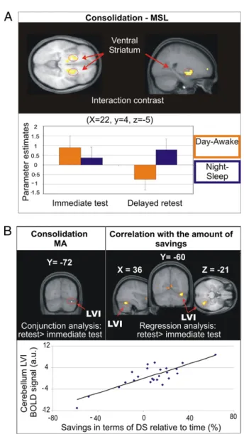
fMRI Data: Motor Sequence Learning.Execution of the sequence at an asymptotic performance level during the immediate post-training test, for both night/sleep and day/awake groups, was associated with increased activity relative to baseline in bilateral M1, right sensorimotor cortex, cerebellum (lobules IV, V on the left side, VIII bilaterally as well as VI and CrusII on the right), and ipsilateral putamen (at the junction of the caudate nucleus) (Table S1). To test for possible time of day effects, the imme-diate posttraining test of the day/awake and night/sleep groups were compared (i.e., morning vs. evening), yet no region showed greater activity in either group. Sleep-dependent consolidation effects on the MSL task were then assessed by measuring the difference in activations between the delayed retest and imme-diate posttraining test sessions in the night/sleep group com-pared with the day/awake group (night/sleepdelayed> immediate> day/awakedelayed> immediate). As predicted, significant activations were found bilaterally in the basal ganglia (Fig. 3A), and more specifically in the globus pallidus (Gp) and putamen ventrally. Other less extended activated regions at PFDR-corr < 0.05 in-cluded the left temporal pole, right superior temporal gyrus, and left superior and middle frontal gyrus (Table 1). Activity in the right putamen further showed that from the immediate to the delayed retest, activation in that region slightly increased for the night/sleep group, but significantly decreased for the day/ awake group (Fig. 3A). Note that a global conjunction analysis of the delayed > immediate test contrast revealed no region for which both night/sleep and day/awake groups had common in-creased activity, further suggesting that sleep had a differential effect on the off-line consolidation process. Finally, multiple regression analyses revealed significant correlations between the change in brain activity recorded in the cerebellum (lobule VIII and Crus I) from the test to the retest session and the subject’s gain in performance after sleep (Table S2).
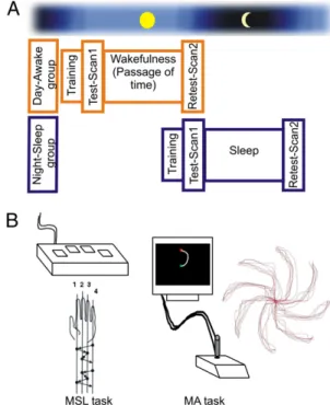
fMRI Data: Motor Adaptation.Execution of the MA task during the immediate posttraining test in both groups was associated with increased activity relative to baseline in cortical and subcortical regions including the M1, supplementary motor area (SMA), cerebellum, primary somatosensory cortex (S1), basal ganglia, and thalamus bilaterally (Table S1). We also tested the possi-bility that time of day could influence the blood oxygenated level dependent (BOLD) signals during thefirst scanning session, but again the latter analysis did not reveal any significant effect, hence suggesting that the pattern of activation associated with early MA learning was not related to circadian effects. Fur-thermore, the group× session interaction revealed no significant brain activity (Table 1), suggesting that the consolidation process yielded similar activated maps in both groups, regardless of the type of delay (i.e., sleep or passage of time). Consolidation was Fig. 1. (A) Experimental design. In orange, the day/awake group was tested
and retested in the scanner while staying awake during the 12-h delay be-tween sessions. In blue, the night/sleep group was tested around 9:00 PM and slept in the laboratory while polysomnographic measures were recorded and retested approximately 2 h after waking up, around 9:00 AM. (B) Illus-trations of the apparatus and order offinger presses used in the motor se-quence learning (MSL) task, as well as the setup and performance of a representative block of trials in the motor adaptation (MA) task.
therefore assessed through a global conjunction analysis of the two groups in the delayed> immediate contrast. This contrast revealed that activity in the right cerebellum (lobule VI) (X = 36, Y = −72, Z = −21), was significantly greater in the retest than in the test session (svc10mm, PFwe-corr = 0.04) (Fig. 3B). Finally, multiple regression analyses revealed that this region of the cerebellum (lobule VI), albeit slightly anterior (X = 36, Y = −60, Z = −21), correlated with the amount of savings found between the two sessions in the two groups (Fig. 3B andTable S2). Discussion
This study aimed at identifying the brain regions associated with the off-line consolidation process of two forms of motor skill learning: MSL and MA. As expected, the behavioral re-sults yielded significant spontaneous gains in performance on an explicitly known MSL task that were observed following a night of sleep, but not after an equivalent diurnal period (2– 5). Importantly, such improvements were present as soon as participants began executing the sequence task in the retest session. This suggests that performance gains were not due to data averaging across trial blocks, hence masking a simple end product of continued learning within that session (12), but rather that they reflect the expression of a real motor memory consolidation process.
By contrast, the amount of savings observed on the MA task was similar in both night/sleep and day/awake groups, thus im-plying that time alone is necessary, but sufficient, for consoli-dation of that memory trace to occur (2, 13, 15). The latter results do not corroborate those of Huber and colleagues (6) who reported evidence of a sleep-dependent consolidation effect on a rotation adaptation task, but this apparent discrepancy may be explained by methodological differences in the two studies, as they differed in terms of their demands on upper-arm effectors and extent of kinematic adaptation needed.
Although the design of the present study does not allow us to exclude entirely the possibility of circadian influences on our
pattern of results, most of the evidence to date suggests that such a confounding factor does not seem to play a major role in motor memory consolidation. Indeed, our own behavioral and imaging data did not yield any significant functional difference between thefirst evening and morning sessions. Gains in performance on motor sequence learning have also been demonstrated after di-urnal sleep (3) or following an afternoon nap of 90 min (16), hence suggesting further that the present results are probably not due to circadian effects, but rather to the sleep-dependent con-solidation process of a motor memory trace.
Motor Memory Consolidation: Functional Imaging Data.The present study demonstrates that, as predicted, the effects of sleep and passage of time on motor memory consolidation of skills mea-suring MSL or MA are associated with distinct neuronal changes. For the MSL task, activity within the basal ganglia (and in the putamen, in particular) was significantly greater during the retest session in subjects who slept than in those who did not. Brain regions involved in the execution of the task after sleep included the striatum, as well as the cerebellum (lobules IV, V, and VIII), bilateral primary motor cortex, the right sensory cortex, and the SMA. Yet, only the striatal activity was strongly influenced by a night of sleep relative to daytime. Because scanning did not take place during sleep, it is not possible to state whether the striatum has an active role during sleep or appears as a consequence of it. Nevertheless, our results suggest that this structure does not only reflect the consolidation of a newly learned sequence of movements (6, 7, 17, 18), but that this physiological process is facilitated by sleep. Indeed, increases of activity within the striatum have previously been related to the acquisition of wrist movement sequences (19–21) per se, as well as the learning of MSL, as opposed to the mere increase in speed of finger movements (22). Moreover, such increase in striatal activity has also been seen following motor memory consolida-tion when sleep (10), or a 24-h delay including sleep is present after initial learning (23). Altogether, thesefindings suggest that Fig. 2. Behavioral results. (A) Behavioral results of the MSL task. Performance of the two groups across blocks in both immediate and retest sessions are illustrated. (B) Illustration of the groups’ performance at the individual sequence level, with each curve representing a session. (Upper) The performance for the day/awake group. (Lower) The night/sleep group. (C) Behavioral results for the MA task. The y axis represents the ratio of DS relative to the time taken to reach each target. Left graph shows averaged subject’s performance for each cycle. Right graph shows performance on each individual movement to a single target. Each curve represents a session. Savings occurred within thefirst four cycles of the retest session. Yet there was no significant interaction or between-group differences (night/sleep vs. day/awake) with respect to the amount of savings, and thus the results of both between-groups were pooled together to look at test– retest differences (consolidation).
PSYCHOL OGICAL AND COGN ITIVE SC IENCES
sleep is critical for the striatum to assure its role following the consolidation of MSL.
Contrary to other researchers (23), our results did not reveal any correlation between the amount of performance gains seen after sleep and the level of BOLD activity within the basal gan-glia. Instead overnight gains were positively correlated with bi-lateral activity within the cerebellum (lobule VIII A and B, Crus I). Although seemingly contradictory, the latter results are thought to reflect the interrelationship existing between the subject’s level of brain activation in those regions and the speed of execution of the motor sequence, rather than the process of
consolidation per se. Indeed activity in lobule VIII of the cere-bellum has been related to the execution of discrete movements (24) and to the motoric implementation of a learned sequence, but not to the learning process as such (22).
In contrast to the sleep-dependent changes in the basal ganglia seen after MSL consolidation, sleep-independent functional changes in lobule VI of the cerebellum were observed following consolidation of the newly acquired adapted motor skill. More-over, activity within this lobule was strongly correlated with the amount of savings that subjects exhibited in the retest session. These findings are consistent with our behavioral data, which demonstrated that both sleep and daytime produce better per-formance in the retest session. They also suggest that sleep does not bear any additional effects on the consolidation process of this form of motor learning. Such results further confirm the view that this region of the cerebellum is not only involved in the building and storing of an internal model necessary to execute the MA task efficiently (25, 26), but that it is also related to the consolidation process of this skill (8). Finally, although increased activity in the right parietal cortex was found during execution of the MA task before and after the 12-h delay, no consolidation-related activity was observed in that region. This is again in-consistent with results from Huber et al. (6), but can be due to the differences in techniques used (fMRI vs. EEG) and the physiological state (awake during the retest session vs. asleep) during which signals were recorded.
The fact that striatal activity related to MSL increased after sleep, but decreased after the passage of time, supports the notion that consolidation of a new motor sequence may rely on the covert reactivation, during sleep, of the brain regions involved in learn-ing the motor skill in thefirst place (27). On the basis of this hypothesis, sleep would allow a“replay” of the neural represen-tation for the sequence mediated by the striatum, thus enhancing the robustness of the initial motor memory trace. Such in-terpretation is consistent with rodents’ work, which showed that reactivation during posttraining sleep can be observed in the ventral striatum after animals are trained on a reward-searching procedural task (28), as well as with previous positron emission tomography studies in humans, which have demonstrated that regional activity recorded during training on a probabilistic serial reaction-time task is reexpressed in the posttraining night (27, 29). Yet cerebral signs of memory reactivation are not limited to sleep, as they have also been observed during postlearning periods of wakefulness in both animals and humans. Reverse replaying of cell activity immediately after maze learning expe-rience has been shown in rodents (30), whereas reorganization of motor sequence related cerebral activity during awake post-training periods has also been demonstrated during an unrelated, attention cognitive task (oddball) in humans (31). The latter Fig. 3. (A) Functional data related to the MSL task. (Upper) Brain regions
showing greater activity in the retest, compared with the immediate post-training test session for the night/sleep group over the day/awake group. The functional data are presented over an average of the anatomical scans (n = 23) acquired in the whole group of subjects. (Lower) Bar graph of beta values from the local maxima in the right putamen. (B) Imaging results for the MA task shown on the averaged brain (n = 24). (Upper Left) Results of both day/awake and night/sleep groups combined, showing an increase of activity from the immediate posttraining to the retest session in lobule VI of the right cerebellum. (Upper Right) Results of the correlation analysis between the increase in activity from the immediate posttraining test to the retest session and the amount of savings observed in each subject. (Lower) Plot graph depicting a positive correlation between the amount of savings (x axis) and the strength of BOLD signal (y axis) in lobule VI of the right cerebellum (x = 36, y =−60, z = −21).
Table 1. Brain regions showing greater BOLD activity in the night/sleep group compared with the day/awake group in the test vs. retest session
Areas x y z PFDR-corr Z
MSL task (N(retest> test)> D(retest> test))
Ventral putamen L −15 15 0 0.044 4.1
Ventral putamen R 23 4 −5 0.044 3.7
L −15 4 −5 0.044 4.1
Temporal lobe (pole) L −41 15 −20 0.044 4.0
(superior) R 45 0 15 0.044 3.8
Frontal lobe (superior) L −23 53 0 0.044 3.8
R 26 56 0 0.049 3.5
Insula R 30 11 −15 0.049 3.5
MA task (N(retest> test)> D(retest> test))
authors reported that the corticocerebellar and corticostriatal systems known to be involved in the acquisition phase of motor skills interact early on during wakefulness after subjects have been trained on a MSL task. Although conjectural, a similar interplay between these two neuronal systems might explain the behavioral savings seen on the MA task after a 12-h delay during daytime. As it is, our experimental design does not allow us to test this hypothesis directly, but it is possible that the cerebellar activation seen in the retest session corresponds to the end result of an interaction between these systems, a supposition that is consistent with Doyon’s model of cerebral plasticity associated with motor skill learning (7, 8).
Finally, why are some motor learning processes dependent on sleep for consolidation to occur, whereas others are not? One possible answer to that question relies on the difference in the acquisition mechanisms necessitated between our two motor tasks. Indeed, MSL was based upon explicit mnemonic processes as the subject had prior declarative knowledge of the sequence, whereas learning during MA was implicit in nature. Indeed it has been suggested that an explicit strategy for MA would be counterproductive (32). Such dissociation has previously re-vealed sleep effect differences in the consolidation of motor sequences (33, 34), and could thus explain the results reported above on the basis of our versions of the MSL and MA tasks. Furthermore, considering that“replay mechanisms” are critical for memory consolidation to take place, this suggests that time alone would permit implicit information acquired during MA practice to be replayed and consolidated, as it could be done in parallel to our everyday conscious activities. By contrast, replay of motor sequence representations acquired through explicit mechanisms could interfere with our thought processing during daytime, and thus requiring sleep for a reorganization of the neural network involved in MSL. Alternatively, another possible answer to the sleep/no-sleep issue in motor memory consolida-tion comes from our own results, which suggest that such effects could be due to differences in neural networks supporting these mnemonic functions. Processes dependent on the striatum would rely on sleep, whereas others dependent upon the cerebellum would rely on daytime alone. Although probable, however, such a working hypothesis awaits further experimental investigation. Methods
Subjects. Forty-eight young healthy subjects (mean age 23.0 y, 32 women) participated in the present study. They were divided into four groups: MSL night/sleep (n = 13; mean age 23 y, eight women), MSL day/awake (n = 11; mean age 24 y, six women), MA night/sleep (n = 12; mean age 22 y, nine women), and MA day/awake (n = 12; mean age 25 y, nine women). Addi-tional details on the specifications for the participants’ selection can be found inSI Methods. They gave their written informed consent to partici-pate in the study. The project was approved by the Regroupement Neuro-imagerie/Québec Ethics Committee at the Montreal Geriatric Institute. Experimental Design. To compare the effects of sleep and passage of time on the consolidation of both motor sequence and motor adaptation tasks, and to identify the neural correlates mediating the consolidation process of each type of motor skill, between-subject (night/sleep–day/awake) and within-subject conditions (test and delayed retest) were implemented. In the night/ sleep group, subjects werefirst trained in the evening (9:00 PM approxi-mately) on one of the two motor tasks in a mock scanner and reached as-ymptotic performance. Following this training session, subjects were moved to the MRI room and scanned using a blocked design paradigm while exe-cuting the recently learned task. They then slept in the laboratory while polysomnographies (PSGs) were acquired (data not reported here) and participants were scanned the following morning 12 h after (9:00 AM ap-proximately) in a retest session. In the day/awake group, subjects were trained around 9:00 AM in the simulator and scanned after. They were then required to spend 12 h in the laboratory under supervision (during which they could only read or watch television) and were retested in a second scanning session around 9:00 PM (Fig. 1A).
Behavioral Paradigms. Motor sequence learning task. A modified version of the finger tapping task (35) with a fixed number of sequences per block was used to control for the number of movements executed during the training and scanning sessions. The sequence 4-1-3-2-4 (1 being the index) was ex-plicitly known to the subjects from the start and was executed with the nondominant hand using an MRI-compatible response box (custom made key pad) (Fig. 1B). The training, immediate posttraining (test), and delayed retest sessions consisted of eight blocks of 20 sequences each. Once the training was completed, subjects entered the MRI room. All experimental blocks started with a 2.5-s instruction where the word“sequence” appeared in the middle of the screen, followed by a green square indicating that subjects could start producing the known sequence as fast and accurately as possible. After having completed 20 sequences, the color of the square changed to red to indicate the beginning of a 15-s rest period. TpS and the number of correct sequences were recorded for each block.
Motor adaptation task. A version of an eight-target tracking task (2) was used to measure motor adaptation (Fig. 1B). In this task, subjects are required to manipulate a joystick with their dominant hand, to move a cursor positioned at the center of the screen to one of eight targets (separated by 45°) fol-lowing an elliptical trajectory. Contrary to the MSL task, the dominant hand was chosen here to be able to compare our results to those from the motor adaptation literature, and because the results of the two tasks were never compared directly. The experimental task consisted of a“reversed mode,” where the relation between movements with the joystick and direction of the cursor were inverted by 180° on each trial. The training was done in the MR simulator room and consisted of 10 blocks of 64 trials to make sure that all subjects had reached asymptotic performance. Once lying down in the scanner with the joystick apparatus on their stomach, subjects could then watch the projection of the instructions and targets to be reached displayed on the screen via the inverted mirrors. During the immediate posttraining test and delayed retest sessions, subjects were required to complete four runs comprising three blocks of 16 trials each. Each block of trials was fol-lowed by a 32-s“perceptual” condition where subjects were simply asked to observe (without making movements) the ideal elliptical trajectories that they needed to execute for reaching each target in the previous block of trials. Each trial began with a white circle (0.75 cm in diameter) in the middle of the screen followed by the appearance of a small green-square cursor superimposed on top of the starting point. The target represented by a red square (1.5 cm large) was displayed 10 cm away from the starting point, and an elliptical line (0.5 cm in thickness; 2.5 cm of radius) joining the starting point and the target were both displayed at the same time. Targets appeared randomly within the eight target locations, constituting one cycle. Subjects were instructed to reach the target as fast and accurately as possible within a time limit of 2,900 ms and to stay on the target for 100 ms. If subjects reached the target on time, the color of the red square changed to green, whereas if subjects had not reached the target on time, the target disappeared and the trial was considered an error. (Please seeSI Methodsfor further information on the behavioral data analysis approaches.) MRI Acquisition and Analysis. Brain imaging data were obtained with a 3T scanner (Magnetom Trio, Siemens AG), equipped with an eight-channel head coil. A high resolution anatomical T1-weighted scan was acquired for each subject (voxel size = 1× 1 × 1 mm3, TR = 23 ms, TE = 2.98 ms, FA = 90°; FOV = 256 × 240 mm2; matrix 256 × 256; 176 slices). Functional T2*-weighted images were also acquired using a gradient echo-planar sequence sensitive BOLD signal (voxel size = 3.75× 3.75 × 5 mm3; TR = 2.5 s for MSL (28 volumes) and 3.2 s for MA tasks (28 volumes); TE = 30 ms; FA = 90°; FOV = 240× 240 mm2, matrix size = 64× 64; 28 slices). A different TR was used for scanning subjects in the two tasks to equate the number of volumes acquired in each block of trials. Data were analyzed with SPM 2 software (http://www.fil.ion.
ucl.ac.uk/spm/software). Preprocessing steps included the realignment, cor-egistration of functional and anatomical images, slice timing correction, spatial normalization into the MNI-152 stereotactic space, and smoothing using a Gaussian kernel of 6 mm full-width at half maximum (FWHM). Consistent with the behavioral analyses, fMRI analyses for the MSL task comprised only the functional volumes obtained in the lastfive blocks of the immediate posttraining test and thefirst five blocks of the retest. By con-trast, all of the volumes were included in the analyses of the MA task.
Statistics were derived on the basis of the general linear model. First, an intraindividual analysis tested the effects of interest, using linear contrasts convolved with a standard canonical hemodynamic response function (HRF), generating statistical parametric maps. Movement parameters derived from realignment of the functional volumes were not included as it is not rec-ommended when using a block design and manual responses (36). Linear contrasts estimated the main effects of either the MSL or MA task, relative
PSYCHOL OGICAL AND COGN ITIVE SC IENCES
to its respective baseline, as well as the main effect of sleep (nightdelayed retest> nightimmediate posttraining) or passage of time (day/awakedelayed retest> day/ awakeimmediate posttraining). For both tasks, the baseline consisted of their respective rest period. The statistical images obtained at the individual level were then entered into a random-effects model. Participants were put in their respective groups (night/sleep and day/awake), and these were mod-eled as two distinct regressors of interest. For both tasks, commonalities between the day/awake and night/sleep groups during the immediate posttraining test were assessed by a global conjunction analysis revealing activity levels that were jointly significant in the two groups (night/ sleepimmediate∩ day/awakeimmediate). We also assessed whether there were common changes between the night/sleep and day/awake groups in both test and retest sessions using another conjunction analysis based on the results of the delayed retest> immediate posttraining test contrast (night/ sleepdelayed> immediate∩ day/awakedelayed> immediate). Similarly, for both tasks, we assessed group× session interactions (night/sleepdelayed> immediate> day/ awakedelayed> immediate) to assess any specific effect of sleep as opposed to the simple passage of time on motor memory consolidation of both motor tasks. To further assess the relationship between brain regions of the motor
network and the behavioral reflection of consolidation, multiple regression analyses were carried out on the interaction and the conjunction contrast analyses for the MSL and MA tasks, respectively. The gain in TpS was used as the predictor for the MSL task, whereas the amount of savings served as the predictor for the MA task. All activation maps reported below are displayed at P< 0.001 to better display the extent of the activity. Results that were significant at P ≤ 0.05 after false discovery rate (FDR) correction (37) for the whole brain volume are also reported in Table 1, andTables S1 andS2. Because we formulated strong a priori hypotheses, we then used small volume correction (svc, radius = 10 mm) for a structure of interest in which correction over the whole brain volume was too strict (cerebellum, 28, −74, −18 mm; ref. 38). In that case, we used familywise error (FWE) cor-rection as it is known to better control for false positives than FDR (39). ACKNOWLEDGMENTS. We thank Vo An Nguyen, Estelle Breton, and Laurence Girouard for their help in data acquisition. Support for this research was provided by a Canadian Institutes of Health Research grant (to J.D., J.C., A.H.T., A.K., H.B., and L.G.U.) and a fellowship from the Fonds de Recherche en Santé du Québec (to K.D.).
1. Diekelmann S, Wilhelm I, Born J (2009) The whats and whens of sleep-dependent memory consolidation. Sleep Med Rev 13:309–321.
2. Doyon J, et al. (2009) Contribution of night and day sleep vs. simple passage of time to the consolidation of motor sequence and visuomotor adaptation learning. Exp Brain Res 195:15–26.
3. Fischer S, Hallschmid M, Elsner AL, Born J (2002) Sleep forms memory forfinger skills. Proc Natl Acad Sci USA 99:11987–11991.
4. Walker MP, Brakefield T, Morgan A, Hobson JA, Stickgold R (2002) Practice with sleep makes perfect: Sleep-dependent motor skill learning. Neuron 35:205–211. 5. Morin A, et al. (2008) Motor sequence learning increases sleep spindles and fast
frequencies in post-training sleep. Sleep 31:1149–1156.
6. Huber R, Ghilardi MF, Massimini M, Tononi G (2004) Local sleep and learning. Nature 430:78–81.
7. Hikosaka O, et al. (1999) Parallel neural networks for learning sequential procedures. Trends Neurosci 22:464–471.
8. Doyon J, et al. (2002) Experience-dependent changes in cerebellar contributions to motor sequence learning. Proc Natl Acad Sci USA 99:1017–1022.
9. Doyon J, Benali H (2005) Reorganization and plasticity in the adult brain during learning of motor skills. Curr Opin Neurobiol 15:161–167.
10. Fischer S, Nitschke MF, Melchert UH, Erdmann C, Born J (2005) Motor memory consolidation in sleep shapes more effective neuronal representations. J Neurosci 25: 11248–11255.
11. Walker MP, Stickgold R, Alsop D, Gaab N, Schlaug G (2005) Sleep-dependent motor memory plasticity in the human brain. Neuroscience 133:911–917.
12. Rickard TC, Cai DJ, Rieth CA, Jones J, Ard MC (2008) Sleep does not enhance motor sequence learning. J Exp Psychol Learn Mem Cogn 34:834–842.
13. Krakauer JW, Ghez C, Ghilardi MF (2005) Adaptation to visuomotor transformations: Consolidation, interference, and forgetting. J Neurosci 25:473–478.
14. Krakauer JW, Shadmehr R (2006) Consolidation of motor memory. Trends Neurosci 29:58–64.
15. Shadmehr R, Brashers-Krug T (1997) Functional stages in the formation of human long-term motor memory. J Neurosci 17:409–419.
16. Korman M, et al. (2007) Daytime sleep condenses the time course of motor memory consolidation. Nat Neurosci 10:1206–1213.
17. Doyon J, Penhune V, Ungerleider LG (2003) Distinct contribution of the cortico-striatal and cortico-cerebellar systems to motor skill learning. Neuropsychologia 41:252–262. 18. Doyon J, et al. (2009) Contributions of the basal ganglia and functionally related
brain structures to motor learning. Behav Brain Res 199:61–75.
19. Rémy F, Wenderoth N, Lipkens K, Swinnen SP (2008) Acquisition of a new bimanual coordination pattern modulates the cerebral activations elicited by an intrinsic pattern: An fMRI study. Cortex 44:482–493.
20. Debaere F, Wenderoth N, Sunaert S, Van Hecke P, Swinnen SP (2004) Changes in brain activation during the acquisition of a new bimanual coodination task. Neuropsychologia 42:855–867.
21. Puttemans V, Wenderoth N, Swinnen SP (2005) Changes in brain activation during the acquisition of a multifrequency bimanual coordination task: From the cognitive stage to advanced levels of automaticity. J Neurosci 25:4270–4278.
22. Orban P, et al. (2010) The multifaceted nature of the relationship between performance and brain activity in motor sequence learning. Neuroimage 49:694–702. 23. Albouy G, et al. (2008) Both the hippocampus and striatum are involved in
consolidation of motor sequence memory. Neuron 58:261–272.
24. Habas C, Cabanis EA (2008) Neural correlates of simple unimanual discrete and continuous movements: A functional imaging study at 3 T. Neuroradiology 50: 367–375.
25. Shadmehr R, Holcomb HH (1997) Neural correlates of motor memory consolidation. Science 277:821–825.
26. Imamizu H, et al. (2000) Human cerebellar activity reflecting an acquired internal model of a new tool. Nature 403:192–195.
27. Maquet P, et al. (2000) Experience-dependent changes in cerebral activation during human REM sleep. Nat Neurosci 3:831–836.
28. Pennartz CM, et al. (2004) The ventral striatum in off-line processing: Ensemble reactivation during sleep and modulation by hippocampal ripples. J Neurosci 24: 6446–6456.
29. Peigneux P, et al. (2003) Learned material content and acquisition level modulate cerebral reactivation during posttraining rapid-eye-movements sleep. Neuroimage 20:125–134.
30. Foster DJ, Wilson MA (2006) Reverse replay of behavioural sequences in hippocampal place cells during the awake state. Nature 440:680–683.
31. Peigneux P, et al. (2006) Offline persistence of memory-related cerebral activity during active wakefulness. PLoS Biol 4:e100.
32. Mazzoni P, Krakauer JW (2006) An implicit plan overrides an explicit strategy during visuomotor adaptation. J Neurosci 26:3642–3645.
33. Robertson EM, Pascual-Leone A, Press DZ (2004) Awareness modifies the skill-learning benefits of sleep. Curr Biol 14:208–212.
34. Robertson EM, Cohen DA (2006) Understanding consolidation through the architecture of memories. Neuroscientist 12:261–271.
35. Karni A, et al. (1995) Functional MRI evidence for adult motor cortex plasticity during motor skill learning. Nature 377:155–158.
36. Johnstone T, et al. (2006) Motion correction and the use of motion covariates in multiple-subject fMRI analysis. Hum Brain Mapp 27:779–788.
37. Genovese CR, Lazar NA, Nichols T (2002) Thresholding of statistical maps in functional neuroimaging using the false discovery rate. Neuroimage 15:870–878.
38. Imamizu H, Kuroda T, Miyauchi S, Yoshioka T, Kawato M (2003) Modular organization of internal models of tools in the human cerebellum. Proc Natl Acad Sci USA 100:5461–5466.
39. Nichols T, Hayasaka S (2003) Controlling the familywise error rate in functional neuroimaging: A comparative review. Stat Methods Med Res 12:419–446.