HAL Id: tel-01592476
https://tel.archives-ouvertes.fr/tel-01592476
Submitted on 25 Sep 2017HAL is a multi-disciplinary open access archive for the deposit and dissemination of sci-entific research documents, whether they are pub-lished or not. The documents may come from teaching and research institutions in France or abroad, or from public or private research centers.
L’archive ouverte pluridisciplinaire HAL, est destinée au dépôt et à la diffusion de documents scientifiques de niveau recherche, publiés ou non, émanant des établissements d’enseignement et de recherche français ou étrangers, des laboratoires publics ou privés.
Systematic assessment of the role of Dynein regulators
in oriented cell divisions by live RNAi screen in a novel
vertebrate model of spindle orientation
Maria Florencia Di Pietro
To cite this version:
Maria Florencia Di Pietro. Systematic assessment of the role of Dynein regulators in oriented cell divisions by live RNAi screen in a novel vertebrate model of spindle orientation. Cellular Biology. Université Pierre et Marie Curie - Paris VI, 2016. English. �NNT : 2016PA066405�. �tel-01592476�
Systematic assessment of the role of
Dynein regulators in oriented cell divisions
by live RNAi screen in a novel vertebrate
model of spindle orientation
Maria Florencia di Pietro
PhD thesis
Université Pierre et Marie Curie
Ecole doctorale Complexité du Vivant
Institut de Biologie de l’Ecole Normale Supérieure,
Team: « Cell division and Neurogenesis »
Systematic assessment of the role of
Dynein regulators in oriented cell divisions
by live RNAi screen in a novel vertebrate
model of spindle orientation
Par Maria Florencia di Pietro
Thèse de doctorat de Biologie Cellulaire
Dirigée par Dr Xavier Morin
Présentée et soutenue publiquement le 23 Septembre 2016
Devant un jury composé de :
Gho, Michel- Research Director- Président du Jury
Gotta, Monica- Professor- Rapportrice
Mapelli, Marina- Research Director- Rapportrice
Bellaiche, Yohanns-Research Director- Examinateur
0
ACKNOWLEDGEMENTS
First of all I want to thank Xavier for welcoming me in his lab and for being an excellent thesis supervisor during all these years. I would like to thank him for the guidance and support during my project, for allowing me to discover the world of cell biology and imaging, and for being also great at the human level. I also want to thank him for trusting me by proposing a huge and ambitious project, for his great contribution to my development as a young researcher and for always proposing solutions and saying the right things at the right moment.
Secondly I want to thank my favorite postdocs, Mehdi and Samuel, for being invaluable colleagues, for sharing with me their experience, for sharing great conversations about science life and life in science. Each of you has enormously contributed to my PhD years in a different manner and I have greatly appreciated having you as colleagues since I joined the lab; you have been awesome.
I want to thank our technicians in the lab for helping me with cell cultures. Raphael, a former technician in the lab, for accompanying me in my first steps in the cell culture room. Rosette, our current technician, for having helped me by maintaining cell lines and assisting me in preparing materials all along these years, and for being a really nice colleague.
Next I want to thank Chooyoung and Evelyne, too more recently incorporated members of the lab, for being great colleagues in everyday life lab and for helping and being kind with me.
I have really spent great times in Xavier’s lab, which I consider a treasure in my career and more generally in life. Thank all of you for creating this amazing atmosphere I could find both at the scientific and human level.
Moreover, I want to thank different people who I worked with especially during the first years. Léo, for having spent very nice times working together, and for his feedback, his interest and collaboration on my project. Simon, for our interesting collaboration working in optogenetics and for further help with other aspects of my project. More generally, I would like to thank all the
1
“optoclubbers”, for our meetings about optogenetics and cell division orientation, and also because we’ve had a lot of fun together. In addition, I thank a lot to Auguste Genovesio, Yingbo Li and France Rose for collaborating on my project by developing software for data analysis.
Furthermore I thank all the people of the imaging platform of the institute for assistance during these years. I also want to thank the people from 7th floor of the IBENS for being nice and helpful these years. More generally, thanks to the people that have contributed with protocols, reagents, cell lines or in other aspects of my project: Olivier Collin, Nicolas Carpi and Matthieu Piel, Salah Elias, Mickael Machicoane and Arnaud Echard, Iain Cheeseman, the Hyman lab and Maria Bagonis.
Moreover, I would like to thank Monica Gotta, Marina Mapelli, Yohanns Bellaiche and Michel Gho for accepting to participate as a jury of my thesis. Also many thanks to the members of my thesis committee, Renata Basto, Marie-Emilie Terret and Matthieu Piel, for the excellent input to my project during our two meetings.
I also want to mention great people I have known in Paris: the friends I have made at Cité U, my mates from M2, my “more Parisian” friends, people from the theater classes and every other nice person that I have met along my way. Thank you all for nice moments spent together. Thanks also to my friends from life in Buenos Aires, for supporting me and for keeping in touch all these years.
Finally I want to thank my brother, my sister and my parents who, while being 11k km far away from me, have unconditionally supported me in every aspect of life during all these years. Thanks Euge and Fede for always being there for me. Thanks Mum and Dad for adapting so well to the distance, for helping me with everything and for visiting me every year. These years would not have been the same without the support of my family.
2 CONTENTS Acknowledgements ... 0 Figures index ... 7 Glossary ... 8 Abstract ... 10 Résumé ... 12
Chapter 1: Mitotic spindle orientation in development and disease ... 14
1-1 Generation of cellular diversity ... 14
1.1.1- Drosophila Neuroblasts ... 17
1.1.2- Mouse Skin progenitors ... 19
1.1.3- Vertebrate neural progenitors ... 20
1.2. Morphogenetic processes... 24
1.3-Tumorigenesis... 28
1.3.1- Deregulation of stem cells compartments ... 28
1.3.2- Epithelial disruption ... 29
1.4. Conclusion ... 31
Chapter 2: Mechanisms of mitotic spindle orientation ... 33
2.1. Introduction ... 33
2.2- The LGN complex ... 34
2.3. Models for studying spindle orientation... 38
2.4. New insights into the molecular regulation of LGN complex recruitment/stability at the cortex ... 40
2.4.1. Molecules regulating the recruitment /stability of the LGN complex at the cortex ... 40
2.4.2. Temporal and spatial regulation of LGN complex localization ... 43
2.4.3. Not a monopoly: Gαi/LGN independent pathways in spindle orientation ... 49
2.5- The emerging role of actin in spindle orientation ... 51
2.5.1. Requirement of an intact actin cortex ... 51
2.5.2. Anthrax receptor and actin polarization ... 52
2.5.3. Polarized subcortical actin clouds ... 53
3
2.6. Modulation of spindle orientation through the specific regulation of astral microtubules... 57
2.6.1. Astral microtubules nucleation ... 58
2.6.2. Astral microtubules dynamics and stability ... 59
2.6.3. Astral MT cortical capture ... 60
2.6.4. Behavior of astral microtubules at the cortex ... 61
2.6.5. Modulation of specific astral MT subpopulations ... 61
2.7. Extracellular stimuli influencing spindle orientation ... 63
2.8. Spindle orientation in context: roles of cell geometry and mechanical forces ... 64
2.8.1. Intrinsic cell geometry in mitosis impacts on spindle orientation ... 65
2.8.2. Role of surrounding forces in spindle orientation ... 67
2.9. Other models of Spindle positioning ... 71
Spindle orientation in budding yeast... 71
Spindle orientation in Oocyte Meiosis... 72
2.10. Conclusion... 74
Chapter 3: Dynein and its regulators ... 76
3.0- Molecular motors... 76
3.1- The Dynein family ... 76
3.1.1 Dynein structure ... 78
3.1.2 Motor characteristics ... 80
3.1.3- Dynein regulation ... 81
3.2- Dynactin... 84
3.2.1-Dynactin structure ... 84
3.2.2- Dynactin Interaction with Dynein ... 87
3.2.3- Functions of Dynein assisted by Dynactin ... 89
3.2.4- Function of individual dynactin subunits ... 92
3.3-LIS1/NDE1/NDEL1 ... 96
3.3.1-Structure and Interaction LIS1- Nde1/NdeL1-Dynein ... 96
3.3.2-Functions of Dynein assisted by LIS1/NDE1/NDEL1 ... 97
4
3.5 RZZ Complex and Spindly ... 100
3.5- The Dynein family in Spindle Orientation in metazoans... 100
3.5.1-Function of Dynein in spindle orientation in C.elegans and Drosophila ... 101
3.5.2- Function of Dynein in spindle orientation in vertebrate cultured cells ... 103
3.5.3- Function of dynein in vertebrate spindle orientation in vivo ... 108
3.6- Conclusion ... 109
Chapter 4: The actin Capping Proteins CAPZ- A/B (CP) ... 110
4.1- CAPZ A/B isoforms and structure ... 110
4.1.2-Structure of CAPZ A/B heterodimer ... 111
4.1-3- Structure of the CP bound to actin ... 112
4.2- CAPZ A/B actin capping activity in vitro ... 112
4.3- CAPZ A/B functions in cells and in vivo ... 113
4.3.1- Role of CAPZ A/B in actin dependent processes ... 113
4.3.2- CAPZ A/B in Dynactin ... 119
4.3.3- CAPZ A/B and Microtubules ... 119
4.4- Conclusion ... 120
Chapter 5: Questions and objectives of the project ... 121
5.1. Questions motivating this project ... 121
5.2- Objectives ... 122
A- Development of a cellular model of LGN-controlled spindle orientation ... 122
B- Screen for new regulators of vertebrate spindle orientation ... 125
C- Characterization of the mechanisms of action of interesting hits in cells ... 125
D- Validation of interesting hit(s) in vivo ... 126
Chapter 6: Results ... 127
6.1. Designing of a spindle orientation model specifically guided by the LGN complex in cultured cells ... 127
6.2. A systematic live RNAi screen identifies essential and dispensable dynein/dynactin complex members downstream of the LGN complex ... 133
6.2.1. Workflow... 133
6.2.2. Candidate choice ... 134
5 6.3. The Actin Capping protein CAPZ-B localizes to the spindle poles and cell cortex in mitosis, and regulates
mitotic spindle orientation in adherent cells ... 142
6.4. CAPZ-B controls spindle orientation in an actin independent manner ... 145
6.5. Regulation of dynactin/dynein complexes by CAPZ-B ... 148
6.6. CAPZ-B controls the dynamics of mitotic microtubules... 152
6.7. CAPZ-B controls planar spindle orientation in the chick neuroepithelium ... 155
Chapter 7: Discussion ... 159
A novel cellular model of oriented divisions: a new tool in the spindle orientation field ... 159
Advantages and potential uses of the model ... 159
Limitations of the Ed-Gαi spindle orientation model ... 162
A live RNAi screen for spindle orientation regulators ... 165
Regulation of Mitotic spindle orientation by CAPZ-B ... 168
CAPZ-B localization during mitosis ... 168
Regulation of dynactin/dynein by CAPZ-B ... 169
Regulation of Microtubules by CAPZ-B ... 170
Differential role of CAPZ-B vs CAPZ-A in spindle orientation ... 174
CAPZ-B regulates spindle orientation in the chick neuroepithelium ... 174
Conclusion ... 176
Résumé de la thèse ... 177
Introduction ... 177
L’orientation du fuseau mitotique ... 177
Le complexes dynein-dynactin ... 178
Objectifs... 179
Résultats ... 180
Développement d’un modèle d’orientation de fuseau en culture cellulaire ... 180
Un crible RNAi pour trouver des nouveaux régulateurs de l’orientation de fuseau ... 181
Caractérisation de la fonction de CAPZ-B dans l’orientation du fuseau mitotique ... 181
Conclusion ... 182
Appendix 1: Supplementary Figures ... 184
6
Cell culture ... 190
Transfection ... 190
RNAi library ... 191
Plasmids and cell lines ... 191
Drug treatment ... 192
Immunofluorescence ... 192
In ovo electroporation ... 193
Image acquisition ... 194
Image analysis... 195
Angle measurement in RNAi screen and Ed-Gαi model development experiments ... 195
Description of Matlab software: Nuclei segmentation, division angle tracking and GFP cluster quantification ... 196
Quantification of cortical signals in mitotic cells ... 197
Analysis of MT dynamics using u-track... 198
Appendix 3: Contribution to additional research projects ... 199
7
FIGURES INDEX Introduction
Figure 1: Spindle orientation in binary fate decision determined by extrinsic cues………..15
Figure 2: Spindle orientation during Neuroblast asymmetric division……….18
Figure 3: Mitotic spindle orientation in mouse embryonic skin progenitors………..20
Figure 4: Mitotic spindle orientation in the vertebrate neuroepithelium……….23
Figure 5: Mitotic spindle orientation in morphogenetic processes………27
Figure 6: Role of spindle orientation in epithelial architecture maintenance and potential role in tumorigenesis………..31
Figure 7: The LGN complex……….35
Figure 8: Models of spindle orientation in 2D or 3D cultured cells………39
Figure 9: Temporal-spatial regulation of LGN complex localization………..48
Figure 10: The role of actin in spindle orientation……….56
Figure 11: Modulation of spindle orientation through regulation of astral microtubules………62
Figure 12: Role of cell geometry and external forces………..70
Figure 13: Functions of Cytoplasmic dynein 1 in interphase and mitosis in metazoans……….77
Figure 14: Crystal structure of human dynein 2………..79
Figure 15: Structure of the Dynein complex, including catalytic and non-catalytic subunits……….79
Figure 16: Structure of the Dynactin complex………..84
Figure 17: Model for the interaction between the CAPZ A/B heterodimer and the Arp1 filament barbed end..87
Figure 18: Interaction of Dynein –Dynactin when bound to MT……….88
Figure 19: Functions and interactions for individual subunits………..95
Figure 20: DHC function in the micropattern-guided spindle orientation model………104
Figure 21: Cellular phenotypes involved in spindle misorientation generated by LIS1 or Spindly depletion….107 Figure 22: Integrin receptor and ILK recruit dynactin to the basal membrane……….108
Figure 23: Structure of the CAPZ A/B heterodimer………111
Figure 24: Actin capping function of CAPZ A/B in different cellular processes……….116
Figure 25: Effect of LGN knockdown on micropattern guided spindle orientation………124
Results Figure 26: Development of a spindle orientation model controlled by the LGN complex………132
Figure 27: Localization of LGNGFP, NuMA and the dynactin subunit p150 in pairs of Ed-Gαi cells………133
Figure 28: A live siRNA screen for spindle orientation regulators: Dynein subunits and regulators……….137
Figure 29: Effect of CAPZ-B siRNA on spindle orientation with respect to the substrate………..143
Figure 30: Localization of CAPZ-B in mitotic cells………144
Figure 31: Effect of CAPZ-B depletion on the actin cytoskeleton in mitotic cells……….146
Figure 32: CAPZ-B regulates Ed-Gαi controlled spindle orientation independently of actin modulation…………147
Figure 33: Effects of CAPZ-B depletion on the Dynactin and Dynein complexes……….150
Figure 34: Effect of CAPZ-B depletion on the spindle displacement towards single DHC-GFP crescents…………151
Figure 35: Effect of CAPZ-B depletion in spindle- and astral- MT density………..153
Figure 36: Effect of CAPZ-B depletion on astral MT dynamics………154
Figure 37: CAPZ-B localization in neuroepithelial progenitors………155
Figure 38: CAPZ-B function during planar spindle orientation of neuroepithelial progenitors………..157
Discussion Figure 39: Model proposed for the recruitment/ stability of Dynein/Dynactin at the cell cortex in mitosis……166
Appendix 1 Figure 40: Development of the Ed-Gαi model and characterization of EdGFP cells………..185
Figure 41: Workflow for automated anaphase angle and GFP level measurements……….186
Figure 42: A live siRNA screen for spindle orientation (second part)………187
8
GLOSSARY
- GSC: Germline stem cells
- LGN: Leucine-Glycine-Asparagine - NuMA: Nuclear and Mitotic Apparatus - Pins: Partner of Inscuteable
- Mud: Mushroom body defect - Baz: Bazooka
-Par6: Partitioning Defective 6 -Par3: Partitioning Defective 3 - aPKC: atypical protein kinase C - Brat: Brain tumor
- Numb?
- Insc: Inscuteable
- mInsc: mouse Inscuteable - MCPH: Microcephaly - Fat-Ds: Fat-Dachsous - PCP: Planar Cell Polarity
- APC: Adenomatous polyposis coli - VHL : von Hippel-Lindau
- MT: Microtubule
- GOA1: Guanine nucleotide-binding protein G (o) subunit alpha
- GPA16: G Protein, Alpha subunit - GPR1/2: G protein regulator 1/2 - LIN5: Spindle apparatus protein lin-5 - TPR: Tetratricopeptide repeats - GPR: G Protein Regulator - GAP: GTPase activating protein - GEF: Guanine exchange factor - MDCK: Madin-Darby canine kidney - Ed: Echinoid
- Dlg: Discs-large
- SOP: sensory organ precursor - Lgl: lethal giant larvae
- NB: Neuroblast - AurA: Aurora A
- Ran: RAs-related Nuclear protein - HTT: Huntingtin
- Plk1: Polo-like kinase 1 - T2055: Threonine 2055
- CDK1: Cyclin dependent kinase 1 - 4.1G: band 4.1-like 2 protein/EPB41L2 - 4.1R: band 4.1 protein/EPB41
- PIP: Phosphatidylinositol phosphate
- PIP2: Phosphatidylinositol 4,5-bisphosphate - CYK4: Rho family GTPase-activating protein
CYK4 / MgcRacGAP
- MKLP1: Mitotic kinesin-like protein 1
- Fz-Dsh: Frizzled-Dishevelled - Dsh DEP domain:
Dishevelled/EGL10/Pleckstrin domain - Antxr2a: Anthrax receptor 2a
- zdia2: diaphanous related formin 2 (in zebrafish)
- Arp3: Actin related protein 3 - ERM: Ezrin-Radixin-Moesin - Pcnt: pericentrin
- Rab11: Ras-related protein Rab-11, recycling endosome GTPase
- EB1: End Binding family member 1 - γ-TuRCs: γ-tubulin ring complexes
- Dgrip75: Drosophila grip-motif-polypeptide 75
- MISP: mitotic interactor and substrate of Plk1
- MAP4: Microtubule associated protein 4 - EB3: End Binding family member 3 - APs: apical progenitors
- Fat/Ds/Fj: Fat/Dachsous/Four jointed - Tre1: trapped in endoderm 1
- ECM: extracellular matrix - NRK: Normal Rat Kidney -EVL: enveloping cell layer - TCJs: tricellular junctions - Num1: NUclear Migration 1 - Bim1: BInding to Microtubules 1 - Myo2: MYOsin
- Kar 9: KARyogamy
- She1: Sensitivity to High Expression - Dyn1: Yeast DHC
- Pac1: Perish in the Absence of Cin8p - Bik1: BIlateral Karyogamy defect - LIS1: Lissencephaly 1
- CLIP-170: Cytoplasmic Linker Protein 170 - Myo V: Myosin 5
- Arp2/3: Actin related proteins 2/3 complex - SAC: Spindle Assembly Checkpoint-
- MTOC: Microtubule Organizing Center - NEB: Nuclear Envelope Breakdown - DHC: Dynein Heavy chain
- DIC: Dynein Intermediate chain - DLIC: Dynein Light intermediate chains - DLC: Dynein Light Chains
9
- Roadblock: Dynein Light Chain Roadblock or LC7 family
- TCTEX: Dynein Light Chain Tctex (T-Complex-Associated-Testis-Expressed 1-Like) family - AAA: ATPASES Associated with diverse cellular Activities
- NDE1: NudE - NDEL1: Nude Like1 - RZZ: ROD ZW10 Zwilch - Arp1: Actin related protein 1 - cryoEM: cryo Electron Microscopy
- CAP-Gly: Cytoskeleton-associated proteins glycine-rich domain
- MTBD: Microtubule Binding Domain
- CAPZ A/B: Capping Actin Protein of Muscle Z-Line isoforms A/B
- Arp11: Actin-Related Protein 11 - ER: Endoplasmic Reticulum
- RILP: Rab Interacting Lysosomal Protein - SPB: Spindle pole bodies
- BICD: Bicaudal D
- BICDR: Bicaudal-D-related protein
- KT: kinetochore
- dyrb1: Dynein Light chain Roadblock type 1 - DYNLL1: Dynein Light Chain LC8-Type 1 - MEFs: Mouse Embryonic Fibroblasts - ILK: Integrin Linked Kinase
- CP: actin capping protein
- TIRF: Total internal reflection fluorescence - Glu MTs: MTs enriched in post-translationally
detyrosinated tubulin (Glu-tubulin) - mDia: Diaphanous-related formin - H2B: Histone 2B
-Patterns=micropatterns -Ed-Gαi= EdGFP-Gαi - Ed-LGN= EdGFP-LGN - NCZ: Nocodazole - Lat A: Latrunculin A
- AGS3: Activator of G-Protein Signaling 3 - Frmpd1: FERM and PDZ domain containing 1 - STMN2: Stathmin 2
- MACF1: Microtubule-Actin Crosslinking Factor 1
- RB1: Retinoblastoma 1
- CLASP1: Cytoplasmic Linker Associated Protein 1
- ASPM: Abnormal Spindle Microtubule Assembly
- STIL: SCL/TAL1 Interrupting Locus - LKB1: Liver Kinase B1
- Dox: Doxycycline - RB: Roadblock
- LT1 and 3: Isoforms of TCTEX DLC type - E3-E4: embryonic day 3 /or 4
- indels: insertion/deletions - gRNA: guide RNA
- TetON: Tetracycline ON transactivator - TRE: Tetracycline response element - NeoR: Resistance to neomycin
10
ABSTRACT
During cell division, the positioning and orientation of the mitotic spindle within the cell is tightly regulated in many cell types. This precise orientation may be involved in cell fate decisions, tissue morphogenesis and maintenance of epithelial structures. Therefore, this process is critical for development and tissue homeostasis, and its deregulation can lead to different pathologies. In several contexts, spindle orientation is controlled by the LGN molecular complex (composed of Gαi, LGN and NuMA), whose subcortical localization determines the axis of spindle orientation. In particular, the localization of the LGN complex determines the site of recruitment of the molecular motor dynein which in turn exerts forces on astral microtubules to orient the spindle. Insights into the molecular mechanisms regulating LGN dependent spindle orientation have been obtained mainly in invertebrate models. In contrast, our understanding of vertebrate spindle orientation is somehow limited to the members of the LGN complex and its simple model of recruitment. There is missing information about the molecules regulating the formation of the complex and those working downstream of it. In particular, how molecular motors function during spindle orientation has been little explored. This prompted us to screen for new regulators of vertebrate spindle orientation. For this, I developed a novel model of spindle orientation specifically controlled by the LGN complex, using human cells cultured on micropatterns and live imaging. Using this model, I performed a live siRNA screen testing 110 candidates including molecular motors and their regulators, MAPs and a set of centrosomal proteins for their function in LGN complex-controlled spindle orientation. Remarkably, this screen revealed that dynein regulators are unequally required for spindle orientation. This reinforces the notion that regulation of this single molecular motor relies on specific subunits for the control of different cellular processes. Furthermore, within the dynactin subunits, I found that the actin capping protein CAPZ-B, whose function in the dynactin complex was previously unknown, is a strong regulator of spindle orientation. Characterization of the mechanisms of action of CAPZ-B in cultured cells revealed that CAPZ-B regulates spindle orientation independently of its classical role in modulating actin dynamics. Instead, my results suggest that CAPZ-B controls spindle
11
orientation by modulating the localization/activity of the dynein/dynactin complexes as well as the dynamics of spindle microtubules. Finally, we demonstrated that CAPZ-B regulates spindle orientation in vivo in the chick embryonic neuroepithelium where progenitors divide with a planar orientation in an LGN complex dependent manner.
I expect that my work will contribute to the understanding of dynein function during vertebrate spindle orientation and will open the path for new investigations in the field. In addition, I hope that our newly developed model of spindle orientation will be of interest for the community working on this question. Better characterizing vertebrate spindle orientation at the molecular level is essential for the understanding of this relevant question in cell and developmental biology.
12
RESUME
Lors de la division cellulaire, le positionnement et l’orientation du fuseau mitotique dans la cellule sont strictement régulés dans de nombreux types cellulaires. L’orientation spécifique du fuseau peut jouer un rôle dans la détermination du destin cellulaire, ainsi que dans la morphogénèse et le maintien des structures épithéliales. En conséquence, ce processus est critique pour le développement et l’homéostasie de tissus, et sa dérégulation peut conduire à diverses pathologies.
Dans certains contextes, l’orientation du fuseau est contrôlée par le complexe moléculaire LGN, dont la localisation sous-corticale détermine l’axe d’orientation du fuseau. En particulier, la localisation du complexe LGN détermine le site de recrutement du moteur moléculaire dynein, lequel exerce des forces sur les microtubules astraux pour orienter le fuseau. Les détails des mécanismes moléculaires régulant l’orientation du fuseau dépendant du complexe LGN ont été obtenus principalement chez les invertébrés. En revanche, notre compréhension de l’orientation du fuseau chez les vertébrés est plutôt limitée aux membres du complexe LGN et à leur simple mode de recrutement. Il y a des informations manquantes concernant les molécules régulant la formation du complexe et celles qui fonctionnent en aval. En particulier, comment les moteurs moléculaires fonctionnent pendant l’orientation du fuseau a été peu exploré. Ces faits nous ont motivés à initier un crible moléculaire pour trouver de nouveaux régulateurs de l’orientation de fuseau chez les vertébrés. Avec cet objectif, j’ai développé un nouveau modèle d’orientation du fuseau spécifiquement contrôlé par le complexe LGN, en utilisant des cellules humaines cultivées sur des micropatrons ainsi que la vidéo-microscopie. Avec ce modèle, j’ai réalisé un crible « siRNA » en évaluant 110 candidats incluant des moteurs moléculaires et leurs régulateurs, des protéines associées aux microtubules et un groupe de protéines centrosomales, pour leur fonction dans l’orientation du fuseau contrôlée par le complexe LGN. De façon remarquable, ce crible a révélé que les régulateurs de la dynein sont inégalement requis pour orienter le fuseau. Ceci renforce la notion que la régulation de ce moteur moléculaire dépend de sous-unités spécifiques pour le contrôle de processus cellulaires différents. De plus, entre les sous-unités de la dynactine, j’ai trouvé que la protéine du « capping » de l’actine, CAPZ-B, dont
13
aucune fonction au sein du complexe dynactine n’avait jusqu’à présent été identifiée, est un régulateur majeur de l’orientation du fuseau. La caractérisation des mécanismes d’action de CAPZ-B dans des cellules en culture a révélé que CAPZ-B régule l’orientation du fuseau indépendamment de son rôle classique comme modulateur de la dynamique de l’actine. En revanche, mes résultats suggèrent que CAPZ-B contrôle l’orientation du fuseau en régulant la localisation et l’activité des complexes dynein et dynactine ainsi que la dynamique des microtubules du fuseau. Finalement, nous avons démontré que CAPZ-B régule l’orientation du fuseau in vivo dans le neuroépithelium de l’embryon de poulet où les progéniteurs se divisent avec une orientation planaire d’une façon dépendante du complexe LGN. Je pense que mes travaux vont contribuer à la compréhension de la fonction de la dynein pendant l’orientation du fuseau chez les vertébrés et vont ouvrir la voie pour de nouvelles recherches dans le domaine. De plus, j’espère que notre nouveau modèle d’orientation du fuseau sera d’intérêt pour la communauté scientifique dédiée à cette question. Une meilleure caractérisation au niveau moléculaire de l’orientation du fuseau chez les vertébrés est essentielle pour la compréhension de cette question pertinente pour la biologie cellulaire et du développement.
14
CHAPTER 1: MITOTIC SPINDLE ORIENTATION IN DEVELOPMENT AND DIS EASE
The development of multicellular organisms composed of functional tissues and organs relies on a series of morphogenetic events, and on mechanisms that generate cellular diversity in a timely manner. In the adult organism, homeostasis of mature tissues requires controlled proliferation to produce new cells in normal or repair conditions, while maintaining tissue architecture. In this chapter I will discuss some examples showing how mitotic spindle orientation contributes to these processes as well as potential links between defective spindle orientation and different diseases.
1-1 GENERATION OF CELLULAR DIVERSITY
Stem cells need to balance between proliferation and differentiation in order to maintain their pool as well as producing differentiating progeny. In this sense, one interesting stem cell feature is that they can divide asymmetrically producing a daughter cell with a distinct fate as well as a self-renewing daughter. This allows the generation of differentiating cells while conserving a precursor able to divide. The generation of different fates in one single division can be controlled both by extrinsic or intrinsic factors. In the first scenario, the positioning of the daughter cells with respect to a surrounding signaling source that dictates cell identity determines the occurrence of symmetric vs asymmetric outputs. A clear example is given by the division of the germline stem cells (GSC) in the Drosophila ovaries. Self-renewal of these cells depends on signals coming from the surrounding cells (cap cells) that constitute the niche. Thus, positioning of one cell away from this niche results in its differentiation while the daughter remaining in contact with the niche self-renews. In contrast, positioning of both daughter cells in parallel to the cap cells allows the generation of two GSC (Spradling et al., 2011; Xie and Spradling, 2000) (Fig.1).
15
Figure 1: Spindle orientation determines the occurrence of symmetric/ asymmetric cell division by defining the
position of the daughter cells with respect to a signaling niche. The example given corresponds to Germline stem cell divisions. Symmetric divisions can occur upon GSC loss, and contribute to repopulate the niche.
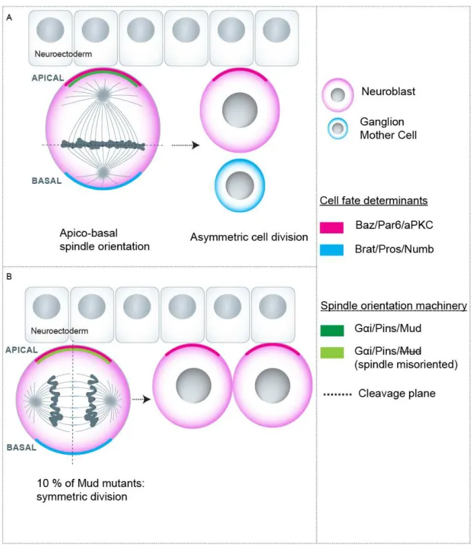
In the second scenario, the unequal segregation of intrinsic cell fate determinants between sibling cells accounts for the generation of two different fates. Probably the best example is constituted by the Drosophila neuroblasts which divide asymmetrically to self-renew and produce a ganglion mother cell. Here, the fate determinants Brat, Prospero and Numb are uniquely segregated into the ganglion mother cell, allowing for asymmetric cell division (reviewed in Homem and Knoblich, 2012; Knoblich, 2008) (Fig. 2a) (see below for additional details).
16
How can the differential positioning or asymmetric segregation of intrinsic fate determinants be achieved? The plane in which cell division occurs constitutes a mechanistic solution for both scenarios. In the first case, if the division plane is perpendicular to the signaling source, then both cells will remain in contact with the signal giving rise to a symmetric cell division. If the division plane is parallel, in contrast, one cell is positioned away from the niche thus generating an asymmetric division (Yamashita et al., 2003b) (Fig. 1a). In the intrinsic mode of cell fate determination, intrinsic fate determinants asymmetrically distribute in mitosis, and a cleavage plane that results in asymmetric segregation of these determinants allows the generation of distinct cell fates (Fig. 2a).
The plane of cell division is mainly controlled by the orientation of the mitotic spindle in anaphase. Thus, differential orientation of the mitotic spindle is a potential mechanism to determine the occurrence of symmetric vs asymmetric cell divisions.
It should be noted, however, that the orientation of the spindle (apico/basal, planar, etc) is not a synonym of the division outcome (asymmetric/symmetric) as I will discuss below.
In the following sections I will present some of the most studied examples linking spindle orientation with differential cell fate generation in higher eukaryotes as well as potential links between spindle misorientation and pathologies. The mechanisms of spindle orientation will be discussed in detail in chapter 2. However, to facilitate the discussion of these examples, I introduce the core mechanisms here. In short, in many tissues, spindle orientation is controlled by the specific subcortical localization of an evolutionary conserved complex composed of Gαi subunits, LGN and NuMA (Gαi, Pins and Mud in Drosophila, hereafter called “LGN complex”) which mediates the recruitment of force generators that in turn orient the spindle towards the cortical sites in which the LGN complex is enriched (reviewed in Morin and Bellaïche, 2011).
17
1.1.1- DROSOPHILA NEUROBLASTS
Neuroblasts are the neural stem cells in the Drosophila nervous system. These cells delaminate from the neuroectoderm and divide continually in an asymmetric fashion giving rise to a self-renewed neuroblast and a ganglion mother cell which will further produce two neurons. Neuroblasts are polarized along the apico basal axis with Bazooka (Par3 homolog), Par6 and aPKC localized to the apical side (Suzuki and Ohno, 2006). During prometaphase the cell fate determinants Brat, Prospero and Numb accumulate to the basal side (Betschinger et al., 2006; Choksi et al., 2006). In this context, the spindle is oriented along the apico-basal axis which allows the segregation of basal cell fate determinants to the basal cell and of the apical determinants to the apical cell, thus resulting in an asymmetric cell division (reviewed in Knoblich, 2008). In terms of mechanisms, an apically localized adaptor called Inscuteable (Insc) provides the link between the apical polarity complex and the spindle orientation machinery i.e. the Gα/Pins/Mud complex, which is recruited to the apical membrane (Bowman et al., 2006; Yu et al., 2000) (Fig. 2a).
In the context of neuroblast division, spindle orientation is seen as one of several mechanisms favoring the occurrence of cell fate decisions. Of note, binary cell fate choices are not much affected in mud mutants in which spindle orientation is specifically perturbed without affecting the apical-basal polarity and the distribution of fate determinants. The fact that asymmetric division still takes place is attributed to a “telophase rescue” in which the fate determinants are redistributed in relation to the final spindle orientation axis in late mitosis (Bowman et al., 2006; Knoblich, 2010). However, Cabernard and Doe have found that a minority of mud mutant cells present defective distribution of cell determinants at the end of mitosis. Within this cell population, these authors have observed that when the spindle is perpendicular to the apico-basal axis, apical determinants are equally inherited and the two daughters become neuroblasts (Fig. 2b). Of note, the basal determinants are still unequally segregated in this case, suggesting that the presence of the apical determinants overrides the inheritance of basal determinants (Cabernard and Doe, 2009).
18
Because of the stereotypic behavior of neuroblast division as well as the powerful genetics available in flies, this model is extensively used for the study of asymmetric cell division and spindle orientation.
Figure 2: Spindle orientation during Neuroblast asymmetric division. a) Spindle orientation allows the
asymmetric segregation of intrinsic cell fate determinants and asymmetric division.. b) A minority of Mud mutants (defective in spindle orientation) in which spindle is misoriented at late mitosis results in symmetric division.
19
1.1.2- MOUSE SKIN PROGENITORS
The mammalian epidermis is a stratified epithelium composed of distinct layers. In early embryonic development the epidermal tissue is constituted by a single layered epithelium that contacts the basement membrane. In this context, basal progenitors divide with a planar orientation, i.e. with the spindle oriented in the plane of the epithelium, which gives rise to symmetric divisions with both cells remaining in the plane of the epithelia. This type of division is linked with tissue expansion during these early stages of development. Later on, as the epidermis becomes multilayered, the progenitors switch to an asymmetric mode of division giving rise to cells with distinct fates and position (basal proliferative and suprabasal commited cells) which is essential for skin stratification (Lechler and Fuchs, 2005; Williams et al., 2011). Here, the switch from a symmetric to an asymmetric mode of cell division coincides with a switch in spindle orientation from planar to apico-basal orientation. Somehow similarly to the Drosophila neuroblast model, the spindle orientation machinery is recruited specifically to the apical domain which allows apico-basal spindle orientation (Lechler and Fuchs, 2005) (Fig. 3). Likewise, mInsc, a distant homologue of Insc makes the link between the apical polarity marker Par3 and LGN as they form a complex in vivo (Lechler and Fuchs, 2005).
Positioning one cell away from the basement membrane, known as a source of growth factors and extracellular matrix signaling would account for the differential cell fate (Lechler and Fuchs, 2005).
Importantly, knock-down of proteins regulating specifically spindle orientation results in divisions occurring mainly planarly, and impairs tissue differentiation and stratification (Fig.3). Hence, these data demonstrated that spindle orientation mediates asymmetric cell division and is essential for skin stratification in mice (Williams et al., 2011).
20
Figure 3: Mitotic spindle orientation is essential for asymmetric cell division and skin stratification during
mouse embryogenesis.
1.1.3- VERTEBRATE NEURAL PROGENITORS
The vertebrate neuroepithelium is organized as a pseudostratified epithelial monolayer. Neuroepithelial progenitors are elongated cells. They have a small apical surface that faces the lumen of the neural tube, which is separated from the molecularly distinct basolateral domain by sub-apical junctions that are important for tissue cohesion and maintenance of polarity. The pseudostratified aspect of the tissue is a consequence of the so-called interkinetic nuclear movement, during which
21
the position of the nucleus varies along the apico-basal axis of the cell in relation to the cell cycle stage. In particular, the nucleus localizes to the apical surface when cells enters mitosis.
In a first stage, neuropithelial cells divide symmetrically to amplify their pool. Later on in development, in particular at the onset of neurogenesis, they switch to an asymmetric mode of division which allows the production of a differentiating neuron or intermediate progenitor and the self-renewal of the progenitor (Fig.4) (Peyre and Morin, 2012). After the onset of neurogenesis, more committed and differentiated cells lose the apical attachment and start to accumulate basally in the “mantle zone”, whereas proliferative cells remain apically in the “ventricular” zone and retain the apico-basal organization.
By analogy with the asymmetric division of fly neuroblasts, it was proposed in the mid-nineties that cells dividing with a spindle oriented parallel to the apico-basal axis were undergoing asymmetric divisions, whereas cells dividing symmetrically would maintain a planar spindle orientation (Chenn and McConnell, 1995), However, careful analysis of the orientation of cell divisions showed that apical progenitors mainly divide with a planar spindle orientation even at the peak of neurogenesis (Kosodo et al., 2004; Noctor et al., 2008). This is difficult to reconcile with the idea that spindle orientation is a driver for asymmetric cell division in this context.
It was therefore proposed that small angle variations from the planar orientation (and thus from a vertical cleavage plane) would be enough for one of the daughter cells to bypass the small apical domain. The differential inheritance of the apical domain which may contain cell fate determinants would then result in different cell fates (Fig. 4) (Huttner and Brand, 1997). Indeed, while both daughters retain apical attachment, differential inheritance of the domain containing the apical polarity proteins has been shown to correlate with markers of binary cell fate decision in mouse cortical progenitors (Kosodo et al., 2004; Marthiens and ffrench-Constant, 2009).
In support of this model, a number of loss of function studies have shown a correlation between defects in spindle orientation (resulting in an increase in the frequency of “oblique” and “vertical”
22
divisions”) with accelerated neurogenesis (e.g. (Feng and Walsh, 2004; Fish et al., 2006; Godin et al., 2010; Lizarraga et al., 2010). Similarly, overexpression of Inscuteable in the chick neural tube or mouse neocortex increases the number of vertical divisions and simultaneously accelerates neurogenesis through an increase of asymmetric (neurogenic) divisions (Das and Storey, 2012; Postiglione et al., 2011).
In line with this model, some authors have proposed that some forms of microcephaly may be a consequence of defective spindle orientation. Primary microcephaly is an autosomal recessive disorder in which patients show small brains. Smaller brains are thought to arise from a defect in the number of neurons due to early exhaustion of the progenitor pool by uncontrolled and premature occurrence of asymmetric divisions. Indeed, most genes associated with primary Microcephaly (MCPH) in humans have been involved to various extent with the regulation of spindle orientation in different experimental systems (Fish et al., 2006; Gruber et al., 2011; Kitagawa et al., 2011; Lizarraga et al., 2010), lending some credit to the hypothesis. However, MCPH1-9 genes are also involved in multiple cellular processes, making difficult to assign a role to spindle orientation in microcephaly. Of note, all MCPH genes code for centrosomal proteins and their depletion often results in defective centriole duplication or centrosome maturation (Noatynska et al., 2012; Thornton and Woods, 2009). Therefore, it is possible that multiple processes linked to centrosome function and the cell cycle contribute to generate microcephaly (Arquint and Nigg, 2014; Marthiens et al., 2013), and that the defects in spindle orientation are only a minor aspect of the phenotype.
Indeed, knocking-down LGN (which normally localizes at the lateral cortex directing planar spindle orientation) in the chick spinal cord and in the mouse cortex also resulted in randomized spindle orientation, but contrary to the models prediction, this did not significantly affect the rate of neurogenesis. Instead, this resulted in the production of ectopic progenitors in the mantle zone (Fig.4) (Konno et al., 2008; Morin et al., 2007). This suggested that oblique or vertical spindle orientations are not sufficient to induce neurogenic divisions. In the same line, both symmetric
23
proliferative and asymmetric neurogenic divisions were shown to be associated with variable angles of division in the chick spinal cord, with no statistical difference between the two groups (Wilcock et al., 2007). Moreover, in rat cortical slice cultures apical progenitors divide symmetrically or asymmetrically independently of the cleavage plane but depending on the developmental stage (Noctor et al., 2008).
Figure 4: Mitotic spindle orientation in the vertebrate neuroepithelium. a) Apical progenitors divide
symmetrically to expand their pool during the proliferative phase. b) During the neurogenic stage they switch to an asymmetric mode of division. The spindle orient mostly planarly during both phases. Subtle deviations are proposed to be sufficient to bypass the apical domain (see the red line indicating an oblique cleavage plane). However, randomization of spindle orientation by LGN loss of function does not impact the rate of neurogenesis but results in ectopic progenitors (c).
24
Overall, the role of spindle orientation in determining asymmetric cell division of apical progenitors and neurogenesis is controversial in vertebrates.
Alternatively, different mechanisms that allow generating asymmetric fates independently on spindle orientation have been proposed. In particular, Paridaen and colleagues have shown that the differential inheritance of cilia remnants associated with the mitotic centrosome contributes to generate distinct cell fates (Paridaen et al., 2013). Along the same line, Wang and colleagues also proposed that the intrinsic asymmetry of the mitotic spindle (due to the different maturation of spindle poles) contributes to the generation of different fates upon division of neural progenitors (Wang et al., 2009).
Finally, a more established role of spindle orientation in the neuroepithelium is to maintain the progenitors in the ventricular zone. Indeed, randomization of spindle orientation by depletion of LGN generates ectopic progenitors that overproliferate in the subventricular zone (Morin et al., 2007). This highlights the importance of understanding how spindle orientation is achieved in the neuroepithelium independently of its unclear role in neurogenesis.
1.2. MORPHOGENETIC PROCESSES
Different cellular processes are proposed to drive the shaping of organs during embryogenesis. Both oriented cell divisions and cell arrangements are predicted to contribute to this process. In principle, cell division orientation along the axis of tissue elongation would contribute to the process of elongation as in this case daughter cells are positioned along the elongation axis. Indeed, orientation of cell division along the elongation axis has been observed in several contexts (reviewed in Gillies and Cabernard, 2011). However, this does not prove a role for spindle orientation in tissue morphogenesis. Of note, higher tension in the direction of tissue elongation could affect the orientation of cell divisions and not the opposite (Campinho et al., 2013). Nevertheless, different studies have demonstrated a role for spindle orientation in different morphogenetic processes. In particular, stereotypic cell division orientations have been observed in the Drosophila wing and eye
25
discs. In the wing blade, division occurs mainly along the proximal/distal axis which coincides with the axis of preferential tissue growth (Fig. 5a). Mutation of Dachsous or Dachs (components of the Fat-Ds PCP pathway) resulted both in cell divisions orienting randomly and a defective wing shape, which suggests a contribution of division orientation to the shaping of organs in Drosophila (Baena-Lopez et al., 2005; Mao et al., 2011). Remarkably, repolarization of Dachs perpendicularly to the proximal/distal axis along which it is normally polarized is sufficient to drive division orientation and tissue growth perpendicularly to the proximal distal axis (Mao et al., 2011).
Cell divisions are also oriented along the axis of kidney tubule elongation in mice. Consistently, loss of function of Fat4 generates cell division misorientation and defective tubule elongation in mouse kidney (Fischer et al., 2006).
In addition, cell division is oriented along the antero-posterior axis in the posterior region of the Drosophila germband, during the fast elongation phase of this tissue which occurs along the anterio-posterior axis (Fig. 5b) Notably, in mutants where cell division is inhibited the elongation of the germband is compromised (da Silva and Vincent, 2007).
Moreover, during Zebrafish gastrulation, cell divisions are oriented along the animal-vegetal axis in the dorsal epiblast, which matches the axis of tissue elongation (Gong et al., 2004). Importantly, loss of function of Dsh –a molecule involved in spindle orientation in different systems- results in random spindle orientation and defective convergence and tissue extension in the Zebrafish gastrula. Of note, contribution of spindle orientation to tissue elongation is significant but only partial, suggesting that other processes such as cell intercalation are involved in tissue elongation in this context (Gong et al., 2004). However, later studies showed that when spindle orientation is perturbed by injecting blocking antibodies against the force generator dynein, this does not result in defects in body axis elongation, contradicting the idea that spindle orientation is involved in tissue elongation during Zebrafish gastrulation (Quesada-Hernandez et al., 2010).
26
Furthermore, cell division orientation is essential for neural tube morphogenesis in Zebrafish (Zigman et al., 2011). The immature epithelium of the Zebrafish neural keel develops into a lumenized neural tube. During the neural keel and rod stage, apical neuroepithelial cell divisions occur with an apico-basal orientation and give rise to two bilaterally distributed progenitors (Fig. 5c) (Geldmacher-Voss et al., 2003; Tawk et al., 2007). Remarkably, induction of spindle misorientation by depletion of Scribble in the neural keel generates drastic defects in morphogenesis, in particular a misaligned configuration of the apical surfaces laying the neural tube lumen in contrast to the straight wild type organization. Of note, these effects occur without problems in the apical domain organization, which suggests that the morphogenetic defects are generated by misoriented cell divisions (Zigman, Trinh le et al. 2011). In the same line, oriented cell divisions are required for midline formation in the neural rod, a later stage of neurulation (Quesada-Hernandez, Caneparo et al. 2010).
In conclusion, the orientation of cell division contributes to several morphogenetic processes. However, in some tissues, there is only a correlative link between these processes. Noteworthy, perturbing certain signaling pathways may alter several cellular processes in addition to cell division orientation, probably explaining the different results obtained with respect to tissue elongation in Zebrafish (Gong et al., 2004; Quesada-Hernandez et al., 2010). Thus, careful analysis of tissue organization and different cellular processes is critical for understanding the actual contribution of spindle orientation to tissue morphogenesis.
Finally, in addition to participate in the shaping of tissues and organs, spindle orientation along the plane of epithelia is essential to maintain the two daughter cells in the epithelia both during growth and homeostasis (Macara et al., 2014; Zheng et al., 2010). Thus, defective planar spindle orientation could lead to epithelial architecture disruption as discussed below.
27
Figure 5: Mitotic spindle orientation in morphogenetic processes. a) Oriented divisions along the proximo-distal
axis are important for directional tissue growth in Drosophila wing discs. The arrows indicate the direction of growth. b) Spindle orientation along the anterio-posterior axis contributes to Drosophila germband extension. c) Apico-basal spindle orientation is critical for correct midline organization during neural tube formation in Zebrafish.
28
1.3-TUMORIGENESIS
Different observations have led to the idea that spindle orientation could be at least a contributor factor to tumorigenesis. First, misoriented spindles are seen in many tumors (Fleming et al., 2009). Second, tumor suppressors often mutated in cancer have been seen to regulate spindle orientation. This is the case of the tumor suppressors APC, E-cadherin and VHL (Pease and Tirnauer, 2011). Here I discuss two possible mechanisms by which spindle misorientation could contribute to tumor development as well as some evidence supporting these mechanisms.
1.3.1- DEREGULATION OF STEM CELLS COMPARTMENTS
Stem cells can divide both symmetrically and asymmetrically at least in some contexts. Asymmetric stem cell division is considered as critical to maintain the size of stem cell compartments by producing one stem cell and one differentiating cell. Notably, many cancers are proposed to arise from a deregulation of the stem cell compartment. Because spindle orientation is one of the mechanisms involved in determining the occurrence of asymmetric vs symmetric divisions, spindle orientation could then control the size of stem cell compartments and therefore its deregulation could contribute to tumorigenesis. The strongest evidence for this hypothesis comes from work in Drosophila neuroblasts (Caussinus and Gonzalez, 2005). In particular, mutation of genes regulating spindle orientation and asymmetric cell division (namely Pins, Numb, Prospero and Miranda) result in hyperproliferation of larval neuroblasts transplanted in adult tissue and generation of highly proliferating invading tumors. In line with this, loss of the cell fate determinant Brat, which is considered as a self-renewal repressor, leads to a massive increase in the number of larval neuroblasts in situ (Betschinger et al., 2006; Lee et al., 2006).
In vertebrates, the potential link between control of asymmetric cell division and cancer has been investigated in the mouse and human gut epithelia (Quyn et al., 2010). Quyn and colleagues found that spindles are preferentially oriented perpendicularly to the apical surface specifically in the stem cell compartment. In mice gut, this orientation correlates with the asymmetric inheritance of DNA
29
strands which is considered as a feature of asymmetric cell division based on studies in muscle cells (Rocheteau et al., 2012). Notably, in precancerous tissue generated by heterozygous loss of the tumor suppressor APC, spindle orientation and asymmetric DNA segregation are defective. While these data do not prove a specific contribution of spindle orientation to cancer, this suggests that APC mutation could contribute to tumor formation by deregulating spindle orientation and asymmetric cell division.
1.3.2- EPITHELIAL DISRUPTION
Epithelial tumors constitute the majority of human cancers (Pease and Tirnauer, 2011). In epithelia, cells normally divide with a planar orientation allowing to maintain both cells in the plane of the tissue (Fig. 6a). In contrast, loss of planar spindle orientation can result in positioning one daughter cell on top of the other leading to disrupted epithelial tissue architecture (Fig. 6b). The cell positioned away from the extracellular matrix could follow different destinies. First, this cell could die as a consequence of losing essential signals and attachment; however apoptotic mechanisms are often perturbed in cancers. Thus, a second possibility is that the cell remains in the tissue leading to vertical tissue expansion and hyperplasia, which are premalignant features (Fig. 6c). Alternatively, if the cell detaches from the tissue, this could lead to dissemination and metastasis (Pease and Tirnauer, 2011).
Interestingly, recent work performed in the Drosophila wing disc epithelia, in which cell divisions occur preferentially in the plane of the epithelium, has brought support to these ideas. In this tissue, Nakajima and colleagues have shown that defective spindle orientation generated by mutation of different genes including mud correlates with basal cell delamination. Remarkably, while the delaminated cells normally die by apoptosis, blocking apoptosis in this context led to the formation of basal tumor-like masses with characteristics of epithelial-mesenchymal transition (Fig. 6b-c) (Nakajima et al., 2013). Whether spindle misorientation combined with apoptosis inhibition lead to epithelial disruption and tumor formation in vertebrate epithelia remains to be elucidated.
30
Noteworthy, the cell behavior observed by Nakajima et al in the wing disk does not apply to all fly epithelia, and Bergstralh and colleagues have shown that abnormally positioned cells generated by apico-basal orientation in different tissues (namely the follicular epithelium, the embryonic ectoderm and the neuroepithelium) reintegrate into the tissue rather than undergoing apoptosis (Fig. 6c) (Bergstralh et al., 2015). Hence different epithelial structures cope differently with defective spindle orientation, and may have different susceptibility to daughter cells mispositioning and tumor formation.
In conclusion, spindle misorientation leads to tissue overgrowth and tumor formation in specific Drosophila tissues and both mechanisms proposed here have somehow found support in this organism. However, clear evidence for a specific role of spindle misorientation in contributing to tumorigenesis or tumor development is lacking in mammalian organisms. In this sense, targeting pathways specifically involved in spindle orientation without perturbing tissue polarity could help to define the potential contribution of spindle orientation to mammalian tumors.
31
Figure 6: Role of spindle orientation in epithelial architecture maintenance and potential role in tumorigenesis.
a) Planar spindle orientation maintains both daughter cells in the epithelia. b) Random or apico-basal spindle orientation leads to mispositioning of one cell out of epithelia. c) Different outcomes are possible: cell death, overproliferation and tumor-like formation or reintegration of the cell into the epithelial layer.
1.4. CONCLUSION
Mitotic spindle orientation is critical for diverse developmental processes in several organisms. In invertebrates and more specifically in Drosophila, spindle orientation is clearly involved in binary cell fate choices and morphogenesis and spindle misorientation can lead to tissue overgrowth. While I discussed only a few examples, it should be noted that in invertebrates we find other classical models in which spindle orientation is critical for asymmetric division. These include the asymmetric cell division of the C.elegans zygote and of Drosophila Sensory Organ Precursors, which will be further discussed in the frame of the mechanisms of spindle orientation in the next chapters. In
32
vertebrates, spindle orientation is linked to the morphogenesis of at least some tissues while its role in asymmetric cell division is only well defined in tissues such as the skin. The relatively better understanding of the role of oriented divisions in Drosophila development is partially justified by a more refined understanding of the molecular mechanisms controlling spindle orientation in this organism. That said, efforts to dissect the molecular mechanisms of vertebrate spindle orientation could be useful for a better comprehension of the function of this cellular process in normal development and disease in higher vertebrates. Notably, to understand the specific contribution of spindle orientation to development and disease, one challenge is to target spindle orientation without affecting cell polarity, centrosome function and tissue architecture. Many of the genes mutated in Microcephaly and cancer have pleiotropic roles, thus complicating the understanding of the contribution of spindle orientation defects to disease in these mutants. Thus, finding molecules that regulate specifically spindle orientation could help to address these issues.
33
CHAPTER 2: MECHANISMS OF MITOTIC SPINDLE ORIENTATION
This chapter is highly based on the review that we have recently published: “Regulation of mitotic spindle orientation: an integrated view” Florencia di Pietro, Arnaud Echard, Xavier Morin. EMBO Reports, Aug. 2016. The figures are adapted from the review figures as well (di Pietro et al., 2016). 2.1. INTRODUCTION
The orientation of the mitotic spindle in animal cells can be influenced by geometric cues, internal cues and external cues. More than a century ago, Hertwig proposed that cells orient their spindles along the long axis of the cell, arguing for a role of cellular geometry in controlling the plane of division (Hertwig, 1884). While this rule applies to many situations, orientation of the spindle is also often set by specific polarity cues. Early studies identified the evolutionary conserved Gαi/LGN/NuMA complex as a key regulator that polarizes cortical force generators that exert pulling forces on astral MT to orient the spindle. Indeed, in most animal cell types oriented cell divisions involve the transmission of localized pulling forces located at the cell cortex to astral microtubules, resulting in the positioning the mitotic spindle. As a consequence, the cell cortex, the specific mechanisms that recruit and localize force generators, and the astral microtubule network have emerged as the three essential levels of regulation for spindle orientation.
An excellent model that contributed to the establishment of spindle orientation principles is the C.elegans zygote. The C. elegans zygote divides asymmetrically with regard to both cell size and fate, and spindle displacement towards to posterior cortex is necessary for this process. Remarkably, elegant studies performed in this embryo have demonstrated that the polarization of force generators on the posterior cortex results in the exertion of higher forces on the spindle pole closest to that cortex (Grill et al., 2001; Grill et al., 2003). In particular, Grill and colleagues used laser ablation to sever spindle poles and considered the velocity of displacement of centrosome fragments as a measure of the force previously exerted on that pole (Grill et al., 2003). With this approach, they found that the posterior pole fragments showed higher velocities than the anterior ones which is linked with the recruitment of more force generators to the posterior cortex.
34
In this chapter I will discuss spindle orientation mechanisms in detail. In particular, I will present the main characteristics of the LGN complex as well as recent data that shed light on how the cortical recruitment and dynamics of this complex are regulated in mitosis. Then, I will discuss a host of data illustrating how modulation of the actin cytoskeleton and astral MT control spindle orientation in different contexts. Finally, I will focus on the regulation of spindle orientation by geometric cues and external forces. The role of Dynein in spindle orientation will be separately discussed in Chapter 3. Of note, the present chapter makes emphasis in the regulation of spindle orientation mainly in Drosophila and vertebrate models. In the end I briefly discuss other models of spindle positioning that are relevant for the next chapter.
2.2- THE LGN COMPLEX
A number of genetic studies have revealed that an evolutionary conserved molecular complex composed of the heterotrimeric Gα protein Gαi, LGN and NuMA (respectively Gαi, Pins and Mud in Drosophila, and GOA1/GPA16, GPR1/2 and LIN5 in C. elegans, “the LGN complex for simplicity, Fig.7) is at the core of spindle orientation and positioning in different tissues both in invertebrate and vertebrate species (Du and Macara, 2004; Gotta and Ahringer, 2001; Gotta et al., 2003; Konno et al., 2008; Lechler and Fuchs, 2005; Morin et al., 2007; Peyre et al., 2011; Schaefer et al., 2001; Schaefer et al., 2000; Srinivasan et al., 2003; Yu et al., 2000) (reviewed in Morin and Bellaïche, 2011). During mitosis, this complex is localized to a particular subcortical domain and directs the recruitment of the minus end-directed microtubule motor dynein (Couwenbergs et al., 2007; Kotak et al., 2012; Nguyen-Ngoc et al., 2007) (Fig. 1a). The directed movement of cortically anchored dynein along astral microtubules generates pulling forces on spindle poles leading to the orientation and/or positioning of the spindle. Therefore, the specific localization of the LGN complex determines the site of force concentration and the axis of spindle orientation. Consistently, the apical localization of Pins/Mud or LGN/NuMA directs spindle orientation along the apico-basal axis in Drosophila neuroblasts (Fig. 7B) and mouse skin progenitors, respectively (Bowman et al., 2006; Izumi et al., 2006; Lechler and Fuchs, 2005; Siller et al., 2006; Williams et al., 2011; Yu et al., 2000).
35
Figure 7: The LGN complex A) Schema showing the LGN domains and its interactions with Gαi membrane
anchored subunits, and with NuMA, as well as the interaction with cortical proteins (Dlg, Afadin) that regulate LGN cortical localization. B) LGN complex localization in different systems, showing the polarity proteins regulating this specific localization when applicable. i) Drosophila embryonic neuroblasts, ii) C. elegans zygote, iii) neural progenitors in the vertebrate neuroepithelium, iv) mammalian cell lines. Adapted from di Pietro et al. 2016.
In the C. elegans zygote, enrichment of GPR1/2 at the posterior cortex is necessary for spindle positioning along the anterio-posterior axis (Gotta et al., 2003; Srinivasan et al., 2003) (Fig.7b).
36
Furthermore, the lateral localization of the LGN complex regulates planar spindle orientation of progenitors in chick and mouse neuroepithelium (Konno et al., 2008; Morin et al., 2007; Peyre et al., 2011) as well as during epithelial morphogenesis of Drosophila and mammalian cells (Bergstralh et al., 2013; Zheng et al., 2010) (Fig.7b).
Biochemical and structural analysis revealed the different components of the LGN complex which are briefly presented below:
Gαi. Gαi subunits localize to the plasma membrane through myristylation, where they serve as an anchor to the complex. By default, Gαi subunits cover the whole cell inner surface, and do not contribute to the polarization of LGN and NuMA crescents.
LGN. LGN was first identified as a biochemical interactor of Gαi subunits (Mochizuki et al., 1996). LGN is a modular protein composed of three main domains. Its N-terminal TPR domain contains 7 (6 or 8 depending on the authors) Tetratricopeptide repeats, which mediate interaction with multiple binding partners, including NuMA, Afadin and Inscuteable. Its central “linker” domain does not show any recognizable organization or binding motif, but is crucially required for its function through its interaction with Dlg (Fig. 7a). The C-terminal GPR (G Protein Regulator) domain contains four (3 in Drosophila Pins and 1 in C. elegans GPR1/2) Goloco domains that mediate interaction with Gαi/o subunits. LGN interacts with Gαi only when it is bound to GDP, and has a guanine dissociation inhibitory (GDI) activity (Willard et al., 2004).
The GTPase activating protein (GAP) RGS14/Loco/RGS-7 (in vertebrates, Drosophila and C. elegans, respectively) and the Guanine exchange factor (GEF) Ric8a control the interaction between LGN and Gαi, and therefore the stability of the complex, by modulating the GTPase activity of Gαi subunits and thereby the phosphorylation state of bound guanosine (Afshar et al., 2004; Couwenbergs et al., 2004; David et al., 2005; Hampoelz et al., 2005; Hess et al., 2004; Wang et al., 2005; Woodard et al., 2010; Yu et al., 2005).
37
NuMA. NuMA is a coiled coil protein that can interact with LGN, dynein as well as with microtubules (Fig. 7a) (Bowman et al., 2006; Du et al., 2001; Haren and Merdes, 2002; Kotak et al., 2012; Merdes et al., 1996). NuMA is also present on the spindle, enriched near the poles, and regulates spindle formation and organisation.
In dividing cells, LGN and NuMA are usually observed as cortical crescents facing one or both spindle poles (Fig. 7b), and it is established that this specific cortical localization is instructive for spindle orientation in many systems.
Because Gαi subunits localize all over the cortex, additional factors, and in particular polarity proteins, must regulate the polarized cortical distribution of LGN and NuMA. Indeed, the LGN homolog Pins (Partner of Inscuteable) was initially identified in Drosophila in interaction screens with Inscuteable (Schaefer et al., 2000; Yu et al., 2000), an apical protein known for its role in apico-basal spindle orientation in neuroblasts (Kraut et al., 1996). Apical localization of Insc, and consequently of Pins, requires the polarity protein Bazooka (Drosophila Par3) (Yu et al., 2000), as well as atypical protein kinase C (aPKC) (Izumi et al., 2004; Wodarz et al., 2000). Similarly, the posterior cortical enrichment of GPR1/2 requires the Par2 and Par3 polarity proteins during the first division of the C. elegans embryo (Gotta et al., 2003).
Later work has revealed a surprising diversity in the mechanisms that control LGN localization at restricted cortical domains. While Pins/LGN localizes apically in Drosophila neuroblasts, it is found in a ring at the lateral cortex during planar spindle orientation in different epithelial contexts (Bergstralh et al., 2013; Peyre et al., 2011; Zheng et al., 2010). Remarkably, while aPKC is required for the apical recruitment of Pins in neuroblasts (Izumi et al., 2004), it inhibits the apical localization of LGN and favours its lateral enrichment during cystogenesis in MDCK cells (Zheng et al., 2010), see section 2.3). The mechanism mediating this inhibition involves the phosphorylation of LGN by apical aPKC, which increases locally LGN affinity to a 14-3-3 protein, competing with the interaction of LGN