HAL Id: inserm-02971228
https://www.hal.inserm.fr/inserm-02971228
Submitted on 19 Oct 2020
HAL is a multi-disciplinary open access archive for the deposit and dissemination of sci-entific research documents, whether they are pub-lished or not. The documents may come from teaching and research institutions in France or abroad, or from public or private research centers.
L’archive ouverte pluridisciplinaire HAL, est destinée au dépôt et à la diffusion de documents scientifiques de niveau recherche, publiés ou non, émanant des établissements d’enseignement et de recherche français ou étrangers, des laboratoires publics ou privés.
Ischemic Core for Mechanical Thrombectomy
Basile Kerleroux, Kevin Janot, Cyril Dargazanli, Dimitri Daly-Eraya, Wagih
Ben-Hassen, François Zhu, Benjamin Gory, Jean François Hak, Charline
Perot, Lili Detraz, et al.
To cite this version:
Basile Kerleroux, Kevin Janot, Cyril Dargazanli, Dimitri Daly-Eraya, Wagih Ben-Hassen, et al.. Per-fusion Imaging to Select Patients with Large Ischemic Core for Mechanical Thrombectomy. Journal of Stroke, Korean Stroke Society, 2020, 22 (2), pp.225-233. �10.5853/jos.2019.02908�. �inserm-02971228�
Copyright © 2020 Korean Stroke Society
This is an Open Access article distributed under the terms of the Creative Commons Attribution Non-Commercial License (http://creativecommons.org/licenses/by-nc/4.0/) which permits unrestricted non-commercial use, distribution, and reproduction in any medium, provided the original work is properly cited.
Original Article
Perfusion Imaging to Select Patients with Large
Ischemic Core for Mechanical Thrombectomy
Basile Kerleroux,
aKevin Janot,
aCyril Dargazanli,
bDimitri Daly-Eraya,
bWagih Ben-Hassen,
cFrançois Zhu,
dBenjamin Gory,
dJean François Hak,
eCharline Perot,
fLili Detraz,
gRomain Bourcier,
gAymeric Rouchaud,
hGéraud Forestier,
hJoseph Benzakoun,
cGaultier Marnat,
iFlorent Gariel,
iPasquale Mordasini,
jJohannes Kaesmacher,
jGrégoire Boulouis,
con Behalf of the JENI Research
Collaborative
aDiagnostic and Therapeutic Neuroradiology, CHRU de Tours, Tours, France
bDepartment of Interventional Neuroradiology, University Hospital Center of Montpellier, Gui de Chauliac Hospital, Montpellier, France
cCentre Hospitalier Sainte Anne, Neuroradiology Department, Paris University, INSERM U1266, Psychiatry and Neurosciences Institute of Paris,
Paris, France
dUniversity Hospital of Nancy, Department of Diagnostic and Therapeutic Neuroradiology, INSERM U1254, Nancy, France
eDepartment of Diagnostic and Interventional Neuroradiology, Timone Hospital, Aix Marseille University, Marseille, France
fNeurology Department, Timone Hospital, Aix Marseille University, Marseille, France
gDepartment of Diagnostic and Interventional Neuroradiology, Guillaume et René Laennec University Hospital, Nantes, France
hDepartment of Interventional Neuroradiology, Dupuytren University Hospital, Limoges, France
iDepartment of Diagnostic and Interventional Neuroradiology, Pellegrin Hospital-University Hospital of Bordeaux, Bordeaux, France
jInstitute of Diagnostic, Interventional and Pediatric Radiology and Institute of Diagnostic and Interventional Neuroradiology, University
Hospital Bern, Inselspital, University of Bern, Bern, Switzerland
Background and Purpose Patients with acute ischemic stroke, proximal vessel occlusion and a large ischemic core at presentation are commonly not considered for mechanical thrombectomy (MT). We tested the hypothesis that in patients with baseline large infarct cores, identification of remaining penumbral tissue using perfusion imaging would translate to better outcomes after MT.
Methods This was a multicenter, retrospective, core lab adjudicated, cohort study of adult patients with proximal vessel occlusion, a large ischemic core volume (diffusion weighted imaging volume ≥70 mL), with pre-treatment magnetic resonance imaging perfusion, treated with MT (2015 to 2018) or medical care alone (controls; before 2015). Primary outcome measure was 3-month favorable outcome (defined as a modified Rankin Scale of 0–3). Core perfusion mismatch ratio (CPMR) was defined as the volume of critically hypo-perfused tissue (Tmax >6 seconds) divided by the core volume. Multivariable logistic regression models were used to determine factors that were independently associated with clinical outcomes. Outputs are displayed as adjusted odds ratio (aOR) and 95% confidence interval (CI).
Results A total of 172 patients were included (MT n=130; Control n=42; mean age 69.0±15.4 years; 36% females). Mean core-volume and CPMR were 102.3±36.7 and 1.8±0.7 mL, respectively. As hypothesized, receiving MT was associated with increased probability of favorable outcome and functional independence, as CPMR increased, a difference becoming statistically significant above a mismatch-ratio of 1.72. Similarly, receiving MT was also associated with favorable outcome in the subgroup of 74 patients with CPMR >1.7 (aOR, 8.12;
Correspondence: Grégoire Boulouis
Centre Hospitalier Sainte Anne, Neuro-radiology Department, Paris University, INSERM U1266, Psychiatry and Neuro-sciences Institute of Paris, 1 rue Caba-nis, 75014 Paris, France
Tel: +33-145658574 Fax: +33-145658574 E-mail: [email protected] Received: October 25, 2019 Revised: March 30, 2020 Accepted: April 28, 2020
Introduction
In recent years, mechanical thrombectomy (MT) has demon-strated its compelling efficacy in reducing mortality and func-tional dependence for patients with acute ischemic stroke (AIS) due to anterior proximal vessel occlusion (PVO).1,2 Patients with
an unfavorable imaging profile at baseline, assessed using magnetic resonance imaging (MRI) diffusion weighted imaging (DWI) volume core (≥70 mL),1 or the computed tomography
(CT)-based-Alberta Stroke Program Early CT score (ASPECTS) <6, were excluded in four of the seven randomized clinical tri-als that validated MT in AIS-PVO,3 precluding to draw strong
conclusions in this subgroup. Hence they are typically not of-fered MT in clinical practice,1 despite converging evidence
sug-gesting a benefit of MT despite large ischemic core (LIC), with almost 25% of patients experiencing favorable functional out-come after MT.3-9
Perfusion imaging is used in the diagnostic work up of AIS to identify hypo-perfused yet not infarcted (e.g., ‘at-risk’ or ‘sal-vageable’) brain tissue,10 and to estimate the core perfusion
mismatch ratio (CPMR).2 Nonetheless, perfusion data remain
very scarce in patients with LIC before MT, while they are criti-cally needed to pragmaticriti-cally design future randomized trials, and better select patients for MT until then.
We hypothesized that perfusion imaging may enhance the effective selection of AIS-PVO patients with LIC by determining those likeliest to benefit from revascularization and tested this hypothesis in a cohort study using data from a multicenter co-hort, by comparing the rates of favorable functional outcome, symptomatic intracerebral hemorrhage (sICH), and case-fatali-ty in patients with DWI assessed baseline LIC (>70 mL) and perfusion who received MT versus patients who were treated with intravenous thrombolysis only. The hypothesis driving this analysis was that in patients with baseline large infarct cores, identification of significant remaining penumbral tissue, as as-sessed using CPMR, would translate to better outcomes after MT.
Methods
Study design
The study was a multicenter, core-lab adjudicated, observa-tional retrospective cohort study. The “Jeunes en Neuroradiolo-gie Interventionnelle Research Collaborative” (JENI-RC), is a re-cently launched trainee-led research network.11 Local JENI-RC
members were asked to provide de-identified data for patients with DWI ASPECTs 0–6 otherwise meeting study inclusion cri-teria (see below). Case report form items included demograph-ics, relevant past medical history, sICH, and 3-month functional outcome. Imaging data were centralized by the internal core lab, for central assessment. Nine academic centers contributed data (eight in France, and one in Switzerland). This report was prepared according to the Strengthening the Reporting of Ob-servational Studies in Epidemiology (STROBE) statement.12
Ethics
As for all non-interventional retrospective studies of de-identi-fied data in France, written informed consent was waived and a commitment to compliance (reference methodology CPMR-3) was filed to the French National Information Science and Liberties Commission prior to data centralization, in respect to the General Data Protection Regulation.
Inclusion criteria
In the MT group, we included consecutive adult patients with AIS after 2015 if they had an occlusion of the intracranial in-ternal carotid artery or of the M1 segment of the middle cere-bral artery; had a large pretreatment ischemic core volume de-fined as 70 mL or more on magnetic resonance-DWI as as-sessed centrally; had no preexisting handicap as defined by a modified Rankin Scale (mRS) of 2 or higher; underwent MT; and if pre-treatment MR-T2* perfusion sequence had been performed. The control group was constituted by retrospective-ly querying the prospective intravenous tissue plasminogen ac-tivator (tPA) stroke data base at a single university hospital, to identify patients treated before MT related guidelines in 2015 95% CI, 1.24 to 53.11; P=0.028). Overall (prior to stratification by CPMR) 73 (42.4%) patients
had a favorable outcome at 3 months, with no difference amongst groups.
Conclusions In patients currently deemed ineligible for MT due to large infarct ischemic cores at baseline, CPMR identifies a subgroup strongly benefiting from MT. Prospective studies are warranted.
Keywords Acute stroke; Ischemic stroke; Thrombectomy; Endovascular treatment; Perfusion imaging
who met the same criteria but did not receive MT due to guidelines compliance.
Imaging analysis and mismatch definition
The post-processing and images interpretation were performed centrally after complete de-identification, by an internal core-lab (B.K. and G.B.), blinded to clinical data.
Ischemic core volumes (DWI volume) were calculated using Olea-Sphere version 3.0 software (Olea Medical, La Ciotat, France), by the semi-automated segmentation of brain tissue ipsilateral to the occlusion with apparent diffusion coefficient of 0.6×10⁻³ mm2/sec or less. Perfusion maps were generated
using the Olea-Sphere version 3.0 software then used to calcu-late critically hypo-perfused brain tissue, with an inferior threshold set, according to recent literature,13 at T max >6
sec-onds. As in previous large studies investigating core penumbral mismatch,14,15 we defined penumbral tissue as the volume of
critically hypo-perfused tissue minus the DWI volume. Similar-ly, we defined the CPMR, as the volume of critically hypo-per-fused tissue divided by the DWI volume, and defined a target mismatch as patients with a CPMR of 1.8 or above.
Assessment criteria
The primary endpoint was functional outcome assessed at 3 months, using the mRS, with a favorable outcome defined by a score of 3 or less, taking into account the inherent severity of AIS with baseline LIC, and in line with recent literature. Sec-ondary end points included functional independence defined as mRS of 0–2. Ninety days mortality, and the rate of symptom-atic intracerebral hemorrhage within 7 days (according to Eu-ropean Cooperative Acute Stroke Study [ECASS] II criteria).16
Substantial reperfusion was defined as a modified Thrombolysis in Cerebral Infarction score of 2B, 2C, or 3.17
Statistical analysis
Baseline characteristics of patients has been compared be-tween MT group and control group. Continuous variables were summarized using mean±standard deviation or median (inter-quartile range) where appropriate, and discrete variables were summarized using counts (percentages). Chi-square test, Fish-er-exact test, t-test, Mann-Whitney test were used as appro-priate for the univariate analysis, with a P<0.05 as the thresh-old for statistical significance.
Clinical outcomes were compared between MT group and control group. First, multivariable logistic regression models were used to determine factors that were independently asso-ciated with clinical outcomes. Variables assoasso-ciated with the outcome in univariate analysis (P≤0.1) were entered into
nomi-nal logistic models, with a prespecified adjustment for age, in-farct core, and CPMR. Backward elimination was then used to remove non-significant variables (P>0.05). The adjusted odds ratios (aOR) and 95% confidence interval (CI) of having favor-able outcome (model 1) and functional independence (model 2) were reported. Then, a sensitivity analysis was performed to assess for changes in the results in patients with a predefined CPMR cutoff of 1.8, as was reported in previous cornerstone studies analyzing the pertinence of CMPR in AIS-PVO pa-tients.13,18 Finally, a shift analysis of mRS score (of 0–6 points)
based on the proportional odds model has been performed. All analyses were done using JMP Pro 14 (Institute Inc., Cary, NC, USA) software. Where needed, we derived 95% CI by boot-strapping (5,000 occurrences) statistical results.18
Results
Study population
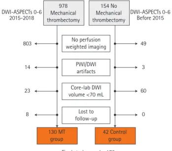
Between January 2015 and July 2018, in the MT group a total of 971 patients with AIS-PVO and ASPECTs 0–6 were screened for inclusion. After applying inclusion criteria, 168 patients were evaluated by the core lab, of which 130 were finally in-cluded and analyzed in the MT group. Before 2015, a total of 154 patients with DWI-ASPECTs 0–6 were screened for inclu-sion in the control group, and 42 met study criteria (Figure 1). A total of 172 were finally included in the present analysis (36% females, mean age 69.0±15.4 years) (Table 1 for baseline clinical-imaging characteristics). Patients in the MT group were
Figure 1. Flow chart. DWI, diffusion weighted imaging; ASPECTS, Alberta Stroke Program Early Computed Tomography Score; PWI, perfusion weight-ed imaging; MT, mechanical thrombectomy.
DWI-ASPECTs 0-6 2015-2018 DWI-ASPECTs 0-6Before 2015 No perfusion weighted imaging PWI/DWI artifacts Core-lab DWI volume <70 mL Lost to follow-up
Final study sample: 172 803 14 23 8 49 3 60 0 978 Mechanical thrombectomy 154 No Mechanical thrombectomy 130 MT
more frequently females (41% vs. 21%, P=0.03), less frequently received intravenous tPA (48.5% vs. 100%, P<0.01), were more frequently referred from an outside primary stroke center hos-pital (25.4% vs. 0%, P<0.01), were younger (66.2±14.9 years-old vs. 77.7±13.5, P<0.01). They did not differ otherwise in terms of baseline clinical parameters, notably for delay be-tween onset and qualifying imaging (P=0.58) and occlusion site (P=0.82). Substantial recanalization was achieved in 111/140 patients in the MT group (79.3%).
Core and penumbra
Mean core DWI volume was 102.3±36.7 mL and did not differ between groups. The volume of critically hypo-perfused tissue was larger in the MT group (mean 180.8±72.4 mL vs. 145.5±52.5 mL, P<0.01), which consequently demonstrated larger penumbral volumes (mean 76.8±63.7 mL vs. 48.8ml±40.9 mL, P<0.01) as well as higher CPMRS (mean 1.8±0.7 vs. 1.5±0.5, P<0.01). A total
of 90 (52%), 65 (38%), and 53 (31%) patients had CPMRS above 1.6, 1.8, and 2.0 respectively (Supplementary Figure 1, in the sup-plemental material for detailed CPMR distribution in the cohort).
Outcomes
Functional outcomes
At 3 months, 73 (42.4%) patients had a favorable functional outcome (mRS 0–3), with no difference amongst groups prior to stratification by CPMR (43.1% in the MT vs. 40.5, P=0.86). Forty-one patients (23.8%) were functionally independent and 54 (31.4%) were deceased (P=1.000) (Table 2). Unfavorable outcome was associated with higher age, higher baseline NI-HSS, history of hypertension, and diabetes mellitus (Supple-mentary Table 1).
Independent of, as well as within, treatment groups both larger core and larger penumbral volumes were associated with
Table 1. Baseline characteristics of included patients
Variable All (n=172) Control (n=42) MT (n=130) P
Age (yr) 69.0±15.4 77.7±13.5 66.2±15 <0.001 Female sex 62 (36) 9 (21.4) 53 (40.8) 0.029 Dyslipidemia 69 (40.1) 18 (42.9) 51 (39.2) 0.718 Diabetes mellitus 26 (15.1) 7 (16.7) 19 (14.6) 0.811 Tobacco use (current or past) 56 (32.6) 14 (33.3) 42 (32.3) 1.000 Hypertension 97 (56.4) 24 (57.1) 73 (56.2) 1.000 NIHSS 18.5±4.5 18±5.1 18.7±4.2 0.435 Left sided stroke 75 (43.6) 16 (38.1) 59 (45.4) 0.476 iv tPA 105 (61) 42 (100) 63 (48.5) <0.001 Drip and ship 33 (19.2) 0 (0) 33 (25.4) <0.001 ICA occlusion 31 (18) 7 (16.7) 24 (18.5) 0.269 Delay till imaging (min) 161.3±129 152.7±108.5 164±135.2 0.583 Volume T max <6 sec (mL) 172.2±69.6 145.5±52.5 180.8±72.4 <0.001 Mismatch ratio 1.8±0.7 1.5±0.5 1.8±0.7 <0.001 Core volume (mL) 102.3±36.7 96.7±33 104±37.7 0.233 Values are presented as mean±standard deviation or number (%).
MT, mechanical thrombectomy; NIHSS, National Institute of Health Stroke Scale; iv tPA, intravenous tissue plasminogen activator; ICA, internal carotid artery.
Table 2. Outcome of included patients
Variable All (n=172) Control (n=42) MT (n=130) P
Substantial recanalization - - 111 (79.3)
90-day, mRS 0–2 41 (23.8) 11 (26.2) 30 (23.1) 0.684 90-day, mRS 0–3 73 (42.4) 17 (40.5) 56 (43.1) 0.857 90-day, mortality 54/172 (31.4) 13 (30.9) 41 (31.5) 1 sICH 31/161 (19.3) 5/35 (14.3) 26/126 (20.26) 0.483 Values are presented as number (%).
poor functional outcome in univariable analysis (185±67.9 mL in patients with mRS 4–6 vs. 154±68.4 mL in those with mRS 0–3, P<0.01; and 76±59 vs. 67±60, P=0.04) (Figure 2).
In the entire population, there was no difference in favorable outcome between treatment groups (P=0.68) (Table 1). After adjusting for age, hypertension, diabetes, core volume, CPMR, delay to imaging, intravenous tPA before MT, and occlusion site, there was a significant interaction between MT effect and CPMR in both models (Model 1: aOR, 0.29; P=0.008; and Mod-el 2: aOR, 0.4; P=0.047), indicating an increasing benefit of MT as CPMR increases (Table 3). In the same model, with mismatch ratio dichotomized as a nominal variable (below or above 1.8) the odds ratio of the interaction term between treatment arm
and mismatch ratio (>1.8), was 0.42 (95% CI, 0 to 0,82;
P=0.02).
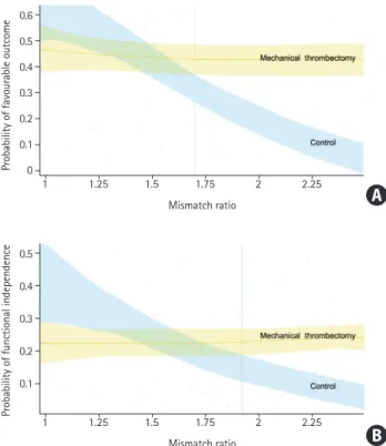
Analyzing the probability for favorable outcome in the fully adjusted model plotted against CPMR, we showed that receiv-ing MT (vs. not receivreceiv-ing MT) was associated with increased probability of favorable outcome and functional independence, as CPMR increased, a difference becoming statistically signifi-cant above a CPMR of 1.72 for favorable outcome, and above 1.93 for functional independence (Figure 3).
As a sensitivity analysis, when stratifying by CPMR; in the sample of 65 patients with a CPMR of 1.8 or above, after ad-justing for group specific outcome predictors (age, core vol-ume, intravenous tPA, and CPMR), receiving MT was associated with a significant increase in the rate of favorable outcome (aOR, 999; 95% CI, 999 to infinite). Similarly, in the subgroup of 74 patients for which CPMR exceeded 1.7 (42% of favorable outcome in the MT group vs. 20% in the control group,
P=0.031; receiving MT was associated with a significantly
in-creased rate of favorable outcome) (aOR, 8.12; 95% CI, 1.24 to 53.11, P=0.028). Using ordinal regression, receiving MT was as-sociated with overall favorable shift in mRS distribution (com-mon risk ratio, 1.83; 95% CI, 1.01 to 3.44; P=0.049) (Figure 4).
Finally, when restricting the sample to patients in the MT group, we found substantial recanalization to be associated with significantly higher odds of favorable outcome and of 3 months functional independence, in fully adjusted models (aOR, 53.7; 95% CI, 5.0 to 573; P<0.001; and aOR infinite,
P<0.001, respectively). Lower age (P<0.001), lower mismatch
ratio (P=0.03), and lower core volume (P<0.001) were also as-sociated with higher odds of favorable outcome, but the inter-action between CPMR and substantial recanalization only tended towards significance (P=0.058).
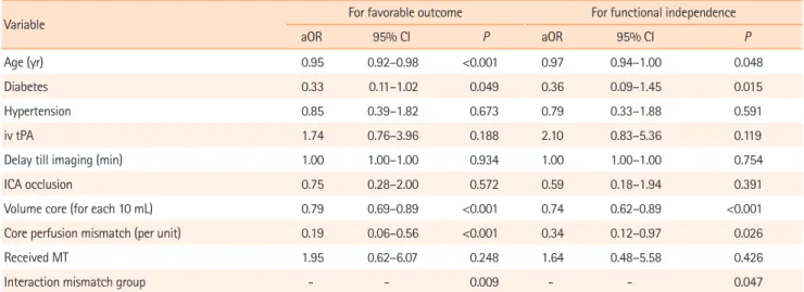
Table 3. Multivariable models for outcome
Variable For favorable outcome For functional independence aOR 95% CI P aOR 95% CI P
Age (yr) 0.95 0.92–0.98 <0.001 0.97 0.94–1.00 0.048 Diabetes 0.33 0.11–1.02 0.049 0.36 0.09–1.45 0.015 Hypertension 0.85 0.39–1.82 0.673 0.79 0.33–1.88 0.591 iv tPA 1.74 0.76–3.96 0.188 2.10 0.83–5.36 0.119 Delay till imaging (min) 1.00 1.00–1.00 0.934 1.00 1.00–1.00 0.754 ICA occlusion 0.75 0.28–2.00 0.572 0.59 0.18–1.94 0.391 Volume core (for each 10 mL) 0.79 0.69–0.89 <0.001 0.74 0.62–0.89 <0.001 Core perfusion mismatch (per unit) 0.19 0.06–0.56 <0.001 0.34 0.12–0.97 0.026 Received MT 1.95 0.62–6.07 0.248 1.64 0.48–5.58 0.426 Interaction mismatch group - - 0.009 - - 0.047 aOR, adjusted odds ratio; CI, confidence interval; iv tPA, intravenous tissue plasminogen activator; ICA, internal carotid artery; MT, mechanical thrombectomy.
Figure 2. Modified Rankin Scale at 3 months and initial diffusion weighted imaging (DWI) core volume and mismatch ratio per group.
3 months modified Rankin Scale score Control
Core/perfusion mismatch
DWI core volume
Mechanical thrombectomy 0 1 2 3 4 5 6 2 1.8 1.6 1.4 0 1 2 3 4 5 6 110 100 90 80 70 0 1 2 3 4 5 6 2 1.8 1.6 1.4 0 1 2 3 4 5 6 110 100 90 80 70
Secondary outcomes
At 3 months, 54 patients were deceased (31.5% in the MT vs. 30.9% in the control group, P=1). Fully adjusted model
identi-fied older age, larger core volume, higher CPMR, and diabetes mellitus as being associated with significantly higher mortality (all P<0.01) (Table 2). Treatment group did not influence 3 months mortality rate (Supplementary Tables 2 and 3).
A total of 31 of 161 patients with available data suffered from a sICH (20.6% in the MT group vs. 14.3%, P=0.48) (Table 2). In our sample, receiving MT was not associated with higher odds of sICH (aOR, 1.94; 95% CI, 0.6 to 6.26; P=0.26). While larger infarct core was associated with more frequent sICH (aOR, 1.01 for each mL increase in core volume; 95% CI, 1 to 1.03; P=0.02), higher CPMR was not (Supplementary Tables 4 and 5). In the MT group, substantial recanalization was not as-sociated with decreased odds of sICH (P=0.49).
Discussion
In this multicenter collaborative study we showed that (1) MT is strongly beneficial on clinical outcome of patients with large in-farct core at baseline, and persisting core/penumbral mismatch (CPMR >1.72 in our sample and by extension >1.8), with no het-erogeneity in treatment effect across strata of CPMR and (2) that MT does not increase the odds of sICH and mortality.
The question of the best treatment approach for AIS-PVO patients with large infarct at baseline remains unanswered, despite being amongst the most timely and relevant in acute stroke care. There is indeed a critical need to assess treatment opportunities expansion to those patients that were excluded from princeps MT trials (especially patients with LIC, that are at critical risk of poor functional outcomes and for which guidelines remain ambiguous), and may in turn not be offered MT, despite a potential benefit. Until the results of dedicated trials such as Exploration of the limits of mechanical throm-bectomy indications in a single action–Large Stroke Therapy Evaluation (IN EXTREMIS–LASTE) or Efficacy and Safety of Thrombectomy in Stroke With Extended Lesion and Extended Time Window (TENSION) become available, the community faces a challenge in the treatment strategy for this subgroup.
Expectedly, our results are in line with previous studies3,4,7,19-21
demonstrating the direct correlation between larger infarct core sizes decreased odds of favorable functional outcome. In our sample as a whole, for every 10 mL increase in core volume, there was a 22% increase in the risk of unfavorable outcome, and a 26% increase in the risk of 90 days functional dependence. Our results, in that sense, confirm that presenting with a large infarct at baseline is of poorer prognosis and do support careful expectations’ management with families and caregivers.
Less intuitively, but substantiating our working hypothesis, in-creasing CPMR was also associated with lower chance of
favor-Figure 3. Probability of favourable functional outcome (A) or functional in-dependence (B) by mismatch ratio, in patient receiving mechanical throm-bectomy (yellow), and in the control group (blue).
Probability of favourable outcome
Probability of functional independence
Mismatch ratio 1 0.6 0.5 0.4 0.3 0.2 0.1 0 1.25 1.5 1.75 2 2.25 Mismatch ratio 1 0.5 0.4 0.3 0.2 0.1 1.25 1.5 1.75 2 2.25
Figure 4. Modified Rankin Scale (mRS) distribution in patients receiving mechanical thrombectomy (MT) and those not receiving MT, stratified by core perfusion mismatch ratio (CPMR). IV, intravenous.
MT MT IV IV 11% 11% 19% 22% 4% 31% 7% 14% 21% 3% 23% 19% 16% 3% 29% 20% 5% 32% 9% 27% 27% 36% Large mismatch P=0.049 P=0.40 CPMR >1.8 (n=65) CPMR ≤1.8 (n=107) Limited mismatch mRS scores 0 1 2 3 4 5 6 A B
able outcome, with an odd decrease of 81% for every mismatch unit increase. This finding was not unexpected, since patients with higher CPMRS are at inherent higher risk for infarct progression within the hypo-perfused area, until recanalization or if recanali-zation doesn’t occur, or not fast enough. In our sample, the benefit of MT over the control group became significant above a CPMR of 1.72 corresponding to a minimal penumbral volume of 50.4 mL (e.g., if the patient has a core volume of exactly 70 mL) and was stable in the stratified subgroup of patients with CPMRS above 1.7. Although the conceptual framework supporting the benefit of MT in patients with PVO and target mismatch has been substanti-ated by a large number of publications, including the cornerstone studies from the diffusion and perfusion imaging evaluation for understanding stroke evolution (DEFUSE) group,13,22 there is still
limited dedicated studies in the specific subgroup of patients with larger infarcts at baseline. Rebello et al.5 demonstrated in a sample
of 24 patients with large infarct cores assessed using CT perfusion (cerebral blood flow <30%; 70 mL) and an penumbra volumes (Tmax >6 seconds) above 40 to 50 mL that MT was associated with significant reduction in final infarct volumes (110±65 mL vs. 319±147 mL, P<0.001) but only a nonsignificant improvement in the overall distribution of mRS scores favoring the treatment group (P=0.18). These neutral results with regards to clinical out-come, are likely due to insufficient power, in this subgroup limited by a binary design that excluded patients with limited mismatch, precluding to further test interaction between MT effects and CPMR. More recently, Campbell et al.15 showed in post hoc
analy-ses of individual patient level data from The Highly Effective Reperfusion evaluated in Multiple Endovascular Stroke Trials (HER-MES) collaboration, that amongst the 583 patients with computed tomography perfusion (CTP), the interaction between CPMR and endovascular treatment effect was not significant (P=0.15), but statistical power was strongly limited by the small number of pa-tients not meeting criteria (less than 5% with a CPMR <1.8 amongst the 583 patients with CTP). In this study, CTP mismatch volume was negatively associated, in univariate analysis, with functional improvement (common odds ratio per 10 mL 0.96; 95% CI, 0.93 to 0.99; P=0.009) reinforcing the conceptual balance par-adox by which larger mismatch volumes are associated with de-creased favorable outcomes due to inde-creased possibilities of infarct progression, and in turn explaining the increasing benefit of MT as CPMR increases, a notion that had not been confirmed before our study in patients with larger infarct cores.
In secondary analyses, we did not show a significant associa-tion between MT and the risk of sICH, and there was conversely a positive interaction between infarct core volume and treat-ment group, in favor of MT. There’s been several reports on the risk of sICH after MT in LIC, none of which showed an increase in
the risk of sICH after MT except in the HERMES collaboration3
where, for patients with ASPECTS 0–4, sICH was more frequent in the MT group, although not significantly (adjusted cOR, 3.94; 95% CI, 0.94 to 16.49; P interaction=0.025), and not reproduced when restricting the sample to patients with DWI volume ≥70 mL were no significant difference of sICH between EVT (1/23, 4.3%) and best medical treatment (2/37, 5.4%) was found.3,15 Of
critical note, the most important predictor of sICH and paren-chymal hemorrhage is core volume, independent of treatment modality,3,7,19,20,23 likely explaining that successful reperfusion was
associated with lower sICH in many “real life” recent studies.7,19,20
Whether the benefits of not revascularizing a patient to prevent sICH, outweigh those of revascularization to prevent infarction extension is unknown, but very unlikely, especially in patients with important mismatch, at highest risk for infarct progression and progression to malignant infarction. Our study was neither powered nor designed to answer this question.
There’s a long ongoing debate on optimal imaging modality (CT or MRI) for AIS-PVO patients’ selection for revascularization strategies.24-26 The main risk of patients’ selection in the context
of AIS is over-selection, that is, to decline a patient a treatment that may have been beneficial. For patients with LIC, the ques-tion of over-selecques-tion is amongst the timeliest in modern stroke care. The first level of over-selection may happen at the core as-sessment level (e.g., dismissing a patient because of large in-farct). Interestingly, in the HERMES collaboration,3 the treatment
benefit in patients with ASPECTs 0–4 derived from the aggrega-tion of CT and MRI ASPECTs data, and was likely contingent on the disproportionately larger effect size seen in the MRI sub-group (aOR, 3.57; 95% CI, 1.22 to 10.39 vs. aOR, 1.68; 95% CI, 0.58 to 4.87 in the CT group). Similarly, Campbell et al.15
demon-strated that CTP was associated with significantly halved propor-tion of patients funcpropor-tional independence (OR, 0.47; 95% CI, 0.30 to 0.72; P=0.0007), and also with less functional improvement (cOR, 0.51; 95% CI, 0.36 to 0.72; P=0.0001) when compared to MRI, both studies reinforcing the notion that MRI may better se-lect patients for MT, but at the inherent risk of oversese-lecting. In that sense, our study provides critical answers with regards to outcome in patients with LIC by the use of MRI with known higher conspicuity and reproducibility to detect and measure in-farct core when compared to CT (especially when ASPECTs is used).27 The second level of over-selection, accounting that
pa-tients are considered for MT even with a LIC, is perfusion imag-ing. Our study showed, using strict post processing method, that patients with a CPMR above 1.72 (and by extension, over 1.8) demonstrated more favorable outcomes when treated with MT but the benefit increase was expectedly linear and not sudden at a discrete threshold of 1.72. While this finding suggests that
pa-tients with lower CPMR are likely to also benefit from MT, more subjects would be needed to demonstrate it, the benefit would likely be lesser, and this would need to be confirmed in a larger scale study. Importantly, we did not demonstrate any harm de-rived from MT in patients with lower CPMR.
Altogether, by comforting patho-physiologically plausible and statistically stable answers the question of the benefit of MT in patients with LIC at baseline and persisting salvageable tissue, our study raises ethical and philosophical considerations. There is growing evidence that by withholding MT by fear of potential harm for some patients, we may actually dismiss a much larger of patients that may have benefited from revascularization. In that sense, by demonstrating the continuum of unfavorable out-come paralleling the increases in core as well as mismatch vol-umes, and by showing the increasing benefit of MT with increas-ing CPMR, we actually fuel the argument that perfusion imagincreas-ing is not needed as it may delay revascularization in eligible pa-tients (and be used to decline MT to papa-tients with CPMRS in the lower ranges, despite potential benefit). With no evidence of harm from MT in any of the explored configurations in our sam-ple and the above-mentioned continuums in treatment benefit increases, it may be reasonable not to withhold treatment based on strict-cut offs (e.g., the study specific 1.72, applicable only to our sample, or the more common 1.8), but to adjust treatment decisions to both outcome and patients/families’ centered ex-pectations. Results from future trials may yield more definite an-swers to these questions, although the authors are not aware of any ongoing large randomized study using perfusion imaging as a selection criterion in patients with LIC.
Our study has limitations, most inherent to its design. It was a retrospective analysis, with a high risk of selection bias in in-cluded cases and important number of exin-cluded cases due to the limited penetration of perfusion imaging for AIS amongst French centers and to the yet unusual use of MT in patients with LIC. For similar reasons, our sample size did not allow for a split into a derivation and a validation cohort, but we aimed at substantiating our estimates by various sensitivity analyses, which proved to be stable. We acknowledge that our control group was biased, by the fact that it included only patients who received intravenous tPA, and that this bias may have yielded underestimated estimates of the benefit of MT over best medical management. Lastly, using CPMR instead of CT allowed for more precise estimates of ischemic infarct cores, but make our results less generalizable beyond the pathophysi-ological rationale it provides.
Conclusions
In patients currently deemed ineligible for MT due to large in-farct ischemic cores at baseline, CPMR identifies patients strongly benefiting from MT. These finding provide a data-driv-en framework supporting both the relevance of CPMR perfu-sion assessment at the acute phase of AIS due to PVO in pa-tients with LIC, and the notion that there is no strict plausible cutoff in core or penumbral volumes above or below which MT may become harmful by comparison to best medical treatment alone. Our results may help at informing the design of future randomized trials and may, further, help inform clinical practice for more individualized decision making in this subgroup until higher level evidence becomes available.
Supplementary materials
Supplementary materials related to this article can be found online at https://doi.org/10.5853/jos.2019.02908.
Disclosure
The authors have no financial conflicts of interest.
References
1. Powers WJ, Rabinstein AA, Ackerson T, Adeoye OM, Bambaki-dis NC, Becker K, et al. 2018 Guidelines for the early manage-ment of patients with acute ischemic stroke: a guideline for healthcare professionals from the American Heart Association/ American Stroke Association. Stroke 2018;49:e46-e110. 2. Turc G, Bhogal P, Fischer U, Khatri P, Lobotesis K, Mazighi M,
et al. European Stroke Organisation (ESO)-European Society for Minimally Invasive Neurological Therapy (ESMINT) guide-lines on mechanical thrombectomy in acute ischemic stroke. J Neurointerv Surg 2019;11:535-538.
3. Román LS, Menon BK, Blasco J, Hernández-Pérez M, Dávalos A, Majoie CBLM, et al. Imaging features and safety and effica-cy of endovascular stroke treatment: a meta-analysis of indi-vidual patient-level data. Lancet Neurol 2018;17:895-904. 4. Desilles JP, Consoli A, Redjem H, Coskun O, Ciccio G, Smajda
S, et al. Successful reperfusion with mechanical thrombecto-my is associated with reduced disability and mortality in pa-tients with pretreatment diffusion-weighted imaging-alberta stroke program early computed tomography score ≤6. Stroke 2017;48:963-969.
5. Rebello LC, Bouslama M, Haussen DC, Dehkharghani S, Gross-berg JA, Belagaje S, et al. Endovascular treatment for patients
with acute stroke who have a large ischemic core and large mismatch imaging profile. JAMA Neurol 2017;74:34-40. 6. Gautheron V, Xie Y, Tisserand M, Raoult H, Soize S, Naggara
O, et al. outcome after reperfusion therapies in patients with large baseline diffusion-weighted imaging stroke lesions: a THRACE trial (mechanical thrombectomy after intravenous alteplase versus alteplase alone after stroke) subgroup analy-sis. Stroke 2018;49:750-753.
7. Gilgen MD, Klimek D, Liesirova KT, Meisterernst J, Klinger-Gratz PP, Schroth G, et al. Younger stroke patients with large pretreatment diffusion-weighted imaging lesions may bene-fit from endovascular treatment. Stroke 2015;46:2510-2516. 8. Mourand I, Abergel E, Mantilla D, Ayrignac X, Sacagiu T, Eker
OF, et al. Favorable revascularization therapy in patients with ASPECTS ≤ 5 on DWI in anterior circulation stroke. J Neuro-interv Surg 2018;10:5-9.
9. Sarraj A, Hassan AE, Savitz S, Sitton C, Grotta J, Chen P, et al. Outcomes of endovascular thrombectomy vs medical man-agement alone in patients with large ischemic cores: a sec-ondary analysis of the optimizing patient’s selection for en-dovascular treatment in acute ischemic stroke (SELECT) study. JAMA Neurol 2019;76:1147–1156.
10. Shih LC, Saver JL, Alger JR, Starkman S, Leary MC, Vinuela F, et al. Perfusion-weighted magnetic resonance imaging thresholds identifying core, irreversibly infarcted tissue. Stroke 2003;34:1425-1430.
11. JENI Research Collaboration. A call for junior interventional neuroradiologists to join the JENI-Research Collaboration. J Neuroradiol 2018;45:341-342.
12. von Elm E, Altman DG, Egger M, Pocock SJ, Gøtzsche PC, Van-denbroucke JP, et al. The Strengthening the reporting of obser-vational studies in epidemiology (STROBE) statement: guidelines for reporting observational studies. Lancet 2007;370:1453-1457.
13. Lansberg MG, Straka M, Kemp S, Mlynash M, Wechsler LR, Jovin TG, et al. MRI profile and response to endovascular re-perfusion after stroke (DEFUSE 2): a prospective cohort study. Lancet Neurol 2012;11:860-867.
14. Goyal M, Menon BK, van Zwam WH, Dippel DW, Mitchell PJ, Demchuk AM, et al. Endovascular thrombectomy after large-vessel ischaemic stroke: a meta-analysis of individual patient data from five randomised trials. Lancet 2016;387:1723-1731. 15. Campbell BCV, Majoie CBLM, Albers GW, Menon BK, Yassi N,
Sharma G, et al. Penumbral imaging and functional outcome in patients with anterior circulation ischaemic stroke treated with endovascular thrombectomy versus medical therapy: a meta-analysis of individual patient-level data. Lancet Neurol
2019;18:46-55.
16. Fiorelli M, Bastianello S, von Kummer R, del Zoppo GJ, Larrue V, Lesaffre E, et al. Hemorrhagic transformation within 36 hours of a cerebral infarct: relationships with early clinical deterioration and 3-month outcome in the European Cooperative Acute Stroke Study I (ECASS I) cohort. Stroke 1999;30:2280-2284. 17. Goyal M, Fargen KM, Turk AS, Mocco J, Liebeskind DS, Frei D,
et al. 2C or not 2C: defining an improved revascularization grading scale and the need for standardization of angiography outcomes in stroke trials. J Neurointerv Surg 2014;6:83-86. 18. Efron B, Tibshirani RJ. An Introduction to the Bootstrap. Boca
Raton, FL: CRC Press, 1994.
19. Kaesmacher J, Chaloulos-Iakovidis P, Panos L, Mordasini P, Michel P, Hajdu SD, et al. Mechanical thrombectomy in isch-emic stroke patients with alberta stroke program early com-puted tomography score 0-5. Stroke 2019;50:880-888. 20. Panni P, Gory B, Xie Y, Consoli A, Desilles JP, Mazighi M, et al.
Acute stroke with large ischemic core treated by thrombec-tomy. Stroke 2019;50:1164-1171.
21. Phan K, Saleh S, Dmytriw AA, Maingard J, Barras C, Hirsch JA, et al. Influence of ASPECTS and endovascular thrombec-tomy in acute ischemic stroke: a meta-analysis. J Neuroin-terv Surg 2019;11:664-669.
22. Albers GW, Marks MP, Kemp S, Christensen S, Tsai JP, Ortega-Gutierrez S, et al. Thrombectomy for stroke at 6 to 16 hours with selection by perfusion imaging. N Engl J Med 2018;378: 708-718.
23. Marsh EB, Llinas RH, Schneider AL, Hillis AE, Lawrence E, Dziedzic P, et al. Predicting hemorrhagic transformation of acute ischemic stroke: prospective validation of the HeRS score. Medicine (Baltimore) 2016;95:e2430.
24. Albers GW. Endovascular thrombectomy in patients with large infarctions: reasons for restraint. Lancet Neurol 2018;17:836-837.
25. Kim JT, Cho BH, Choi KH, Park MS, Kim BJ, Park JM, et al. Magnetic resonance imaging versus computed tomography angiography based selection for endovascular therapy in pa-tients with acute ischemic stroke. Stroke 2019;50:365-372. 26. Provost C, Soudant M, Legrand L, Ben Hassen W, Xie Y, Soize S,
et al. Magnetic resonance imaging or computed tomography before treatment in acute ischemic stroke. Stroke 2019;50:659-664.
27. Farzin B, Fahed R, Guilbert F, Poppe AY, Daneault N, Durocher AP, et al. Early CT changes in patients admitted for throm-bectomy: intrarater and interrater agreement. Neurology 2016;87:249-256.
Supplementary Table 1. Univariate analysis of outcome predictors (mRS 0–3)
Variable Unfavorable (n=99) Favorable (n=73) P
Age (yr) 72.9±14.2 63.8±15.5 <0.001 Female sex 36 (36.4) 26 (35.6) 1 Dyslipidemia 49 (49.5) 20 (27.4) 0.003 Diabetes mellitus 20 (20.2) 6 (8.2) 0.005 Tobacco use (current or past) 28 (28.3) 28 (38.4) 0.187 Hypertension 65 (65.7) 32 (43.8) 0.005
NIHSS 19.3±4.5 17.5±4.3 0.011
Left sided stroke 46 (46.5) 29 (39.7) 0.355
iv tPA 56 (56.6) 49 (67.1) 0.205
Drip and ship 22 (22.2) 11 (15.1) 0.327 ICA occlusion 21 (21.2) 11 (15.1) 0.329 Delay till imaging (min) 167.3±137.9 153.1±116.4 0.468 Volume T max <6 sec (mL) 185.1±68 154.7±68.4 0.004 Mismatch ratio 1.8±0.7 1.7±0.6 0.166 Core volume (mL) 108.4±41.7 94±26.6 <0.001 Received MT 74 (74.7) 56 (76.7) 0.851 Values are presented as mean±standard deviation or number (%).
mRS, modified Rankin Scale; NIHSS, National Institute of Health Stroke Scale; iv tPA, intravenous tissue plasminogen activator; ICA, internal carotid artery; MT, mechanical thrombectomy.
Supplementary Table 2. Univariable analysis of 90-day mortality
Variable Dead (n= 54) Alive (n=118) P
Age (yr) 77.63±11.99 65.09±15.21 <0.001 Female sex 19 (35.2) 43 (36.4) 1 Dyslipidemia 27 (50) 42 (35.6) 0.094 Diabetes 15 (27.8) 11 (9.3) 0.003 Hypertension 37 (68.5) 60 (50.8) 0.033 NIHSS 19.78±4.7 17.96±4.25 0.019 Tobacco use 11 (20.4) 45 (38.1) 0.023 ICA occlusion 10 (18.5) 22 (18.6) 1.000 Delay till imaging (min) 164.87±155.96 159.61±115.33 0.825 Drip and ship 12 (22.2) 21 (17.8) 0.534
iv tPA 30 (55.6) 75 (63.6) 0.401
Volume core (mL) 111.3±43.4 98.11±32.49 0.025 Volume T max >6 sec (min) 192.02±64.11 163.13±70.38 0.009 Mismatch ratio 1.86±0.75 1.7±0.65 0.173 MT 41 (31.5) 89 (68.5) vs. control, 1.000 Control 13 (30.9) 29 (69.1)
Values are presented as number (%) or mean±standard deviation.
Supplementary Table 3. Multivariable model of 90-day mortality predictors (pre specified adjustment for core volume, mismatch ratio, and treatment group)
Variable aOR 95% CI P
Age 1.11 1.07–1.16 <0.001
Diabetes 4.34 1.54–12.27 0.006
Volume core (each 10 mL) 1.32 1.16–1.53 <0.001 Core perfusion mismatch 2.38 1.26–4.47 0.006 MT vs. Control 0.49 0.19–1.37 0.177 aOR, adjusted odds ratio; CI, confidence interval; MT, mechanical thrombectomy.
Supplementary Table 4. Univariable analysis of sICH (ECASS II) predictors
Variable sICH (n=31) No sICH (n=130) P
Age (yr) 72.89±12.15 67.74±15.98 0.051 Female sex 9 (29.0) 50 (38.5) 0.409 Dyslipidemia 12 (38.7) 51 (39.2) 1.000 Diabetes mellitus 9 (29.0) 14 (10.8) 0.002 Hypertension 22 (71.0) 67 (51.5) 0.068 NIHSS 18.53±3.46 18.6±4.67 0.925 Tobacco use 6 (19.4) 45 (34.6) 0.133 ICA occlusion 7 (22.6) 22 (16.9) 0.444 Delay till imaging (min) 149.71±113.66 163.7±131.43 0.548 Drip and ship 7 (22.6) 24 (18.5) 0.609
iv tPA 19 (61.3) 78 (60.0) 1.000
Volume core (mL) 112.43±38.47 100.6±35.67 0.126 Volume T max >6 sec (mL) 188.75±59.87 171.58±72.5 0.175 Mismatch ratio 1.8±0.72 1.76±0.7 0.795 MT (P for vs. control) 26 (20.6) 100 (76.4) 0.475 Control 5 (14.3) 30 (85.7)
Values are presented as number (%) or mean±standard deviation.
sICH, symptomatic intracerebral hemorrhage; ECASS, European Cooperative Acute Stroke Study; NIHSS, National Institute of Health Stroke Scale; ICA, internal carotid artery; iv tPA, intravenous tissue plasminogen activator; MT, mechanical thrombectomy.
Supplementary Table 5. Multivariable model of symptomatic intra-cranial hemorrhage predictors (pre specified adjustment for core volume, mismatch ratio and treatment group)
Variable aOR 95% CI P
Age (yr) 1.03 1.00–1.07 0.051
Hypertension 1.15 0.43–3.03 0.77
Diabetes 3.58 1.21–10.64 0.02
Volume core (each 10 mL) 1.01 1.00–1.03 0.02 Core perfusion mismatch 1.29 0.68–2.46 0.43 MT vs. Control 1.94 0.59–6.26 0.26 aOR, adjusted odds ratio; CI, confidence interval; MT, mechanical thrombectomy.
Supplementary Figure 1. Distribution of core perfusion mismatch ratios (CPMRs) in the entire sample (A) and proportion of patients with CPMRs above 1.6, 1.8, and 2 (B).
Entire sample
0.5 1 1.5 2 2.5 3 3.5 4
Patient distribution according CPMR levels
CPMR >1.6 60 50 40 30 20 10 0 CPMR >1.8 CPMR >2
Core-perfusion mismatch ratios
A