HAL Id: tel-01380601
https://tel.archives-ouvertes.fr/tel-01380601
Submitted on 13 Oct 2016
HAL is a multi-disciplinary open access
archive for the deposit and dissemination of sci-entific research documents, whether they are pub-lished or not. The documents may come from teaching and research institutions in France or abroad, or from public or private research centers.
L’archive ouverte pluridisciplinaire HAL, est destinée au dépôt et à la diffusion de documents scientifiques de niveau recherche, publiés ou non, émanant des établissements d’enseignement et de recherche français ou étrangers, des laboratoires publics ou privés.
Laurianne Davignon
To cite this version:
Laurianne Davignon. Identification and characterisation of new genes associated to multiminicore disease. Biochemistry, Molecular Biology. Université Pierre et Marie Curie - Paris VI; Helmholtz-Gemeinschaft, 2015. English. �NNT : 2015PA066696�. �tel-01380601�
Université Pierre et Marie Curie
Freie Universität
Ecole Doctorale Complexité du Vivant
Laboratory Pathophysiology of striated muscles
Identification and characterisation of new genes associated
to multiminicore disease
By Laurianne Davignon
Doctorate of Molecular Biology
Co Directed by Dr Ana Ferreiro and Pr Michael Gotthardt
Thesis defense: January 20th, 2015Jury Members:
Ana Ferreiro, MD, PhD PhD Supervisor
Florence Ruggiero, PhD External Scientifc Expert Susana Quijano Roy, MD, PhD External Scientifc Expert Michael Gotthardt, MD, Pr Internal Scientific Expert Simone Spuler, MD, Pr FU Internal Scientific Expert Stefanie Grunwald, PhD Internal Scientific Expert Onnik Agbulut, Pr UPMC President of the jury
Identification and characterisation of new genes associated
to multiminicore disease
Inaugural-Dissertation to obtain the academic degree Doctor rerum naturalium (Dr. rer. nat.)
Submitted to the Department of Biology, Chemistry and Pharmacy of Freie Universität Berlin
by
Laurianne Davignon
from Chambray-Les-Tours (France)
2015
2011-2013 Dr A. FERREIRO, UMRS787 Université Pierre et Marie Curie, Paris 2014 Dr A. FERREIRO, UMR8251 Université Paris Diderot, Paris
2011-2014 Pr M. GOTTHARDT MDC Freie Universität, Berlin
1st Reviewer: Pr M. GOTTHARDT 2nd Reviewer: Pr S. SPULER
Date of defense: January 20th, 2015
ACKNOWLEDGMENTS
I would like to thank the members of my jury – Florence Ruggiero, Susana Quijano-Roy, Simone Spüler, Stefanie Grunwald and Onnik Agbulut – who accepted to read this manuscript and judge my work. I would also like to thank them for their availability, as fixing a date and a location has not been that simple! I would also like to express my gratitude to Florence Ruggiero and Susana Quijano-Roy who accepted to be my scientific experts and for the time they dedicated to the editing of my manuscript.
I am using the opportunity to express my gratitude to the MyoGrad program for providing me funding and the scientific network to conduct these investigations for the past three years. I would also like to thank David Sassoon and Jean Marie Dupret for welcoming me in their respective laboratories for my PhD. I would like to thank Patrick Vicart and more particularly Ana Ferreiro for welcoming me into their newly fused group. Ana thank you for initiating me to the genetics of congenital myopathies and sharing the realities of patients conditions. Getting « stuck » is also part of the learning process and I am grateful for your patience, which gave me the time and the independence to manage this project on my own. I am looking forward submitting our paper now! I would also thank Michael Gotthardt, who has always been available and actively involved in scientific discussion; while I was showing up in Berlin, especially on short notice. This manuscript is also a mix of several comments and suggestions. I am grateful to the people who contributed to this final version: Sonia, Fany, Nathalie and Corinne for their help in refining the introduction. Thanks to Isabelle who has been going through the English and inconsistencies.
I am grateful to the people of Ana’s group, and especially Claire, without whom, this project would have never seen the day! Le 105, a huge THANK YOU! It has been an amazing two years with you guys. I would like more particularly to thank « les Relaixxx » and « les petits poulets » for their expertise, kindness and their time for brainstorming. Obviously, Daniel, Vincent, Bruno, Maria, Sonia, Shin, Sestina, Nathalie… I’ve to say that I will miss our scientific beer sessions, where we discussed the craziest ideas that were a goldmine for me! I would also
cells, you are my cell culture fairies! Frederic, thank you for sharing a bit of the « magic » you do have with molecular biology. Vanessa, thank you for being amazingly supportive that is impossible to transpose on paper. John and Antoine, well … thank you for pushing me; I will always be an appreciative monkey!
To the BFA unit, thank you for demonstrating me that a 6-month-lengh period is sufficient to move an entire lab and restart efficient work! Thank you for easing our settling and making us feeling comfortable and part of the group; Alain and Eva, for providing me a spot in your office while we were in transition. Isabelle, wish you that the Mister T challenge would be up to your expectations! Brigitte, thank for your time, discussing protocols and professional prospects. I’m glad that we finally succeeded to set up the PLA experiment together. Corinne, Nathalie and Anne, « mes mamans poules » for your support !! Alice, Fany and Zabeth: le chocolat Lindt est mon ami !!
A PhD also requires the support of people from the « outside ». Thank you to Zunho, Yak, Steeve and Ben, for their incredible support while I was writing. To my buddies from Strasbourg: Lena, Claire, Serena, Hub, Olga and Karim. My friends in Paris: Anne-Lyse, Kirik, Lapin, Julien & Angelica, Le Gouas. My deepest THANK YOU to Sylvia who has been there, days and nights listening and giving advices... I wish for you to find out the postdoc position you deserve. Last but not least, my family. They can be sometimes overwhelmed with my crazy ideas, but would invariably being supportive.
Papa, Maman, Cyrielle … croyez le ou non, mais j’ai fini mes études !
TABLE OF CONTENTS
ACKNOWLEDGMENTS ... 7
TABLE OF CONTENTS ... 9
ABBREVIATIONS ... 13
LIST OF FIGURES ... 15
LIST OF TABLES ... 17
PREFACE ... 19
INTRODUCTION ... 21
I. SKELETAL MUSCLE TISSUE ... 23
I.1 Muscle tissue (s) ... 25
I.2 Skeletal muscle ... 26
II. SKELETAL MUSCLE CONTRACTION ... 29
II.1 Muscle contraction unit: the sarcomere ... 31
II.1.a Thick filaments and associated proteins ... 32
II.1.b Thin filaments and associated proteins ... 34
II.1.c Titin filament as a third component of the sarcomere ... 36
II.2 The triadic junction ... 38
III. MYOGENESIS ... 41
III.1 Muscle specific transcriptional factors ... 43
III.1.a Paired box (Pax) transcription factors ... 43
III.1.b Muscle Regulatory Factors ... 44
III.2 Myogenic determination and differentiation ... 47
III.2.a Induction of MRF ... 47
III.2.b Cell cycle and progenitors expansion ... 48
III.2.c Cell cycle withdrawal ... 50
III.2.d Induction of differentiation and fusion ... 50
III.3 Embryonic myogenesis ... 51
III.3.a Specification of domains ... 51
III.3.b Specification of muscle primitive progenitors ... 51
III.3.c Embryonic pathways regulating muscle progenitor cell fate ... 53
III.3.d Foetal myogenesis and muscle growth ... 54
III.4 Adult myogenesis ... 57
III.4.a Quiescent satellite cells ... 59
III.4.b Activation ... 59
III.4.c Satellite cell heterogeneity ... 60
III.4.d Satellite cells and their niche ... 62
IV. CONGENITAL MUSCULAR DISORDERS ... 65
IV.1 General presentation ... 67
IV.2 Congenital muscular dystrophies ... 68
IV.2.a General presentation ... 68
IV.2.b Muscle collagenopathies ... 70
IV.3 Congenital myopathies ... 72
IV.3.a General presentation ... 72
IV.3.b Cap disease ... 73
IV.4 Specific focus: Multiminicore disease ... 74
V. METHODS OF INVESTIGATIONS IN MONOGENIC DISORDERS ... 77
V.2.a Linkage analysis ... 80
V.2.b Positional cloning ... 81
V.2.c Concrete case: Investigation in consanguineous families ... 81
V.3 Second generation sequencing ... 83
V.4 Third generation sequencing ... 86
VI. AIMS OF MY PROJECT ... 87
MATERIAL & METHODS ... 89
I. GENETIC INVESTIGATIONS ... 91
I.1. Patients DNA samples and consent ... 91
I.2. Linkage analyses ... 91
I.3. Next Generation Sequencing ... 91
I.4. Positional cloning and Sanger sequencing ... 92
II. CELL CULTURE ... 92
II.1. Human material ... 92
II.2. Murine myoblastic cell line ... 93
II.3. RNA silencing ... 93
III. TRANSCRIPTOMIC ANALYSES ... 93
III.1. RT PCR & q RT PCR ... 93
III.2. Microarray ... 94
III.3. Luciferase assays ... 95
IV. PROTEIN ANALYSES ... 95
IV.1. Western Blotting ... 95
IV.2. Animals tissues sampling ... 96
IV.2. Immunofluorescence ... 96
VI. STATISTICAL ANALYSES ... 96
RESULTS ... 97
I. COHORT PRESENTATION ... 99
II. IDENTIFICATION AND CHARACTERISATION OF A TRANSCRIPTIONAL COACTIVATOR MUTATED IN AN UNREPORTED FORM OF CONGENITAL MYOPATHY ... 101
II.1 Original phenotypical presentation ... 101
II.2 Novel locus associated to a human condition ... 108
II.3 Positional candidate genes ... 109
II.4 Relevance of the TRIP4 mutation ... 110
II.4.a A nonsense mutation leading to messenger RNA degradation ... 110
II.4.b The nonsense mutation leads to the complete absence of protein ... 112
II.4.c Absence of ASC-‐1 impairs the intracellular localisation of known protein partners ... 113
II.5 ASC-‐1 as a novel actor in muscle physiology ... 115
II.6 Function of ASC-‐1 in an in vitro model ... 117
II.6.a Transcriptomic analysis of a transient knock down model ... 117
II.6.b ASC-‐1 has no major impact on myoblast proliferation in vitro ... 120
II.6.c ASC-‐1 transient knock down induces a delay in late myogenic differentiation ... 122
II.7 TRIP4 mutation as a privative condition ... 124
III. INVESTIGATIONS IN A SERIE OF PATIENTS WITH MmD AND SCOLIOSIS: IDENTIFICATION OF NEW CAUSATIVE GENES ... 125
III.1 Strategies used ... 125
III.2 Families status and candidate genes ... 129
DISCUSSION ... 132
Identification of TRIP4 as a novel MmD gene: towards a reassessment of the classification of congenital muscle disorders ... 133
TRIP4 deficiency and muscle disease: potential genotype-‐phenotype correlations ... 135
ASC-‐1 as a novel key player in muscle physiology and pathophysiology ... 137
Search for new genes in MmD: efficiency and limitations of the current genetic methods of investigation ... 143
CONCLUSION & PERSPECTIVES ... 149
BIBLIOGRAPHY ... 155
ANNEXES ... 183
ANNEXE 1: Congenital muscular dystrophies ... 185
ANNEXE 2: Congenital myopathies ... 187
ANNEXE 3: Primers for TRIP4 sequencing and quantification ... 189
ANNEXE 4: Antibodies ... 190
ANNEXE 5: PLEKHO2 variant predictions ... 191
ANNEXE 6: Modulation of transcripts of interest in a muscle context ... 193
ABSTRACT ... 195
RESUME ... 197
ABSTRAKTE ... 199
ABBREVIATIONS
ASC-1 Activating Signal Cointegrator 1
BSA Bovin Serum Albumin
CM Congenital myopathies
CMD Congenital muscular dystrophies DMEM Dulbecco's Modified Eagle's medium E-C coupling Excitation-contraction coupling ECM Extra Cellular Matrix
FBS Fetal Bovine Serum
GAPDH Glyceraldehyde 3-‐phosphate dehydrogenase HS Horse serum
MmD Multiminicore disease MRF Muscle Regulatory Factors
NGS Next Generation Sequencing or Masse Parallel Sequencing NIMA Never In Mitosis A
NMD Nonsense Mediated Decay PBS Phosphate Buffered Saline PCR Polymerase Chain Reaction
qPCR quantitative Polymerase Chain Reaction SC Satellite cell
SRF Serum Response Factor
SNP Single Nucleotide Polymorphism TCA Transcriptional CoActivator
TRIP4 Thyroid Receptor Interacting Protein 4 WB Western Blot
LIST OF FIGURES
Figure 1 Smooth, cardiac and skeletal muscle tissues Figure 2 Skeletal muscle organisation
Figure 3 The sarcomere as the minimal contraction unit of striated muscles Figure 4 Assembly of myosin II-containing thick filaments
Figure 5 Muscle contraction and proteins participating to the motion of actin and myosin filaments
Figure 6 Titin binding sites and partners along the sarcomere
Figure 7 Modelisation of the Ca2+ release mechanism in skeletal muscle Figure 8 Key myogenic transcription factors during myogenesis and muscle
regeneration
Figure 9 Cell cycle phases and associated proteins in a myogenic context Figure 10 Somite segmentation
Figure 11 Amniotes delamination of trunk and limb muscle progenitors in a mouse embryo
Figure 12 Embryonic pathways mediating positional location during embryonic myogenesis
Figure 13 Developmental stages in mouse embryo and contribution of progenitor cells
Figure 14 Satellite cells are the main muscle stem cells Figure 15 Committed satellite cells
Figure 16 Proteins involved in congenital muscular dystrophies and their location within muscle fibres
Figure 17 Collagen 6 assembly
Figure 18 Preparation of fragments libraries and exonic enrichments Figure 19 AB SOLiD sequencing strategy and 2 base encoding processing Figure 20 Pedigree of the 5 families investigated
Figure 21 Clinical presentation in Family C Figure 22 Histological presentation
Figure 23 Family C pedigree and identification of a novel locus associated to human condition
Figure 24 The homozygous TRIP4 nonsense mutation is associated to a major reduction of mRNA expression
Figure 26 Mislocalisation of ASC-1 protein partners
Figure 27 Relative expression of ASC-1 in adult tissues and protein expression during in vitro C2C12 differentiation
Figure 28 Microarray analysis performed in a C2C12 transient knock down model
Figure 29 Trip4 knockdown impact on C2C12 proliferation and cell cycle exit
Figure 30 ASC-1 transient silencing altered C2C12 late differentiation Figure 31 Summary of the genetic investigations conducted and the current
diagnosis status of each family Figure 32 Enlarged pedigree for the family S
Figure 33 Schematic representation of ASC-1 domains and predicted posttranslational modifications
LIST OF TABLES
Table 1 Clinical features
Table 2 List of candidate genes investigated in the 3Mb locus at Chr15q22 Table 3 List of the three different cohorts investigated
Table 4 Homozigosity mapping and loci identified
Table 5 Results of the WES investigations for homozygous variants Table 6 Blind analysis of the WES investigations for homozygous variants Table 7 Additional low coverage variants not retained for the NextGENE
PREFACE
The object of my PhD was the study of a heterogenous congenital myopathy termed multiminicore disease. This condition affects mostly skeletal muscles although its most severe form can also present with cardiac involvement. My work is based on the molecular and post-gene investigations of a previously unreported form of multiminicore disease without cardiac involvement. Thus, I will present in this manuscript the skeletal muscle tissue, its contractile function and the protein network participating to this mechanism (Section I and II). Since our findings underline the contribution of the muscle repair to the pathophysiology of congenital myopathies, the main factors regulating myogenesis and muscle regeneration are compiled in Section III.
The current knowledge regarding genes mutated in congenital muscular disorders and their classification is provided in Section IV. The strategies used for the identification of causative genes and mutations have drastically evolved within the last 10 years. I provide an overview of the different sequencing “generations” and discuss their limitations in Section V.
The second part of this manuscript reports the work I conducted during my PhD. It is organised with first a presentation of the material and methods I used and developed in the laboratory during my thesis. The investigations conducted and the results I obtained are mentioned prior to a discussion of my current interpretations and the perspectives of this project.
INTRODUCTION
I. SKELETAL MUSCLE TISSUE
This manuscript is treating about congenital disorders affecting specifically and exclusively the skeletal muscle tissue. Theferore, here is a short introduction regarding the muscle tissues classification. The contribution of skeletal muscle to human body movements and the healthy and resting histological presentation of this tissue is also hereafter provided.
I.1 Muscle tissue (s)
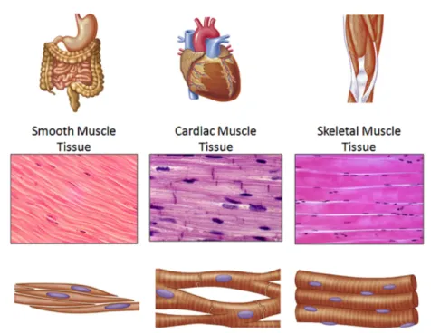
Muscle tissue is one of the four major soft tissues of the human body. The exact number of muscles is difficult to assess due to the different classifications used, but there are approximately 642 muscles mostly organised in pairs due to the bilateral organisation of the human body. According to their organisation, muscles can be subdivided into smooth and striated muscles (Figure 1). Differing by their cellular organisation but showing the same organised contractile filament architecture, cardiac and skeletal muscles are assembled together in the striated muscle subgroup. The skeletal muscles represents the most important fraction of muscle tissue and correspond to a third of our body mass.
Figure 1: Smooth, cardiac and skeletal muscle tissues. Here are presented schematically the differences between the subgroups of muscles. Example of the location and organs constituted by the different types of muscles (top panel), longitudinal histological sections providing an overview of the tissue structure (middle panel) and scheme of the cellular organisation of each type muscle (bottom panel). Figure adapted from © The McGraw Hills companies, Inc.
I.2 Skeletal muscle
Skeletal muscles are voluntary muscles and therefore their contraction is controlled by the voluntary nervous system. Skeletal muscle contraction is responsible for skeleton or body motion that refers to voluntary movements. Skeletal muscles also mediate automatic or semi-automatic functions such as posture, head maintenance and most importantly breathing resulting in the strict requirement of this tissue for human survival. The neuromuscular junction (NMJ) makes the connection between a motor nerve and a muscle fibre and represents the first chemical synapsis described (Dale et al., 1936) and one of the best-studied
(Sanes and Lichtman, 1999).
Skeletal muscle is a unique tissue composed of fused muscle cells which can be considered as forming individual syncytia termed muscle fibres or myofibres (Konigsberg et
al., 1960). These cells share the same highly organised cytoplasm, whereas nuclei are located in a peripheral position under a basal membrane called sarcolemma. Rare quiescent cells located in a satellite position (between the sarcolemma and the basal lamina), termed thereafter satellite cells, can be also observed along the myofibres. Satellite cells represent the main pool of muscle progenitor cells, already committed into the muscular lineage and contributing to muscle regeneration (Chargé and Rudnicki, 2004; Collins et al., 2005).
Each myofibre is wrapped individually in a connective tissue envelope, termed the endomysium (Figure 2). The perimysium is the connective tissue surrounding a fascicle composed of 20 to 40 myofibres. Finally, a third connective tissue envelope termed epymisium protects each whole muscle. While blood vessels circulating between fascicles ensure the distribution of oxygen and nutrients, nerve fibres ensure the transduction of the motor signal from motor nerves to each muscle fibre. At both extremities, the myotendinous junction links muscle fibres to collagen filaments. Grouped, these filaments form the tendon, which is attached to bone’s periosteum (Figure 2).
Figure 2: Skeletal muscle organisation. a) A scheme of the skeletal muscle and b) hematoxylin eosin staining of a transversal muscle section presenting the organisation of the different surrounding envelopes of connective tissues. Also, blood vessels localisation is mentioned and the tendinous structure linking muscle to bone is presented. Figure from © 2006 Pearson Education, Inc.
II. SKELETAL MUSCLE CONTRACTION
Skeletal muscle is composed of a unique highly structured and contractile network required for muscle contraction. Impairment of the proteins constituting these filaments is associated with muscular disorders. An overview of the three types of sarcomeric filaments and the excitation-contraction coupling mechanism mediating voluntary muscle contraction are presented in the object of this section.
Figure 3: The sarcomere as the minimal contraction unit of striated muscles. From the top to the bottom panel, see the magnification of the muscle fibre to the sarcomere and its key components. Panel A represents a muscle fibre and the alternation of dark and light bands. Panel B presents the sarcomere structure and its different zones. Panel C presents the antiparallel arrangement of filaments of myosin (pink) and actin (blue). Note the presentation of titin protein as the third major component of the sarcomere (yellow). Figure adapted from ©Pearson Education, Inc.
As mentioned previously, the muscle fibre is a highly organised syncytium. The sarcoplasm (muscle cytoplasm) includes numerous parallel and aligned myofibrils. These myofibrils compose the contractile protein network providing the specific striated pattern in skeletal and cardiac muscles (Figure 3A). This pattern is due to the alternation of dark and light bands visible by electron microscopy: A-bands (Anisotropic) and I-bands (Isotropic) respectively. Anchoring platforms located in the middle of each band and termed M-line and Z-disk respectively, ensure the maintenance of the striated structure (Figure 3B). These anchoring platforms are essential for both contractile and non-contractile protein localisation, contributing to muscle contraction.
II.1 Muscle contraction unit: the sarcomere
The sarcomere is defined as the 2 µm-length minimal contraction unit delimited by two Z-disks. A sarcomere is thus composed of an A-band and two half I-bands. Thin filaments, mainly composed of actin, form the I-bands, while the main component of the A-bands are thick filaments of myosin (Hanson and Huxley, 1953). The giant protein titin is the third major component of the sarcomere and is responsible for the elasticity of the structure (Figure 3C).
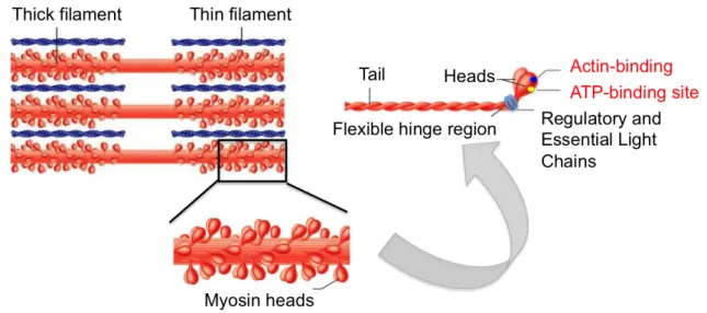
II.1.a Thick filaments and associated proteins
Myosin II (also termed conventional myosin) is a hexamer composed of a dimer of heavy chains (MyHC, approximately 200 kDa) and two pairs of light chains (MyLC, approx. 20 kDa) termed RLC and ECL for Regulatory and Essential light chains (Figure 4). The myosin heavy chains are composed of 2 parts: the C-terminal α-helical coiled-coil tails, able to dimerize and assembly into thick filaments, and the mobile N-terminal heads (Rayment et
al., 1993), which emerge at the surface of the structure to bind the light chains and contain
the ATP- and actin-binding sites.
According to the development phase and muscle fibre specificity, different isoforms of “conventional” Myosin II can be expressed. Two developmental isoforms MYHC-embryonic (MYH3) and MYHC-perinatal (MYH8) can be considered, while there are 3 adult skeletal muscle isoforms: MYHC IIa (MYH2), MYHC IIb (MYH4) and MYHC IIx/d (MYH1). The MYHC-beta/slow (MYH7) is also expressed in cardiac muscle. Myosin isoforms differ by their ATPase activities and therefore defined muscle fibre functional properties. Slow myosin heavy chains are found in type I fibres rich in oxidative enzymes and higly resistant to fatigue. Myosins of type II characterised fast contracting muscle fibres, rich in glycolytic markers.
Also, the myosin binding protein C (MyBP-C, Starr and Offer, 1971) and the highly homologous MyBP-H proteins, known to localise at the A band. MyBP-C has been involved in thick filaments assembly and accessibility of myosin heads, thus participating in muscle contraction (Gilbert et al., 1999; Gruen et al., 1999). Recently, MyBP-C N-terminal domains have been described as decorating thin filaments (Luther et al., 2011; Mun et al., 2011) enhancing its critical requirement for muscle contraction (van Dijk et al., 2014).
Figure 4: Assembly of myosin II-‐containing thick filaments. Thick filaments are the rigid skeletons localised in the middle of the sarcomere (top panel) and composed of a network of multiple myosin hexamers due to myosin heavy chain tail domains, from where emerge myosin heavy chain heads. Visualisation of the assembly of myosin II hexamer (right panel): tail domains ensure the rigidity of the complex, while the mobile domains (the heads) are responsible for the binding to the Light Chains subunits. Myosin heavy chains are also the sites for ATP-‐ and actin-‐binding. Figure adapted from © Pearson Education.
II.1.b Thin filaments and associated proteins
The actin monomer is a 42 kDa globular protein (G-Actin) that can polymerise to form F-Actin, the actin filament. This microfilament consists of a two-stranded helical polymer
(Hanson and Lowy, 1963) stabilised on both sides (helical grooves) by a “backbone” of
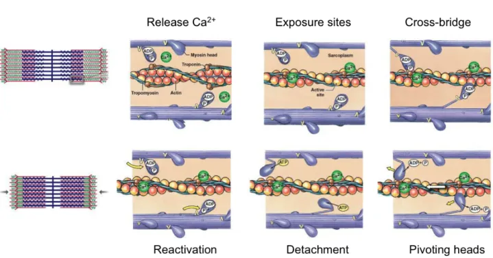
tropomyosin (Corsi and Perry, 1958). Tropomyosin is a homodimer filament capable of movement along the actin filament, thus uncovering myosin-binding sites. Another component of the thin filament is troponin which consists of 3 subunits termed T (tropomyosin binding), I (inhibitory) and C (calcium binding) and is essential for the complex function (Greaser and Gergely, 1971). When the complex conformation changes upon Ca2+ influx, tropomyosin slides and releases myosin-binding sites (Narita et al., 2001, Figure 5).
As actin polymerisation is a continual process, thin filaments are also a polarised structure. Thus, actin has a fast growing “barbed” end localised in the Z disk, and a slow growing “pointed” end (Carlier and Pantaloni, 1997). Tropomodulin is an actin-capping protein of the pointed end (Weber, 1994) and therefore contributes to maintain the final length of F-actin. Tropomodulin binds to tropomyosin and nebulin (Kostyukova et al., 2006;
McElhinny et al., 2001). Nebulin is a giant protein, which spans the entire length of the thin
filament, stabilising and scaffolding the entire structure (Pappas et al., 2010, 2011). Nebulin interacts also with the barbed-end actin-capping protein Cap Z (Pappas et al., 2008).
During Excitation-Contraction (E-C) coupling, the Ca2+ released triggers the accessibility of myosin-binding domains at the surface of actin filaments (mechanism more
extensively described in II.2). In an ATP dependent manner, myosin heads are then able to
generate new cross bridges on actin (Figure 5). This motion in an antiparallel manner of the filaments is responsible for the shortening of the sarcomere.
Figure 5: Muscle contraction and proteins participating to the motion of actin and myosin filaments. Upon Ca2+ realease and its binding to the trimeric troponin complex, tropomyosin filaments located in the groove of the actin filament slide and release myosin binding domains at the surface of actin proteins. In an ATP dependent manner, myosin heads are capable of anchoring to further actin molecules. Therefore, the actin and myosin filaments moving in an antiparallel manner shorten the sarcomere, which leads to muscle contraction. Figure adapted from ©2011 Pearson Education, Inc.
II.1.c Titin filament as a third component of the sarcomere
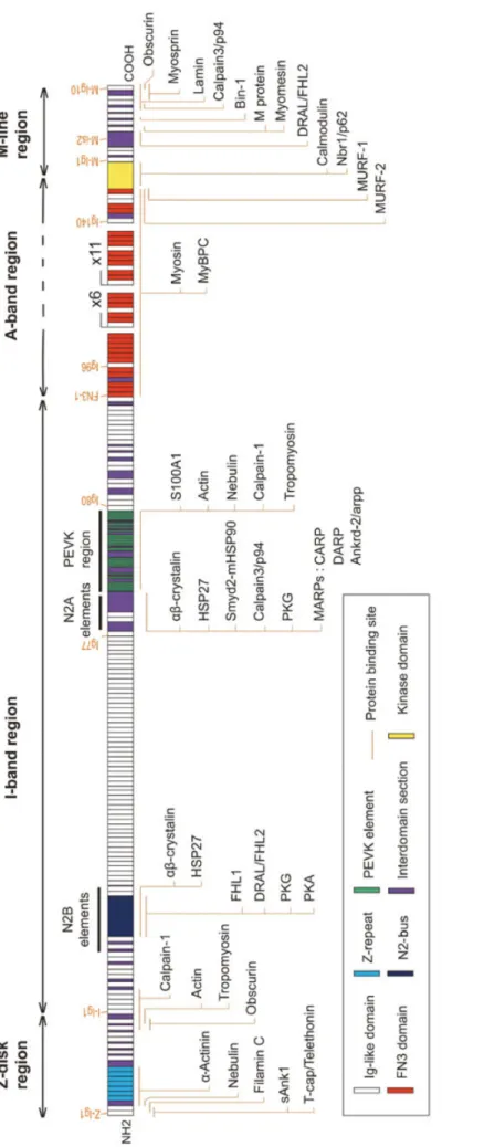
The giant titin protein is the largest protein known in the animal kingdom (its canonical form has a predicted size of 4200 kDa, Bang et al., 2001) and is considered as the third major component of the muscular myofibrillar system. Titin is a 1 µm elastic molecule that anchors to the Z-disk and the M-line and therefore covers half a sarcomere (Labeit et al., 1990). Due to its size, titin binds numerous structural and functional partners along the sarcomere (Figure
6, Chauveau et al., 2014). First, the N-terminal part anchors in the Z disk where it binds,
among others, telethonin and actin (Gautel et al., 1996; Gregorio et al., 1998; Linke et al.,
1997; Mues et al., 1998). The I-band includes the titin PEVK domain (termed after the amino
acid presentation in the motif), critical for sarcomere stiffness (Freiburg et al., 2000; Linke et
al., 1998) and responsible for titin-actin binding (Linke et al., 2002). The A-band domain of
titin consists mainly of repeats of two sequence motifs named type I and type II homologous to fibronectin type III and immunoglobulin-C2 domains respectively (Benian et al., 1989). At the A-band, titin is able to bind myosin (Houmeida et al., 1995) and also its partner MyBP-C partner (Labeit et al., 1992). Finally, the M-line includes the titin C-terminal pseudo-kinase domain, whose activity is widely discussed (Bogomolovas et al., 2014; Gotthardt et al., 2003;
Mayans et al., 1998). The M-line is also stabilised by the ternary complex formed by titin and
its partners myomesin, obscurin and obscurin-like (Agarkova and Perriard, 2005; Fukuzawa
et al., 2008; Pernigo et al., 2010).
Titin is known to play a major role as a blueprint for myofilament assembly and also contributes to sarcomere maintenance (Freiburg and Gautel, 1996; Gotthardt et al., 2003;
Labeit et al., 1992; Miller et al., 2003). However, titin remains a complex and giant protein
Fi gu re 6 : Ti ti n bi n di ng s it es a nd p ar tn er s al on g th e sa rc om er e. Ti ti n i s th e la rg es t m am m al ia n pr ot ei n an d th er ef or e in te ra ct s wi th n um er ou s pa rt ne rs . Th e la te st o ve rv ie w is p ro vi de d in t hi s sc he m e. F ig ur e fr om (C ha uv ea u et a l. , 20 14 ).
II.2 The triadic junction
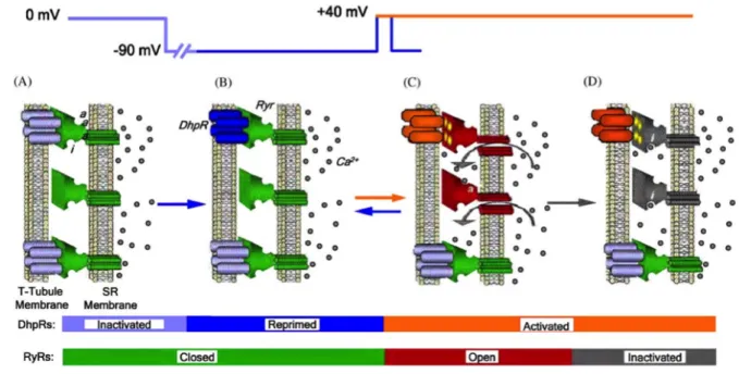
Muscle contraction is driven by the voluntary nervous system. Thus, muscle contraction is initiated by an action potential generated at the neuromuscular junction and propagated along the muscle fibre. This action potential requires an intermediate messenger to mediate muscle contraction; this role is played by calcium ions. Due to the length and the thickness of myofibres, the simultaneous release of calcium along the whole fibre needs a precise excitation – calcium release coupling. These spacial constraints are overtaken by a specific membrane transtalk structure: the triad. The triadic junction is a highly organised interface between the myoplasm (muscle cytoplasm) and the extracellular compartment. A triad is composed of the sarcolemmal membrane invagination termed T-tubule surrounded by two sarcoplasmic reticulum (SR; muscle endoplasmic reticulum) cisternae buttons. Two calcium channels mediate calcium release: the Dihydhropyridine Receptor (DHPR) a voltage-gated calcium channel and the Ryanodine Receptor (RyR) a calcium-gated release channel (Figure 7). DHPR and RyR are localised face to face in the T-tubule and the SR membranes respectively (Curtis and Catterall, 1984; Fleischer et al., 1985; Fosset et al., 1983;
Inui et al., 1987). RYR1, RYR2, and RYR3 encode for RyR channels. RYR1 and RYR2 are
respectively expressed in skeletal muscle and myocardium (Fill and Copello, 2002), whose activation mechanisms are different. In skeletal myofibers, RyR1s and DHPRs are physically coupled (Figure 7), while in cardiac fibres, RyR2s are activated by the influx of Ca2+ through DHPRs after depolarisation; this mechanism is termed Calcium Induced Calcium Release (CIRC) (Protasi, 2002). In both cases, RyRs activation leads to the release of Ca2+ from the SR lumen into the cytosol. Released Ca2+ thereafter mediates the motion of contractile filaments, which in turn is responsible for muscle contraction; this process is therefore termed Excitation-Contraction Coupling (E-C Coupling). The calcium homeostasis of the system is mediated by calsequestrin that sequesters Ca2+ in the SR lumen (MacLennan and Wong, 1971), and the Sarco-Endoplasmic Reticulum Calcium ATPase pumps (SERCA), responsible for Ca2+ recapture (MacLennan et al., 1985).
Figure 7: Modelisation of the Ca2+ release mechanism in skeletal muscle. Two main actors of this triad structure are the DHPRs and RYRs calcium channels located in the T-‐tubule and SR membranes respectively. A) At resting potential, DHPRs are inactivated and RYRs are closed. B) Upon membrane action potential, a part of DHPRs channels are reprimed. After this repriming event, C) activated DHPRs would in turn activate RYRs channels, responsible for Ca2+ release. RYRs channels can be activated upon DHPR activation (coupled channels) or by local elevation of Ca2+ (visualised by Ca2+binding to “a”). According to depolarisation
length, DHPRs can be desactivated and RYRs channels closed – visualised by the reverse transition C) to B). Over a longer depolarisation event, DHPRs remain activated, by a Ca2+
induced inactivation (visualised by Ca2+binding to “i”) RYRs close. Scheme from Klein and
Schneider, 2006
III. MYOGENESIS
Muscle regeneration takes place after muscle injury and recapitulates partially the myogenic process that occurs in pre- and post-natal development to generate skeletal muscle structures. This process is structured in multiple ordered steps that require the sequential activation of cell autonomous muscle-specific transcription factors. The sequential activation of these factors and their requirement for myogenesis have mostly been investigated during muscle development. In vitro and in vivo these factors are crucial for differentiation and myogenesis respectively.
Knowledge about myogenesis has dramatically increased during the past decade. However, most of the precise mechanisms leading to this sequential process remain largely unknown. I will present in this section the current knowledge regarding muscle repair and myogenesis, a physiological mechanism potentially involved in the pathophysiology of multiminicore disease and investigated during my PhD.
III.1 Muscle specific transcriptional factors
III.1.a Paired box (Pax) transcription factors
Pax3 and Pax7 are structurally closely related transcription factors and constitute the subgroup III of the Paired-homeobox (Pax) family. Pax3/7 contribute to the expansion of muscle progenitors and are also required for differentiation during developmental and adult regeneration myogenesis (Collins et al., 2009). Pax3/7 may recruit a H3K4 methyltransferase complex, activating myogenic genes (Diao et al., 2012; McKinnell et al., 2008).
Although both proteins are expressed in muscle primitive progenitors, their contribution to muscle commitment is not redundant. Pax3 is required for early processes such as migration of myogenic precursors in limb buds and body wall. Splotch mutant mice lacking functional Pax3 present limb structures devoid of muscle due to impaired migration of precursors (Daston et al., 1996). Pax7 contribution is restricted to adult age. Pax7 -/-animals develop normally but fail to grow and regenerate (Seale et al., 2000). Recent studies are in agreement with the requirement of Pax7 to muscle progenitor survival (Oustanina et
al., 2004; Relaix et al., 2006). Indeed, Pax3 can be expressed in some satellite cells but cannot compensate the absence of Pax7 (Kuang et al., 2006; Relaix et al., 2005, 2006). An elegant study presents the spacio-temporal contribution of both proteins to the fates of myogenic progenitors during mouse development (Hutcheson et al., 2009).
III.1.b Muscle Regulatory Factors
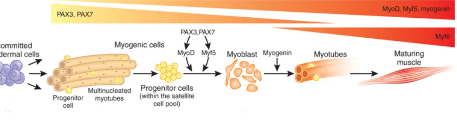
A subgroup of basic Helix-loop-Helix (bHLH) transcription factors constitutes the Muscle Regulatory Factors (MRFs) family (Davis and Weintraub, 1992). Originating from a common ancestral gene, their spatial and temporal expression has been analysed to establish the hierarchy between them (Atchley et al., 1994). The specification and differentiation of myogenic progenitors also depend on the sequential expression of these factors (Figure 8). MRFs bind E-box motifs (CANNTG, where N denotes any base) within muscle-specific gene enhancers and interact with non-myogenic MEF2 factors (Molkentin et al., 1995) to induce transcription. Although the consensus E-box motif is highly represented into the genome (more than 10 million sites), only a small fraction is effectively bound by MRFs in both overlapping and distinctive manners (Blais et al., 2005; Cao et al., 2010). Most of the knockout experiments show the requirement of the MRFs for muscle differentiation, while knock-in experiments highlight their non-overlapping, although sometimes partially redundant, functions.
Figure 8: Key myogenic transcription factors during myogenesis and muscle regeneration. Scheme of expression pattern adopted of Pax3/7 and MRFs. Progenitor cells derivate from the mesodermal cell pool and therefore express Pax3/7. At the adult stage, these cells contribute to the satellite cell pool and maintain solely Pax7 expression. Upon Pax3/7 action, MyoD and Myf5 determining MRF are expressed. Cells proliferate and give rise to myoblasts that will exit cell cycle while expressing the differentiation MRF myogenin, undergo terminal differentiation and fuse in myotubes. During development, these newly formed myotubes generate a mature muscle. When muscle injury occurs, the myoblasts will either repair existing myofibres or generate new muscle fibres. Figure adapted from (Hettmer and Wagers, 2010)
MyoD and Myf5 are the earliest factors involved in the determination of myogenic cells and thus are considered as determinant MRFs. Myogenin (MyoG) appears later during myogenic differentiation and determines a “non-return” state closely associated with terminal differentiation, and therefore is termed differentiation factor. Myf6 (previously MRF4) function is not yet clearly elucidated and appears both as a determination and a differentiation factor (Kassar-Duchossoy et al., 2004).
MyoD was the first bHLH factor identified, able to ectopically induce the expression of skeletal muscle genes in non-muscle cells (Davis et al., 1987; Weintraub et al., 1989). In murine embryos, Myf5 (E8.0) is expressed prior to MyoD (E10.5) in somatic primitive progenitors (Cossu et al., 1996), while the opposite occurs in avian embryos. However, determinant Myf5 and MyoD transcriptional factors exhibit a redundant function: while independent inactivation of MyoD or Myf5 results in a relatively normal myogenesis process, combined inactivation leads to a complete absence of skeletal muscles (Braun et al., 1992;
Rudnicki et al., 1992, 1993). Although MyoD and Myf5 present some overlapping function,
both mark alternative lineages. The epaxial myotome, that leads to trunk musles, preferentially expresses Myf5 while the hypaxial myotome, at the origin of limb muscles, would express MyoD in larger amounts (Kablar et al., 1997). These alternative and independent lineages have been highlighted in recent Myf5-/- mice models (Gensch et al.,
2008; Haldar et al., 2008)
Myogenin (MyoG) is a marker of myogenic commitment and terminal differentiation as evocated by the mutually exclusive expression of MyoG and Pax7 (Olguin and Olwin, 2004). Myogenin knock out mice present perinatal lethality due to a severe impairment of all muscle structures (Hasty et al., 1993; Nabeshima et al., 1993). Myogenin expression is initiated after the determinant MRFs as double knockout Myf5-/-,MyoG-/- or MyoD-/-,MyoG
-/-animals recapitulate the drastic phenotype observed in the MyoG-/- model (Rawls et al., 1995). Thus, Myogenin acts a unique and downstream MRF, mediating terminal differentiation during foetal myogenesis.
Myf6 function has remained elusive, but seems to have a role in both determination and differentiation, consistently with its biphasic expression: first from E9.0 to E11.5 and then
mutants show a severe muscle deficit similar to the one observed in myogenin mutants, anticipating a potential compensatory role for Myf6 with regard to MyoD (Rawls et al., 1998). However, due to the close vicinity of Myf5 and Myf6 genes, the transgenic mice generated often recapitulate a combined Myf5-/-,Myf6-/- phenotype (Braun and Arnold, 1995). Several mutants show the requirement of Myf6 to generate skeletal muscle in Myf5-/-,MyoD -/-model (Kassar-Duchossoy et al., 2004).
Also, Myf6 expression is maintained in mature myofibres highlighting its role in both differentiation and maintenance. Indeed, Myf6 possibly downregulates MyoG since Myf6 knock out mice present a strong upregulation of MyoG (Zhang et al., 1995).
III.2 Myogenic determination and differentiation
Most of the spatial information regarding MRFs expression has been provided by in
vivo experiments. However, the hierarchical relationship and function of the different MRF
members has been established in ex vivo studies using satellite cells and in in vitro C2C12 myoblastic cell line. Adult muscle regeneration is characterised by the activation of the satellite cells, which are the quiescent resident muscle stem cells. Once activated, these cells become myogenic precursors. Activation is followed by the expansion of the myogenic progenitor cell pool prior to cell cycle withdrawal, which is concomitant with differentiation. Terminal differentiation is characterised by the fusion of mononucleated cells and the expression of contractile proteins.
III.2.a Induction of MRF
Upon activation, myogenic precursors are rapidly characterised by the expression of both Myf5 and MyoD, by dissociation of Myf5 sequestered mRNP granules (Crist et al., 2012) and initiation of MyoD transcription (Smith et al., 1994), promoting expansion of the myogenic precursors. Knock out of either MyoD or Myf5 in mdx mice induces muscle regenerative defects (Megeney et al., 1996; Ustanina et al., 2007). In addition to the promotion of myogenic progenitor expansion, MyoD is responsible for the initiation of the muscle differention process, depending on the cellular environment (Blais et al., 2005; Cao et
al., 2010). Indeed, in vitro experiments performed on MyoD-/- and Myf5-/- cells showed
proliferative defects ultimately leading to differentiation impairments (Gayraud-Morel et al.,
2007; Sabourin et al., 1999).
On the opposite, Myogenin induces cell cycle exit and promotes terminal differentiation (Liu et al., 2012). Indeed, knock-in expression of Myogenin driven under the control of Myf5 locus, in Myf5-/-,MyoD-/- background, leads to the generation of skeletal muscles but mice die perinatally due to a reduced number of healthy muscle fibres (Wang
and Jaenisch, 1997).
III.2.b Cell cycle and progenitors expansion
The cell cycle is divided in four periods (or phases), called G1, S, G2 and M (Figure 9) leading to the division of a cell into 2 daughter cells. The cell fate transition (G1) is followed by DNA replication (S), synthesis of mitotic proteins (G2) and cell division (M) providing each daughter cell with the same genetic inheritance (or material) prior to return into G1 for both generated cells. A specific G0 phase refers to non-cycling cells that can, upon activation, reenter into G1 and cycle. Cyclins and cyclin-dependent-kinases (CDKs), whose functions are modulated by CDK inhibitors (CKIs), play an important role in regulating these phase transitions. Two classes of inhibitors are involved: INK4 (inhibitor of CDK4), inhibiting the catalytic subunits of CDK4 and CDK6, and the Cip/Kip family, which acts more broadly on cyclin D-, E- and A-dependent kinases by binding to both cyclin and CDK subunits.
The early myogenic transcription factors MyoD and Myf5 present distinct and contrasting expression patterns during the cell cycle (Kitzmann et al., 1998) with non redundant functions as shown by the defects observed in MyoD-/- or Myf5-/- mdx animals
(Megeney et al., 1996; Ustanina et al., 2007). Myf5 protein levels peak in G0, prior to an
important drop in early G1. Its expression finally rises again at the end of G1 and remains stable through mitosis (Kitzmann et al., 1998). At the end of mitosis, Myf5 phosphorylation leads to its degradation. The modulation of this MRF is supposed to promote initiation of differentiation (Lindon et al., 1998).
MyoD is preferentially expressed during mid-G1 (Kitzmann et al., 1998) and degraded by the ubiquitin proteasome system in late G1 in a CyclinE/CDK2 dependent manner (Song et al., 1998; Tintignac et al., 2000). The maintenance of MyoD beyond the G1 phase can interfere with cell cycle progression and blocks the G1/S transition (Crescenzi et
al., 1990; Sorrentino et al., 1990).
Figure 9: Cell cycle phases and associated proteins in a myogenic context. The four phases of the cell cycle are represented. G1 restricts cell to resting condition or entering the cell cycle. The S phase allows the replication of the genomic material and G2, the synthesis of mitotic proteins. During the M phase, the cell undergoes numerous changes ranging from chromatin condensation to cytokinesis. Cell cycle exit is mediated in a myogenic context by MyoD, which promotes p21 and Rb transcription (black arrows). Rb phosphorylation is mediated by p21 activity. P21 inhibits CDKs activity and prevens Rb phosphorylation required for E2F cell cycle progression transcription factor release.
III.2.c Cell cycle withdrawal
The cell cycle plays a crucial role in myogenesis. Cell cycle components mediating transitions through the different cycle checkpoints are now well known. However, signalling events associated to cell cycle exit are not yet clearly understood. They may drive the cell to quiescence, senescence or differentiation depending on so far poorly defined mechanisms. Upon terminal differentiation, the ultimate fusion of myoblasts and the expression of muscle-specific proteins such as contractile proteins lead to the formation of muscle fibres. It is commonly accepted that terminal differentiation requires cell cycle exit. This statement has been verified in vitro. Indeed, overexpression of cyclins D, E or A inhibits MRF transcriptional activity (Rao et al., 1994; Skapek et al., 1995, 1996) leading to inhibition of terminal differentiation. Conversely, MyoD is responsible for the transcriptional induction of Rb and also Cdkn1a (p21) (Halevy et al., 1995; Martelli et al., 1994), which favour the establishment of a postmitotic status within the cell ( Figure 9, Walsh and Perlman, 1997).
III.2.d Induction of differentiation and fusion
Terminal differentiation is ultimately characterised by an irreversible loss of proliferative capacity coupled to the activation of skeletal muscle related genes. This activation is based on the combined regulation of MyoD and Myogenin (Blais et al., 2005). MyoD favours chromatin accessibility at muscle-specific loci by recruiting ATP-dependent chromatin-remodelling complexes such as SWI/SNF (de la Serna et al., 2005) and the p300 histone acetyltransferase (Dilworth et al., 2004; Puri et al., 1997). Importantly, while binding to a large number of genes along the genome (Cao et al., 2010), MyoD facilitates DNA accessibility for Myogenin (Blais et al., 2005; Cao et al., 2006). Thereby the remodelling function of MyoD promotes the transcriptional activation mediated by Myogenin (Bergstrom
and Tapscott, 2001; Ohkawa et al., 2006). MyoD targets include late muscle differentiation
genes as well as cell cycle withdrawal actors such as Cdkn1a (Cao et al., 2010; Puri et al.,
1997).
III.3 Embryonic myogenesis
III.3.a Specification of domains
Vertebrate myogenesis has been extensively investigated in three main vertebrate models: birds (chick and quails), mammals (mouse) and fish (zebrafish). In vertebrates embryos, trunk and limbs skeletal muscles derive from somites, segments of the paraxial mesoderm formed following the anterior-posterior axis on either side of the neural tube and notochord (Bryson-Richardson and Currie, 2008). Adjacent tissues signalling are responsible for determination of somitic derivates. The paraxial mesoderm changes to transitory somatic compartments: dermomyotome, and sclerotome (Figure 10) leading to the specification of several deriving tissues such as skeletal muscle, cartilage, endothelia and connective tissue. Later the dermomyotome splits in dermatome and myotome to generate trunk, dermis and muscle. This last specification seems to depend upon cell division and spindle orientation
(Ben-Yair and Kalcheim, 2005). Signalling pathways promoting these various tissues start to
emerge. Notch signalling promotes smooth muscle differentiation at skeletal muscle expense
(Ben-Yair and Kalcheim, 2008). High levels of Sonic Hedgehog (Shh), expressed by the
neural tube and the notochord, favour Nkx3.2 expression in the sclerotome and cartilaginous differentiation (Cairns et al., 2008).
III.3.b Specification of muscle primitive progenitors
Vertebrate muscles structures are composed of the trunk, limbs and head muscles. Head muscles originate from the unique convergence of various cell populations and respond to different specification pathways. Limbs and trunk muscles originate from the same multipotent cells population. However, cells movement and the onset of lineage initiating markers differ and would be described thereafter.
Figure 10: Somite segmentation. Following the anterior posterior axis, somites give rise to dermomyotome and sclerotome. The dermomyotome is at the origin of muscle cell progenitors. Figure adapted from (Parker et al., 2003).
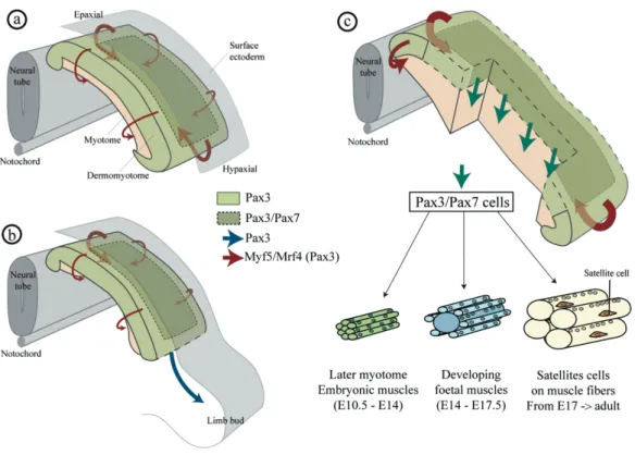
Myogenesis is initiated by delamination of Pax3+ myogenic progenitors cells from dermomyotome lips to the myotome, first skeletal muscle in amniotes (Figure 11a). At E10.5 in mice and 3.5 days in chicks, the central domain of the dermomyotome undergoes an epithelial-to-mesenchymal transition (EMT) following the anterio-posterior gradient (Gros et
al., 2004; Hutcheson et al., 2009). Resident muscle progenitor cells Pax3+ engulf the
myotome (Gros et al., 2005; Kassar-Duchossoy et al., 2005; Relaix et al., 2005) that either proliferate to generate a maintained pool of progenitors or undergo myogenic differentiation to constitute the primitive trunk muscles (Figure 11c). While delamination occurs, Pax3+ multipotent progenitors cells from the hypaxial dermomyotome migrate as single cells (Figure 11b) into limb buds mesenchyme (Birchmeier and Brohmann, 2000; Schienda et al., 2006). In chick embryos these cells also participate to vascular and lymphatic endothelia as well as muscle lineage (He et al., 2003). In mice, the expression of Pax7 restrict the Pax3+/Pax7+ to muscle lineage and thus, to limb muscles (Hutcheson et al., 2009).
Figure 11: Amniotes delamination of trunk and limb muscle progenitors in a mouse embryo. (a) Dermomyotome is composed of Pax3+ multipotent cells that express Myf5 and Myf6
(Mrf4). The edge of this epithelial structure delaminates to form the myotome. (b) Also, cells from the hypaxial dermomyotome migrate and colonise the limb bud. (c) Cells from the central region of the dermomyotome express Pax3/7 engulf the myotome. The following phases of muscle growth and progenitors proliferation depend of this population. Figure from Lagha et al., 2008.
III.3.c Embryonic pathways regulating muscle progenitor cell fate
Embryonic myogenesis is dependent upon numerous pathways also termed positional signals arising from neighbouring tissues: neural tube, notochord and ectoderm (Figure 12). Activation of myogenesis depends of the activation of Muscle Regulatory Factors, such as Myf5 and MyoD. The neural tube preferentially express Wnt1 promoting activation of Myf5 in an axial manner, while Wnt7a is expressed in the dorsal ectoderm and enhances MyoD expression in the hypaxial region (Tajbakhsh et al., 1998). Also sonic hedgehog (Shh), expressed in the floor plate of the neural tube and the notochord, promotes Myf5 in the epaxial region (Borycki et al., 1999a; Gustafsson et al., 2002). Bone Morphogenetic Proteins (BMP) are expressed in the mesoderm and the limb bud ectoderm and mediates the
proliferation maintenance by Pax3 expression while inhibiting MyoD (Amthor et al., 1998). To note, Pax3 expression has also a complementary function in the regulation and activation of MyoD under axial (neural tube and notochord) and surface ectoderm signals (Borycki et
al., 1999b).
Figure 12: Embryonic pathways mediating positional location during embryonic myogenesis. Among them, Wnt, sonic hedgehog (Shh) and bone morphogenetic proteins (BMP) pathways have been widely investigated. Wnt promotes muscle lineage in the epaxial and hypaxial regions of the dermomyotome as well as Shh. BMP promote preferentially proliferation by activating Pax3 and repressing MyoD in the hypaxial region. Pax3 (Pax protein) is quoted in yellow, early MRF factors are labelled in red. Pathways are quoted in blue. Dm dermomyotome, My: myotome, Sc: sclerotome.
III.3.d Foetal myogenesis and muscle growth
Muscle progenitors cells derived from a progenitor pool of Pax3+/Pax7+ committed cells restricted to the central part of the dermomyotome and limb buds, that contribute to myogenesis (Gros et al., 2005; Kassar-Duchossoy et al., 2005; Ben-Yair and Kalcheim, 2005). Double mutant Pax3/Pax7 mice show a startling muscle deficit (Relaix et al., 2005). Cell lacking Pax3/7 expression do not initiate myogenic determination program and die or differentiate in different lineages. Downstream effectors of Pax3/7 genes drive developmental myogenesis. During foetal myogenesis, a fraction of the muscle progenitors would undergo cell cycle withdrawal and provide the first “wave” of fusion, leading to the development of embryonic myofibres at the origin of muscles. Then, during postnatal myogenesis, muscle