ORIGINAL ARTICLE
Oral microbiota in Swiss adolescents
Sigrun Eick&Malgorzata Pietkiewicz&Anton Sculean
Received: 30 September 2011 / Accepted: 13 February 2012 / Published online: 28 February 2012 # Springer-Verlag 2012
Abstract
Objectives The purpose of the study was to determine the prevalence of different oral microbes in gingival plaque samples and in samples from the dorsum of the tongue in a Swiss adolescent population.
Materials and methods Ninety-nine adolescents between 15 and 18 years were enrolled. Plaque index, bleeding on probing (BOP), the periodontal screening index, and decayed missed filled tooth (DMFT) index were recorded. Samples from sub-gingival plaque and swabs from the tongue were analyzed by the Checkerboard DNA-DNA hybridization method. Addi-tionally, counts of Streptococus mutans and Aggregatibacter actinomycetemcomitans, Porphyromonas gingivalis, Tanner-ella forsythia, and Treponema denticola were determined by real-time PCR.
Results Periodontitis was not diagnosed in any of the sub-jects but all of them presented signs of gingival inflamma-tion displaying a mean BOP of 28%. Ten (10.1%) subjects were tested positive for P. gingivalis, each 22 (22.2%) for A. actinomycetemcomitans and T. forsythia, (47.5%) for T. denticola. T. denticola and S. mutans showed a high affinity to the gingival plaque, whereas T. forsythia was often detected from the dorsum of the tongue. DMFT was associated with S. mutans counts, and BOP correlated with counts of P. gingivalis and T. denticola.
Conclusions The present data indicate that: (a) gingivitis but not periodontitis is a common finding among Swiss adoles-cents, and (b) bacteria associated with periodontitis were frequently detected in the subgingival dental plaque and on the dorsum of the tongue in Swiss adolescents with gingivitis. Clinical relevance Although gingivitis was a frequent find-ing in Swiss adolescents, periodontitis was not detected in this population. The dorsum of the tongue appears to repre-sent an important reservoir for periodontopathic bacteria. Keywords Adolescents . Periodontal health . Microflora . Periodontitis-associated bacteria . Streptococcus mutans
Introduction
Initiation and progression of periodontal disease is closely related with the colonization of the microorganisms, including Aggregatibacter actinomycetemcomitans, as well as the mem-bers of the so called“red complex”: Porphyromonas gingivalis, Tannerella forsythia, and Treponema denticola [1–3]. An in-verse relationship between caries and periodontitis has been suggested, underlined by the fact that counts of P. gingivalis and Streptococcus mutans correlate negatively in saliva [4]. Bacteria associated with periodontitis are detectable already in early childhood and adolescents [5,6]. Not only plaque seems to be a reservoir for bacteria associated with periodontitis and S. mutans but also tongue might be colonized by those species already in the very early age of 6 months [7,8].
In older children and adolescents, the detection rates of the periodontopathogens vary. Studies in the 1980s reported a rare detection of P. gingivalis in a study population in London [9] and a presence of A. actinomycetemcomitans in 10% of the subjects in London and Finland [9, 10]. In contrast, detection rates of 100% of those species were S. Eick
:
A. Sculean (*)Department of Periodontology, Dental School, University of Bern, Freiburgstrasse 7,
CH-3010 Bern, Switzerland e-mail: [email protected] M. Pietkiewicz
School Dental Service, Bern, Switzerland
described recently in a Greece population [8]. In a previous study, 33 subjects were included in a follow-up study in Switzerland during puberty [11]. In that study, culture tech-niques have failed to detect P. gingivalis through the whole observation period while A. actinomycetemcomitans was only rarely detected in some of the subjects developing a puberty gingivitis [11].
The purpose of the present study was to evaluate the prevalence of oral bacteria primarily associated with perio-dontitis in subgingival plaque and on the tongue dorsum of Swiss adolescents in relation to clinical variables by using nucleic-acid based methods.
Materials and methods
The ethical committee of the Canton of Bern, CH approved the study (KEK-BE: 235/08). Written informed ascent and consent was obtained from each subject and from their parents respectively prior to participation.
Assessment of epidemiological and clinical data
Ninety-nine adolescents aged between 15 and 18 years and involved in the oral health monitoring protocol of a dental service for children and adolescents in Bern (Switzerland) were enrolled in the present study. Subjects were asked about their smoking habits as well as for significant system-ic disease (e.g., diabetes mellitus, coronary heart disease). Subjects with antibiotic therapy within the last 6 months and pregnant or lactating females were excluded. Further, an ongoing orthodontic treatment at the time-point of the in-vestigation was an exclusion criterion.
From all participants of the study, decayed missed filled teeth (DMFT) index was recorded. Further, periodontal screening index (PSI [12]) was used to characterize periodon-tal health. Probing depths were measured with a periodonperiodon-tal probe (PCP-UNC 15, Hu Friedy, Leimen, Germany). Bleed-ing on probBleed-ing (BOP) was calculated as the percentage of positive sites per subject based on a measurement of four sites/ tooth. Oral hygiene was recorded by using the plaque index described by O’Leary et al. [13].
All clinical recordings were performed by the same cal-ibrated examiner (MP). Examiner calibration was performed as follows: five adolescents, not enrolled in the study, were evaluated by the examiner on two separate occasions, 48 h apart. Calibration was accepted if measurements at baseline and at 48 h were similar to the millimeter at≥90%. Sample collection
For sampling, each of the mesiobuccal site of the first molar was selected. Without removing supragingival plaque [14],
the test site was air dried and kept dry using cotton rolls. Each one paper point (ISO 50) was inserted into the selected gingival sulcus for 20 s to obtain plaque. The samples from the four different sites were pooled in the transport vials. Furthermore, samples from the tongue were taken by swab-bing of about 1 cm2 of the center of the dorsum of the tongue with sterile cotton sticks (Applimed SA, Chàtel-St-Denis, Switzerland). Samples were sent to the laboratory, where they were stored at−20°C until analyzed.
Microbiological analysis
Counts of 21 oral species were determined in each sample using a modification of the checkerboard DNA-DNA hy-bridization technique [15,16]. DNA was extracted by using Chelex method [17] and then 100μl of the extract (half of the total extract) were placed in lanes on a nylon membrane using a Minislot device (Immunetics, Cambridge, MA, USA). (Before applying Chelex to checkerboard, the suit-ability of the method has been tested. This method is easy to make and allows direct comparison of different methods by using the same DNA.) After fixation of the DNA to the membrane, the membrane was placed in a Miniblotter 45 (Immunetics, Cambridge, MA, USA) with the lanes of DNA at 90° to the lanes of the device. Digoxigenin-labeled whole genomic DNA probes to 21 bacterial species were hybrid-ized in individual lanes of the Miniblotter. After hybridiza-tion, the membranes were washed at high stringency, and the DNA probes were detected using antibody to digoxige-nin conjugated with alkaline phosphatase and chemifluor-escence detection. The probes and their source strains were compared as described in a methodological paper by Ximenez-Fyvie et al. [18]. Signals were detected using AttoPhos substrate (Amersham Life Science, Arlington Heights, Illinois, USA) and a Storm Fluorimager (Molecular Dynamics, Sunnyvale, CA, USA). Two lanes in each run contained standards at concentrations of 105and 106cells of each species. The sensitivity of the assay was adjusted to permit detection of 7.5×10⁴ cells of a given species by adjusting the concentration of each DNA probe. Signals were evaluated using the Storm Fluorimager and converted to absolute counts by comparison with the standards on the same membrane. Because of the methodology, cross-reactions cannot be completely excluded between closely related taxa [19]. Following, that closely related species were grouped (Table1) by using the highest signal as result. Additionally to the Checkerboard technique, real-time PCRs of four bacterial species associated with periodontitis (A. actinomycetemcomitans, P. gingivalis, T. forsythia, and T. denticola) and of S. mutans as a cariogenic species were performed as described recently [20,21] modified by using GoTaq® qPCR Master mix (Promega Corporation, Madi-son, WI, USA). The detection level (cut-off) was set to 102
bacteria, although often, lower counts were also measured. In the final analysis, only results of real-time PCR for these species were used. Due to technical problems, the obtained DNA from tongue samples did not cover all PCRs from all subjects. Thus, in part, a few samples are missing in analy-sis; the exact number of included samples is always given in the results.
Data analysis
All data were entered into the PASW 18.0 (SPSS Inc, Chicago, IL, USA) program, and were analyzed using parameter-free methods. Tongue samples and plaque were compared by Wilcoxon test. Mann–Whitney U test was used for comparisons of subgroups within the study population. The level of significance was set to p<0.05.
Results Clinical data
Demographic and clinical data are presented in Table2. All subjects were in the age range between 15 and 18 years. Seventeen (17.1%) were already smokers. Thirty-three (33.3%) of the adolescents were immigrants (13 from
former Yugoslavia, 6 from South Europe, 5 of the other Europe, 6 from Asia and 3 from Africa). The study popula-tion consisted of adolescents aged between 15 and 18 years and enrolled in a dental service program. The adolescents were students of regular schools and schools that provided vocational education. Therefore, the age range was relative-ly small.
None of the study subjects was diagnosed with periodon-titis. All subjects were diagnosed with gingivitis; the BOP was 27.52%±12.42%. Only nine (9.1%) adolescents had PSI01, in 90 (90.9%) additionally calculus and/or defected margins were present (PSI02). The high mean BOP was associated with a mean plaque index of nearly 20%.
In thirty-eight (38.4%) of the participants (DMFT00), no tooth with clinical evidence of tooth decay or respective treatment was detected. In three (3%) of the adolescents, a DMFT >10 was found, the highest score was DMFT015. DMFT differed significantly between natives (1.35±1.56) and immigrants (3.94±4.28; p00.003).
Microorganisms identified in samples from gingival sulci and from tongue samples
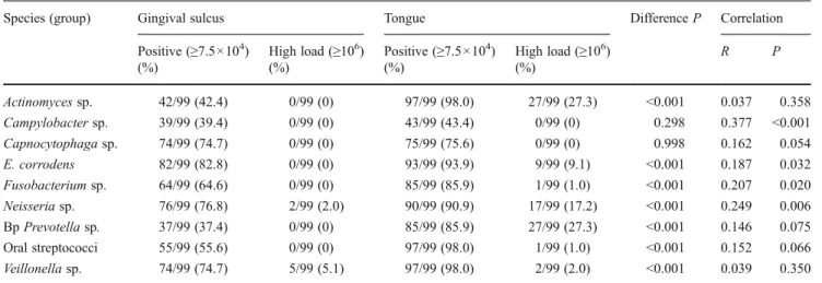
Using a detection level of about 7.5 × 10⁴ microbes in Checkerboard technique, the percentages of the positive samples range between 37.4% (black pigmented Prevotella sp.) and 82.4% (Eikenella corrodens) by analyzing gingival plaque samples, and between 43.3% (Campylobacter sp.) and 97.9% (Actinomyces sp., oral streptococci, Veillonella sp.) by analyzing tongue swabs. Most groups of microbes were detected in higher amounts on tongue than in gingival sulcus. This might depend on the sample size. No difference was found for Campylobacter sp. and Capnocytophaga sp. suggesting an affinity of those for gingival region (Table3).
The proportions of subjects defined as carrying the stud-ied species at different detection levels by real-time PCR are presented (Fig. 1). Using real-time PCR as reference and considering the detection level of Checkerboard technique, comparison of both results showed a sufficient sensitivity Table 1 Grouping of microorganisms used as probes in Checkerboard technique
Group Species
Oral Streptococci S. mitis ATCC 49456, S. oralis ATCC 35037, S. gordonii ATCC 10558
Actinomyces sp. A. odontolyticus ATCC 17929, A. viscosus ATCC 43146/ A. naeslundii ATCC 12104
Neisseria sp. N. mucosa ATCC 19696
Veillonella sp. V. parvula ATCC 10790
Black pigmented (Bp) Prevotella sp. P. intermedia ATCC 25611, P. melaninogenica ATCC 25845
Fusobacteria sp. F. nucleatum ssp. nucleatum ATCC 25586, F. periodonticum ATCC 33693
Capnocytophaga sp. C. gingivalis ATCC 33612, C. ochraceae ATCC 33596
Campylobacter sp. C. rectus ATCC 33238, C. gracilis ATCC 33236
Eikenella corrodens E. corrodens ATCC 23834
Table 2 Demographic and clinical data of the study population Study group
n 99
Gender (m:f) 52:47
Age (mean ±SD) (years) 16.8±1.1
Smoking habits (smoker:non-smoker) 17:82
Plaque index (mean±SD) 19.34%±15.48%
BOP (mean ±SD) 27.52%±12.42%
DMFT (mean±SD) 2.21±3.02
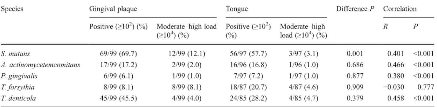
(close to 100%) but in part an inadequate specificity for Checkerboard technique (A. actinomycetemcomitans 34%). Real-time PCR results showed that 78 (78.8%) of all sub-jects were positive when tested for S. mutans; in 22 subsub-jects, the species was found only in plaque; in nine, only on the tongue; whereas in 47 adolescents, it was detectable both in plaque and tongue swabs. Among the ten subjects being positive for P. gingivalis, and the 22 being positive for A. actinomycetemcomitans, each about one third is colonized only on the tongue, in another one third, the species is found only in plaque, and in the last third, in both samples. In case of T. forsythia, only the tongue samples are mostly positive (14 (16.1% of all subjects)); in four subjects (4.6%), the species was detected in plaque and on tongue; whereas in only one subject, the plaque sample exclusively had T. forsythia. Contrary, only in two of the 47T. denticola posi-tive subjects, the species was detectable only on tongue. Except for T. forsythia, the counts in gingival plaque corre-lated with those on tongue (Table4).
Clinical data and microflora
Clinical data were set into association with the microflora. The microflora did not depend on age and gender of the adolescents. Data analysis failed to demonstrate differences
in bacterial counts and smoking status. The BOP scores were, however, higher in smokers (p00.05).
The counts of S. mutans correlated with DMFT index (Table 5, Fig. 2). Subjects with PSI02 had significantly
more Fusobacteria sp. (p00.022) and bp Prevotella sp. (p00.020) in their plaque compared with those having PSI01. The BOP was found to be correlated with P. gingivalis and T. denticola (Fig. 3), whereas the PI was associated with increasing counts of Capnocytophaga sp., T. denticola, and Veillonella sp. (Table 5). The counts of S. mutans were positively correlated with the numbers of T. forsythia (R00.370, p<0.001).
Discussion
The present study aimed to collect information about the colonization of bacteria associated with periodontitis in subgingival sulci and from the dorsum of the tongue in Swiss adolescents. A. actinomycetemcomitans was found by real-time PCR in approximately 20% of the study subjects. Using culture techniques, a similar prev-alence of A. actinomycetemcomitans was found in patients without periodontitis in USA [22] and in Fin-land [10]. Lower detection rates were reported in
1–15-Table 3 Detection of different species by Checkerboard technique
Species (group) Gingival sulcus Tongue Difference P Correlation
Positive (≥7.5×10⁴) (%) High load (≥106) (%) Positive (≥7.5×10⁴) (%) High load (≥106) (%) R P Actinomyces sp. 42/99 (42.4) 0/99 (0) 97/99 (98.0) 27/99 (27.3) <0.001 0.037 0.358 Campylobacter sp. 39/99 (39.4) 0/99 (0) 43/99 (43.4) 0/99 (0) 0.298 0.377 <0.001 Capnocytophaga sp. 74/99 (74.7) 0/99 (0) 75/99 (75.6) 0/99 (0) 0.998 0.162 0.054 E. corrodens 82/99 (82.8) 0/99 (0) 93/99 (93.9) 9/99 (9.1) <0.001 0.187 0.032 Fusobacterium sp. 64/99 (64.6) 0/99 (0) 85/99 (85.9) 1/99 (1.0) <0.001 0.207 0.020 Neisseria sp. 76/99 (76.8) 2/99 (2.0) 90/99 (90.9) 17/99 (17.2) <0.001 0.249 0.006 Bp Prevotella sp. 37/99 (37.4) 0/99 (0) 85/99 (85.9) 27/99 (27.3) <0.001 0.146 0.075 Oral streptococci 55/99 (55.6) 0/99 (0) 97/99 (98.0) 1/99 (1.0) <0.001 0.152 0.066 Veillonella sp. 74/99 (74.7) 5/99 (5.1) 97/99 (98.0) 2/99 (2.0) <0.001 0.039 0.350 Tongue Gingival plaque 0% 20% 40% 60% 80% 100% negative 100 1000 10000 100000 100000 0% 20% 40% 60% 80% 100%
S.mutanz A.actinom. P.gingivalis T.forsythia T.denticola S.mutanz A.actinom. P.gingivalis T.forsythia T.denticola
negative 100 1000 10000 100000 100000 Fig. 1 Detection of S. mutans,
A. actinomycetemcomitans, P. gingivalis, T. forsythia and T. denticola in gingival sulcus samples and swab samples obtained from the dorsum of the tongue
year-old children in Japan [23] using PCR techniques. Remarkable higher rates have been determined in 13-year-old children in Thailand [24] and in less than 3-year-old children in Northern Mariana Islands [7], in a Greek population [8], and in USA [25]. Taken together, these findings appear to suggest geographic or ethnical differences in susceptibility. It should, however, be pointed out that the highest prevalence was found fol-lowing the use of hybridization assays. On the other hand, our group has found a low specificity for A. actinomycetemcomitans when using Checkerboard tech-nique. The method which allows determination of sev-eral species at the same time uses the whole genomic DNA of selected species for hybridization. In our lab-oratory, it was found that cross-reactivity occurs when genetically similar species are present in high numbers (data not shown). As recently demonstrated, the real-time PCRs used in the present study has shown a good correlation with another nucleic-based method [20]. Furthermore, the method was additionally validated by using primers against other regions in the A. actino-mycetemcomitans genome (leukotoxin, cytolethal tox-ins), thus confirming the correct identification of that species. In this study, we did not determine the sero-types of A. actinomycetemcomitans. In general, serotype b is thought to be associated particularly with periodon-tal disease [26] especially with aggressive periodontitis
[27], but serotype distribution depends also on geo-graphical regions and ethnicity of the subjects [27, 28]. The presence of P. gingivalis in about 10% of the subjects is consistent with other data [6,23,29]. It has been shown that in children, the prevalence and load of P. gingivalis increases with age [30]. While in children, P. gingivalis colonizes only transiently; in adolescents, colonization becomes stable [25]. In our study, nearly half of the subjects were colonized by T. forsythia and T. denticola, which were the most frequently identified major pathogens.
Cortelli et al. [5] analyzed T. forsythia, P. gingivalis, and A. actinomycetemcomitans within gingival sulci and on tongue in adolescents; prevalence of the species in gingival sulcus was similar to our results, whereas they found an affinity of those species to gingival region which is not confirmed in the present study. The anatomy of the tongue provides favorable conditions for anaerobes. Six months after full-mouth tooth extraction, periodontopathogens were still detectable on tongue [31]. The high prevalence of periodontopathic bacteria (e.g., fusobacteria, E. corrodens, and T. forsythia) on tongue might not only play a role in the development of periodontal disease. Halitosis is clearly seen in association with tongue coatings containing a high Table 4 Detection of different species by Real-time PCR
Species Gingival plaque Tongue Difference P Correlation
Positive (≥102) (%) Moderate–high load
(≥104) (%) Positive (≥102) (%) Moderate–high load (≥104) (%) R P S. mutans 69/99 (69.7) 12/99 (12.1) 56/97 (57.7) 3/97 (3.1) 0.001 0.401 <0.001 A. actinomycetemcomitans 17/99 (17.2) 2/99 (2.0) 16/96 (16.8) 1/96 (1.0) 0.686 0.466 <0.001 P. gingivalis 6/99 (6.1) 1/99 (1.0) 7/97 (7.2) 1/97 (1.0) 0.877 0.380 <0.001 T. forsythia 8/99 (8.1) 8/99 (8.1) 18/87 (20.7) 4/87 (4.6) 0.909 −0.030 0.777 T. denticola 45/99 (45.5) 4/99 (4.0) 24/85 (28.2) 4/85 (4.7) 0.379 0.458 <0.001
Table 5 Significant correlations of clinical indices with counts of selected microorganisms in gingival plaque
R P
DMFT S. mutans (real-time) 0.225 0.013
BOP P. gingivalis (real-time) 0.218 0.018
T. denticola (real-time) 0.207 0.020 PI Capnocytophaga sp. 0.254 0.011 T. denticola (real-time) 0.301 0.001 Veillonella sp. 0.409 <0.001 0% 20% 40% 60% 80% 100% 0 1 3 4 DMFT negative 100 1'000 10'000 10'000 Fig. 2 Detection of S. mutans in gingival sulcus samples in depen-dence of DMFT index (DMFT 0: n038; DMFT01–3: n036; DMFT≥ 4: n023)
number of anaerobes [32]. The proportion of T. forsythia in tongue dorsum samples was higher in patients with malodor than in those without [33]. In this respect, T. denticola and F. nucleatum have been shown to be significantly associated with indicator measurements of halitosis [34]. The problem of halitosis has been reported already in adolescents [35].
The present study has failed to show any gender-related differences in terms of clinical and microbiological param-eters which is in contrast to an earlier report [10]. On the other hand, our data are comparable to those from a recent study, which has found higher BOP values in adolescent smokers [36]. In the present study, however, smoking was not associated with different microflora. One reason for this discrepancy may be related to the young age of the subjects. It is obvious that due to their age, the examined subjects were not able to smoke for a long time. On the other hand, it should be kept in mind that also in adult periodontitis patients, no clear difference in the subgingival microflora between smokers and non-smokers was observed [37,38].
Very recent epidemiological data suggest that the DMFT is 0.9 in 12-year-old children in Switzerland and among the lowest in Europe [39]. The relatively high mean DMFT measured in this study was, most probably, influenced by the included immigrants. Even in adolescents, the DMFT is higher in immigrants compared to Swiss residents confirm-ing the higher DMFT values in 7-year-old children [40]. The counts of S. mutans as an important causative agent for caries correlated positively with DMFT and with the counts of T. forsythia. An inverse association of S. mutans with clinical indices as well as with other species was never found.
In the present study population, no periodontal pocket depth exceeded 3 mm, but signs of gingival inflammation were visible in each subject. A study by Asikainen et al. [10] has also failed to detect periodontitis in 12–17-year-old residents of Finland. In contrast, Schiffner et al. [41] reported a rate of 13.4% of periodontitis patients in German adolecsents. Previous reports have also indicated that gingi-vitis is a common disease in adolescents [9,10,42,43]. In a study analyzing 624 Swiss army recruits in the age of 18– 24 years, at least one site with pathological periodontal
probing depth and a mean rate of 27% on bleeding at gentle sulcular probing were found in 5.1% of the study population [44]. These findings are in accordance with those from the present study, despite the fact that the study was conducted in a slightly younger Swiss population.
Onset and severity of gingivitis in children has been associated with P. gingivalis and A. actinomycetemcomitans [45]. In a study group of 14–15-year-old subjects with
gingivitis, P. gingivalis, spirochetes, and black pigmented Prevotella sp. but not A. actinomycetemcomitans have been detected in high counts [9]. In a Swiss study analyzing 33 subjects over 6 years during puberty, high counts of spiro-chetes were detected in subjects developing a pubertal gin-givitis [11]. In the present study, counts of P. gingivalis and T. denticola were associated with BOP. Especially, the high prevalence of T. denticola in gingival plaque supports the role of spirochetes in the etiology of gingivitis. Although periodontopathic bacteria are an important causative agent for periodontitis, follow-up studies supporting or neglecting a risk in development are extremely rare. This view is supported by findings from a large, long-term (i.e., up to 7 years), longitudinal epidemiological study in 167 untreat-ed Indonesian adolescents where the presence or absence of A. actinomycetemcomitans in subgingival plaque was asso-ciated with disease development and progression [46,47].
In conclusion, the present data have provided evi-dence that gingivitis but not periodontitis is a common clinical finding among Swiss adolescents. Bacteria asso-ciated with periodontitis are frequently present in this population on the dorsum of the tongue, representing an important reservoir for those species. Follow-up studies should focus on the prevalence of periodontitis-associated species as a possible predictive marker for development of periodontitis.
Acknowledgments We are grateful to Marianne Weibel and Regula Hirschi for their excellent assistance in performing the microbiological analysis. Professor Dr. Wolfgang Strübig is acknowledged for support-ing the adolescents' examinations at the Dental Service Bern. The Department of Periodontology at the University of Bern, Bern, Swit-zerland funded the study.
0% 20% 40% 60% 80% 100% 20 21-30 31-40 40 BOP(%) negative 100 1'000 10'000 100'000 100'000 0% 20% 40% 60% 80% 100% 20 21-30 31-40 40 BOP(%) negative 100 1'000 10'000 10'000 P.gingivalis T.denticola Fig. 3 Detection of P. gingivalis and T. denticola in gingival sulcus samples in dependence of BOP (BOP≤20: n033; BOP 21–30: n030; BOP 31–40: n021; BOP >40: n015)
Conflicts of interest The authors declare that they have no conflicts of interest.
References
1. Holt SC, Ebersole JL (2005) Porphyromonas gingivalis, Trepone-ma denticola, and Tannerella forsythia: the “red complex”, a prototype polybacterial pathogenic consortium in periodontitis. Periodontol 2000 38:72–122
2. (1996) Consensus report. Periodontal diseases: pathogenesis and microbial factors. Ann Periodontol 1:926-32
3. Meng S, Zhao L, Yang H, Wu Y, Ouyang Y (2009) Prevalence of Actinobacillus actinomycetemcomitans in Chinese chronic perio-dontitis patients and periodontally healthy adults. Quintessence Int 40:53–60
4. Iwano Y, Sugano N, Matsumoto K, Nishihara R, Iizuka T, Yoshinuma N, Ito K (2010) Salivary microbial levels in relation to periodontal status and caries development. J Periodontal Res 45:165–169
5. Cortelli JR, Aquino DR, Cortelli SC, Fernandes CB, de Carvalho-Filho J, Franco GC, Costa FO, Kawai T (2008) Etiological analysis of initial colonization of periodontal pathogens in oral cavity. J Clin Microbiol 46:1322–1329
6. Kulekci G, Leblebicioglu B, Keskin F, Ciftci S, Badur S (2008) Salivary detection of periodontopathic bacteria in periodontally healthy children. Anaerobe 14:49–54
7. Tanner AC, Milgrom PM, Kent R Jr, Mokeem SA, Page RC, Riedy CA, Weinstein P, Bruss J (2002) The microbiota of young children from tooth and tongue samples. J Dent Res 81:53–57
8. Papaioannou W, Gizani S, Haffajee AD, Quirynen M, Mamai-Homata E, Papagiannoulis L (2009) The microbiota on different oral surfaces in healthy children. Oral Microbiol Immunol 24:183–189 9. Ashley FP, Gallagher J, Wilson RF (1988) The occurrence of
Actinobacillus actinomycetemcomitans, Bacteroides gingivalis, Bacteroides intermedius and spirochaetes in the subgingival mi-croflora of adolescents and their relationship with the amount of supragingival plaque and gingivitis. Oral Microbiol Immunol 3:77–82
10. Asikainen S, Alaluusua S, Kari K, Kleemola-Kujala E (1986) Subgingival microflora and periodontal conditions in healthy teen-agers. J Periodontol 57:505–509
11. Mombelli A, Rutar A, Lang NP (1995) Correlation of the peri-odontal status 6 years after puberty with clinical and microbiolog-ical conditions during puberty. J Clin Periodontol 22:300–305 12. Cutress TW, Hunter PB, Beck DJ, de Souza P (1978) A
compar-ison of WHO periodontal status index with the periodontal and oral hygiene indices. Community Dent Oral Epidemiol 6:245–252 13. O’Leary TJ, Drake RB, Naylor JE (1972) The plaque control
record. J Periodontol 43:38
14. Beikler T, Schnitzer S, Abdeen G, Ehmke B, Eisenacher M, Flemmig TF (2006) Sampling strategy for intraoral detection of periodontal pathogens before and following periodontal therapy. J Periodontol 77:1323–1332
15. Socransky SS, Smith C, Martin L, Paster BJ, Dewhirst FE, Levin AE (1994)“Checkerboard” DNA-DNA hybridization. Biotechni-ques 17:788–792
16. Haffajee AD, Cugini MA, Dibart S, Smith C, Kent RL Jr, Socransky SS (1997) The effect of SRP on the clinical and microbiological parameters of periodontal diseases. J Clin Periodontol 24:324–334 17. Yang JL, Wang MS, Cheng AC, Pan KC, Li CF, Deng SX (2008)
A simple and rapid method for extracting bacterial DNA from intestinal microflora for ERIC-PCR detection. World J Gastroen-terol 14:2872–2876
18. Ximenez-Fyvie LA, Haffajee AD, Socransky SS (2000) Microbial composition of supra- and subgingival plaque in subjects with adult periodontitis. J Clin Periodontol 27:722–732
19. Socransky SS, Haffajee AD, Smith C, Martin L, Haffajee JA, Uzel NG, Goodson JM (2004) Use of checkerboard DNA-DNA hybrid-ization to study complex microbial ecosystems. Oral Microbiol Immunol 19:352–362
20. Eick S, Straube A, Guentsch A, Pfister W, Jentsch H (2011) Comparison of real-time polymerase chain reaction and DNA-strip technology in microbiological evaluation of periodontitis treatment. Diagn Microbiol Infect Dis 69:12–20
21. Yoshida A, Suzuki N, Nakano Y, Kawada M, Oho T, Koga T (2003) Development of a 5′ nuclease-based real-time PCR assay for quantitative detection of cariogenic dental pathogens Strepto-coccus mutans and StreptoStrepto-coccus sobrinus. J Clin Microbiol 41:4438–4441
22. Slots J, Reynolds HS, Genco RJ (1980) Actinobacillus actino-mycetemcomitans in human periodontal disease: a cross-sectional microbiological investigation. Infect Immun 29:1013–1020 23. Umeda M, Miwa Z, Takeuchi Y, Ishizuka M, Huang Y, Noguchi K,
Tanaka M, Takagi Y, Ishikawa I (2004) The distribution of perio-dontopathic bacteria among Japanese children and their parents. J Periodontal Res 39:398–404
24. Dahlen G, Konradsson K, Eriksson S, Teanpaisan R, Piwat S, Carlen A (2010) A microbiological study in relation to the pres-ence of caries and calculus. Acta Odontol Scand 68:199–206 25. Lamell CW, Griffen AL, McClellan DL, Leys EJ (2000)
Acquisi-tion and colonizaAcquisi-tion stability of Actinobacillus actinomycetemco-mitans and Porphyromonas gingivalis in children. J Clin Microbiol 38:1196–1199
26. Socransky SS, Haffajee AD, Smith C, Dibart S (1991) Relation of counts of microbial species to clinical status at the sampled site. J Clin Periodontol 18:766–775
27. Jentsch H, Cachovan G, Guentsch A, Eickholz P, Pfister W, Eick S (2012) Characterization of Aggregatibacter actinomycetemcomi-tans strains in periodontitis patients in Germany. Clin Oral Inves-tig. 2012 Jan 14. [Epub ahead of print]
28. Kim TS, Frank P, Eickholz P, Eick S, Kim CK (2009) Serotypes of Aggregatibacter actinomycetemcomitans in patients with different ethnic backgrounds. J Periodontol 80:2020–2027
29. Sirinian G, Shimizu T, Sugar C, Slots J, Chen C (2002) Periodon-topathic bacteria in young healthy subjects of different ethnic backgrounds in Los Angeles. J Periodontol 73:283–288
30. Bimstein E, Sapir S, Houri-Haddad Y, Dibart S, Van Dyke TE, Shapira L (2004) The relationship between Porphyromonas gingi-valis infection and local and systemic factors in children. J Perio-dontol 75:1371–1376
31. Van Assche N, Van Essche M, Pauwels M, Teughels W, Quirynen M (2009) Do periodontopathogens disappear after full-mouth tooth extraction? J Clin Periodontol 36:1043–1047
32. Quirynen M, Avontroodt P, Soers C, Zhao H, Pauwels M, van Steenberghe D (2004) Impact of tongue cleansers on microbial load and taste. J Clin Periodontol 31:506–510
33. Tanaka M, Yamamoto Y, Kuboniwa M, Nonaka A, Nishida N, Maeda K, Kataoka K, Nagata H, Shizukuishi S (2004) Contribu-tion of periodontal pathogens on tongue dorsa analyzed with real-time PCR to oral malodor. Microbes Infect 6:1078–1083 34. Yasukawa T, Ohmori M, Sato S (2010) The relationship between
physiologic halitosis and periodontopathic bacteria of the tongue and gingival sulcus. Odontology 98:44–51
35. Kanli A, Kanbur NO, Dural S, Derman O (2008) Effects of oral health behaviors and socioeconomic factors on a group of Turkish adolescents. Quintessence Int 39:e26–e32
36. Hermann P, Gera I, Borbely J, Fejerdy P, Madlena M (2009) Periodontal health of an adult population in Hungary: findings of a national survey. J Clin Periodontol 36:449–457
37. Apatzidou DA, Riggio MP, Kinane DF (2005) Impact of smoking on the clinical, microbiological and immunological parameters of adult patients with periodontitis. J Clin Periodontol 32:973–983 38. Van der Velden U, Varoufaki A, Hutter JW, Xu L, Timmerman MF,
Van Winkelhoff AJ, Loos BG (2003) Effect of smoking and periodontal treatment on the subgingival microflora. J Clin Perio-dontol 30:603–610
39. Malmö, University (2011) http://www.mah.se/CAPP/Country-Oral-Health-Profiles/EURO/#5
40. Marthaler TM (2004) Changes in dental caries 1953-2003. Caries Res 38:173–181
41. Schiffner U, Hoffmann T, Kerschbaum T, Micheelis W (2009) Oral health in German children, adolescents, adults and senior citizens in 2005. Community Dent Health 26:18–22
42. Campus G, Cagetti MG, Senna A, Spano G, Benedicenti S, Sacco G (2009) Differences in oral health among Italian adolescents related to the type of secondary school attended. Oral Health Prev Dent 7:323–330
43. Ericsson JS, Abrahamsson KH, Ostberg AL, Hellstrom MK, Jonsson K, Wennstrom JL (2009) Periodontal health status in
Swedish adolescents: an epidemiological, cross-sectional study. Swed Dent J 33:131–139
44. Rothlisberger B, Kuonen P, Salvi GE, Gerber J, Pjetursson BE, Attstrom R, Joss A, Lang NP (2007) Periodontal conditions in Swiss army recruits: a comparative study between the years 1985, 1996 and 2006. J Clin Periodontol 34:860–866
45. Morinushi T, Lopatin DE, Van Poperin N, Ueda Y (2000) The relationship between gingivitis and colonization by Porphyromo-nas gingivalis and Actinobacillus actinomycetemcomitans in chil-dren. J Periodontol 71:403–409
46. Timmerman MF, Van der Weijden GA, Arief EM, Armand S, Abbas F, Winkel EG, Van Winkelhoff AJ, Van der Velden U (2001) Untreated periodontal disease in Indonesian adolescents. Subgingival microbiota in relation to experienced progression of periodontitis. J Clin Periodontol 28:617–627
47. Timmerman MF, Van der Weijden GA, Abbas F, Arief EM, Armand S, Winkel EG, Van Winkelhoff AJ, Van der Velden U (2000) Untreated periodontal disease in Indonesian adolescents. Longitudinal clinical data and prospective clinical and microbio-logical risk assessment. J Clin Periodontol 27:932–942