*Corresponding author: Wassim Eid, Biochemistry Department, Medical Research Institute, University of Alexandria, 165 Horreya Street, 21311 Alexandria, Egypt; and Division of Endocrinology, Department of Medicine, University of Fribourg, 1700 Fribourg, Switzerland, e-mail: wassim.eid@unifr.ch
Wafaa Abdel-Rehim: Biochemistry Department, Medical Research Institute, University of Alexandria, 165 Horreya Street, 21311 Alexandria, Egypt
Wassim Eid* and Wafaa Abdel-Rehim
Vitamin C promotes pluripotency of human
induced pluripotent stem cells via the histone
demethylase JARID1A
Abstract: Somatic cells can be reprogramed into induced
pluripotent stem (iPS) cells by defined factors, which pro-vide a powerful basis for personalized stem-cell based therapies. However, cellular reprograming is an inefficient and metabolically demanding process commonly associ-ated with obstacles that hamper further use of this tech-nology. Spontaneous differentiation of iPS cells cultures represents a significant hurdle that hinder obtaining high quality iPS cells for further downstream experimentation. In this study, we found that a natural compound, vita-min C, augmented pluripotency in iPS cells and reduced unwanted spontaneous differentiation during iPS cells maintenance. Gene expression analysis showed that vita-min C increased the expression of the histone demethylase JARID1A. Furthermore, through gain- and loss-of- function approaches, we show that JARID1A is a key effector in promoting pluripotency and reducing differentiation downstream of vitamin C. Our results therefore highlight a straightforward method for improving the pluripotency and quality of iPS cells; it also shows a possible role for H3K4me2/3 in cell fate determination and establishes a link between vitamin C and epigenetic regulation.
Keywords: iPS cells; JARID1A; reprograming;
spontane-ous differentiation; vitamin C.
Introduction
Animal development starts with the fertilized egg undergoing a programed process of cell proliferation
and differentiation that generates all cell types of an individual. This process of nuclear reprograming to pluri-potent state was first achieved by the transfer of somatic cell nucleus into an enucleated egg (Wilmut et al., 1997). More recently, Yamanaka and colleagues demonstrated that somatic cells can acquire pluripotent state after the introduction of a defined combination of transcription factors that are highly enriched in embryonic stem cells (ESCs), which were termed induced pluripotent stem (iPS) cells (Takahashi et al., 2007). iPS cells possess the ability to differentiate into the three different germ layers, ecto-derm, mesoderm and endoecto-derm, raising the possibility of clinical application of personalized stem-cell based therapies without immune rejection or ethical concerns. Human iPS cells also provide a unique platform for study-ing genetic diseases in vitro (Park et al., 2008). However,
the reprograming of somatic cells to a pluripotent state is prone to errors that could impede the use of this tech-nology (Esteban and Pei, 2012). One important obstacle hampering the utilization of iPS cells is the spontaneous differentiation of iPS colonies (Belinsky and Antic, 2013), which represents a significant handicap for mechanistic studies and high throughput screening, and makes bona fide colony isolation time consuming and costly. In fact, from a practical perspective, a colony with too much dif-ferentiation, should be discarded and not used for further downstream applications.
Improving the quality of iPS cells has been tackled by recent work. In one study, mild hypothermia led to decreased differentiation in ESCs and iPS cells (Belin-sky and Antic, 2013), while another study showed that vitamin C enhances the quality of somatic cell reprogram-ing in mice and human cells (Esteban and Pei, 2012), highlighting the possibility that further manipulation of culture conditions could improve the technology of iPS cells for regenerative medicine. Vitamin C has received a great deal of interest because of its involvement in the generation of induced pluripotent stem cells from both mouse and human somatic cells (Shi et al., 2008). The molecular regulation network of vitamin C during repro-graming seems to be quite versatile: vitamin C has been reported to improve iPS cells generation by demethylation
http://doc.rero.ch
Published in "Biological Chemistry 397(11): 1205–1213, 2016"
which should be cited to refer to this work.
also found to be an agonist of the histone demethylase Jhdm1a/1b (Wang et al., 2011) and to keep normal Dlk1-Dio3 cluster imprinting status (Stadtfeld et al., 2012). In addition, vitamin C inhibits activation of the NF-κB sign-aling (Carcamo et al., 2002, 2004) and decreases reactive oxygen species (ROS), protecting cells from senescence and death (Shi et al., 2010). Moreover, vitamin C decreases p53-p21 signaling, a barrier for iPS cells generation (Hong et al., 2009). The epigenetic state of a cell is determined by the unique pattern of DNA methylation and histone modifications, and is responsible for cell- and tissue-specific gene expression patterns. Reprograming must involve mechanisms responsible for changing the epi-genetic status of both DNA and histones genome-wide. Interestingly, despite the fact that the acquisition of the pluripotent state during reprograming entails both DNA methylations at the promoters of somatic genes (Lister et al., 2009) and the DNA demethylations at the promoters of pluripotent genes, the two de novo DNA
methyltrans-ferases Dnmt3a and Dnmt3b are dispensable (Pawlak and Jaenisch, 2011), suggesting that additional epigenetic modifiers play a relevant role.
In this study, we demonstrate that vitamin C signifi-cantly reduces the incidence of spontaneous differentia-tion in iPS cells, promotes pluripotency and maintains an undifferentiated state of the colonies, at least in part by facilitating the expression and function of histone dem-ethylase JARID1A and promoting the demethylation of H3K4me2/3 histone marks.
Results
Generation of human iPS cells from adult
dermal fibroblasts
Human dermal fibroblasts were transduced with retro-viruses designed to express the human cDNAs encod-ing OCT4, SOX2, KLF4 and C-MYC genes, as previously described (Takahashi et al., 2007). Six days after trans-duction, the cells were plated onto mitotically inactivated mouse embryonic fibroblasts (MEFs) which served as a feeder layer. Two days later, the medium was replaced by iPS medium supplemented with 4 ng/ml basic fibroblast growth factor (bFGF). After approximately 4 weeks, colo-nies that resembled iPS cells were observed. To check the stemness of the iPS cell line, the expression of core tran-scription factors and surface markers including octamer-binding 3/4 (OCT4), SRY-box containing gene 2 (SOX2),
TRA1-60 and TRA1-81 was tested using reverse transcrip-tion PCR (RT-PCR) or immunofluorescence. The results showed that the expression pattern of these pluripotency markers in the generated iPS cell line is similar to that of the well-established human ES cell line H1, further-more, neither the iPS nor H1 cells expressed stage specific embryonic antigen-1 (SSEA-1) (Supplementary Figure 1).
Vitamin C maintains self-renewal and colony
morphology of iPS cells
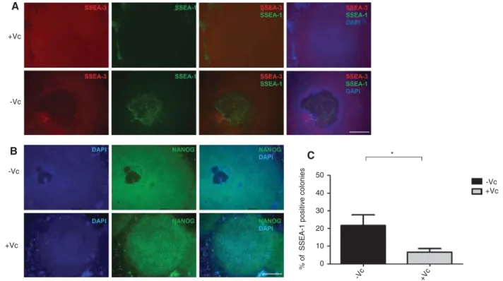
To examine the effect of vitamin C on spontaneous differ-entiation of iPS cells and whether vitamin C is effective in maintaining pluripotency, we assessed the expression of NANOG, the expression of the pluripotency surface marker SSEA-3 as well as the expression of the stage- specific embryonic antigen (SSEA)-1, SSEA-1 can be used as a positive surface marker for mouse undifferentiated stem cells and negative surface marker for human undif-ferentiated stem cells. Expression is down-regulated fol-lowing differentiation of murine stem cells, while the differentiation of human stem cells is accompanied by an increase in SSEA-1 expression (Draper et al., 2002; Takahashi et al., 2007). After 7 days of culturing in the presence or absence of vitamin C, we observed that the number of iPS colonies developing spontaneous differ-entiation was significantly lower when the medium was supplemented with vitamin C, compared to colonies grown in basal iPS medium (Figure 1); the differenti-ated spots were hallmarked by the loss of expression of NANOG and SSEA-3 accompanied by an increased expression of SSEA-1 (Figure 1A and B). Collectively, the results indicate that vitamin C promotes the mainte-nance of pluripotency and inhibits differentiation.
Vitamin C augments the expression of
pluri-potency genes
To evaluate the effect of vitamin C on the expression of pluripotency genes, we measured the relative expres-sion levels of several pluripotency genes in iPS cells following vitamin C treatment for 7 days by qualitative real-time PCR (qRT-PCR), cells grown in basal medium were used as controls. We observed that numerous pluri-potency genes (Takahashi et al., 2007), such as OCT3/4, NANOG, growth and differentiation factor 3 (GDF3), reduced expression 1 (REX1), undifferentiated embryonic cell transcription factor 1 (UTF1) and fibroblast growth
factor 4 (FGF4) showed slight but significant increase of expression in vitamin C-treated cells compared to cells grown in basal medium (Figure 2A). These results
revealed that vitamin C may promote pluripotency gene expression and facilitate maintenance of iPSCs cells in an undifferentiated state. +Vc * 50 -Vc 40 % of SSEA-1 positiv e colonies 30 20 10 0 +Vc +Vc -Vc -Vc +Vc -Vc A B C
Figure 1: Vitamin C promotes pluripotency of iPS cells and protects from differentiation.
(A) iPS colonies grown on a feeder layer in basal medium or basal medium supplemented with vitamin C. Cells were fixed and co- immunostained with SSEA-1 and SSEA-3 and analyzed by fluorescence microscopy. (B) Cells treated as in (A) were immunostained with a NANOG antibody. (C) The number of colonies showing SSEA-1 expression was quantified in the absence of vitamin C (-Vc, black) or its presence (+Vc, gray). A total of 50 colonies per condition were counted in three independent experiments. Shown are representative images of SSEA-1, SSEA-3 and NANOG expression. Nuclei were visualized by DAPI. Scale bar 50 μm (A) and 5 μm (B). Error bars represent mean±standard deviation (n = 3). The asterisk (*) indicates a statistically significant difference (p < 0.05). Vc, Vitamin C.
Figure 2: Vitamin C increases pluripotency gene expression.
(A) qRT-PCR quantification of mRNA levels for pluripotency genes in iPS cells grown in basal medium (control, white) or basal medium supplemented with vitamin C (gray). Relative expression levels of target genes were determined by qRT-PCR after normalization to GAPDH as an endogenous control. Primers used for Oct3/4 specifically detect the transcript from the endogenous genes, but not from the retroviral tran-script. (B) qRT-PCR quantification of mRNA levels of various differentiation markers for the three germ layers in iPS cells grown in basal medium (white) or basal medium supplemented with vitamin C (black). The data in all graphs are the average of three independent experiments, error bars represent standard deviation from the mean and values are expressed as relative to control = 1. ***p < 0.001; **p < 0.01; *p < 0.05.
Effect of vitamin C on differentiation markers
Induced pluripotent stem cells possess the ability to dif-ferentiate into the three different germ layers: ectoderm, mesoderm and endoderm. To further investigate the role of vitamin C in maintaining an undifferentiated state of iPS colonies, we used qRT-PCR to measure the mRNA levels of markers of the three germ layers following vitamin C treatment for 7 days. Cells grown in basal medium showed increased expression of forkhead box A2 (FOXA2, a marker of endoderm), α feto protein (endoderm), BRA-CHURY (mesoderm), Msh homeobox-1 (MSX1, mesoderm), microtubule-associated protein 2 (MAP2, ectoderm), and paired box 6 (PAX6, ectoderm), compared to cells supple-mented with vitamin C (Figure 2B). Taken together, these results revealed that vitamin C decreases the expression of differentiation markers and preserves iPS colonies in an undifferentiated state.
Vitamin C promotes the demethylation of
H3K4me2/me3 through JARID1A
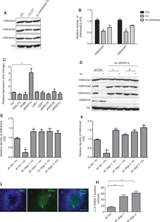
DNA methylation and histone demethylases play a central role in the reprograming process and induction of pluripo-tency (Pawlak and Jaenisch, 2011); vitamin C is a known cofactor for multiple histone demethylases (Horton et al., 2010; Young et al., 2015) and is known to modulate gene expression of several genes (Carinci et al., 2005; Belin et al., 2010; Canali et al., 2014). This led to the hypoth-esis that spontaneously differentiated colonies undergo changes in the histone methylation status which even-tually abolish the acquired pluripotency status and that the presence of vitamin C might inhibit this process. To test this idea, we extracted total histones from iPS colo-nies untreated or treated with vitamin C and analyzed the methylation status by Western blotting using antibodies against mono-, di- and tri-methylated histones H3 at K4, K9, K27 and K36. Vitamin C reduced the levels of H3K4 me2/3 at day 7 posttreatment (Figure 3A), whereas other tested histone methylation marks did not change notice-ably (data not shown). Upon removal of vitamin C from the culture medium, the level of H3K4 me2/3 recovered in 3 days (Figure 3A), indicating that the effect of vitamin C on H3K4 methylation is reversible. We then questioned which DNA demethylase(s) could be responsible for reduced DNA methylation after vitamin C treatment. To address this, we extracted total RNA from iPS colonies grown with or without vitamin C and checked for the expression levels of demethylases that are known to di- and tri-methylate H3K4 (Kooistra and Helin, 2012). Expression analysis
showed that expression of the DNA demethylase JARID1A, also known as KDM5A/RBP2, was significantly higher upon treating cells with vitamin C; the expression of some other demethylases were slightly increased after vitamin C treatment, however, this increase was not significant (Figure 3B). These results suggested that vitamin C influ-ences pluripotency by removing H3K4me2/3 through modulating the expression of the histone demethylase JARID1A. JARID1A is a member of the Jumonji family of proteins and has been shown to demethylate H3K4me2/3 (Kooistra and Helin, 2012). To analyse the role of JARID1A in maintaining pluripotency, we knocked it down using two different shRNA lentiviral vectors. Knocking down JARID1A resulted in a concomitant increase in the levels of H3K4 me2/3, in agreement with vitamin C reducing this histone mark through JARID1A (Figure 3C). Importantly, knocking down JARID1A also increased differentiation in the absence of vitamin C (Figure 3D and E). This shows that JARID1A is important for maintaining a pluripotent state of iPS cells and protecting against spontaneous differentiation.
We then performed gain-of-function studies by trans-fecting JARID1A into the cells. Overexpressing JARID1A reduced the spontaneous differentiation in the presence (more significantly) or absence of vitamin C (Figure 4A–C). Our results thus suggest that JARID1A play a relevant role in vitamin C-maintenance of pluripotency.
Discussion
The reprograming of somatic cells to a pluripotent state marks the dawn of a new era in biomedical research and personalized regenerative medicine as well as disease modeling. However, the conversion of somatic cells to induced pluripotent stem cells by forced expression of defined factors is an inefficient process by which somatic cells need to overcome numerous physiological barriers to reach the pluripotent state (Esteban and Pei, 2012; Qi et al., 2015). In recent years, some studies have revealed that vitamin C enhances the quality of somatic cell repro-graming (Esteban et al., 2010; Shi et al., 2010) by modu-lation of signaling pathways and influencing epigenetic modification to reset the epigenome to an embryonic-like state (Wang et al., 2011), highlighting the possibility that further manipulation of culture conditions could improve this technology for regenerative medicine and downstream applications. To this goal, we generated human iPS cells and evaluated the role of vitamin C, a common nutrient vital to human health, on the spontaneous differentiation of human iPS colonies. Our results revealed an enhanced
1.5 1.0 5 H3K4me1 H3K4me2 H3K4me3 H3 H3K4me1 H3K4me2 H3K4me3 JARID1A H3 Vc sh Scr 1 2 sh JARID1A + + + - - -Ctr l +Vc D7 Vc withdr aw al D3 ** 4 3 2 1 0 2.0 1.5 1.0 0.5 0.0 80 60 % of SSEA-1 positiv e colonies 40 20 0 sh Scr sh oligo 1 sh oligo 2 2.0 1.5 1.0 0.5 0.0 ** ** ** ** ** ** ** * * * * * * Relativ e density of H3K4me2 (H3) Relativ e density of H3K4me3 (H3) sh Scr -Vcsh Scr +Vc
sh oligo 1 -Vcsh oligo 1 +Vcsh oligo 2 -Vcsh oligo 2 +Vc
Relativ
e e
xpression (f
old change)
sh Scr -Vcsh Scr +Vc
sh oligo 1 -Vcsh oligo 1 +Vcsh oligo 2 -Vcsh oligo 2 +Vc
Ctr l
FBXL10NO66JARID1A LSD1 LSD1
JARID1DJARID1BJARID1C
0.5 0.0 H3K4me2 H3K4me3 Ctrl Vc Vc withdrawal Relativ e density of H3K4me2/3 (H3) A B C D E F G H ** ** sh Scr 1 2 + + + - - -** * * * * * * ** ** **
Figure 3: Vitamin C promotes the demethylation of H3K4me2/3 and augments pluripotency of iPS cells via JARID1A.
(A) Expression of H3K4me1/2/3 was assessed in iPS cells grown in basal iPS medium (Ctrl, lane 1) or cells grown with vitamin C (Vc) for 7 days (D7, middle lane) or cells grown for 3 days (D3, right lane) after removal of vitamin C from the medium. Total histone 3 (H3) was used as loading control. (B) The relative density of H3K4me2/3 in (A) normalized to H3 (C) qRT-PCR quantification of mRNA levels for histone demethylases that use H3K4me2/3 as a substrate. Relative expression levels of target genes were determined by qRT-PCR after normalization to GAPDH as an endogenous control. Ctrl: cells grown in basal iPS medium without vitamin C. (D) Expression of H3K4me1/2/3 and JARID1A in iPS cells transduced with either scrambled shRNA (sh Scr) or two different shRNA oligos (sh oligo 1 and sh oligo 2) targeting JARID1A. (E and F) The rela-tive density of H3K4me2/3 in (D) normalized to H3 (G) SSEA-1 expression in iPS cells upon knocking down JARID1A using shRNA, shown is a representative image. (H) The number of colonies showing SSEA-1 expression was quantified after knocking down JARID1A. Scale bar 50 μm. A total of 50 colonies per condition were counted in three independent experiments. The data in all graphs are the average of three independent experiments, error bars represent standard deviation from the mean and values are expressed as relative to control = 1. **p < 0.01; *p < 0.05.
pluripotency status and diminished spontaneous differ-entiation in colonies grown with vitamin C compared to colonies grown in basal medium. A hallmark of iPS cells colonies is their ability to differentiate into the three germ layers (Takahashi et al., 2007). We show that vitamin C significantly reduces the expression of various differentia-tion markers, helping maintain the colonies in an undif-ferentiated state.
We then questioned the mechanism by which vitamin C might be promoting pluripotency and inhibiting spon-taneous differentiation. Cellular reprograming involves extensive changes to the epigenetic status of DNA and his-tones genome-wide, in particular acquiring pluripotency involves DNA methylations at the promoters of several somatic genes a simultaneous demethylation at the pro-moters at pluripotent genes (Lister et al., 2009; Pawlak and Jaenisch, 2011). This made us hypothesize that a spontaneous loss of pluripotency might involve, a ‘rever-sal’ of this process and that the effect of vitamin C may rely at least in part on its ability to enhance histone demethyl-ase function and/or expression.
Intriguingly, it has been reported that vitamin C pro-motes widespread DNA CpG demethylation in human ESCs through an unknown mechanism (Chung et al., 2010), further suggesting a link between vitamin C and epige-netic remodeling. On a separate note, it has been shown that vitamin C can modulate gene expression (Carinci et al., 2005; Belin et al., 2010; Canali et al., 2014). In this study, we present evidence that vitamin C enhances the expression of the histone demethylase JARID1A, which
is accompanied by reduced spontaneous differentiation and, as such, JARID1A knockdown in iPS cells increased spontaneous differentiation significantly. Moreover, our results show that the overexpression of JARID1A effec-tively reduces spontaneous differentiation and that this effect is augmented in the presence of vitamin C. At this point, we cannot exclude the possibility that vitamin C and/or JARID1A promote pluripotency through alterna-tive mechanisms. The role of histone demethylases in cell fate decisions is well established in multiple complex contexts. For example, JMJD1A is important for OCT4 reac-tivation in cell-fusion-mediated reprograming (Ma et al., 2008). Likewise, LSD1 regulates self-renewal and differen-tiation in human ESCs (Adamo et al., 2011). In the same line of evidence, vitamin C has been shown to act as a cofactor for histone demethylases such as JMJD1A, which demethylates histone 3 lysine 9 (H3K9) (Shi et al., 2010). Recent reports have also associated vitamin C with H3K36 demethylase Kdm2b during generation of iPS cells (Wang et al., 2011), these findings highlight the complexity of epi-genetic regulation during cellular reprograming.
Collectively, our data shows that vitamin C promotes pluripotency and protects iPS cells from spontaneous dif-ferentiation. We also provide a mechanistic insight into how vitamin C exerts this function which is, at least in part, through its ability to enhance the expression of the histone demethylase JARID1A in iPS cells. This is relevant from a practical point of view because for downstream analysis, only pluripotent iPS cells with no spontaneous differentiation should be used; hence our findings provide (A) SSEA-1 expression in iPS cells after overexpression of JARID1A, shown is a representative image. (B) The number of colonies showing SSEA-1 expression was quantified after overexpressing JARID1A. (C) Western blot of JARID1A overexpression in iPS cells. Error bars represent mean±standard deviation (n = 3). A total of 50 colonies were counted in three independent experiments. **p < 0.01; *p < 0.05. Scale bar 50 μm. EV, Empty vector.
a practical and low-cost method for reducing spontaneous differentiation in human iPSCs colonies, generating high quality pluripotent cells that can be used for downstream applications. It also serves as a basis for further investi-gations into the molecular networks of vitamin C during reprograming and pluripotency maintenance.
Materials and methods
Cell culture
Human dermal fibroblasts (HDF), Phoenix-AMPHO 293T, mouse embryonic fibroblasts (MEFs) and 293T cells (ATCC, Manassas, VA, USA) were maintained in Dulbecco’s modified eagle medium (DMEM, Sigma, St. Louis, MI, USA) containing 10% FBS and 1% penicillin and streptomycin (Invitrogen, Carlsbad, CA, USA). For MEFs the medium were supplemented with 1 × nonessential amino acids (NEAA, Invitrogen). Mitotic inactivation of MEFs was achieved by exposing cells to 4000 rads of γ-radiation. When needed, MEF feeder cells were grown on gelatin-coated dishes, 0.1% (w:v) gelatin solution was prepared by dissolving 0.1 g of gelatin powder (Sigma) in 100 ml ddH2O and autoclaved. The solution was added to dishes,
incubated for 1 h at 37°C then removed and the dishes were then used immediately. The human ES cell line H1 was obtained from the WiCell research Institute (Madison, USA). hES and iPS cells were maintained in iPS medium, composed of DMEM/F-12- GlutaMAX-I (Invitrogen) supplemented with 20% knockout serum replace-ment (KSR, Invitrogen), 100 μm β-mercaptoethanol (Invitrogen), 1 × NEAA and 25 ng/ml recombinant human basic fibroblast factor (bFGF, Invitrogen). The passage number of the H1 cells used ranged from 12 to 22 and the passage number of the iPS used ranged from 5 to 18. All cells were cultured at 37°C with 5% CO2.
For passaging, human iPS cells were washed once with PBS and then treated with 1 mg/ml collagenase IV (Invitrogen) dissolved in DMEM/F12-GlutaMAX-I. When the edges of the colonies started dis-sociating from the bottom, the DMEM-F12-collagenase was removed, cells were then washed with iPS medium, scraped and collected into 15 ml conical tube, colonies were left to settle by gravity. Supernatant was removed and an appropriate volume of iPS medium was added, colonies were resuspended by gently pipetting up and down to form cell clumps. Cell clumps were transferred to a new dish on MEF feeder cells. For vitamin C treatment, vitamin C (Sigma) was added to the medium at a final concentration of 50 μg/ml (Reidling et al., 2008).
Plasmids
Plasmids used in this study were obtained from Addgene (Cambridge, MA, USA). pMXs-hOCT3/4 (Addgene, #17217) coding for the OCT3/4 gene, pMXs-SOX2 (Addgene, #17218) coding for SOX2, pMXs-hKLF4 (Addgene, #17219) coding for KLF4 and pMXs-hc-MYC (Addgene, #17220) coding for C-MYC, pcDNA3/HA-RBP2 (Addgene, #14799) coding for JARID1A. Lentiviral vectors containing shRNA sequences against JARID1A were obtained from Origene (Origene, Rockville, MD, USA). Human JARID1A was amplified from pcDNA3/HA-RBP2 and cloned into pLJM1 (Addgene, #19319).
Virus production and infection
Phoenix-AMPHO 293T cells were plated at 3 × 10⁶ cells per 100 mm
dish; four dishes were prepared and incubated overnight. Cells were separately transfected with 9.0 μg of each of the four plasmids (pMXs-hOCT3/4, pMXs-SOX2, pMXs-hKLF4 and pMXs-hc-MYC) using calcium phosphate. Forty-eight hours after transfection, the super-natant of transfected cells was collected as the first virus-containing supernatant and replaced with fresh medium, which was collected after 24 h as the second virus-containing supernatant. Human fibro-blasts were seeded at 5 × 10⁶ cells per 100 mm dish 1 day before
trans-duction. The virus-containing supernatants were filtered through a 0.45 μm pore-size cellulose acetate filter (Whatman, Maidstone, UK) and supplemented with 4 ug/ml polybrene (Sigma). Equal volumes of supernatants containing each of the four retroviruses were com-bined together, transferred to the fibroblast dish, and incubated over-night. Twenty-four hours after transduction, the virus-containing medium was removed and the second supernatant was added to the fibroblasts. Four days after transduction, fibroblasts were harvested by trypsinization and seeded on mitotically inactivated SNL feeder layer. Forty-eight hours later, the medium was replaced with iPSC medium supplemented with 4 ng/ml bFGF, the medium was changed daily. Thirty-five days after re-seeding on SNL feeder cells, colonies were picked and mechanically dissociated to small clamps by pipet-ting up and down. The cell suspension was transferred on a fresh SNL feeder layer in six-well plates. JARID1A shRNA lentiviral particles were generated similarly to retrovirus production but using pMD.G, pCMV and pRSV as packaging vectors and 293T cells for packaging.
Western blotting
Whole cell extracts were prepared as previously reported ( El-Shemerly et al., 2008; Bologna et al., 2015). Total histones were extracted as pre-viously reported (Shechter et al., 2007). Proteins were separated by running whole cell extracts on SDS- polyacrylamide gels. Proteins were transferred to polyvinylidene difluoride (PVDF) (GE-Healthcare, Little Chalfont, UK), probed with appropriate antibodies and immune com-plexes were revealed using the enhanced chemiluminescence (ECL) system (GE-Healthcare). Antibodies used in study were purchased from GeneTex (Irvine, CA, USA): H3K4me1 (GTX60818), anti-H3K4me2, (GTX121915), anti-H3K4me3,GTX128954), anti-H3K9me1 (GTX60894), anti-H3K9me2 (GTX54102), anti-H3K9me3 (GTX121677), H3K27me1 (GTX60350), H3K27me2 (GTX54105), anti-H3K27me3 (GTX129774), anti-H3K36me1 (GTX60354), anti-H3K36me2 (GTX630555), anti-H3K36me3 (GTX60817), anti-β-tubulin (GTX101279); Santa Cruz Biotech (Dallas, TX, USA): anti-Histone 3 (sc-10809); and Abcam (Cambridge, UK): JARID1A (ab194286). Secondary anti-bodies included horseradish peroxidase (HRP)-conjugated anti-mouse and anti-rabbit antibodies (GE-Healthcare).
Immunostaining
For immunocytochemistry, cells were grown in Nunc Lab-Tek II Chamber slide system (Thermo Scientific, Waltham, MA, USA). For staining with antibodies, cells were processed as previously described (Eid et al., 2010). Cells were fixed with PBS containing 4% formaldehyde for 15 min at room temperature. After washing with
temperature, cells were then washed with PBS and treated with 3% low-fat dry milk in PBS for 30 min at room temperature. Primary antibodies included anti-NANOG (Santa Cruz, sc-374103), anti-SSEA1 (Santa Cruz, sc-21702), anti-SSEA3 (Abcam, ab16286), anti-OCT-4 (Abcam, ab181557), anti-TRA-1-60 (Abcam, ab16288), anti-TRA-1-81 (Abcam, ab16289). Secondary antibodies used were Texas Red and FITC (Invitrogen). DNA was stained with DAPI (Sigma).
RNA isolation and qPCR
Total RNA was isolated using Trizol reagent (Invitrogen) according to manufacturer instructions. For mRNA analysis, 1 μg of total RNA was reverse transcribed using PrimeScript RT-PCR kit (Clontech, Mountain View, CA, USA) according to the manufacturer’s instruc-tions. Quantitative PCR was performed with SYBR Premix Ex Taq (Tli RnaseH Plus) (Clontech, USA) and analyzed with the LightCycler 96 Real-time PCR system (Roche, Basel, Switzerland). The reference mRNA GAPDH was used for normalizing the data. All samples were run in triplicates. Unpaired t-test was performed using GraphPad
Prism version 5.04 for Windows, GraphPad Software, San Diego Cali-fornia USA (www.graphpad.com). Primer sequences are available upon request.
Acknowledgments: The authors wish to thank Dr. Stefano
Ferrari for his technical assistance.
References
Adamo, A., Sese, B., Boue, S., Castano, J., Paramonov, I., Barrero, M.J., and Izpisua Belmonte, J.C. (2011). LSD1 regulates the balance between self-renewal and differentiation in human embryonic stem cells. Nat. Cell Biol. 13, 652–659.
Belin, S., Kaya, F., Burtey, S., and Fontes, M. (2010). Ascorbic acid and gene expression: another example of regulation of gene expression by small molecules? Curr. Genomics 11, 52–57. Belinsky, G.S. and Antic, S.D. (2013). Mild hypothermia inhibits
dif-ferentiation of human embryonic and induced pluripotent stem cells. Biotechniques 55, 79–82.
Bologna, S., Altmannova, V., Valtorta, E., Koenig, C., Liberali, P., Gentili, C., Anrather, D., Ammerer, G., Pelkmans, L., Krejci, L., et al. (2015). Sumoylation regulates EXO1 stability and process-ing of DNA damage. Cell Cycle 14, 2439–2450.
Canali, R., Natarelli, L., Leoni, G., Azzini, E., Comitato, R., Sancak, O., Barella, L., and Virgili, F. (2014). Vitamin C supplementation modulates gene expression in peripheral blood mononuclear cells specifically upon an inflammatory stimulus: a pilot study in healthy subjects. Genes Nutr. 9, 390.
Carcamo, J.M., Pedraza, A., Borquez-Ojeda, O., and Golde, D.W. (2002). Vitamin C suppresses TNF α-induced NF-κB activation by inhibiting IκB αl phosphorylation. Biochemistry 41, 12995– 13002.
Carcamo, J.M., Pedraza, A., Borquez-Ojeda, O., Zhang, B., Sanchez, R., and Golde, D.W. (2004). Vitamin C is a kinase inhibitor: dehydroascorbic acid inhibits IκBα kinase β. Mol. Cell Biol. 24, 6645–6652.
Farina, E., Illiano, F., Stabellini, G., Perrotti, V., et al. (2005). Effect of Vitamin C on pre-osteoblast gene expression. Arch. Oral. Biol.
50, 481–496.
Chung, T.L., Brena, R.M., Kolle, G., Grimmond, S.M., Berman, B.P., Laird, P.W., Pera, M.F., and Wolvetang, E.J. (2010). Vitamin C promotes widespread yet specific DNA demethylation of the epigenome in human embryonic stem cells. Stem. Cells 28, 1848–1855.
Draper, J.S., Pigott, C., Thomson, J.A., and Andrews, P.W. (2002). Surface antigens of human embryonic stem cells: changes upon differentiation in culture. J. Anat. 200, 249–258. Eid, W., Steger, M., El-Shemerly, M., Ferretti, L.P., Pena-Diaz, J.,
Konig, C., Valtorta, E., Sartori, A.A., and Ferrari, S. (2010). DNA end resection by CtIP and exonuclease 1 prevents genomic instability. EMBO Rep. 11, 962–968.
El-Shemerly, M., Hess, D., Pyakurel, A.K., Moselhy, S., and Ferrari, S. (2008). ATR-dependent pathways control hEXO1 stability in response to stalled forks. Nucleic Acids Res. 36, 511–519.
Esteban, M.A. and Pei, D. (2012). Vitamin C improves the quality of somatic cell reprogramming. Nat. Genet. 44, 366–367.
Esteban, M.A., Wang, T., Qin, B., Yang, J., Qin, D., Cai, J., Li, W., Weng, Z., Chen, J., Ni, S., et al. (2010). Vitamin C enhances the generation of mouse and human induced pluripotent stem cells. Cell Stem. Cell 6, 71–79.
Hong, H., Takahashi, K., Ichisaka, T., Aoi, T., Kanagawa, O., Naka-gawa, M., Okita, K., and Yamanaka, S. (2009). Suppression of induced pluripotent stem cell generation by the p53-p21 pathway. Nature 460, 1132–1135.
Horton, J.R., Upadhyay, A.K., Qi, H.H., Zhang, X., Shi, Y., and Cheng, X. (2010). Enzymatic and structural insights for substrate specific-ity of a family of jumonji histone lysine demethylases. Nat. Struct. Mol. Biol. 17, 38–43.
Kooistra, S.M. and Helin, K. (2012). Molecular mechanisms and potential functions of histone demethylases. Nat. Rev. Mol. Cell Biol. 13, 297–311.
Lister, R., Pelizzola, M., Dowen, R.H., Hawkins, R.D., Hon, G., Tonti-Filippini, J., Nery, J.R., Lee, L., Ye, Z., Ngo, Q.M., et al. (2009). Human DNA methylomes at base resolution show widespread epigenomic differences. Nature 462, 315–322.
Ma, D.K., Chiang, C.H., Ponnusamy, K., Ming, G.L., and Song, H. (2008). G9a and Jhdm2a regulate embryonic stem cell fusion-induced reprogramming of adult neural stem cells. Stem. Cells
26, 2131–2141.
Park, I.H., Arora, N., Huo, H., Maherali, N., Ahfeldt, T., Shimamura, A., Lensch, M.W., Cowan, C., Hochedlinger, K., and Daley, G.Q. (2008). Disease-specific induced pluripotent stem cells. Cell
134, 877–886.
Pawlak, M. and Jaenisch, R. (2011). De novo DNA methylation by
Dnmt3a and Dnmt3b is dispensable for nuclear reprogram-ming of somatic cells to a pluripotent state. Genes Dev. 25,
1035–1040.
Qi, S., Fang, Z., Wang, D., Menendez, P., Yao, K., and Ji, J. (2015). Concise review: induced pluripotency by defined factors: prey of oxidative stress. Stem. Cells 33, 1371–1376.
Reidling, J.C., Subramanian, V.S., Dahhan, T., Sadat, M., and Said, H.M. (2008). Mechanisms and regulation of vitamin C uptake: studies of the hSVCT systems in human liver epithelial cells. Am. J. Physiol. Gastrointest. Liver Physiol. 295,
G1217–G1227.
Shechter, D., Dormann, H.L., Allis, C.D., and Hake, S.B. (2007). Extraction, purification and analysis of histones. Nat. Protoc. 2, 1445–1457.
Shi, Y., Desponts, C., Do, J.T., Hahm, H.S., Scholer, H.R., and Ding, S. (2008). Induction of pluripotent stem cells from mouse embryonic fibroblasts by Oct4 and Klf4 with small-molecule compounds. Cell Stem. Cell 3, 568–574.
Shi, Y., Zhao, Y., and Deng, H. (2010). Powering reprogramming with vitamin C. Cell Stem. Cell 6, 1–2.
Stadtfeld, M., Apostolou, E., Ferrari, F., Choi, J., Walsh, R.M., Chen, T., Ooi, S.S., Kim, S.Y., Bestor, T.H., Shioda, T., et al. (2012). Ascor-bic acid prevents loss of Dlk1-Dio3 imprinting and facilitates generation of all-iPS cell mice from terminally differentiated B cells. Nat. Genet. 44, 398–405, S391–S392.
Takahashi, K., Tanabe, K., Ohnuki, M., Narita, M., Ichisaka, T., Tomoda, K., and Yamanaka, S. (2007). Induction of pluripotent
stem cells from adult human fibroblasts by defined factors. Cell
131, 861–872.
Wang, T., Chen, K., Zeng, X., Yang, J., Wu, Y., Shi, X., Qin, B., Zeng, L., Esteban, M.A., Pan, G., et al. (2011). The histone demethylases Jhdm1a/1b enhance somatic cell reprogramming in a vitamin- C-dependent manner. Cell Stem. Cell 9, 575–587.
Wilmut, I., Schnieke, A.E., McWhir, J., Kind, A.J., and Campbell, K.H. (1997). Viable offspring derived from fetal and adult mamma-lian cells. Nature 385, 810–813.
Young, J.I., Zuchner, S., and Wang, G. (2015). Regulation of the epig-enome by vitamin C. Annu. Rev. Nutr. 35, 545–564.
Supplemental Material: The online version of this article (DOI: 10.1515/hsz-2016-0181) offers supplementary material, available to authorized users.