HAL Id: tel-01124236
https://tel.archives-ouvertes.fr/tel-01124236
Submitted on 6 Mar 2015
HAL is a multi-disciplinary open access archive for the deposit and dissemination of sci-entific research documents, whether they are pub-lished or not. The documents may come from teaching and research institutions in France or abroad, or from public or private research centers.
L’archive ouverte pluridisciplinaire HAL, est destinée au dépôt et à la diffusion de documents scientifiques de niveau recherche, publiés ou non, émanant des établissements d’enseignement et de recherche français ou étrangers, des laboratoires publics ou privés.
Conditional gene knockout approach to investigate Delta
opioid receptor functions in the forebrain
Paul Chu Sin Chung
To cite this version:
Paul Chu Sin Chung. Conditional gene knockout approach to investigate Delta opioid recep-tor functions in the forebrain. Neurobiology. Université de Strasbourg, 2013. English. �NNT : 2013STRAJ054�. �tel-01124236�
Thèse de Doctorat de l’Université de Strasbourg En Aspects Moléculaires et Cellulaires de la Biologie
Mention Neurosciences
Présentée par Paul Chu Sin Chung pour l’obtention du grade de Docteur de l’Université de Strasbourg
Conditional gene knockout approach to investigate Delta
Opioid Receptor functions in the forebrain
Thèse soutenue publiquement le 4 octobre 2013
A l’Institut de Génétique et de Biologie Moléculaire et Cellulaire
Membres du Jury :
Pr Brigitte L. Kieffer Directeur de thèse, Strasbourg
Dr Rafael Maldonado Rapporteur externe, Barcelone
Pr Andreas Zimmer Rapporteur externe, Bonn
Dr Pierre Veinante Rapporteur interne, Strasbourg
2
« Et il n’est rien de plus beau que
l’instant qui précède le voyage, l’instant
où l’horizon de demain vient nous rendre
visite et nous dire ses promesses »
Milan Kundera
3
Remerciements…
Ce travail de thèse a été effectué sous la direction du professeur Brigitte Kieffer. Je tiens à la remercier chaleureusement de m’avoir donné la formidable opportunité de travailler au sein de son laboratoire, pour sa confiance au quotidien, pour avoir su me transmettre un goût certain pour la science mais surtout pour son optimisme à toute épreuve. J’ai appris énormément de chose durant ces années de thèses et travailler ici fut un réel plaisir.
Je souhaiterai également remercier Ian Kitchen, Raphael Maldonado, Pierre Veinante et Andreas Zimmer pour avoir accepté de juger ce travail ainsi que pour leurs commentaires aviser.
Je remercie vivement tout les membres présents et passés de l’équipe sans qui ce travail n’aurait jamais été possible. A ceux qui sont partis avant moi et mon accueillit dans une ambiance sympathiques et bonne enfant, un grand merci à Olivier, Lauren, Xavier, Sercan, Pierre-eric, Raphael, Alexandru, Chihiro, Yvan, Carolina, Manu. A tous ceux qui me supportent encore quotidiennement et sont toujours disponibles pour discuter, aider ou partager des choses, un grand merci à Sami, Gülebru, Carole, Audrey, Pauline, David, Alice, Jérôme, Julie, Anne, Aline, Michel, Cristian.
J’adresse un remerciement tout particulier à Dom pour m’avoir supporté durant ces années par nos échanges musicaux ou d’expérience de voyage, mais aussi pour son humour.
Je souhaiterai aussi remercier en particulier Abdel, Claire, Katia et Domi pour leurs disponibilités, soutien et conseils avisés durant cette thèse.
Je souhaite remercier tous les membres des plateformes de l’institut qui nous offrent des conditions de travail fantastiques et sont toujours disponibles pour discuter ou répondre à mes nombreuses interrogations. Je remercie en particulier les membres de la plateforme de phénotypage et de la plateforme d’imagerie qui ont grandement contribués à ce travail.
Je remercie tout particulièrement la Dream Blue Team pour leur joie de vivre et en particulier un grand merci à Aurore qui me rend la vie si agréable et facile !! Merci à tous mes amis qui ont toujours été derrière moi, pour être toujours très intéressés par mon travail mais surtout pour tous ces moments de franche rigolade qui seront encore nombreux...
Enfin, un grand merci à toute ma famille qui de loin ou de près ont toujours été présent pour moi.
4
TABLE OF CONTENT
General Introduction
I. T
HE ENDOGENOUS OPIOID SYSTEM... 9
A. History: From opium to the discovery of endogenous opioid receptors ... 9
B. The opioid receptors ... 11
1. Molecular properties ... 11
2. Neuroanatomical distribution ... 12
C. The endogenous opioid peptides ... 13
1. Biosynthesis ... 13
2. Neuroanatomical distribution ... 14
D. Physiological roles of the endogenous opioid system ... 15
E. Opioid receptor / ligand interaction and intracellular signaling ... 16
F. Desensitization and receptor trafficking ... 17
1. Desensitization mechanisms ... 17
2. Receptor trafficking ... 18
II. T
HED
ELTAO
PIOIDR
ECEPTOR... 19
A. Ligands ... 19
1. Pharmacology ... 19
2. Biased agonism ... 20
B. Genetically engineered mutant mouse lines ... 21
1. Null mutant mice line: DOR constitutive KO mice ... 21
2. Conditional knockout of Oprd1 gene ... 22
C. Physiological functions ... 23
1. Main DOR functions ... 24
2. Pain ... 24
3. Learning and memory ... 25
III. N
EUROBIOLOGY OFA
NXIETY... 26
A. Definition ... 26
B. Animal models ... 27
C. Neurocircuitry of anxiety ... 28
D. The amygdala ... 29
Review: "Delta opioid receptors in brain functions and disease" Chu Sin Chung P. and Kieffer B.L., Pharmacology and Therapeutics, in press
A
IMS OF THE THESIS... 32
5
First Part
Delta opioid receptors expressed in forebrain GABAergic
neurons: new function in emotional regulation
M
ANUSCRIPT1 ... 38
S
UPPLEMENTARY EXPERIMENTS... 68
Introduction ... 68
Material and methods ... 69
Results ... 71
Discussion ... 74
Second Part
Delta opioid receptors expressed in forebrain GABAergic
neurons: contribution to other physiological processes
M
ANUSCRIPT2 ... 80
S
UPPLEMENTARY EXPERIMENTS... 89
Introduction ... 89
Material and methods ... 90
Results ... 92
Discussion ... 94
Third Part
Delta opioid receptors expressed in the basolateral nucleus
of the amygdala
Introduction ... 99Material and methods ... 103
Results ... 106
Discussion ... 108
GENERAL DISCUSSION ... 113
6
Abbreviations
aa: amino acids
AC: adenylate cyclase
ACTH: adrenocorticotropin
ADL: adolor
AMG: amygdala
Arc: arcuate nucleus of the hypothalamus
ATP: adenosine triphosphate
BLA: basolateral nucleus of the amygdala
BNST: bed nucleus of the stria terminalis
cAMP: cyclic adenosine monophosphate
cDNA: complementary desoxyribonucleic acid
cKO: conditional knockout
CoA: cortical nucleus of the amygdala
CPA: conditioned place aversion
CPP: conditioned place preference
CPu: caudate putamen nucleus
CRF: corticotropin-releasing factor
CTB: cholera toxin subunit B
DOR: delta opioid receptor
DPDPE: [D-Cys2, L-Pen5]-and [D-Cys2, D-Pen5]enkephalin
DR: dorsal raphe nucleus
EA: extended amygdala
eGFP: enhanced green fluorescent protein
EPM: elevated plus-maze
ERK: extracellular signal regulated kinase
FST: forced swim test
GABA: -aminobutyric acid
GAD: generalized anxiety disorder
GAD: glutamic acid decarboxylase
GFP: green fluorescent protein
GIRK: G-protein inwardly rectifying potassium conductance
GP: globus pallidus
GPCR: G protein coupled receptor
GRKs: G protein-coupled receptor kinases
GTP: guanosine triphosphate
Hb: habenula
HDB: horizontal nucleus of the diagonal band
HPA: hypothalamic-pituitary-adrenal axis
Hyp: hypothalamus
InsCx: insular cortex
7
KO: knockout
KOR: kappa opioid receptor
LC: locus coeruleus
LD: light/dark box
LH: lateral hypothalamus
MAPK: mitogen-activated protein kinase
MeA: medial nucleus of the amygdala
MOR: mu opioid receptor
NAc: nucleus accumbens
NOR: novel object recognition
NSF: novelty suppressed feeding
NTS: nucleus of the solitary tract
OCD: obsessive-compulsive disorder
OF: open field
OFC: orbitofrontal cortex
PAG: periacqueductal gray
PDYN: prodynorphin
PENK: proenkephalin
PFC: prefrontal cortex
PKA: protein kinase A
PKC: protein kinase C
POA: preoptic area
POMC: proopiomelanocortin
PTSD: post-traumatic stress disorder
SN: substantia nigra
ST: stria terminalis
Th: thalamus
TM: transmembrane domain
TST: tail suspension test
Tu: olfactory tubercule
VAFP: ventral amygdalofugal pathway
VP: ventral pallidum
VTA: ventral tegmental area
7
Figure 1:
(A) Picture of opium poppy seed pods.
(B) Picture of Opium poppy (Papaver somniferum) with (left) mature fruit and seed and (right) detail of flower. (J. Fujishima--B.W. Halstead, World Life Research Institute)
(C) Latex trickling from incisions on a green immature capsule. (D) Drawing of Papaver somniferum pods, flowers, seeds and plant.
A.
B.
8
I.
The endogenous opioid system
The term “Opioid” corresponds to endogenous peptides while “Opiates” more classically refers to exogeneous molecules. The opioid receptors recognize both opioids and opiates. The discovery and consumption of opiates date from several centuries. Since the use of opiates for clinical and recreational purposes expanded, their chemical action aroused interest which helped for the more recent discovery of the opioid system.
A.
History: From opium to the discovery of endogenous
opioid receptors
The opium poppy is considered as the oldest opiate used and from which all other opiates derive. Opium is extracted from the opium poppy (Papaver somniferum) following a simple incision on the green capsules (seed pods) (Figure 1). The pods may be incised three or four times with intervals of two to three days. The “poppy tears” are collected the following day as dried brown latex. The dried latex contains several alkaloids responsible for the pursuit effects of opium such as the morphine, codeine, noscapine, papaverine and thebaine.
Opium is used since several centuries for his sedative, analgesic and euphoric effects. The geographic origin of the opium poppy is not well-known. The opium poppy does not really exist as a wild form, and then opium cultivation likely followed the man migrations at the latest ages. Archeological evidence suggest that the opium poppy has been domesticated in Asia Minor, by the Sumerians (4000 B.C.). Lately, the opium poppy cultivation spread to the Persian region and Egypt. At this time, the first written proofs of opium consumption, mainly for medical purposes, were reported (1500 B.C., Ebers Papyrus). The opium poppy cultivation reached the eastern part of Asia between 400 and 1200 C.E., in India and China, likely from the Arab traders. The opium use remained rare until the 17th century. At this time, the opium addiction began to provoke serious troubles and started to be recognized. China prohibited opium consumption in 1729. However, the opium use increased at this period, partially encouraged by the British and the East India Company. After the two opium
Figure 2:
(A) Canvas relating the existence of the famous Ebers papyrus.
(B) Ancient fossil evidence for Papaver somniferum in the Swiss Foreland and surrounding areas (Merlin 2003).
(C) A Minoan goddess, her hair adorned with poppy-capsules.
(D) Apothecary vessel for storage of opium as a pharmaceutical, Germany, 18th or 19th century.
A.
B.
9
wars in 1839 and 1858, opium use in China continued to increase and in 1905 almost 25 % of the male population was counted as regular drug users.
In 1805, the German pharmacist Friedrich W. Sertürner isolated and described the principal active alkaloid in opium. He named it morphine after Morpheus, the Greek god of dreams. Other active ingredients were isolated from opium in the following years; noscapine (1817), codeine (1832 by Pierre Jean Robiquet), thebaine (1833) and papaverine (1848). During the 19th century, the emergence of medical material like the hypodermic syringe as well as a refine production of morphine allowed a larger clinical use of this alleviating pain drug. However, because of a stronger efficiency than opium, morphine appeared to induce even more addiction problems. Therefore, hundreds of morphine analogues, including heroin which was synthesized in 1874 by Charles Robert Alder Wright, were produced with the intention of developing drugs with analgesic properties without the addictive side effects (Figure 2). Unfortunately, these new compounds failed to be as efficient as morphine to relieve pain and were even more prone to induce addiction.
An increasing interest of chemists and physicians to develop drugs that would be analgesic without being addictive drove the scientific community to better understand the action mechanism of these drugs. The endogenous opioid system was discovered in the 1970’s, simultaneously by three groups, and with radiolabeled opiate binding experiments (Pert and Snyder 1973; Simon, Hiller et al. 1973; Terenius 1973).
Subsequently to the discovery of stereospecific binding sites for opiates, the endogenous opioid system was further explored by pharmacological characterization of these receptors. In 1976, a study defined three different opioid receptors (Martin, Eades et al. 1976) based on the ability to reverse their activation with high doses of the opioid antagonist, naloxone. These receptors were named depending on their preferred ligand: mu (μ), kappa (κ) and sigma (σ) for their preferential binding of morphine, ketocyclazocine and SKF 10,047, respectively. However, a few years later a study showed that the sigma receptor was able to bind phencyclidine in a non reversal way and this finding led to the exclusion of this receptor from the opioid receptor family (Vincent and Engelke 1979). At the same time, another group identified a new opioid receptor in the mouse vas deferens which was consequently named delta (δ) (Lord, Waterfield et al. 1977).
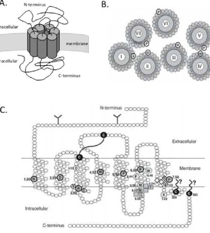
Figure 3: Opioid receptors structure
(A) Schematic view in 3D of opioid receptor conformation
(B) Seven transmembrane domains organizations. Amino acids (aa) indicated by circles are the conserved aa in class A GPCR receptors.
(C) Organization of the delta opioid receptor (Massotte and Kieffer, 2005). Circled aa
correspond to highly conserved residues. N-glycosylation in N-terminus are depicted (Y). The disulfide bridge between cysteine of the helix III and the extracellular loop 2 is represented.
10
At the beginning of the 1980’s, specific synthetic agonists for the three opioid receptors were produced (DAMGO for mu opioid receptor (Handa, Land et al. 1981), DPDPE for DOR (Mosberg, Hurst et al. 1983) and U-50488 (Vonvoigtlander, Lahti et al. 1983) for the kappa opioid receptor. Rapidly, these specific agonists permitted to define the pharmacological properties of each opioid receptor as well as their distribution through the nervous system.
In the early 1990’s, two groups simultaneously reported the first expression cloning of an opioid receptor, the mouse delta opioid receptor (Evans, Keith et al.
1992; Kieffer, Befort et al. 1992). The human and rodent cDNA of mu and kappa
opioid receptors were cloned based on their homology with the delta opioid receptor (for a review see (Kieffer 1995; Kieffer and Evans 2009)). In 1994, a fourth opioid receptor was cloned by sequence homology, the nociceptin/ORFQ (or ORL-1) receptor (Bunzow, Saez et al. 1994).
B.
The opioid receptors
1. Biosynthesis
The genes coding for the opioid receptors are Oprm1, Oprd1 and Oprk1, respectively for mu, delta and kappa receptors (MOR, DOR and KOR in The International Union of Basic and Clinical Pharmacology IUPHAR nomenclature). They are located on separate chromosomes (Oprm1 on chromosomes 10 in mouse and 6 in human, Oprd1 on chromosomes 4 in mouse and 1 in human, Oprk1 on chromosomes 1 in mouse and 8 in human) but exhibit a similar genomic organization. The genes coding for delta and kappa opioid receptors are composed of three exons (Befort, Mattei et al. 1994; Yasuda, Espinosa et al. 1994; Simonin, Gaveriaux-Ruff et al. 1995), while Oprm1 contains four exons (Wang, Johnson et al. 1994).
The cloning of genes coding for opioid receptors allowed determining their protein sequences. Mu, delta and Kappa receptors are composed of 398, 372 and 380 amino acids (aa), respectively. They belong to the superfamily of the G protein
Figure 4:
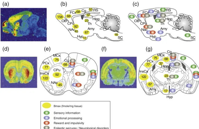
Neuroanatomical distribution of opioid receptor on a sagittal section of rodent brain (Le Merrer et al., 2009).
Abbreviations
Amb, nucleus ambiguus; AD, anterodorsal thalamus; AL, anterior lobe, pituitary; AON, anterior olfactory nucleus; Arc, arcuate nucleus, hypothalamus; BLA, basolateral nucleus, amygdala; BNST, bed nucleus of the stria terminalis; CeA, central nucleus, amygdala; Cl, claustrum; CL, centrolateral thalamus; CM, centromedial thalamus; CoA, cortical nucleus, amygdala; CPu, caudate putamen; CrbN, cerebellar nuclei; DMH, dorsomedial hypothalamus; DMR, dorsal and medial raphe ; DTN, dorsal tegmental nucleus; En, endopiriform cortex; Ent, entorhinal cortex; FrCx, frontal cortex; G, nucleus gelatinosus, thalamus; G/VP, globus pallidus/ventral pallidum; HbL, lateral habenula; HbM, medial habenula; HPC, hippocampus; IL, intermediate lobe, pituitary; IP, interpeduncular nucleus; LC, locus coeruleus; LD, laterodorsal thalamus; LG, lateral geniculate, thalamus; LH, lateral hypothalamus; LRN, lateral reticular nucleus; MD, mediodorsal thalamus; Me, median eminence; MEA, median nucleus, amygdala; MG, medial geniculate; MM, medial mammillary nucleus; MV, medial vestibular nucleus; NAc, nucleus accumbens; NL, neuronal lobe, pituitary; NRGC, nucleus reticularis gigantocellularis; NTS, nucleus tractus solitarius; OCx, occipital cortex; PAG, periaqueductal gray; PCx, parietal cortex; Pir, piriform cortex; PN, pontine nucleus; PnR, pontine reticular; PO, posterior thalamus; POA, preoptic area; PPTg, pedunculopontine nucleus; PrS, presubiculum; PV, paraventricular thalamus; PVN, paraventricular hypothalamus; RE, reuniens thalamus; RN, red nucleus; RM, raphe magnus; SON, supraoptic nucleus; SN, substancia nigra; SNT, sensory trigeminal nucleus; STN, spinal trigeminal nucleus; TCx, temporal cortex; Th, thalamus; Tu, olfactory tubercle; Tz, trapezoid nucleus; VL, ventrolateral thalamus; VM, ventromedial thalamus; VMH, ventromedial hypothalamus; VPL, ventroposterolateral thalamus; VTA, ventral tegmental area; ZI, zona incerta
11
coupled receptor (GPCR) and were classified within the class A GPCR family because of their sequence homology with rhodopsin (Figure 3) (Fredriksson,
Lagerström et al. 2003). As a member of the GPCRs, opioid receptors are
characterized by seven transmembrane domains (TM) connected by three intracellular and three extracellular loops, an N-terminal extremity that specifically interacts with ligands and a C-terminal extremity linked to the downstream intracellular effectors such as the G proteins (Surratt and Adams 2005). The transmembrane domains are composed of seven α-helices and present a highly homologous sequence across the three different receptors (73 to 76 %). They participate to ligand binding as well as receptor signaling (Befort, Tabbara et al. 1996). The extra- and intracellular loops also share highly homologous sequences (86 to 100 %) while the N-terminal and C-terminal diverge in the three opioid receptors (N-terminal: 9 to 10 % of identity; C-terminal: 14 to 20 % of identity). The extracellular loops and N-terminal extremity are responsible for selective ligand binding (Gether 2000). The C-terminal extremity contributes to the receptor stability and the intracellular signaling. Moreover, this extremity contains sites for post-translational modifications that modulate the receptor activity as well as the G protein coupling (Decaillot, Befort et al. 2003). Recently, the structure of opioid receptors has been determined at high-resolution by X-Ray crystallography (Granier, Manglik et al.
2012; Manglik, Kruse et al. 2012; Wu, Wacker et al. 2012) and represent an
important discovery for better understanding receptor-ligand interactions. In the future, it will also help for the bioinformatic modeling of receptor interactions.
2. Neuroanatomical distribution
The opioid receptors are broadly expressed in the central nervous system (Figure 4). They are also expressed in the peripheral nervous system as well as in other organs and systems (Townsend, Portoghese et al. 2004; Schramm and Honda 2010). For instance, the expression in immune cells has been reported (Gaveriaux, Peluso et al. 1995). Here, we will focus on opioid receptors location within the brain. The anatomical distribution of opioid receptors was determined by in situ hybridization and autoradiographic binding experiments, which allowed for a precise characterization of each mRNA (Mansour, Fox et al. 1994; Kitchen, Slowe et al.
Figure 5:
Endogenous peptides (from Faget L.). Schematic representation of genes coding for
endogenous opioid precursor peptides. Pomc, Penk and Pdyn genes are composed of 267, 267 and 254 aa respectively. In the table is represented the sequence of the main endogenous peptides with the common “opioid motif” bolded.
12 1997; Slowe, Simonin et al. 1999; Goody, Oakley et al. 2002). While the endogenous peptides distribution has been assessed by immunohistochemistry assays, the commercial antibodies used to determine the opioid receptors distribution showed a low specificity. Therefore, this questioned the accuracy of reported receptor distributions. More recently, knockin mutant mice expressing the DORs in fusion with the green fluorescent protein (GFP) were developed (Scherrer, Tryoen-Toth et al. 2006). The emergence of such genetically engineered mice will allow mapping the in vivo location of opioid receptors.
The opioid receptors are mostly rexpressed in the cortex, limbic system and brain stem (Le Merrer, Becker et al. 2009). The mu opioid receptor is abundantly expressed in the thalamus (Th), habenula (Hb), substantia nigra (SN), striatum, ventral tegmental area (VTA) and the nucleus of the solitary tract (NTS). The kappa opioid receptor is enriched in the basal anterior forebrain, olfactory tubercule (Tu), striatum, preoptic area (POA), hypothalamus (Hyp) and pituitary gland. The DOR is highly expressed in the olfactory bulb (OB), striatum (CPu and NAc), the globus pallidus, the nucleus of the diagonal band of Broca, septal nuclei, the hippocampus and in subregions of the amygdala (BLA, CoA and MeA). DOR is also the most represented opioid receptor in the olfactory tract and in cortical regions (including the PFC) in particular in the insular cortex (InsCx).
C.
The endogenous opioid peptides
1. Biosynthesis
The opioid system is composed of three families of endogenous peptides: the enkephalins, endorphins and dynorphins (Figure 5). In 1975, Hughes and colleagues identified two molecules from brain extracts that displayed similar action as morphine and was reversed by naloxone (Hughes, Smith et al. 1975). Following this study, about 30 endogenous opioid peptides were discovered. They are composed of 5 to 31 amino acids (aa) and share a common N-terminal sequence Tyr-Gly-Gly-Phe, so called the “opioid motif” (Akil, Owens et al. 1998). The endogenous peptides are synthesized from three precursors: the preproenkephalin (PENK),
Figure 6:
Selectivity windows of some opioid agonists and antagonists (William et al., 2001). At the top are represented compounds highly selective for each opioid receptor. At the bottom are represented endogenous peptides and other commonly used opioids.
13
preproopiomelanocortin (POMC) and preprodynorphine (PDYN). The genes encoding these precursors were cloned before the cloning of the genes coding for opioid receptors, in the early 1980’s (Nakanishi, Inoue et al. 1979; Comb, Seeburg et al. 1982; Kakidani, Furutani et al. 1982).
The cleavage by endopeptidases of these endogenous peptide precursors occurs during the post-translational maturation, in a tissue dependent manner, and leads to the production of about 30 functional peptides (Fricker and Devi 1993; Akil,
Owens et al. 1998). The Penk gene codes for a 267 aa polypeptide precursor
containing four copies of met-enkephalin, one copy of leu-enkephalin and other enkephalins (Rossier 1993). The Pomc gene codes for a 267 aa polypeptide precursor which after proteolytic cleavage provides one copy of β-endorphin, one copy of adrenocorticotropin (ACTH) and several other peptides activating the melanocytes (Young, Bronstein et al. 1993). The Pdyn gene encodes a 245 aa polypeptide precursor cleaved in leu-enkephalin, dynorphin A and dynorphin B (Day, trujillo et al. 1993). The enkephalins, β-endorphin and dynorphin are the main endogenous active peptides and each is able to bind the three different opioid receptors (Figure 6). However, enkephalins and β-endorphin present a lower affinity for the kappa opioid receptor (Loh, Tseng et al. 1976) which is more strongly bound by dynorphin (Goldstein, Tachibana et al. 1979). Recently, some studies discovered and characterized a biosynthesis pathway for morphine production in mice suggesting that morphine could be synthesized endogenously (Grobe, Lamshoft et al. 2010; Laux, Muller et al. 2011).
2. Neuroanatomical distribution
The neuroanatomical distribution of Penk, Pomc and Pdyn mRNA has been described using in situ hybridization experiments (for review see (Le Merrer, Becker et al. 2009)). Moreover, distribution of the active peptides has been determined by immunohistochemistry. Opioid peptide immunoreactivity overlaps largely with the localization of opioid receptors (Figure 7). PENK is largely distributed and the most abundant opioid precursor. It is strongly detected in the striatum and globus pallidus where it overlaps with DOR. PDYN is located in most brain regions with the highest
Figure 7:
Neuroanatomical distribution of endogenous opioid peptides on a sagittal section of rodent brain (Adapted from, Le Merrer et al., 2009).
Abbreviations
Amb, nucleus ambiguus; AD, anterodorsal thalamus; AL, anterior lobe, pituitary; AON, anterior olfactory nucleus; Arc, arcuate nucleus, hypothalamus; BLA, basolateral nucleus, amygdala; BNST, bed nucleus of the stria terminalis; CeA, central nucleus, amygdala; Cl, claustrum; CL, centrolateral thalamus; CM, centromedial thalamus; CoA, cortical nucleus, amygdala; CPu, caudate putamen; CrbN, cerebellar nuclei; DMH, dorsomedial hypothalamus; DMR, dorsal and medial raphe ; DTN, dorsal tegmental nucleus; En, endopiriform cortex; Ent, entorhinal cortex; FrCx, frontal cortex; G, nucleus gelatinosus, thalamus; G/VP, globus pallidus/ventral pallidum; HbL, lateral habenula; HbM, medial habenula; HPC, hippocampus; IL, intermediate lobe, pituitary; IP, interpeduncular nucleus; LC, locus coeruleus; LD, laterodorsal thalamus; LG, lateral geniculate, thalamus; LH, lateral hypothalamus; LRN, lateral reticular nucleus; MD, mediodorsal thalamus; Me, median eminence; MEA, median nucleus, amygdala; MG, medial geniculate; MM, medial mammillary nucleus; MV, medial vestibular nucleus; NAc, nucleus accumbens; NL, neuronal lobe, pituitary; NRGC, nucleus reticularis gigantocellularis; NTS, nucleus tractus solitarius; OCx, occipital cortex; PAG, periaqueductal gray; PCx, parietal cortex; Pir, piriform cortex; PN, pontine nucleus; PnR, pontine reticular; PO, posterior thalamus; POA, preoptic area; PPTg, pedunculopontine nucleus; PrS, presubiculum; PV, paraventricular thalamus; PVN, paraventricular hypothalamus; RE, reuniens thalamus; RN, red nucleus; RM, raphe magnus; SON, supraoptic nucleus; SN, substancia nigra; SNT, sensory trigeminal nucleus; STN, spinal trigeminal nucleus; TCx, temporal cortex; Th, thalamus; Tu, olfactory tubercle; Tz, trapezoid nucleus; VL, ventrolateral thalamus; VM, ventromedial thalamus; VMH, ventromedial hypothalamus; VPL, ventroposterolateral thalamus; VTA, ventral tegmental area; ZI, zona incerta
14
concentration in the nucleus accumbens. POMC distribution is more restricted and absent from cortical structures except for the amygdala. Penk and Pdyn expressing cell bodies show an extensive distribution in the whole brain, while Pomc expressing cell bodies are limited to three regions of the brain; the arcuate nucleus of the hypothalamus (Arc), nucleus of the solitary tract (NTS) and in the pituitary gland. Overall, despite discrepancies in some regions, the anatomical distribution of opioid peptides and receptors is in agreement with the notion that enkephalins and endorphins preferentially bind to delta and mu receptors and that dynorphins preferentially activate kappa receptors.
D.
Physiological roles of the endogenous opioid system
The endogenous opioid system is involved in many different physiological functions. The different functions of the opioid system were explored by using pharmacological approaches and genetically engineered mutant mouse lines. The receptors expressed in peripheral nervous system and other organs are described to be involved in the regulation of autonomic vegetative constants like the cardiovascular responses (Saraiva, Oliveira et al. 2004), regulation of the body temperature (Rawls, Hewson et al. 2005), gastro-intestinal transit (Mehendale and Yuan 2006) as well as hepatic and renal functions (Atici, Cinel et al. 2005).
The three opioid receptors are described as major actors of regulation pain perception, or nociception (Gaveriaux-Ruff 2013). Indeed, three mouse lines genetically deleted for either MOR, DOR or KOR showed modifications of pain perception suggesting a tonic inhibition of pain responses. However, they regulate different aspects of nociception. MORs are involved in acute mechanical and chemical pain (Martin, Matifas et al. 2003; Zollner and Stein 2007). KORs mainly contribute to the regulation of visceral pain (Simonin, Valverde et al. 1998; Chavkin 2011). The DORs participate essentially to the management of chronic pain (Gaveriaux-Ruff and Kieffer 2011). Moreover, it has been recently suggested that DORs specifically expressed on Nav 1.8-positive nociceptive neurons in the dorsal
root ganglia tonically inhibit mechanical hypersensitivity in inflammatory and neuropathic pain (Gaveriaux-Ruff, Nozaki et al. 2011).
15
The opioid system plays a critical role in the control of behavioral responses stimulated by natural rewards and drugs of abuse (Bodnar 2004; Smith and Berridge 2005). The rewarding properties of addictive drugs are mainly dependent on the mu opioid receptor (Matthes, Maldonado et al. 1996; Ghozland, Matthes et al. 2002; Le
Merrer, Becker et al. 2009). KOR is involved in the negative emotional state
experienced during withdrawal periods (Shippenberg, Zapata et al. 2007; Gillett, Harshberger et al. 2013). The role of DOR in drug addiction remains poorly studied despite a potential role in drug craving and relapse. However, the contribution of DORs in several processes such as anxiety-related behaviors (Filliol, Ghozland et al. 2000), drug-context association (Le Merrer, Faget et al. 2012) or motor impulsivity (Olmstead, Ouagazzal et al. 2009; Befort, Mahoney et al. 2011) may indirectly participate to the regulation of addictive responses.
Most studies on the role of the opioid system have focused on the regulation of pain and addictive responses mainly because of the reported effects of opiates reported since the latest ages. Nevertheless, the opioid system is also implicated in mood and well-being (Filliol, Ghozland et al. 2000; Chu Sin Chung and Kieffer 2013), learning and memory (Robles, Vivas-Mejia et al. 2003; Holahan, Nichol et al. 2008; Rodefer and Nguyen 2008), motor control (Le Merrer, Rezai et al. 2013).
E.
Opioid receptor / ligand interaction and intracellular
signaling
Agonists interact with the receptor at the level of the binding pocket composed of the extracelluar loops and the N-terminal extremity. Granier and colleagues recently proposed that the upper binding pocket that diverges among receptor subtypes is responsible for the ligand selectivity, whereas the lower portion of the binding pocket is well-conserved in both sequence and structure (Granier, Manglik et al. 2012; Manglik, Kruse et al. 2012). Moreover, mutagenesis studies suggested that all ligands were not binding the delta receptor at the same site (Befort, Zilliox et al.
1999; Decaillot, Befort et al. 2003). Then, ligand binding to GPCRs induces
conformational changes of the TM domains (Visiers, Ballesteros et al. 2002). These conformational changes ultimately lead to the uncoupling of the G proteins from the
Figure 8:
Intracellular signaling of opioid receptors (from Faget L., adapted from Williams et al., 2001). Opioid receptor activation by ligand binding induces activation of protein Gi/oα and β/γ subunits. Then, these protein G subunits modulate numerous channel activity (K+, Ca2+and Ih currents)
16
C-terminal of the receptor and exposure to other effectors proteins (Waldhoer, Bartlett et al. 2004).
Opioid receptors are GPCRs coupled to Gi/o proteins and their activation leads
to a global decrease of the cell excitability and neurotransmitter release (Figure 8). Gi/o proteins are composed of three subunits: α, β and γ. Subsequently to the ligand
binding, GTP is hydrolyzed leading to G proteins subunits αi/o and β/γ uncoupling
(Oldham and Hamm 2008).
The activation of αi/o subunit inhibits the adenylate cyclase (AC) activity
responsible for cAMP production. Decreased cAMP concentration induces a decreased activity of the protein kinase A (PKA) and consequently of many others downstream signaling pathways. The decreased AC activity also modulates the activity of a voltage-dependent current Ih (Ingram and Williams 1994; Svoboda and Lupica 1998). This current is normally responsible for the repolarization of the membrane potential after a strong hyperpolarization and then allows future activation of the cell. Opioid receptors activation leads to a diminished amplitude of this potassium inward current (also called pacemaker current). In addition, the AC inhibition may induce a decrease of neurotransmitter release via a PKA-dependent mechanism (Chieng and Williams 1998; Ingram, Vaughan et al. 1998).
The β/γ subunits following opioid receptor activation enhance three potassium conductance: a G-protein inwardly rectifying conductance (GIRK) (Sodickson and
Bean 1998), a voltage-dependent potassium current (Madamba, Schweitzer et al.
1999) and a calcium-sensitive potassium conductance (Twitchell and Rane 1993). In parallel, these subunits inhibit voltage-sensitive Ca2+ channels (N, P/Q and T types) (Wilding, Womack et al. 1995). Opioid receptor activation also leads to long term modifications such as changes in gene expression (Bilecki, Wawrzczak-Bargiela et al. 2004). Indeed, β/γ subunits are able to activate the mitogen-activated protein kinases (MAPK) pathway, mainly the Extracellular signal Regulated Kinase 1 and 2 (ERK 1/2), via the Ras-GRF membrane protein, the phosphatidylinositol 3-kinase (Pi-3 kinase) and the phospholipase C (Williams, Christie et al. 2001). Consequently, MAPK phosphorylate several transcription factors such as the CREB (CA2+/cAMP responsive element binding protein), Elk-1 (Ets LiKe gene 1), estrogen receptor, c-jun, c-fos, or AP-1 (activator protein 1 - heterodimeric protein composed of c-fos and c-jun) depending on the opioid receptor subtype. In conclusion, opioid receptor
Figure 9:
Receptor internalization process (Williams et al., 2001). Activated opioid receptor can be phosphorylated at the C-terminal. The phosphorylated sites are bind by arrestin proteins which then recruit c-Src adaptor proteins. Then, the proteic complex is recognized by clathrin to promote endocytosis leading to receptor recycling or degradation.
17
activation induces some short term effects such as a reduction of the cell excitability as well as a decrease of neurotransmitter release; but also triggers long term modifications of gene expression.
F.
Desensitization and receptor trafficking
1. Desensitization mechanisms
Receptor desensitization is a cellular mechanism that regulates the activity of the GPCRs and likely plays a critical role in some physiological functions (Bohn, Gainetdinov et al. 2004). Following receptor activation by a selective agonist, the desensitization process begins with the phosphorylation of this receptor at the C-terminal extremity. Hence, the phosphorylated receptors are no more able to bind G proteins and enter the internalizing process to be either recycled at the membrane surface or degraded in lysozyme vesicles (Figure 9). This mechanism disrupts the GPCR signal transduction.
The phosphorylation process is mediated by G protein-coupled receptor kinases (GRKs) (Law, Kouhen et al. 2000) or by protein kinase A or C (PKA or PKC) (Xiang, Yu et al. 2001). These two effectors can be recruited differentially depending on agonists and may target separate pathways leading to different physiological responses. Recent studies suggested that the same GPCR activated by different agonists could provide diverse cellular responses (Kenakin 2011; Reiter, Ahn et al. 2012). This concept, called biased agonism, will be reviewed later in the introduction for the delta opioid receptor (see section B.1.2).
The stimulation of delta opioid receptor by an agonist induces the phosphorylation of the Serine and Threonine residues, Ser344 and Ser363 (Guo, Wu et al. 2000). Moreover, it has been shown that substitution of the Serine and Threonine residues by Alanine prevents the internalization of DORs (Whistler, Tsao et al. 2001). Cytoplasmic proteins called arrestins specifically recognize phosphorylated residues. In vitro binding of arrestins to MORs and DORs has been correlated to desensitization (Kovoor, Nappey et al. 1997). This arrestins-dependent desensitization has been described in vivo for MORs. Indeed, the desensitized
18
response after morphine injection was not detected in β-arrestin knockout mice (Bohn, Gainetdinov et al. 2000). Binding of arrestins on phosphorylated receptors therefore prevents further coupling to G proteins. This desensitization mechanism has been described as playing an important role in physiological responses such as opioid tolerance (Koch and Hollt 2008).
2. Receptor trafficking
After agonist stimulation, opioid receptors are subsequently internalized in intracellular vesicles. This internalization phenomenon requires previous binding of arrestins on the activated receptors. MORs and DORs internalize via a clathrin-coated pits (Trapaidze, Keith et al. 1996; Zhang, Xiong et al. 2009). Clathrins are responsible for the stabilization and endocytosis of vesicles containing receptors. Once GPCRs are internalized in vesicles, they can be either recycled at the cell surface or degraded by fusion with lysosomes vesicles (von Zastrow 2003). Recycling of GPCRs can be a fast or slow process depending on the receptors as well as the activating ligand. MOR has been described as a fast-recycling receptor under peptidic activation (Koch, Widera et al. 2005). In contrast, DOR post-endocytic fate has been shown as slow-recycling process (Pradhan, Becker et al. 2009). Some studies showed that it is recycled after non-peptidic activation (Lecoq, Marie et al. 2004; Marie, Aguila et al. 2006). Therefore, an activated receptor will follow different trafficking pathways depending on the stimulating agonist. This argument in favor of a biased agonism suggests that different post-endocytic fates may induce diverse physiological responses.
II. The Delta Opioid Receptor
A.
Ligands
Figure 10:
19
As previously mentioned, the opioid receptors can be activated by the three endogenous peptides enkephalins, β-endorphins and dynorphins despite differential selectivity (Williams, Christie et al. 2001). It is well accepted that enkephalins present the highest affinity for the DOR (≈ 2 nM) (Figure 10).
Additionally, several exogenous agonists have been described to activate the DORs (Pradhan, Befort et al. 2011). DOR ligand can be divided in two main classes: the peptidic and the alkaloids molecules.
The peptidic ligands correspond to cyclic analogs of the enkephalin and exhibit a very high affinity for the receptor compared to other opioid receptors. One of the most classically used exogenous ligand is the [Cys2, L-Pen5]-and [Cys2, D-Pen5]-enkephalin (DPDPE) and presents a 100 fold higher affinity for delta than mu opioid receptor (Mosberg, Hurst et al. 1983). The deltorphin I and II are exogenous ligand extracted from frog skin (Phyllomedusa bicolor) and show high affinity and specificity for the DOR (Kreil, Barra et al. 1989). Moreover, specific peptidic antagonists were synthetized such as the TIPP-Ψ (Schiller, Weltrowska et al. 1993).
The alkaloid ligands are also exogenous molecules exhibiting a significant affinity and selectivity towards DOR activation. For instance, SNC80 offers affinity Kd values for 1.73, 882 and 442 nM for delta, mu and kappa binding respectively (Bilsky, Calderon et al. 1995). BW373U86 is another delta opioid agonist obtained from the degradation of the SNC80 that exhibits similar affinity and specificity (Chang, Rigdon
et al. 1993). Several novel delta opioid agonists have been developed recently
(Pradhan, Befort et al. 2011). Regarding the alkaloid antagonists, naltrindole has been described as the compound with the highest affinity for the DORs compared to the more global opioid anatagonists such as the naloxone (Portoghese, Sultana et al. 1988). Later on, more selective DOR antagonists were developed (Bryant, Salvadori et al. 1998).
2. Biased agonism
The biased agonism corresponds to the fact that two different agonists activating the same receptor may produce several different cellular and/or behavioral responses (Kenakin 2007; Pradhan, Walwyn et al. 2010).
Figure 11:
In vivo example of DOR biased agonism (Pradhan et al., 2011). At the top, SNC80 triggers DOR-eGFP internalization in primary culture and tissue sections. Chronic administration of SNC80 result in a generalized tolerance. At the bottom, ARM-390 does not affect DOR-eGFP internalization in primary culture and tissue sections. Chronic administration of ARM-390 induces only tolerance to analgesia.
20
SNC80 has been reported to induce DOR internalization. Conversely, the agonist ARM-390, ADL5747 and ADL5859 selectively activate DOR without stimulating receptor internalization (Marie, Lecoq et al. 2003; Pradhan, Becker et al. 2009; Nozaki, Le Bourdonnec et al. 2012). Moreover, the dissociation between high- and low- internalizing agonists has been shown to underlie some physiological processes such as the development of analgesic tolerance (Pradhan, Walwyn et al. 2010). This study provided a significant illustration of biased agonism in vivo (Figure 11). Additionally, ligand-biased agonism at DOR may occur after the internalization process, to favor either receptor recycling or degradation (Audet, Charfi et al. 2012). New DOR agonists were developed in order to avoid the deleterious consequences of DOR activation by agonists like SNC80. Indeed, while SNC80 induced epileptic seizures and anxiolytic effects, KNT-127 has been shown to produce on anxiolytic effects without provoking any convulsions (Saitoh, Sugiyama et al. 2011). This in vivo illustration of biased agonism provides innovative strategies to develop new drugs for the treatment of several pathologies. Interestingly, the Adolor5859 (ADL5859) (Le
Bourdonnec, Windh et al. 2008) and Adolor 5747 (ADL5747) (Le Bourdonnec, Windh
et al. 2009) are also two DOR agonists currently in phase 2 clinical trials to treat patient suffering from mood disorders.
B.
Genetically engineered mutant mouse lines
The physiological role of DOR has been first assessed following the development of selective ligands such as DPDPE and naltrindole. Nevertheless, the development of genetic models targeting DOR (Filliol, Ghozland et al. 2000; Scherrer, Tryoen-Toth et al. 2006) or PENK (Konig, Zimmer et al. 1996) genes brought new tools to explore the physiological role of DORs. In the future, the construction of refined genetically engineered mouse lines will help to evaluate the participation of DOR in some subtle phenotypes. Additionally, new reporter mouse lines may allow determining more precisely the localization of the receptor in neuronal circuits.
Figure 12:
(A) Genetic construction of constitutive DOR knockout mice (Filliol et al., 2000). Exon 1 coding for Oprd1 gene is replaced by Neomycine cassette. The recombinant allele obtained is a 9.6 kb fragment (wild-type fragment of 8.6 kb).
(B) Southern-blot analysis of mouse tail DNA sample from Oprd1-/-offsprings mice.
A.
21
The DOR constitutive KO mice were generated in our laboratory by using a homologous recombination strategy (Figure 12) (Filliol, Ghozland et al. 2000). In the targeting vector, the first coding exon, encoding the extracellular N-terminal and the first TM, as well as the translation-initiation codon of the Oprd1 gene were replaced by a Neomycine cassette. Then, the sequence was integrated into embryonic stem cells. The selected embryonic cells were implanted into C57BL/6 blastocysts. Finally, homozygous mutant mice were obtained under a hybrid 129 SvPas/C57BL/6J (50%/50%) genetic background.
Knockout animals have provided crucial informations in the identification of proteins functions involved in variety of pathologies. However, constitutive knockout present several limits.
This technology does not allow having a temporal control over the inactivation of the gene of interest. It might be relevant to assess the contribution of a particular molecule at a precise moment, such as for instance during adolescence. Furthermore, some compensatory mechanisms could take place and then hamper the identification of the protein of interest function. Lastly, the nervous system is highly complex and composed of a variety of neuronal populations and circuits. A given protein could be expressed in many different areas and networks playing different roles, even opposite, depending on its localization. Consequently, the total deletion of a specific protein does not enable to evaluate its contribution in a specific circuit, brain region or neuronal population. Additionally, subtle phenotype may be hard to detect in this fully excised models.
In order to overcome these issues and have a significant spatial and/or temporal control of the gene inactivation, the conditional knockout approach appears as the next relevant strategy. The use of recombinant virus, such as adeno-associated virus, is also an innovative technology to specifically target regions or neuronal populations.
Figure 13:
(A) Genetic construction of homozygous floxed mice(Gavériaux-Ruff et al., 2011). Exon 2 of
Oprd1 gene is replaced by homologous recombination with floxed allele containing
Hygromycine cassette as well as the exon 2 surounded by loxP sites.
22
The conditional knockout gene approach has been mainly developed based on the Cre/loxP system. This technology is classically used in neurogenetics. It was developed in order to inactivate gene in precise regions or cell populations (Gaveriaux-Ruff and Kieffer 2007). This tool gives the opportunity to target a gene of interest with a very high spatial control and has been also used to rescue the expression of a gene in specific regions. The Cre recombinase is a tyrosine recombinase enzyme obtained from the bacteriophage P1. The enzyme mediates the specific recombination between two loxP sites. The loxP site corresponds to a 34 base pair sequence composed of two 13 base pair palindromic sequences that flank an 8 base pair spacer region. Depending on the two loxP sites orientation, the surrounded gene can be excised (same loxP sites orientation) or inverted (opposite loxP sites orientation). Additionally, Cre recombinase can also induce translocation between two DNA fragments that both comprise one loxP site. The conditional knockout mouse lines are obtained by crossing two different mouse lines. The first mutant mouse line present loxP sites surrounding a part of the gene of interest. The second transgenic mouse line expresses the Cre recombinase in a tissue or cell population specific manner. Currently, more than 500 different transgenic mouse lines expressing the Cre recombinase under the control of a specific promoter have been developed (Nagy, Mar et al. 2009) and among them, about 70 provide a specific targeting of neurons (Gaveriaux-Ruff and Kieffer 2007). A database with all available information about the properties of these Cre transgenic lines has been created (the ‘‘CreXmice’’ database; http://www.mshri.on.ca/nagy/).
Full knockout could also be obtained by taking advantage of the Cre/loxP system. Indeed, the excision of the flanked sequence of the gene can be achieved by breeding the floxed mice with a mutant mouse line expressing the Cre recombinase under the control of a ubiquitously active promoter. For this purpose, the cytomegalovirus (CMV) is classically used (Feil, Brocard et al. 1996).
The Oprd1 floxed mice were generated in our laboratory (Gaveriaux-Ruff, Nozaki et al. 2011). In this mouse line, the exon 2 of the Oprd1 gene is flanked by two loxP sites (also called floxed) (Figure 13). The homozygous Oprd1 floxed mouse line was obtained on a 50% C57BL/6J–50% 129SvPas genetic background. Furthermore,
23
DOR activation has been checked by [35S]-GTPγS binding experiment and showed a functional DOR.
The Nav 1.8 conditional knockout mice for DOR represent the first and
currently the only reported conditional approaches of DOR (Gaveriaux-Ruff, Nozaki et al. 2011). They were obtained by crossing the Oprd1 floxed mice described above with a transgenic mouse line expressing the Cre recombinase under the control of Nav 1.8 promoter. The Nav 1.8–Cre mutant line specifically expresses the enzyme in
peripheral nociceptive neurons, unmyelinated C and thinly myelinated A∆ fibers, and has been previously successfully used (Abrahamsen, Zhao et al. 2008).
Recently, new technologies used for the study of in vivo gene functions emerges such as the zinc-finger nucleases (ZFNs) or Transcription Activator-Like Effector (TALE) Nucleases (TALENs) and will likely provide alternatives to the Cre/lox system in the future (Sung, Baek et al. 2012)
C.
Physiological functions
As previously mentioned, the opioid system is involved in many physiological processes in particular pain control, hedonic homeostasis (maintenance of the rewarding/aversive balance processes in a physiological range), mood and well-being. Studies on the DOR revealed its role in emotional control (Filliol, Ghozland et al. 2000), in processes that may modulate drug addiction (Roberts, Gold et al. 2001; Le Merrer, Plaza-Zabala et al. 2011; Faget, Erbs et al. 2012), in the development of seizures (Broom, Nitsche et al. 2002; Jutkiewicz, Baladi et al. 2006), in the regulation of locomotor activity (Filliol, Ghozland et al. 2000; Le Merrer, Rezai et al. 2013) and in neuroprotective processes (Gao, Niu et al. 2012; He, Sandhu et al. 2013). The contribution of DOR in these different functions will be discussed later in this manuscript.
Moreover, DOR appeared to be involved in the modulation of immune function (Weber, Gomez-Flores et al. 2004), in cardioprotection process (Maslov, Lishmanov et al. 2009; Shen, Ben et al. 2012) and in gastro-intestinal function (Bueno and Fioramonti 1988; Townsend, Portoghese et al. 2004). These regulatory roles will not be discussed here.
24
In addition, DOR is a major player in pain perception (Gaveriaux-Ruff and Kieffer 2011) and in memory processes (Robles, Vivas-Mejia et al. 2003; Le Merrer, Faget et al. 2012) which will be discussed in the following parts.
1. Pain
Pain is defined by the International Association for the Study Pain’s as “an unpleasant sensory and emotional experience associated with actual or potential tissue damage, or described in terms of such damage” (Bonica 1979). Acute pain is characterized by different modalities such as thermal, mechanical or chemical pain and can be distinguished from chronic pain such as inflammatory or neuropathic pain.
The constitutive DOR knockout mice showed no differences in acute thermal, mechanical or chemical pain perceptions (Kieffer and Gaveriaux-Ruff 2002), whereas MORs are implicated in the regulation of these responses (Martin, Matifas et al. 2003). However, evidence supports the contribution of DOR in chronic pain. Indeed, it has been shown that pharmacological activation of DOR by SNC80 was able to reduce inflammatory pain perception (Gaveriaux-Ruff, Karchewski et al. 2008). In addition, the DOR knockout mice displayed reduced pain thresholds in a classical inflammatory model using Freund adjuvant injections to induce inflammatory conditions (Gaveriaux-Ruff, Karchewski et al. 2008) and also in a classical neuropathic model using the sciatic nerve injury surgery (Nadal, Banos et al. 2006; Benbouzid, Gaveriaux-Ruff et al. 2008).
Altogether, these studies support a role of DOR in decreasing chronic pain perception.
2. Learning and memory
The DORs are expressed in regions involved in learning and memory such as the hippocampus (Erbs, Faget et al. 2012). The pyramidal cells of hippocampus are regulated by GABAergic interneurons which express DOR, suggesting that DOR participate to the modulation of hippocampal outputs.
25
Robles and colleagues showed that animals performing successfully in a spatial discrimination paradigm, the holeboard task, present increased DOR mRNA expression (Robles, Vivas-Mejia et al. 2003). This study emphasizes the potential contribution of DOR in spatial memory skills. A recent study in our laboratory showed that pharmacological inactivation or genetic deletion of DOR in mice altered performances in the spatial object recognition task (Le Merrer, Rezai et al. 2013). Moreover, some results obtained in our laboratory indicate that DOR knockout mice displayed decreased context-induced freezing in a fear conditioning task supporting a deficit in fear memory processes (Scherrer et al., in preparation
In addition, it has been shown that mice deficient for DOR also present a deficit in a drug-context association paradigm. Indeed, they exhibit a decrease of morphine conditioned place preference (CPP) and lithium conditioned place aversion (CPA) tests (
).
Le Merrer, Plaza-Zabala et al. 2011), while they self-administered morphine at a similar level compared to WT mice. Interestingly, the morphine CPP was restored in these animals by exposing them to cues predicting morphine (Le Merrer, Faget et al. 2012). Then, DOR appears crucial for the modulation of spatial contextual cue-related responses.
These data emphasize that DOR may facilitate spatial memory processes and play a major role of DOR in drug-context associations likely crucial in the persistence of addictive behaviors.
3. Summary of other DOR functions
The contribution of DOR in the control of emotional processes, in reward and addiction, in the onset of epileptic seizures, in the control of locomotor activity as well as in hypoxic/ischemic conditions is discussed in the following review (Chu Sin Chung and Kieffer 2013).
26
D.
Definition
Anxiety is defined as an unpleasant mental state which breaks out in anticipation of potential threat (Gross and Hen 2004), whereas fear arises in anticipation of a real or imminent threat.
The non-pathological anxiety is a physiological process necessary for the survival and the adaptation of an organism to its environment. Anxiety can be decomposed in two classes: the state anxiety corresponding to the acute reactivity towards a potentially threatening situation and the trait anxiety which reflects the natural tendency of an organism to express an increase anxiety response over time (Endler and Kocovski 2001; Kennedy, Schwab et al. 2001).
Pathological anxiety is responsible for the incidence of several diseases. According to the Diagnostic and Statistical Manual of American Psychiatric Association (DSM-4th edition TR-2000), anxiety disorders are divided in 7 major classes: generalized anxiety disorder (GAD), social phobia, simple phobia, panic disorder, agoraphobia, post-traumatic stress disorder (PTSD) and obsessive-compulsive disorder (OCD). In the DSM 5th edition, the latter two are removed from the anxiety disorder category and are defined in their own chapters. The diagnostic criteria remain similar to the previous edition, except that patients do not need to declare their fear as irrational or excessive.
Definitions of anxiety disorders:
1) Generalized Anxiety Disorders are the most largely diagnosed anxiety disorder and usually affects young adults. They are characterized by excessive, uncontrollable and often irrational worry.
2 and 3) Social and simple Phobias are defined as an intense and irrational fear (“out of proportion”) toward a precise object or situation that the individual try to avoid, even at the cost of enormous efforts. The specific object or situation is not necessary threatening or noxious for the individual
4) Panic disorders are mainly characterized by the manifestation of a panic attack associated with the fear of another attack.
27
5) Agoraphobia is similar to the panic disorders and defined as an irrational fear of places or situations in which another attack may occur and the patient may be unable to leave or find someone to help.
5) Post-traumatic stress disorders are considered as a symptomatic response to a previous traumatic experience.
6) Obsessive Compulsive Disorders are characterized by undesirable, insistent and repetitive behaviors. The individual had to perform these behaviors or else will feel an intense anxiety.
E.
Animal models
Anxiety disorders are among the most prevalent psychiatric diseases in Europe and North America. They represent a dramatic health problem for individual as well as a major cost for societies. Therefore, there is an important need for the development of therapies and for a better understanding about genetic and environmental risk factors that trigger these pathologies (Cryan and Sweeney 2011). Numerous animal tests of anxiety have long been validated to assess anxiolytic potential of novel drugs (Pellow, Chopin et al. 1985; File, Lippa et al. 2004).
These models should present a reasonable analogy to the human disorder in manifestation or symptoms (Face Validity) like for instance an excessive avoidance of threatening situation. They must also induce objective, measurable behavioral changes that are due to similar physiological mechanisms as for the human pathology (Construct Validity). Finally, animal model for anxiety should display sensitivity to effective clinical treatments such as diazepam (Predictive Validity).
Anxiety tests can be divided in three categories: exploratory behavior models, acute behavioral stress responses test and conditioned responses (Cryan and Sweeney 2011; Haller and Alicki 2012; Kumar, Bhat et al. 2013).
In the first category, anxiety tests are generally based on approach-avoidance reflected by natural tendency of rodent to avoid potentially dangerous environment such as open and/or lit environment. They present a strong ethological relevance (i. e. open field, light-dark box, elevated plus maze, elevated zero maze, social interaction, T-maze, hole board tests).
28
The second category regroups conflict-based tests (i. e. Geller-Seifter test, Vogel punished drinking test, defensive marble burying), hyponeophagia paradigm (i. e. novelty suppressed feeding, novelty induced hypophagia) and physiological tests like stress-induced hyperthermia or autonomic telemetry measures.
The last category of anxiety tests was designed to overcome the effect of motor output and animal reactivity toward conditioned stimuli (i. e. active/passive avoidance, fear potentiated startle, pavlovian fear conditioning, conditioned emotional response, conditioned taste aversion).
As emphasized by the large variety of anxiety tests existing and their variety of stressor applied and parameters measured, animal models of anxiety assess several neurobiological processes involved in anxiety. Therefore, it is inappropriate to consider that one model may serve to detect compounds for a disease that is mediated through multiple and diverse mechanisms. Similarly, it is likely relevant to use several tests in order to evaluate neurobiological processes underlying anxiety in a given study (Ramos 2008).
F.
Neurocircuitry of anxiety
Over the past decades, many studies investigated the neuroanatomical substrates underlying anxiety. Neuroimaging approach has been importantly used to identify brain regions contributing to anxiety disorders (Kent and Rauch 2003). Experiments performed on rodent mainly focused on neurocircuits involved in fear responses. Key brain regions identified in these studies include the amygdala, nucleus accumbens, bed nucleus of the stria terminalis, hippocampus, ventromedial hypothalamus, periaqueductal gray, some brainstem nuclei, thalamus, insular cortex and some prefrontal regions (Davis 2006; Shin and Liberzon 2010). In parallel, in vivo electrophysiological recording, tracing and lesions approaches allowed to characterize the specific contribution of these areas in basic components of fear circuitry.
29
Interestingly, some evidence suggest that fear and anxiety networks might be orchestrated by distinct systems. A contribution of the olfactory bulb (Saitoh, Hirose et al. 2006; Saitoh and Yamada 2012), prefrontal cortex (Bechara, Damasio et al. 2000; Davidson 2002), insular cortex (Paulus and Stein 2006; Lamm and Singer 2010), ventral hippocampus (Deacon, Bannerman et al. 2002; Fournier and Duman 2013) and amygdala (Baxter and Murray 2002; Cardinal, Parkinson et al. 2002) has been evidenced in emotional processing circuits.
Since this will be of interest of the third part of this work, we next reviewed evidence about the contribution of amygdala in emotional responses.
G.
The amygdala
In the early 19th century, Burdach is credited to the first description of the amygdala, a brain area close to the human temporal cortex. The amygdala has long been established to be a key structure for the regulation of emotions as well as for the modulation of memory (LeDoux 2000; Ehrlich, Humeau et al. 2009; Roozendaal, McEwen et al. 2009). In addition, an extensive literature studied the contribution of the amygdala in fear conditioning processes (Johansen, Wolff et al. 2012; Pare and
Duvarci 2012). The basolateral and lateral nuclei of the amygdala (BLA) are
established as the main site for conditioned stimulus (for instance cues or context) and unconditioned stimulus (reward or punishment) associations. On the other hand, the BLA is transmit informations of such associations to the central et centromedial nuclei of the amygdala CeA which in turn may orchestrate adapted autonomic and behavioral responses (Everitt, Cardinal et al. 2003).
The amygdala has been considered as a major limbic area in the neuronal circuits supporting the anxiety-related behaviors. It has been demonstrated that chronic stress enhances the reactivity of projecting neurons of the amygdala by in vivo electrophysiological recordings of pyramidal neurons of the lateral nucleus (Rosenkranz, Venheim et al. 2010). Three models of anxiety (foot shock avoidance, elevated plus maze and puff-induced ultrasonic vocalization test) (Silveira, Sandner et al. 1993; Duncan, Knapp et al. 1996) as well as four anxiogenic drugs (FG-7142, yohimbine, mCPP and caffeine) (Singewald, Salchner et al. 2003) induced an
30
increase of c-fos immunoreactivity in the amygdala. Affective sensory stimuli are essentially provided to the amygdala from associative or sensory cortical areas and lead to an increase of dopamine release in the BLA (Inglis and Moghaddam 1999). This increase of dopamine is reversed by the classical anxiolytic drug diazepam
(Coco, Kuhn et al. 1992). Moreover, the crosstalk between the amygdala and the
PFC has been demonstrated as critical for the modulation of sensory informations, through dopaminergic projections, coming from the temporal cortex (Rosenkranz and
Grace 2001; Rosenkranz and Grace 2002). The optogenetic activation of
glutamatergic projections from the BLA into the CeA produced a reversible anxiolytic effect measured in the elevated plus-maze and the open-field tests in mice, while the opposite effect has been observed by inhibition of the same connections (Tye, Prakash et al. 2011).
The classical fear conditioning paradigm increases c-fos immunoreactivity in the cingulate cortex and amygdala (Huang, Shyu et al. 2013). Moreover, the same study showed that fear conditioning extinction, known as an active process of learning, is related to the amygdala as well. It is well-accepted that the amygdala is a critical brain structure for the acquisition, storage and retrieval of fear memory. The lesion of the lateral nucleus of the amygdala in rats disrupted the freezing-induced by an auditory conditioned stimulus in the classical fear conditioning paradigm (LeDoux, Cicchetti et al. 1990).
Although the amygdala has been essentially studied in the context of aversive conditioning, evidence also support a major role in appetitive conditioning (Everitt, Cardinal et al. 2003). The lesion of the BLA altered the approach to a conditioned stimulus that predicts the apparition of sucrose reinforcement (Burns, Everitt et al. 1999). Interestingly, the BLA is required for the firing of dopamine neurons in the NAc in response to cue-evoked reward (Ambroggi, Ishikawa et al. 2008). Recently, specific optogenetic activation of the glutamatergic projections from the BLA to the NAc reinforced the self-stimulation of light to reactivate the same pathway, suggesting a role in reward-seeking behaviors. Conversely, the inhibition of the same pathway decreased the cue-evoked intake of sucrose and thus confirmed that this connection is critical for the cue-reward association (Stuber, Sparta et al. 2011). Moreover, a local microinjection of the opioid antagonist, naloxone methiodid, into the


![Table 1: Quantification of specific [3H] deltorphin-1 binding. Values represent mean ± SEM fmol/mg of tissue equivalent in brain regions of Ctrl and Dlx-DOR mice](https://thumb-eu.123doks.com/thumbv2/123doknet/14471325.522394/144.892.105.816.122.1074/quantification-specific-deltorphin-binding-values-represent-equivalent-regions.webp)