HAL Id: hal-02879110
https://hal.inrae.fr/hal-02879110
Submitted on 23 Jun 2020
HAL is a multi-disciplinary open access
archive for the deposit and dissemination of
sci-entific research documents, whether they are
pub-lished or not. The documents may come from
teaching and research institutions in France or
abroad, or from public or private research centers.
L’archive ouverte pluridisciplinaire HAL, est
destinée au dépôt et à la diffusion de documents
scientifiques de niveau recherche, publiés ou non,
émanant des établissements d’enseignement et de
recherche français ou étrangers, des laboratoires
publics ou privés.
Distributed under a Creative Commons Attribution| 4.0 International License
translation linked to genomic instability and therapy
resistance in glioblastoma
Pauline Herviou, Morgane Le Bras, Leïla Dumas, Corinne Hieblot, Julia
Gilhodes, Gianluca Cioci, Jean-Philippe Hugnot, Alfred Ameadan, Erik Dassi,
Anne Cammas, et al.
To cite this version:
Pauline Herviou, Morgane Le Bras, Leïla Dumas, Corinne Hieblot, Julia Gilhodes, et al.. hnRNP
H/F drive RNA G-quadruplex-mediated translation linked to genomic instability and therapy
resis-tance in glioblastoma. Nature Communications, Nature Publishing Group, 2020, 11 (1), pp.1-17.
�10.1038/s41467-020-16168-x�. �hal-02879110�
hnRNP H/F drive RNA G-quadruplex-mediated
translation linked to genomic instability and
therapy resistance in glioblastoma
Pauline Herviou
1,2,3,9
, Morgane Le Bras
1,2,3,9
, Leïla Dumas
1,2,3
, Corinne Hieblot
1,2,3
, Julia Gilhodes
4
,
Gianluca Cioci
5
, Jean-Philippe Hugnot
6
, Alfred Ameadan
7
, François Guillonneau
7
, Erik Dassi
8
✉
,
Anne Cammas
1,2,3
✉
& Stefania Millevoi
1,2,3
✉
RNA G-quadruplexes (RG4s) are four-stranded structures known to control mRNA
transla-tion of cancer relevant genes. RG4 formatransla-tion is pervasive in vitro but not in cellulo, indicating
the existence of poorly characterized molecular machinery that remodels RG4s and
main-tains them unfolded. Here, we performed a quantitative proteomic screen to identify cytosolic
proteins that interact with a canonical RG4 in its folded and unfolded conformation. Our
results identi
fied hnRNP H/F as important components of the cytoplasmic machinery
modulating the structural integrity of RG4s, revealed their function in RG4-mediated
trans-lation and uncovered the underlying molecular mechanism impacting the cellular stress
response linked to the outcome of glioblastoma.
https://doi.org/10.1038/s41467-020-16168-x
OPEN
1Cancer Research Center of Toulouse (CRCT), INSERM UMR 1037, 31037 Toulouse, France.2Université Toulouse III Paul Sabatier, 31330 Toulouse, France. 3Laboratoire d’Excellence “TOUCAN”, Toulouse, France.4Institut Universitaire du Cancer de Toulouse-Oncopole, 31100 Toulouse, France.5TBI, Université de Toulouse, CNRS, INRA, INSA, Toulouse, France.6INSERM U1051, Institute for Neurosciences, Hôpital Saint Eloi, Université de Montpellier 2, 34090 Montpellier, France.7Plateforme Protéomique 3P5, Université de Paris, Inserm U1016-institut Cochin, Labex GReX, 22 rue Méchain, 75014 Paris, France. 8Department of Cellular, Computational and Integrative Biology (CIBIO), University of Trento Via Sommarive 9, 38123 Trento, Italy.9These authors
contributed equally: Pauline Herviou, Morgane Le Bras. ✉email:erik.dassi@unitn.it;anne.cammas@inserm.fr;stefania.millevoi@inserm.fr
123456789
M
ost steps in the gene expression pathway are regulated
by the ability of specific RNA regions to form duplexes
and other types of RNA conformations involving both
Watson-Crick and non-canonical interactions. RNA-binding
proteins (RBPs) establish highly dynamic interactions with such
RNA elements, creating functional ribonucleoprotein complexes
that are essential for every step of post-transcriptional control,
including mRNA processing, stability, transport and translation.
Accumulating evidences showed that RBPs are dysregulated
in cancers, with a major proportion altered in glioblastomas
(GBM)
1, one of the deadliest forms of brain cancer, and impact
on the expression and function of oncogenic and
tumor-suppressor proteins
2. A detailed knowledge of the interactions
between RBPs and their cancer-related RNA targets is vital to
better understand tumor biology and potentially unveil new
tar-gets for anti-cancer therapy.
Among the many unusual RNA conformations, RNA
G-quadruplex (RG4) structures are intriguing not only because they
possess unique properties and have been implicated in key
cel-lular functions and gene expression mechanisms but also, and
more importantly, their dysregulation has been proposed to have
a tremendous impact on human diseases, including cancer
3. RG4s
are extremely stable structures formed by stacking of two or more
G-quartets, each composed of four guanines interacting via
Hoogsteen bonding. RG4 motif hotspots include both 5′ and 3′
untranslated regions (5′UTR and 3′UTR, respectively)
4,
suggest-ing an important role in mRNA translation. Their formation is
regulated by intrinsic properties (e.g., the nature of the
coordi-nating ion, the loop sequence and length, the number of
G-quartets) and extrinsic interacting factors, with RBPs being
critical regulators of RG4 conformation and function in cancer
cells. This notion is supported by studies demonstrating the role
of RG4-protein interactions on the expression of cancer-relevant
genes
3as well as by affinity proteomic approaches identifying
RG4-binding proteins (or RG4-BPs)
5–9known to modulate
multiple cancer traits. Important insights on the impact of RBPs
on RG4 formation have been recently provided by
high-throughput RG4 mapping studies which showed that RG4
for-mation is pervasive in vitro
10,11but not in cellulo
10. This led to
propose that RBPs might be critical to maintain RG4s unfolded in
eukaryotic cells
10. However, the notion of global in cellulo
unfolding is in contrast with cellular imaging studies showing
RG4 formation in cellulo as well as with functional in cellulo
analysis of RG4-driven endogenous or reporter gene expression
3.
This view has been recently revisited by in vitro transcription
experiments
12and in cellulo RG4s capturing approaches
13which provided evidence of transient RG4 formation. These
observations, together with other
findings suggesting that the
rate of protein-RNA complex assembly is faster than RG4
structuration
14, reinforced the view that RBPs play a major role in
shifting RG4s toward an unfolded state, yet the RG4s dynamics
and function remain poorly investigated. Recent unbiased affinity
proteomic
approaches
identified several RG4 interactors,
including RBPs (e.g., hnRNP H, hnRNP F, FMRP) and RNA
helicases (e.g., DDX21, DDX3X, DHX36)
5–8. However, given that
the strategy used in these studies consisted in comparing RBP
binding either to folded G4s or to their mutated version
(har-boring substitutions of the Gs), the question of which RBPs bind
the unfolded RG4s and of whether, how and by what extent they
impact on post-transcriptional gene expression in cancer cells
have not been fully addressed. Answering these questions is
essential to gain a better understanding of the role of RBP-RG4
interactions in translational control where RG4s function as
strong repressors by different poorly elucidated mechanisms
15.
Here, we identify hnRNP H and hnRNP F as important
components of the cytoplasmic molecular machinery that
specifically bind RG4s in their unfolded state. Our findings
establish a role for hnRNP H/F as translational regulators acting
in synergy with the RNA helicase DHX36 and impacting
the biology of GBM. This activity appears to be involved in the
resistance mechanisms of GBM, possibly accounting for the
failure of current treatments.
Results
Identi
fication of the protein machinery binding to folded or
unfolded RG4s. Previous work demonstrated that the canonical
RG4 sequence G3A2G3A2G3A2G3 (hereafter referred to as the
G3A2) is highly prone to form a RG4 structure in vitro
10,11but
remained largely unfolded when ectopically expressed in cells
10.
This led to propose that RNA helicases and RBPs unfold RG4s
and maintain them in an unfolded state. To identify the protein
machinery that recognizes RG4 forming G-rich sequences and
modulates their function in mRNA translation, we used an
unbiased proteomic approach based on RNA affinity purification
of cytoplasmic proteins (refer to Supplementary Fig. 1a for
fractionation control) with immobilized biotinylated RNAs
fol-lowed by mass spectrometry (RP-MS). Unlike other studies using
RP-MS to identify proteins bound to wild-type RG4-forming or
mutated G-less sequences
5–8, we compared affinity enrichment
between the G3A2 RNA (G3A2 WT) folded into a RG4 (as
described in the Methods section) and its modified version
(hereafter referred to as G3A2 7dG) in which replacement of
guanines by 7-deaza-guanines prevented Hoogsteen base-pairing
and RG4 formation (Supplementary Fig. 1b), as revealed by
cir-cular dichroism spectra (Supplementary Fig. 1c). Gel
electro-phoresis followed by silver staining displayed different complex
protein patterns between the native and 7-deaza modified G3A2
RNAs, whereas mock pull-downs with control beads were
remarkably clean (Supplementary Fig. 1d). Proteins bound to the
G3A2 WT and 7dG RNAs were subjected to tryptic digestion
followed by HCD-MS/MS allowing quantitative label free
pro-teomic analysis of RNA-protein interaction data
16. RG4-BPs (i.e.,
proteins binding to the G3A2 WT) and G-rich-BPs (i.e., proteins
binding to the G3A2 7dG) were defined by the ratio WT/7dG and
high confidence proteins (false discovery rate (FDR < 0.05)) were
ranked according to an arbitrary 1.5-fold enrichment cutoff after
subtraction of the background proteins resulting from
non-specific protein binding to the bait RNA sequences
(Supple-mentary Data 1). This quantitative analysis performed with four
biological replicates revealed 370 significant G3A2 protein
interactors (with 237 proteins found in all replicates), among
which we experimentally characterized 328 RG4-BPs and 42
G-rich-BPs. The RP-MS screen (Fig.
1
a, Supplementary Data 1)
selectively enriched known RG4-BPs, revealed RBPs that have not
previously been reported to interact with RG4s and, more
importantly, underscored the RBPs that preferentially bind folded
or unfolded RG4s. As expected, RNA helicases were found
pre-ferentially associated to structured RG4s (Fig.
1
a). We compared
these results with a recent qualitative RP-MS data set
6identifying
cytoplasmic proteins associated to the RG4 inhibiting NRAS
mRNA translation
17. Of the 370 high-confidence proteins
iden-tified in our screen, 27 overlapped with the 80 high-confidence
proteins bound to the NRAS RG4
6, resulting in 343 additional
cytoplasmic RG4 binders, of which 320 were assigned to specific
functional pathways, including translation and RNA metabolism
(Fig.
1
b, c). In addition, the intersection of our RP-MS and the
RNA-binding total proteome (using a compilation of recent RNA
interactome capture methods
18–22), revealed that 260 out of the
370 identified proteins were annotated as RBPs (Fig.
1
b and
Supplementary Data 2). It is noteworthy that several recently
identified m6A-responsive RBPs (based on ref.
20) were found
among the RG4-binders, highlighting a possible interplay
between RG4s and epitranscriptomic modifications of RNAs
23.
To confirm the RP-MS results and further validate preferential
binding to the RG4 sequence or structure, RNA pull-down
experiments were repeated by incubating cytoplasmic extracts
with RNAs containing the G3A2 WT, the G3A2 7dG or the
mutated (G-tract-less or G-less) G3A2 sequence (Mut)
(Supple-mentary Fig. 1b). The recovered proteins, for which the silver
stained SDS-PAGE gel displayed distinct protein profiles
(Supplementary Fig. 1d), were then probed for some RBPs and
f
hnRNP I hnRNP H/F WT 7dG Mut WT 7dG Mut NT cPDS Bait RNA WT 7dG Mut G3A2: 0 20 40 60 80 100 120 140 Number of proteins Reactome/KEGG pathways Translation (128) Metabolism of RNA (80) Infectious disease (42) Cell cycle (17) Cytosolic tRNA aminoacylation (15) PolII transcription termination (15) Protein processing in ER (13) L1CAM interactions (10) 37.3% 23.3% 12.2% 5% 4.4% 4.4% 3.8% 2.9%b
c
RG4-BP Herviou G-rich-BP Herviou 1 7 15 98 RBP list Perez-Perri/Gerstberger/ Queiroz/Trendel/Urdaneta 206 20 2 38 4 32 RG4-BP Herdyd
Input Mock WT 7dG G3A2 Mut KSRP hnRNP H/F Bait RNA DHX36 DHX9 DDX3X LARP1 100 kDa 150 kDa 75 kDa 150 kDa 50 kDa 75 kDaa
RBM12 MBNL1 KSRP FUBP1 hnRNP H3 hnRNP H2 MBNL2 HARS EIF4H HDLBP hnRNP H1 EEF1G VARS SLBP EEF1D CCT8 FUBP3 EEF1B2 CCT6A TARDBP
CHD4
SAMD4B LRRC59 PPIL4 IARS MATR3 RPL24
PPFIBP1 SEC61B HEXIM1 hnRNP
K
HMGB2
CAPRIN1 PARP12
RPS6
RPL23A DNAJC9 ZC3H4 UPF3B
LARS LFQ intensity (log2 WT/7dG) RG4-BP G-rich-BP RBP CCT4 hnRNP F QKI SNRPD2 SRAS2
EXOC4 AP2A1 TCOF1 AP2B1 HADHA
LFQ intensity (log2 WT/7dG) Helicases DHX36 DHX9 DDX6 DDX5 DDX17 GBP1 SNRNP200 DDX3X DDX58 10 8 6 4 2 0 –2 –4 –6 2 0
"!
! "#
"
$"!
$"#
"
( * Relative hnRNP H/F protein levels * NS NT cPDS 0 1 0 1 2 %e
* * NS * * NS ** * NS ** ** NS ** * * NS * ** WT 7dG Mut DHX36 DHX9 DDX3X LARP1 hnRNP H/F KSRP 0Relative protein levels
2 4 6
RNA helicases found to bind preferentially either the native or the
7dG G3A2 RNAs (Fig.
1
d, e). We focused on proteins known to
bind RG4s, as for instance DHX36
24,25, DHX9
24, DDX3X
6, and
additional proteins, as KSRP or LARP1, whose function was not
associated to RG4 binding. Similar bead-bound RNA elution
profiles suggested that the differential interaction was not related
to the amount of loaded RNA (Fig.
1
d, f). As expected, RG4
helicases recently identified as translational regulators, specifically
DHX36
24, DHX9
24, DDX3X
6, were enriched by pull-down with
the G3A2 WT and showed less interaction with the G3A2 7dG or
the Mut sequence, corroborating the requirement of a RG4 for
RNA binding. Similar results were obtained for the translational
regulator LARP1 (Fig.
1
d, e), who was also found at the NRAS
RG4
6, suggesting that this is an uncharacterized RG4-BP possibly
binding high G-content 3′UTR motifs
26. In contrast, the RBPs
hnRNP H/F showed a remarkable selectivity towards the 7dG
RNA but a weaker interaction with the G-less RNA (Fig.
1
d, e),
indicating that these proteins bind G-rich sequences incapable of
RG4 folding. As observed for hnRNP H/F, KSRP (top 3 hit in
Supplementary Data 1), a RBP previously reported to regulate
miRNA biogenesis through binding G-rich motifs
27, exhibited
stronger binding to the 7dG RNA (Fig.
1
d, e) but whether this
factor is involved in the RG4 network will require further
validation. Overall, these results extended the number of proteins
binding the RG4-forming G-rich sequences and provided the
first
comprehensive evidence of which proteins bind structured RG4s
and which ones prefer to bind the G-rich sequence per se.
To bring further insights into the role and mechanism of action
of the machinery preferentially binding unfolded RG4s, we
focused on two closely (structurally and functionally) related
RBPs
28–31, hnRNP H and hnRNP F (or hnRNP H/F), since these
factors have been reported to regulate mRNA expression through
binding RG4-forming sequences
29,32,33but their role in
transla-tion via these motifs or structures has not been investigated yet.
As observed for the G3A2, hnRNP H/F binding to the NRAS
RG4
17depended on RG4 unfolding and the presence of
G-stretches (Supplementary Fig. 2). It is interesting to note that the
overall binding protein profile was similar between NRAS and
G3A2 but differed between cytoplasmic and total extracts
(Supplementary Fig. 2b, c). The RG4 structuration-dependency
of hnRNP H/F binding was further analyzed by RNA-pull down
with RNA baits pre-incubated with either the small-molecule
ligand carboxypyridostatin (cPDS) or pyridostatin (PDS) known
to specifically stabilize cytoplasmic RG4s
34or RNA/DNA G4s
35,
respectively. We found that the binding of hnRNP H/F, but
not that of the control polypyrimidine tract-binding protein
hnRNP I (described in the Methods section), to both the G3A2
RG4 (Fig.
1
f and Supplementary Fig. 3a) and NRAS RG4
(Supplementary Fig. 2d) was decreased upon cPDS or PDS
treatment. Similar results were obtained by reversed pull-down
(i.e., RBP/helicase immunoprecipitation of G3A2 RNAs (WT or
7dG), followed by RNA detection; Supplementary Fig. 3b, c) and
surface plasmon resonance (Supplementary Fig. 3d, e), further
validating that the binding of hnRNP H/F and helicases depend
on RG4 structuration.
hnRNP H/F localization and association with translationally
active fractions. The ability of cytoplasmic hnRNP H/F to bind to
unfolded RG4s prompted us to study the function and
mechan-ism of action of these interactions in regulating mRNA
transla-tion in cancer cells. We focused on high-grade glioma or GBM,
highly aggressive, angiogenic and treatment-resistant brain
tumors, for the following reasons. First, previous studies showed
that RBPs are highly dysregulated in GBM
1, with hnRNP H/F
being over-expressed both at the protein and mRNA level
28,36.
Then, the nuclear activity of these factors appeared to be involved
in the pathogenesis and progression of malignant gliomas
28.
Finally, it is well known that mRNA translation dysregulation
contributes to GBM progression and response to current
ther-apeutic treatments
37,38, yet the molecular mechanisms and
therapeutic targets remain to be fully elucidated. To address
whether hnRNP H/F drive translational control of genes
con-tributing to GBM progression and treatment, we
first evaluated
the expression level of hnRNP H/F in tumor and normal tissues
from the TCGA database. We found that hnRNP H/F family
members displayed higher expression levels in GBM compared to
normal brain (Supplementary Fig. 4a), suggesting a potential role
for both RBPs in GBM gene expression reprogramming. In
addition, data from REMBRANDT (Repository for Molecular
Brain Neoplasia Data), a publicly available dataset with
infor-mation on tumor gene expression, treatment history, and
survi-val, demonstrated that high hnRNP H or hnRNP F expression is
correlated with poor survival (Supplementary Fig. 4b), indicating
that hnRNP H/F are likely clinically relevant molecular target in
GBM. To gain insight into the role of hnRNP H/F in translation
regulation in GBMs, we
first addressed their specific localization
by subcellular fractionation of three GBM cell lines (U251, LN18
and U87) that differ in their response to chemo- and radiotherapy
treatments and in the mutational profiles (Supplementary
Fig. 4c). In addition to being present in nuclear fractions, hnRNP
H/F co-distributed with proteins associated with active
transla-tion (eIF4A) and was enriched in microsomal fractransla-tions,
con-taining endoplasmic reticulum-associated proteins (Fig.
2
a). This
result is consistent with previous
findings showing moderate to
high cytoplasmic expression for both hnRNP H and hnRNP F,
Fig. 1 Analysis of RG4 sequence or structure binding preferences by RP-MS reveals hnRNP H/F binding to unfolded RG4s. a RNA affinity chromatography using the G3A2 sequence either native (WT, which forms RG4s) or 7-deaza-modified (7dG, unable to form RG4s) and U251 cytoplasmic cell extracts, followed by mass spectrometry (RP-MS). Proteins identified from RP-MS were sorted according to the ratio G3A2 WT/7dG (top 20 were shown) and to whether they are RBPs (based on refs.18–22) or RNA helicases. Data are presented as mean values ± SEM ofn = 4 independent experiments, FDR < 0.05 (two-sided pairedt-test). Highlighted in red are the different members of the hnRNP H/F subfamily. b Venn diagram showing the overlap of this study (Herviou, red and orange), which identified proteins bound to RG4s either folded (RG4-BPs) or unfolded (G-rich-BP), with the RG4-BPs identified in Herdy6(blue) and the RBPs identified in at least 2 RNA capture methods18–22(white).c Functional enrichment analysis of the identified high confidence 343 factors not known as RG4 binders. d, e Validation of RP-MS by performing RNA affinity chromatography using G3A2 WT, 7dG or Mut RNAs, followed by western blot analysis (d) quantified and normalized to the WT (e). Data are presented as mean values ± SEM of n = 5 independent experiments for hnRNP H/F andn = 3 independent experiments for the other proteins, *P < 0.05, **P < 0.005, NS: Non-Significant (two-sided paired t-test). Bait RNA: RNAs retained on beads. Source data and exactP-values are provided as a Source Data file. f RNA affinity chromatography using the G3A2 RNAs as in (d), treated with carboxypyridostatin (cPDS) or untreated (NT), followed by western blot analysis, quantification and normalization of the hnRNP H/F protein levels to the control (hnRNP I). Data are presented as mean values ± SEM ofn = 3 independent experiments, P-value = 0.02276 andP-value = 0.3228 for the WT and 7dG RNAs, respectively, NS: Non-Significant (two-sided paired t-test). Shown is a representative result from n = 3 independent experiments. Source data are provided as a Source Datafile.depending on the tissues and on the normal/tumoral status
39.
Then, we performed polysomes profiling combined with
immu-noblotting to monitor the distribution of hnRNP H/F between
translational inactive (non-polysomes, NP) and active
(poly-somes, P) fractions, in the absence or presence of puromycin, a
drug that causes ribosome dissociation. We found that hnRNP H/
F and other RG4 helicases (e.g., DHX36, DHX9 and DDX3X)
co-sedimented with translating polyribosomes and that their
asso-ciation depended on polysome integrity (Fig.
2
b). The
co-sedimentation profiles observed and their modifications after
treatment with puromycin were similar to those of the initiation
factor eIF4A. Specifically, the fraction of hnRNP H/F loaded on
a
hnRNP H/F eIF4A PERK Histone H3 C U251 M Nb
OD 254 nm 40S 60S 80S P NT Puromycin NP 16 15 14 13 12 11 10 9 8 7 Input C U87 M N C LN18 M N Tubulin 50 kDa 20 kDa 150 kDa 50 kDa 50 kDac
OD 254 nm 60S 80S P NT cPDS NP hnRNP H/F cPDS: RPL22 40S 15 14 13 12 11 10 9 8 16 7 25 kDa 25 kDa 50 kDa 50 kDa hnRNP H/F DHX36 DHX9 DDX3X eIF4A EEA1 RPS6 Puro: 150 kDa 100 kDa 100 kDa 50 kDa 50 kDa 50 kDa 50 kDa 75 kDa 150 kDa 75 kDa 37 kDa 37 kDa 250 kDa 250 kDa – + – + – + – + – + – + – + – + – +d
–2 –1 0 1 11 12 13 14 15 cPDS/NT Polysomal fractions –1 0 1 11 12 13 14 15 cPDS/NT hnRNP H/FRelative protein quantity (log2)
Relative protein quantity (log2)
the polysomes was 5% and, similarly to Sauer et al.
25, those of
DHX36 and eIF4A were 9.5 and 7%, respectively. These results
suggest the involvement of hnRNP H/F in the regulation of the
initial steps of mRNA translation.
Consistent with the observation that RG4 structuration
reduced hnRNP H/F RNA-binding (Fig.
1
), we observed that
the cPDS- (or PDS- (Supplementary Fig. 5a)) induced
stabiliza-tion of RG4s resulted in the dissociastabiliza-tion of hnRNP H/F from
translating ribosomes in U251 (Fig.
2
c, d, Supplementary Fig. 5a)
and U87 (Supplementary Fig. 5b) GBM cells. Taken together,
these results suggest that hnRNP H/F localize to sites of active
translation and associate to translating ribosomes in a way that
depends on the ability of RG4s to adopt an unfolded
conformation.
Role for hnRNP H/F in translational regulation of DNA
damage response genes. To demonstrate a functional role for
hnRNP H/F in translational regulation, we transfected U87 or
LN18 GBM cells with hnRNP H and/or hnRNP F specific or
control siRNAs for 48 h, followed by quantification of global
protein synthesis rates by pulse-labeling with puromycin and
immunoblotting using an anti-puromycin antibody (i.e., SUnSET
assay). We found that hnRNP H/F silencing induced only minor
reduction of global translation rates (Supplementary Fig. 6a–d).
Consistent with this, the polysomal profile was slightly altered by
hnRNP H/F depletion (Fig.
3
a, Supplementary Fig. 6e), indicating
that cells deficient in hnRNP H/F are not globally defective in
protein synthesis. Neither apoptosis nor proliferation were
affected under these treatment conditions (Supplementary Fig. 6f,
g), suggesting that changes in translational efficiency after hnRNP
H/F silencing were not directly related to these processes. Based
on these results and our previous
findings (Figs.
1
and
2
), we
reasoned that hnRNP H/F might selectively control translation of
sub-groups of mRNAs containing RG4-forming sequences. To
test this hypothesis, we
first mapped RG4-forming sequences
within hnRNP H/F-binding regions in 5′UTRs, 3′UTRs and CDSs
by combining the bioinformatic prediction of RG4 formation
(using QGRS Mapper
40) and the reanalysis of previously
pub-lished in cellulo RNA-protein interactions using CLIP-seq
(cross-linking immunoprecipitation (CLIP) combined with deep RNA
sequencing) data
41,42(Supplementary Fig. 7a). Strikingly, hnRNP
H/F bound an important fraction of RG4s over all the RG4s
predicted in the transcriptome (11% of 5′UTR, 2.7% of CDS, and
11.4% of 3′UTR) (Fig.
3
b). Similar results were obtained by
intersecting experimentally validated RG4s (based on
11) with
hnRNP H/F CLIP-seq data (Supplementary Fig. 7b, c), although
the magnitudes of the enrichment were different and reflected the
shifted abundance of RG4s identified in the different regions of
the mRNA by the rG4-seq method
11(Supplementary Fig. 7b).
Overall, these results support the notion of widespread regulation
of RG4-containing mRNAs by hnRNP H/F. In addition, RG4s
were significantly enriched in the binding regions of hnRNP H/F
relative to random control sequences (Fig.
3
c, Supplementary
Fig. 7d and Supplementary Data 3). Most hnRNP H/F sites in
those regions contain a high-scoring RG4-forming sequence
(Supplementary Fig. 7e), with hnRNP F sites being less dense but
still highly enriched, especially in 5′UTRs (Fig.
3
c). These results
extend the notion of a physical link between hnRNP F and RG4s
(recently investigated in ref.
32) to translational regulatory
regions, but most notably underscore the extent of hnRNP
H-RG4 interactions, which has not been reported so far. Gene
Ontology enrichment analysis showed that hnRNP H and hnRNP
F bind RG4-containing RNAs associated with genes regulating
cell stress response, including those involved in the response to
DNA damage (DDR) (Supplementary Fig. 7f). This result is
particularly relevant to GBM since chemo- and radio-resistance
of these tumors is largely influenced by the expression of DDR
genes
43. We then asked whether RG4-containing mRNAs bound
by hnRNP H/F were candidates for direct translational control by
these factors. To this end, we performed polysomal fractionation
of hnRNP H/F-depleted cells followed by RNA isolation from
non-polysome (NP), light (LP) and heavy (HP) polysome
frac-tions and RT-qPCR analysis. Based on our bioinformatic analysis
(Fig.
3
b, c and Supplementary Fig. 7), we selected 5 mRNAs
involved in the DDR and/or playing a function in GBM that
contained an hnRNP H/F binding site overlapping RG4-forming
sequences. Among them, the mRNA encoding VEGF (vascular
endothelial growth factor) was chosen as positive control due to
its pivotal role in regulating tumor angiogenesis in human
glio-mas
44. Also, the VEGF mRNA is regulated at the translational
level by a variety of mechanisms relying on different sequence/
structure elements, including RG4s
45. Furthermore, we previously
demonstrated that RG4 stabilization strongly represses VEGF
mRNA translation and protein expression in living cells
45. The
ability of these mRNAs to form RG4s was validated by
per-forming RNA-immunoprecipitation (RIP) assays with
cyto-plasmic extracts and the BG4 antibody, known to recognize
folded RG4s
34. In agreement with the bioinformatic analysis of
RG4 formation, we found that these mRNAs were prone to form
RG4s in cellulo (Fig.
3
d and Supplementary Fig. 8a). The
trans-lational efficiency of these mRNAs and the control HPRT mRNA,
following hnRNP H/F silencing, was quantified either by
ana-lyzing the ratio HP/total RNA (Fig.
3
e) or by measuring the
distribution of each mRNA across the gradient (Supplementary
Fig. 8b). We observed that hnRNP H/F depletion induced a
significant modification in mRNAs association with translating
polysomes, indicating a role of hnRNP H/F in both translational
activation (MECP2 and PRR5) and repression (VEGF, USP1,
CCNA2, BABAM1) (Fig.
3
e and Supplementary Fig. 8b).
Importantly, cPDS cellular treatments over short periods of time
(1 h) also modified the translation efficiency of these targets
(Fig.
3
f), without affecting the mRNA amounts for all except the
USP1 mRNA, for which the effect on transcripts levels was
reversed compared to the translational effect (Supplementary
Fig. 8c). For this target, and in agreement with previous
findings
obtained with the VEGF mRNA
45, we further validated the direct
effect of cPDS on RG4-dependent translation using USP1 RNA
Fig. 2 hnRNP H/F subcellular localization and association with polysomal fractions. a Subcellular fractionation of GBM cell lines, followed by western blot analysis of hnRNP H/F, eIF4A (cytosolic and microsomal marker), PERK (microsomal marker), histone H3 (nuclear marker) and tubulin (cytosolic marker associated to microsomes). Nuclear (N), microsomal (M), and cytosolic fractions (C). Shown is a representative result fromn = 2 independent experiments. Source data are provided as a Source Datafile. b Polysome profile of U251 cells untreated (NT) or puromycin treated (Puro), followed by western blot analysis from individual non-polysomal (NP) and polysomal (P) fractions by probing for hnRNP H/F, DHX36, DHX9, DDX3X, eIF4A. EEA1: negative control. RPS6: positive control. Shown is a representative result fromn = 3 independent experiments. Source data are provided as a Source Data file. c As in b, except that cells were NT or treated with 20 μM carboxypyridostatin (cPDS) for 1 h, and probing for hnRNP H/F or RPL22 (negative control). Shown is a representative result fromn = 2 independent experiments. Source data are provided as a Source Data file. d Repartition of hnRNP H/F proteins in polysomal fractions was quantified with n = 2 independent experiments.reporters and in vitro translation assays (Supplementary Fig. 8d).
It is important to note that hnRNP H/F depletion and cPDS
treatment resulted in similar positive/negative effects in
transla-tion efficiency (Fig.
3
e, f) that were consistent with the ability of
cPDS to both diminish hnRNP H/F binding to RG4 forming
sequences (Fig.
1
and Supplementary Figs. 1–3) and dissociate
hnRNP H/F from translating ribosomes (Fig.
2
c, d). To further
explore the translational regulation mediated by hnRNP H/F
bound to RG4 forming sequences, we transfected GBM U87
((Fig.
3
g) or U251, Supplementary Fig. 8e) cells with in
vitro-transcribed USP1 RNA reporters containing the RG4 sequence
WT (USP1 RG4 WT), 7dG-modified (USP1 RG4 7dG) or
mutated (USP1 RG4 Mut). We observed that hnRNP H/F
silencing significantly decreased the expression of the USP1 RG4
WT, but also, by a greater extent, that of the USP1 RG4 7dG,
while leaving the USP1 RG4 Mut reporter unaffected (Fig.
3
g,
Supplementary Fig. 8e). Moreover, ligand-induced
RG4-stabili-zation resulted in significant inhibition of the USP1 WT, not the
USP1 RG4 7dG, expression (Supplementary Fig. 8f). These
functional effects fully mirrored both the efficiency of hnRNP H/
F binding to the RG4 RNAs (WT, 7dG, Mut) (Fig.
1
d, e,
Sup-plementary Figs. 2 and 3) and the effect of a RG4-stabilizing
a
b
OD 254 nm 40S 60S 80S HP LP NP siCtr CDS 5′UTR 3′UTR 0 10 20 30 hnRNP F hnRNP H 0 100 200 –log10 p value 10 8 6 4 2 0 12 % of H/F-bound RG4/ total RG4 0 200 400 600RG4 per Mb in CLIP H sites
0 1 2 3
RG4 per Mb in CLIP F sites
" # $
d
*
NS NS NS NS NS**
***
*
*
Fold enrichment over IgG and relative to HPRT
IgG BG4 6 4 2 0
e
f
g
*
siCtr siH/F HPRT MECP2 PRR5 VEGF USP1 BABAM1cPDS/NT CCNA2
*
*
**
*
**
*
0.5 0.0 –0.5 –1.0 –1.5Relative Rluc/Fluc activity of
USP1 mRNA reporter
in cellulo
c
MECP2 siH/F / siCtr**
*
*
**
*
1.0 0.5 0.0 –0.5 –1.0 NS**
***
NS WT 7dG Mut 0.5 1.0 0 siH/F –log10 p value NT cPDS NT cPDS NT cPDS NT cPDS NT cPDS MECP2 VEGF USP1 BABAM1 CCNA2RG4 in USP1 RNA reporter
Relative mRNA quantity (log2 (HP)/(NP+LP+HP))
Relative mRNA quantity (log2 (HP)/(NP+LP+HP))
HPRT BABAM1 USP1 VEGF PRR5
ligand on these interactions (Fig.
1
f, Supplementary Figs. 2 and
3). The observations that the translation of the USP1 mRNA
driven by the unfolded RG4 (7dG-modified) was insensitive to
stabilizing ligands (Supplementary Fig. 8f) but much more
responsive to hnRNP H/F loss compared to the USP1 RG4 WT
(Fig.
3
g, Supplementary Fig. 8e) suggest that the dynamic
equi-librium between RG4s and linear G-rich sequences in cellulo
results in low binding of hnRNP H/F to RG4s but, when
pre-venting RG4 from folding, hnRNP H/F strongly bind the G-rich
RNA to potentiate translation. In agreement with the dual role of
RG4s in translation
15, these results also demonstrate that,
RG4 stabilization, resulting from either the absence of hnRNP H/
F or from the addition of RG4 stabilizing ligands, can either
activate or suppress mRNA translation.
Cooperation between hnRNP H/F and DHX36 in translational
regulation. We then sought to define the molecular mechanism
underlying the function of hnRNP H/F in translation regulation
involving RG4 motifs. Previous work speculated on the
possibility that hnRNP H/F-RG4 interaction could be facilitated
by helicases, specifically DHX36
46. This unwinding factor has
been shown to bind RG4s both in vitro
47and in cellulo
48, and
to be required for optimal translation of two mixed lineage
leu-kemia proto-oncogenes in synergy with Aven
49. Furthermore,
DHX36 is associated with translating polysomes (Fig.
2
b) and
regulates mRNA translation by specifically targeting RG4s
24. To
investigate the possibility that hnRNP H/F and DHX36 cooperate
to regulate RG4-dependent translation, we
first performed
co-immunoprecipitation assays using total (TE) or cytoplasmic (CE)
extracts from U87 (Fig.
4
a) or U251 (Supplementary Fig. 9a)
GBM cells, in the presence of RNase and DNase to exclude
nucleic acid-mediated interactions. In agreement with previous
large-scale protein-protein interaction studies
50, we found that
hnRNP H/F co-immunoprecipitated with DHX36 in both total
and cytoplasmic extracts, irrespective of which protein was
immunoprecipitated (Fig.
4
a and Supplementary Fig. 9a). Unlike
DHX36, DHX9 and DDX3X were co-immunoprecipitated with
hnRNP H/F in total extract, but weakly in the cytoplasmic extract,
suggesting the formation of different RBP-helicase-RG4
com-plexes depending on their subcellular localization. However,
neither hnRNP H/F nor DHX36 antibodies precipitated the
translation initiation factor eIF4A (Fig.
4
a and Supplementary
Fig. 9a), recently proposed as an RG4 regulator
51. To analyze the
formation of ribonucleoprotein complexes involving hnRNP H/F,
DHX36 and RG4-containing mRNAs, we performed a series of
RIP assays using cytoplasmic extracts from U87 cells. In
agree-ment with CLIP-data
41,42, we found that the hnRNP H/F
antibody immunoprecipitated endogenous mRNAs (Fig.
4
b)
previously identified as hnRNP H/F translational targets (Fig.
3
).
Since these mRNAs were also found in DHX36 RIP samples
(Fig.
4
b), we concluded that hnRNP H/F-DHX36 interactions
might be involved in the translation regulation of RG4-containing
mRNAs. However, as shown above, even if the two proteins
shared similar distribution profiles in polysomes (Fig.
2
b) and in
microsomes (Fig.
2
a and Supplementary Fig. 9b), they display
opposite RNA-binding preferences, with hnRNP H/F
pre-ferentially associated to unfolded RG4s while DHX36 showing an
improved association to structured RG4s (Fig.
1
and
Supple-mentary Data 1). By combining RIP with the depletion of either
of these factors, we tested the possibility of a sequential
mechanism that would
first unfold the RG4s and then keep them
unfolded. As shown in Fig.
4
c, d and Supplementary Fig. 9c, d,
while DHX36 silencing reduced the binding of hnRNP H/F to
RG4 targets, the recruitment of DHX36 was not affected by
hnRNP H/F depletion, indicating that DHX36 is necessary for
hnRNP H/F to bind to RG4s targets but not the opposite.
Together, these results suggest that hnRNP H/F is recruited onto
G-rich elements through direct interaction with DHX36 once the
latter has bound and unfolded RG4s. To further test this model,
we verified the in cellulo RG4 structuration after depletion of
hnRNP H/F or DHX36 in LN18 (or U251), using the BG4
antibody and the treatment with cPDS as a positive control
34. For
both cell lines, we observed that depletion of either of the two
factors induced a similar increase in the BG4 signal, which was
RNAse-dependent and comparable in magnitude to that
pre-viously observed for DHX36
25(Fig.
4
e, f and Supplementary
Fig. 9e). Therefore, hnRNP H/F and DHX36 might cooperate
to maintain RG4s in an unfolded conformation, thus facilitating
or repressing mRNA translation depending on whether the
specific RG4 plays a negative or a positive role in this process,
respectively.
Impact of hnRNP H/F-RG4 mediated translational regulation
on the DDR. Based on the observation that a sub-group of
mRNAs containing RG4 and interacting with hnRNP H/F are
associated with stress response (Supplementary Fig. 7f), we
hypothesized that the RG4 formation induced by hnRNP H/F
silencing or RG4 stabilization (Fig.
4
e, f and Supplementary
Fig. 9e) could interfere with the cells’ ability to synthesize proteins
playing a cytoprotective role, resulting in cellular DNA damage
stress. Combined analysis of two markers of genetic instability,
γ-H2AX (i.e., phosphorylated γ-H2AX) and 53BP1
52, by
immuno-fluorescence microscopy revealed that hnRNP H/F removal from
LN18 cells induced the appearance of nuclear foci of both factors
(Fig.
5
a). Consistent with this result, increased phosphorylation of
H2AX was observed after treatment of LN18 cells with cPDS
Fig. 3 hnRNP H/F drive mRNA translation of stress-response genes. a Polysome profile of U87 cells treated with control (siCtr) and hnRNP H/F (siH/F) siRNAs. The positions of the 40S, 60S and 80S ribosomal subunits and non-polysomal (NP) and light (LP) and heavy (HP) polysomal fractions are indicated.b Fraction of RG4s (in 5′UTR, CDS, and 3′UTR regions) bound by hnRNP H/F over all RG4s predicted in the transcriptome. c Density of RG4s per Mb of hnRNP H and F binding sites, along with the -log10(P-value) of the enrichment with respect to random sites. d Immunoprecipitation (IP) of in cellulo RNA-protein complexes in U87 cells (cytoplasmic fraction) untreated (NT) or treated with 20μM carboxypyridostatin (cPDS) for 2 h using the BG4 antibody or control IgG, followed by RT–qPCR analysis. The relative mRNA levels for each IP sample were normalized to the corresponding IP IgG and to the corresponding input sample and were plotted relatively to the HPRT mRNA (negative control). Data are presented as mean values ± SEM ofn = 3 independent experiments.e As in a, but followed by RT–qPCR analysis from pooled NP, LP, HP fractions, for the indicated mRNAs and quantification by analyzing the ratio HP/total mRNAs. Data are presented as mean values ± SEM ofn = 3 independent experiments. f NP, LP, HP fractions were extracted from U87 cells NT or treated with 20μM cPDS for 1 h and RT-qPCR was performed using primers for the indicated mRNAs. Quantification and plot as in d. Data are presented as mean values ± SEM ofn = 4 independent experiments. g Ratio of Renilla/Firefly luciferase activities (Rluc/Fluc) determined using U87 cells treated with siCtr and siH/F siRNAs, followed by cotransfection with USP1 RNA reporters containing the RG4 unmodified (WT), 7dG-modified (7dG) or mutated (Mut) and an internal control mRNA encoding the Fluc. Data are presented as mean values ± SEM ofn = 4 independent experiments. For all the panels, *P < 0.05, **P < 0.005, ***P < 0.0005, NS: Non-Significant (two-sided paired t-test). For a, d, e, f, g data and exact P-values are provided as a Source Datafile.(Fig.
5
b, c). Ionizing radiation radiotherapy (IR) and
temozolo-mide chemotherapy (TMZ) are part of the standard treatment for
GBM patients. The ability of hnRNP H/F to induce DDR markers
prompted us to determine whether hnRNP H/F silencing could
enhance the cytotoxic effects of IR or TMZ. To this end, we
depleted hnRNP H/F in the chemo- and radio-resistant GBM cell
line LN18 and either analyzed DNA damage repair by monitoring
H2AX phosphorylation or measured the cell survival fraction
using clonogenic assays, after treatment with IR (Fig.
5
d, e and
Supplementary 10a–c) or TMZ (Supplementary 10d, e). Our
results showed that hnRNP H and/or hnRNP F silencing induced
a marked increase in
γ-H2AX after exposure to IR (Fig.
5
d and
Supplementary Fig. 10a, b). Similar results were observed in the
presence of the chemotherapeutic agent TMZ (Supplementary
Fig. 10d). This reduced ability to cope with genotoxic stress
correlated with reduced survival fraction upon IR (Fig.
5
e and
Supplementary Fig. 10c) or TMZ (Supplementary Fig. 10e). To
define whether the LN18 cells’ ability to recover after genotoxic
stress is dependent on translational regulation by hnRNP H/F, we
analyzed the expression of
γ-H2AX after treatment with TMZ, in
the presence or absence of ectopically expressed hnRNP H/F and/
or the translational inhibitor cycloheximide (CHX). As indicated
in Fig.
5
f, we observed that
γ-H2AX was increased after CHX
treatment, indicating that the recovery from TMZ-induced DNA
a
Input TE Mock hnRNP H/F DHX36 DHX9 DDX3X eIF4A IP H/F IP DHX36 Input CE Mock IP H/F IP DHX36e
f
BG4siCtr siH/F siDHX36 siCtr + cPDS
Cytoplasmic BG4 50 kDa 75 kDa 150 kDa 50 kDa 100 kDa
c
d
* *** * ** NS NS NS NSFold enrichment over IgG
and relative to HPRT
MECP2 VEGF USP1 BABAM1 CCNA2 1
0.5 0
IP hnRNP H/F: siCtr siDHX36
Fold enrichment over IgG and relative to HPRT
MECP2 VEGF USP1 BABAM1 CCNA2 1 0.5 0 1.5 2.0
b
** ** ***** ** * * ** ** **Fold enrichment over IgG
and relative to HPRT
MECP2 VEGF USP1 BABAM1 CCNA2 0 2 4 6 IP IgG IP H/F IP DHX36 10 μm NS IP DHX36: siCtr siDHX36 NS 0 1 2 3
siCtr siH/F siDHX36 siCtr + cPDS
Normalized cytoplasmic
BG4 foci per cell
*** *** *** *** NS *** ** – RNAse + RNAse
damage was dependent on protein synthesis. Overexpression of
hnRNP H/F markedly reduced H2AX phosphorylation,
suggest-ing that these factors play a role in the recovery after
TMZ-induced DNA damage. The observation that this effect is
coun-teracted by CHX, led us to propose that hnRNP H/F control the
cell response to a genotoxic insult by regulating the synthesis of
proteins involved in the DDR.
USP1 translational regulation by hnRNP H/F and DHX36 in
GBM cells and tumors. To strengthen the notion that hnRNP H/
F and DHX36 cooperate to regulate the translation of
RG4-containing DDR genes, we decided to focus on the mRNA
encoding USP1, an ubiquitin peptidase with important functions
in DNA repair
53,54. USP1 was chosen for further investigation
also because its increased expression in GBM has been associated
to resistance to treatments
55, providing a rationale for USP1
inhibition as a potential therapeutic approach against GBM.
Furthermore, the USP1 mRNA translational regulation has been
recently identified as a major mechanism of cisplatin resistance in
non-small-cell lung cancer, yet the molecular mechanisms remain
to be investigated
56. We
first validated that, as shown for hnRNP
H/F (Fig.
3
), USP1 is a DHX36 translational target by performing
polysomal analysis combined RT–qPCR analysis of the USP1
mRNA. We found that the polysome profile of U87 was only
slightly altered by DHX36 depletion (Fig.
6
a), in agreement with
previous
findings reporting a mRNA specific role of this helicase
in translational regulation
24. In agreement with polysomal
ana-lysis of USP1 mRNA translation regulation by hnRNP H/F
(Fig.
3
e and Supplementary Fig. 8b) or DHX36 (Fig.
6
b and
Supplementary Fig. 11a), silencing of either of these factors or
treatment with cPDS reduced USP1 protein expression (Fig.
6
c–e
and Supplementary Fig. 11b), providing further support for a
RG4-dependent translational mechanism in which both hnRNP
H/F and DHX36 cooperate to activate USP1 protein synthesis. In
addition, loss of DHX36 or hnRNP H/F induced an increase in
protein ubiquitination, in agreement with USP1 deubiquitinating
function (Fig.
6
c).
Finally, to investigate the potential clinical importance of our
findings, we analyzed the expression of hnRNP H/F, DHX36 and
USP1 in human glioma patient tissues. Gliomas are classified into
low-grade (LGG) types with slow growth, and high-grade types
(HGG) (or GBM), with fast growth and spread into normal brain
tissue
57. Analysis of the protein expression of the three factors in
four LGG and three GBM human tumor samples revealed that
hnRNP H/F, DHX36 and USP1 were markedly more expressed in
GBM compared to LGG. In HGG, the
fluctuation in the protein
expression of USP1 appeared to correspond to that of hnRNP H/
F and DHX36 (Fig.
6
f). These results, together with the
observation that hnRNP H/F and DHX36 interacted in the
cytoplasm (Fig.
4
a, Supplementary Fig. 9a), that both factors
bound the USP1 mRNA and controlled its protein expression
(Figs.
4
b–d and
6
b–d and Supplementary Fig. 11), strongly
support a role for hnRNP H/F and DHX36 in coordinating USP1
expression in GBM.
Discussion
Recent data proposed that RG4s tend to massively form
in vitro
10,11, in accordance with their great stability, but their in
cellulo formation was proposed to be highly dynamic due to the
presence of a protein machinery that drive them to an unfolded
state
10,13. In contrast to previous RP-MS data sets
5–9, we were
able to capture and identify proteins binding to folded and
unfolded RG4s by comparing native and 7dG-substituted G3A2
RNAs. Of note, incorporation of 7dG was instrumental in the
identification of functionally relevant G4s in long RNAs
58.
Our RP-MS screen (Supplementary Data 1) selectively
enri-ched several RNA helicases (e.g., DHX36, DHX9, DDX3X,
DDX5, DDX17) (Fig.
1
), reinforcing the concept of a dynamic
equilibrium between the formation and resolution of RG4
structures. Surprisingly, while eIF4A, who was previously found
to be required for translation of two-quartet RG4-forming
(CGG)
4motifs
51, did not associate with RG4s (as in ref.
6), its
co-factor, eIF4H, selectively bound the 7dG G3A2 RNA. In
agree-ment with
59, eIF4H could help to destabilize the RG4 by binding
to the newly formed single-stranded region after partial strand
structure unfolding by eIF4A. However, the observation that
hnRNP H/F did not interact with eIF4A (Fig.
4
a and
Supple-mentary Fig. 9a) and bind G triplets
60susceptible to structure in
three-quartet RG4s, suggests the intriguing possibility that the
requirement of a specific helicase-RBP pairs (eIF4A-eIF4H or
DHX36-hnRNP H/F) depends on the number of quartets stacked
to form RG4s. The intersection of our RP-MS data with the
NRAS RG4-binding cytoplasmic proteome
6and the
RNA-binding total proteome
18–22(Supplementary Data 2) revealed
cytoplasmic RBPs whose function was not associated to
RG4-binding, including known translation factors, such as LARP1
26.
We also identified additional RG4-binding proteins, including
known RNA-interactors but also proteins that have not been
annotated as RBPs, thus extending the number of proteins
binding the RG4-forming G-rich sequences (Fig.
1
,
Supplemen-tary Data 1 and 2). Future studies will be needed to fully
char-acterize the RG4/G-rich binding proteome in terms of specificity,
selectivity, RG4/G-rich topology and mode of binding (direct
or indirect).
Fig. 4 hnRNP H/F collaborate with DHX36 to regulate RG4-dependent translation. a Immunoprecipitation (IP) of U87 total (TE) or cytoplasmic (CE) extracts, followed by western blot analysis and probing with the indicated antibodies. Shown is a representative result fromn = 3 independent experiments. Source data are provided as a Source Datafile. b IP of in cellulo RNA-protein complexes (RIP) in cytoplasmic extracts from U87 cells with the hnRNP H/F or DHX36 antibody, followed by RT-qPCR analysis of MECP2, VEGF, USP1, BABAM1, CCNA2, HPRT mRNAs. Data are presented as mean values ± SEM of n = 5 independent experiments for MECP2 and n = 3 independent experiments for the other mRNAs, *P < 0.05, **P < 0.005, ***P < 0.0005, (two-sided pairedt-test). c, d RIP as in b but after treatment with control (siCtr) siRNAs and either DHX36 (siDHX36) (c) or hnRNP H/F (siH/F) (d) siRNAs, followed by RT-qPCR analysis. The relative mRNA levels for each RIP sample in (b–d) were normalized to the corresponding IP IgG and input sample, and were plotted relatively to the HPRT mRNA. Data are presented as mean values ± SEM ofn = 4 independent experiments, *P < 0.05, **P < 0.005, ***P < 0.0005, NS: Non-Significant (two-sided paired t-test). e Immunofluorescence experiments in LN18 cells using the BG4 antibody after treatment with siCtr, siH/F, siDHX36 siRNAs and carboxypyridostatin (cPDS). Phase contrast served to mark the cytoplasm and the nucleus. Panels with masked nuclear signal allow visualization of the BG4 signal in the cytoplasm. Shown is a single representativefield from one experiment over n = 2 independent experiments. f Quantification of BG4 cytoplasmic foci number per cell observed in e. Number of cells counted in the -RNase conditions: 7132 cells for siCtr, 4945 cells for siH/F, 7877 cells for siDHX36, 6843 cells for siCtr+cPDS; Number of cells counted in the +RNase conditions: 6844 cells for siCtr, 5901 cells for siH/F, 6770 cells for siDHX36, 6893 cells for siCtr+cPDS. Data are presented as mean values ± SEM, statistical significance was performed on the full cell populations *P < 0.05, **P < 0.005, ***P < 0.0005, NS: Non-Significant (two-sided Kolmogorov–Smirnov test). For b–d, f source data and exact P-values are provided as a Source Datafile.
More importantly, our work underscored the RBPs that
pre-ferentially bind unfolded RG4s, which included all hnRNP H/F
family members (Supplementary Data 1). This result is consistent
with RNA-protein interaction studies using either purified
recombinant proteins
14,61or extracts from GBM (U87) cells
33showing that hnRNP H
33and hnRNP F
14,61preferentially binds
linear G-tracts. The Drosophila hnRNP H/F homolog, Glorund,
also recognizes G-tracts RNA in a single-stranded
conforma-tion
62. In contrast, other sets of studies demonstrated that hnRNP
H and/or hnRNP F
29,32bind RG4s, but not the mutated
version, and that the small molecule TMPYP4 modulates this
interaction
29,32. To reconcile this whole set of results, and based
on the observation that hnRNP H/F binding is modulated by
DHX36 silencing but not the opposite (Fig.
4
), we propose a
two-step mechanism of binding in which RNA helicases
first resolve
RG4s and then recruit hnRNP H/F driving their binding to the
linear G-rich regions. Thus, our
findings refine the model of RBP
recruitment by RNA helicases recently proposed
49by defining the
RG4 folding status in the regulatory mechanism. A key question
regarding the mechanistic of translational regulation was whether
hnRNP H/F simply bind unfolded RG4s or had a function
once bound to the linear G-rich regions. The last hypothesis is
0
a
e
d
Ctr H/F 0 2 6 24 0 2 6 24 hnRNP H/F GAPDH γH2AX siRNA : Time post-IR (h):f
0 3 Time post-TMZ (h) γH2AX Flag GAPDH – + – + – + – + – + – + pICE Flag H/F pICE Flag H/F pICE Flag H/F siCtr 53BP1 γH2AX DAPI siH/F γH2AX 53BP1 Mean Intensity 0 250 750 1000 siCtr *** ***b
GAPDH 0 cPDS (μM): 2 10 γH2AX 1 0.88 +/– 0.07 1 1.05 +/– 0.38 1 0.50 +/– 0.11 1 1.20 +/– 0.08 15 kDa 37 kDa 37 kDa 50 kDa 15 kDa 50 kDa 15 kDa 15 kDa 15 kDa 15 kDa 37 kDa 37 kDa 50 kDa 50 kDa 50 kDa 37 kDa 37 kDa Relative γH2AX protein levels cPDS (μM):*
*
c
2 4 6 2 10 0**
***
IR Dose (Gy) Survival fraction siCtr 0 2 4 0 50 100 150 10 μm CHX: 500 siH/F siH/Fsupported by our results showing that unfolded RG4s (7dG) still
require the presence of hnRNP H/F for their function in
trans-lational regulation (Fig.
3
g and Supplementary Fig. 8e). While our
results suggest that hnRNP H and hnRNP F behave similarly in
their interactions (RNA-protein (Fig.
1
) or protein-protein
(Fig.
4
)) and function (Fig.
3
) (as previously reported
29,30),
recent data showing that the two factors do not fully share the
same set of protein interactors
50, raise important questions about
the possibility of differential translational effects discernable at
the level of individual mRNAs or in specific translational
com-partments (cytosol versus microsomes). Finally, DHX36 and
DHX9 were shown to stimulate mRNA translation by unfolding
RG4s at upstream open reading frames (uORFs)
24. These results
together with our
findings support interesting future
investiga-tions to determine whether hnRNP H/F are involved in this
regulatory mechanism.
In addition to highlighting the possibility that this mechanism
may be important for splicing
32or polyadenylation
29,46, our
study extends the functions of hnRNP H/F to translational
reg-ulation and assigns to this mechanism a key role in the regreg-ulation
of genes involved in resistance to treatments in GBM (Fig.
6
).
Although further work is needed to understand and characterize
the full hnRNP H/F translatome, we found that RG4s are
over-represented in hnRNP H/F-binding sites at translational
reg-ulatory regions of mRNAs involved in pathways associated to
genome instability and DNA damage and that hnRNP H/F bind
an important fraction of predicted (Fig.
3
b) or experimentally
validated RG4s (based on ref.
11) (Supplementary Fig. 7c).
Therefore, we predict that hnRNP H/F drive a substantial part of
the RG4-dependent translational regulation and impact on the
maintenance of genome integrity. In line with this view,
RG4 stabilization by hnRNP H/F silencing or treatment with
cPDS, induced the expression of markers of genome instability
(Fig.
5
). Although it could not be excluded that these effects are
associated with the nuclear functions of hnRNP H/F
29,46, we
provided evidence that the link between hnRNP H/F and genome
stability depends in part on mRNA translational regulation
(Fig.
5
). Moreover, hnRNP H/F inhibition not only induced but
also enhanced chemo- and radio-therapy-induced DNA damage
correlated with reduced cell survival (Fig.
5
, Supplementary
Fig. 10), indicating that targeting the RG4-dependent and hnRNP
H/F-sensitive regulatory mechanism sensitizes cancer cells to
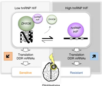
treatments currently used to treat GBM patients (Fig.
7
). Mining
GBM TCGA and REMBRANDT data sets (Supplementary Fig. 4)
as well as analyzing the protein expression in human glioma
protein samples (Fig.
6
), we found that hnRNP H/F is increased
in GBM and correlates with poor survival, extending the notion
of a key role of hnRNP H/F family members in cancer
develop-ment and progression
63. Our results support a model (Fig.
7
) in
which hnRNP H/F overexpression in GBM coordinately regulate
the translation of RG4-containing mRNAs encoding proteins
involved in maintaining genome stability and in the response to
genotoxic damage. The observation that 74 mRNAs coding for
stress response factors are targeted by both hnRNP H/F and
DHX36 (Supplementary Fig. 12) opens up new avenues for future
research to investigate whether and how these regulations induce
adaptive changes crucial for tumor cell survival during treatment
and the development of resistance. Our results not only extend
the notion of a link between G4 and genomic instability
64to
mRNA translational regulation but also associates it with a role in
resistance to treatments in GBM. Given that 1) our results were
similar regardless of the GBM cell line (e.g., Fig.
2
a, c, d and
Supplementary Fig. 5b; Fig.
3
g and Supplementary Fig. 8e, Fig.
5
d
and Supplementary Fig. 10b) or the cancer cell-type
(Supple-mentary Fig. 13), 2) hnRNP H/F were found to be deregulated in
many tumors (Supplementary Fig. 14), and 3) hnRNP H/F
RG4-containing mRNA targets significantly enriched genes associated
to GBM (adjusted P-value
= 0.03284 and 0.001729 for H and F
targets, respectively) but also to other cancers, including breast
(adjusted P-value 0.033 and 1.2E-06) and ovarian cancers
(adjusted P-value 0.013 and 1.8E-05), we propose that the link
between hnRNP H/F and cancer mediated by RG4-dependent
translational regulation could apply to other cancer cells and
tumors, thus making hnRNP H/F a potential target for
ther-apeutic intervention.
Overall, our results support the notion that hnRNP H/F are an
essential regulatory hub in GBM networks that drives
transla-tional control of RG4-containing genes contributing to GBM
progression and response to treatments. Moreover, our RP-MS
screen raises interesting future investigations to determine how
modulation of RG4 structural integrity impacts cellular functions
related to cancer hallmarks.
Methods
Cell culture and treatment. Glioblastoma cells (LN18, ATCC CRL-2610; U251-MG ECACC #;09063001 U87, SIGMA, #89081402-1VL) were grown in DMEM media (4.5 g/l glucose) supplemented with 10% FBS, 2 mML-glutamine, 100 U/ml penicillin, and 100 µg/ml streptomycin. Cells were tested for mycoplasma con-tamination by PCR. Cells were incubated/exposed at 37 °C with: 20 µM PDS (Selleckchem S7444) or 20 µM cPDS (Sigma-Aldrich SML1176) or 10 µM PhenDC3 (Polysciences, #26000-1) for the indicated time, 100 µg/ml Puromycin (Sigma P8833) for 1 h, 500 µM or dose scale of TMZ for 24 h, 4 Gy or dose scale of γ-irradiation (Gammacell 40 Exactor).
Cell transfection. siRNAs were transfected using the Lipofectamine RNAiMAX (Life Technologies) according to the manufacturer’s instructions. In brief, cells were reverse-transfected with 2.5 nM siRNA for 48 h. siRNA oligonucleotides Control (5′-GGUCCGGCUCCCCCAAAUG dTdT-3′), against hnRNP H (5′-GG
UAUUCGUUUCAUCUACA dTdT-3′), hnRNP F (5′-GGUGUCCAUUUCAU
CUACA dTdT-3′) and DHX36 (5′-GGUGUUCGGAAAAUAGUAA dTdT-3′) Fig. 5 hnRNP H/F drive genomic instability and therapy resistance. a Immunofluorescence experiments in LN18 cells using the γ-H2AX, 53BP1 antibodies and DAPI staining. Mean intensities ofγ-H2AX and 53BP1 in 2322 cells were plotted; the bottom and top of the box present the first and third quartile, respectively; the band inside the box shows the mean and the whiskers show the upper and lower extremes. Statistical significance was performed on the full cell populations.n = 2322 cells examined. Shown is a single representative field from one experiment over n = 2 independent experiments. For γ-H2AX: ***P-value = 4.26e-10, for 53BP1: ***P < 2.2e-16 (two-sided Mann & Whitney test). b Western blot analysis of γ-H2AX in LN18 cells treated with dose scale of carboxypyridostatin (cPDS) for 24 h. Shown is a representative result fromn = 3 independent experiments. c Quantification of the γ-H2AX levels in LN18 treated with cPDS normalized to GAPDH levels and plotted relatively to the untreated condition. Data are presented as mean values ± SEM ofn = 3 independent experiments, P-value = 0.0157 and P-value = 0.0457 for the 2 µM and 10 µM cPDS treatment respectively (two-sided paired t-test). d Quantification of DNA repair kinetics by western blot analysis of γ-H2AX after 4 Gy γ-irradiation in LN18 cells treated with control (siCtr) or hnRNP H/F (siH/F) siRNAs. Shown is a representative result fromn = 2 independent experiments. e Plating efficiency assays measuring the cell survival fraction in LN18 treated with siCtr or siF siRNAs and submitted to a radiation dose scale. Data are presented as mean values ± SEM of 6 wells,P-value = 0.0003 and P-value = 0.0006 for the 2 Gy and 4 Gy dose, respectively (two-sided paired t-test). f Quantification of DNA repair kinetics by western blot analysis of γ-H2AX after temozolomide (TMZ) treatment in LN18 cells transfected with an empty plasmid (pICE) or a plasmid expressing Flag-hnRNP H/F. Shown is a representative result fromn = 2 independent experiments. For all panels, source data are provided as a Source Data file.
were synthesized by SIGMA. For DNA plasmid transfections, 3.7 µg of plasmids was transfected in 60 mm diameter dishes using jet-PEI reagent (Polyplus) according to the manufacturer’s instructions. For Luciferase mRNA transfections, 250 ng of reporter mRNA was transfected in 48-well plates using lipofectamine 2000 reagent according to the manufacturer’s instructions. Cells were subsequently incubated at 37 °C for 48 h or 16 h following DNA plasmid or mRNA reporter transfections respectively, before harvesting and analysis.
Cell fractionation. For cell fractionation, cells were gently resuspended in 500 µl of hypotonic lysis buffer (10 mM Tris pH 8.0, 1.5 mM MgCl2, 10 mM NaCl, 1 mM
DTT) and vortexed for 4 s. After centrifugation at 1000 g (4 °C) for 5 min, supernatant (cytosolic fraction) was recovered. Pellet fraction (washed twice with hypotonic lysis buffer) was resuspended in 500 µl lysis buffer A (10 mM Tris pH 8.0, 140 mM NaCl, 1.5 mM MgCl2, 0.5% NP40, 1 mM DTT). The supernatant
(microsomal fraction) was recovered. Pellet-nuclear fraction (washed twice and
resuspended in 500 µl of lysis buffer A) was transferred to a 5-ml round-bottom tube and 50 µl of detergent mix (3.3% (w/v) sodium deoxycholate, 6.6% (v/v) Tween 40) were added. After incubation on ice for 5 min, the supernatant-postnuclear fraction was recovered (perinuclear fraction). The pellet-nuclear fraction (washed with buffer A) was resuspended in 500 µl of lysis buffer A sup-plemented with 0.1% SDS and sonicated. After centrifugation at 1000 g (4 °C) for 5 min, supernatant (nuclear fraction) was transferred into a fresh tube.
Mass spectrometry. Proteins were lysed and denatured in Tris 50 mM pH 8.5 and SDS 2% while disulfide bridges were reduced using TCEP 10 mM and subsequent free thiols groups were protected using chloroacetamide 50 mM for 5 min at 95 °C. Proteins were trypsin-digested overnight using the suspension trapping (S-TRAP) method to collect peptides as described in65. Eluted peptides were vaccum-dried
while centrifuged in a Speed Vac (Eppendorf). C18 liquid nanochromatography and Mass Spectrometry (MS) nanoflowHPLC and MS analyses were performed at
hnRNP H/F Actin USP1 DHX36
a
b
f
d
e
GAPDH 0 cPDS (μM): 2 10 USP1 OD 254 nm 40S 60S 80S siCtr HP LP NP 37 kDa 150 kDa Low Grade Gliomas High Grade GBM 100 kDa 150 kDa 50 kDa 50 kDa USP1 HPRT –0.2 0.2 0 –0.4 –0.6 siDHX36 0.0 0.2 0.4 0.6 0.8 1.0 H/F DHX36 * * siRNA: CtrRelative USP1 protein level
normalized to USP1 mRNA level
Relative mRNA quantity (log2 (HP)/(NP+LP+HP))
c
GAPDH hnRNP H/F USP1 Ctr H/F DHX36 siRNA: DHX36 Ubiquitin 100 kDa 250 kDa 100 kDa 50 kDa 150 kDa 25 kDa 40 kDa 55 kDa 70 kDa 37 kDaFig. 6 hnRNP H/F and DHX36 regulate USP1 translation in glioblastoma cells and tumors. a Polysome profile of U87 cells treated with control (siCtr) and DHX36 (siDHX36) siRNAs.b As in a, followed by RT–qPCR analysis from pooled non-polysomal (NP), light (LP) and heavy (HP) polysomal fractions, using specific primers for USP1 and HPRT mRNAs, and quantification by analyzing the ratio HP/total mRNAs from n = 2 independent experiments. Source data are provided as a Source Datafile. c Western blot analysis of USP1 and ubiquitination in U87 cells treated with siCtr, siRNAs against hnRNP H/F (siH/ F) or DHX36 (siDHX36). Source data are provided as a Source Datafile. d USP1 protein levels in c were normalized first to GAPDH protein levels and then to USP1 mRNA levels and plotted relatively to the siCtr condition. Data are presented as mean values ± SEM ofn = 3 independent experiments, P-value = 0.0291 andP-value = 0.05 for siH/F and siDHX36 respectively (two-sided paired t-test). e Western blot analysis of USP1 in LN18 cells treated with carboxypyridostatin (cPDS) dose scale for 24 h. Shown is a representative result fromn = 3 independent experiments. Source data are provided as a Source Datafile. f Western blot analysis of USP1, DHX36 and hnRNP H/F levels in protein extracts from Diffuse Low Grade Gliomas (Grade II) and High Grade GBM (grade IV). Shown is a representative result fromn = 3 independent western blot. Source data are provided as a Source Data file.