HAL Id: inserm-00144336
https://www.hal.inserm.fr/inserm-00144336
Submitted on 28 Oct 2008HAL is a multi-disciplinary open access
archive for the deposit and dissemination of sci-entific research documents, whether they are pub-lished or not. The documents may come from teaching and research institutions in France or abroad, or from public or private research centers.
L’archive ouverte pluridisciplinaire HAL, est destinée au dépôt et à la diffusion de documents scientifiques de niveau recherche, publiés ou non, émanant des établissements d’enseignement et de recherche français ou étrangers, des laboratoires publics ou privés.
Evidence of a functional role for interaction between
ICAM-1 and nonmuscle alpha-actinins in leukocyte
diapedesis.
Lionel Celli, Jean-Jacques Ryckewaert, Elisabeth Delachanal, Alain Duperray
To cite this version:
Lionel Celli, Jean-Jacques Ryckewaert, Elisabeth Delachanal, Alain Duperray. Evidence of a functional role for interaction between ICAM-1 and nonmuscle alpha-actinins in leukocyte diapedesis.. Journal of Immunology, Publisher : Baltimore : Williams & Wilkins, c1950-. Latest Publisher : Bethesda, MD : American Association of Immunologists, 2006, 177 (6), pp.4113-21. �inserm-00144336�
Evidence of a functional role for interaction between
ICAM-1 and non-muscle α-actinins in leukocyte
diapedesis
*
Running Title: ICAM-1/α-actinin interactions in inflammation
Lionel Celli, Jean-Jacques Ryckewaert, Elisabeth Delachanal, and Alain Duperray†
INSERM, U578, Grenoble, France; Université Grenoble I, Groupe de Recherche sur le Cancer du Poumon, Institut Albert Bonniot, Grenoble, France;
* This work was supported by the Association pour la Recherche sur le Cancer.
† Corresponding author: Alain Duperray, INSERM U578, Institut Albert Bonniot, Domaine de la Merci, 38706 La Tronche Cedex, France. Tel.: +334.76.54.94.20. Fax: +334.76.54.94.13. E-mail address: Alain.Duperray@ujf-grenoble.fr.
Keywords: inflammation, diapedesis, ICAM-1, α-actinin, endothelium
1
HAL author manuscript inserm-00144336, version 1
HAL author manuscript
Abstract
Intercellular adhesion molecule 1 (ICAM-1) is involved in both adhesion and extravasation of leukocytes to endothelium during inflammation. It has been shown that the ICAM-1 cytoplasmic domain is important for transendothelial migration of leukocytes but the precise molecular mechanisms involving the intracytoplasmic portion of ICAM-1 is not known. To characterize precisely the molecular scaffolding associated with ICAM-1, we have used the yeast two hybrid system, and we have identified six different proteins interacting with the ICAM-1 cytoplasmic domain. In this paper, we report that the two forms of non-muscle α-actinin (i.e. α-actinin 1 and α-actinin 4) associate with ICAM-1, and that these interactions are essential for leukocyte extravasation. These interactions were further confirmed by co-immunoprecipitation and immunofluorescence in endothelial cells and in ICAM-1 transfected CHO cells. The function of these interactions was analyzed by point mutation of charged amino acids located on ICAM-1 cytoplasmic domain. We have identified three charged amino acids (Arginine 480, Lysine 481 and Arginine 486) which are essential in the binding of α-actinins to the ICAM-1 cytoplasmic tail. Mutation of these amino-acids completely inhibited ICAM-1-mediated diapedesis. Experiments with siRNA inhibiting specifically α-actinin 1 or α-actinin 4 on endothelial cells indicated that α-actinin 4 had a major role in this phenomenon. Thus, our data demonstrate that ICAM-1 directly interacts with cytoplasmic α-actinin 1 and 4 and that this interaction is required for leukocyte extravasation.
2
Introduction
To eradicate invading pathogens, host defense mechanisms and immune inflammatory responses mainly consist in leukocyte transfer from blood to inflammatory tissues, a process called diapedesis which is regulated, at least in part, by adhesion proteins expressed at the cell surface (1, 2).
Diapedesis is divided into a three step sequence, (i) selectin-mediated-rolling of circulating leukocyte on endothelial cells (EC) (3); (ii) followed by β2 integrin-mediated
leukocyte adhesion (4) and finally (iii) transmigration across endothelial junctions (5, 6). Among the different molecular actors implicated in diapedesis, Intercellular Adhesion Molecule-1 (ICAM-1) (7) is a key molecule in leukocyte-endothelium adhesion through its recognition of β2 integrin counter receptors (6, 7). ICAM-1 belongs to the Immunoglobulin
superfamily and its expression level on endothelial cells (ECs) can be induced by inflammatory mediators such as TNFα. This protein is also expressed on neutrophils, fibroblasts and epithelial cells. Structurally, ICAM-1 is an integral membrane protein divided into three distinct regions (8): (i) an extracellular region containing five immunoglobulin domains, (ii) a transmembrane region and (iii) a short cytoplasmic tail of 28 amino acids, which interacts with actin binding protein such as non-muscular α-actinin, ERM (ezrin, radixin, moesin) proteins, β-tubulin, GAPDH and PIP2, a molecule implicated in signaling cascade (9-12).
The ICAM-1 extracellular region interacts with leukocyte integrins CD11/CD18 (LFA-1 and Mac-(LFA-1) ((LFA-13, (LFA-14). In addition ICAM-(LFA-1 recognizes fibrinogen (fg) ((LFA-15), an abundant plasma glycoprotein. Fibrinogen is also a ligand for β2 integrin Mac-1 (16) and we have
previously shown that fg enhances leukocyte-endothelium adhesion by acting as a molecular bridge between ICAM-1 and Mac-1 expressed on the two cell types (15). In addition we have
3
shown that during diapedesis, ICAM-1 is directly implicated in the transmigration process. This function is mediated by ICAM-1 cytoplasmic domain and a Rho-dependent signaling pathway (6, 17, 18). This implication of the ICAM-1 cytoplasmic tail and a Rho-dependent signaling pathway in leukocyte extravasation were recently confirmed in two models of brain endothelial cells (17, 18).
Our data implicating the ICAM-1 cytoplasmic domain during the inflammation process are in agreement with different reports showing that the engagement of ICAM-1 with its ligands can lead to intracellular signals (reviewed in (19)). How the ICAM-1 cytoplasmic tail is implicated in leukocyte transmigration remains unknown. Whether the involvement of ICAM-1 during diapedesis is due to a direct interaction of its cytoplasmic domain with actin binding proteins and/or a signaling pathway involving molecular switches such as Rho, a small GTPase implicated in regulating actin cytoskeleton (20), remain to be clarified.
Recently, Lyck et al (17), using ICAM-1/ICAM-2 double-deficient brain endothelium, demonstrated that the cytoplasmic tail of endothelial ICAM-1 is necessary for transendothelial migration of T cells. One of their conclusions was that the understanding of the ICAM-1 cytoplasmic tail function will necessitate an in-depth characterization of the intracellular binding partners for ICAM-1 in endothelial cells.
In order to explain the functional role of ICAM-1 cytoplasmic domain in leukocyte extravasation, we focused our attention on identifying novel cytoplasmic binding partners of this molecule, using a yeast two-hybrid system. With this technique, we identified the two non-muscle α-actinins (α-actinin 1 and 4) as major cytoplasmic partners of ICAM-1. We confirmed these interactions in vivo by immunofluorescence microscopy which showed colocalization between these proteins both in human endothelial cells and in ICAM-1 transfected CHO cells. A direct interaction between ICAM-1 and α-actinins was demonstrated
4
1/ by GST pull-down assays and 2/ by co-immunoprecipitation experiments of endogenous proteins.
Finally, the functional role of these interactions was studied by the identification of amino acids on the ICAM-1 cytoplasmic tail that were essential in the ICAM-1/non-muscle α-actinin interactions. We found that three charged residues (in position 480, 481 and 486) in ICAM-1 cytoplasmic domain were necessary for this association. Interestingly, each of the three ICAM-1 mutations was able to inhibit α-actinin 1 as well as α-actinin 4 interaction, suggesting that the same amino acids on ICAM-1 were involved in the binding of α-actinin 1 or α-actinin 4 to ICAM-1.
Using recombinant CHO cell lines expressing ICAM-1 mutants, we showed that inhibition of α-actinins association with ICAM-1 decreased extravasation. Small-interfering RNA (siRNA) silencing of α-actinin 4 expression induced a significant inhibition of neutrophils diapedesis, while actinin 1 silencing was without effect, suggesting that α-actinin 4 plays a major role in this phenomenon. These results indicated that ICAM-1/non-muscle α-actinin interactions are required for diapedesis induced by fg/ICAM-1 association. To our knowledge, this is the first time that a direct relationship is demonstrated between the association of cellular proteins with ICAM-1 and its function.
5
Materials and Methods
Antibodies and reagents
All organic chemicals were from Sigma-Aldrich (St-Louis, USA) unless otherwise specified. Restriction enzymes and DNA-modifying enzymes were from Invitrogen Life-technologies (Cergy-Pointoise, France). Synthetic oligonucleotides were prepared by Genosys Biotechnologies (Cambridge, UK). The following mAbs were used: mouse mAbs 3D6 and 2D5 to human intercellular adhesion molecule-1 (ICAM-1) were produced in our laboratory, as described previously (21); mouse mAb directed against α-actinin 1 (BM-75.2) was from Sigma. In some experiments, 3D6 was labeled with cy3, according to the manufacturer’s instructions (Amersham Biosciences, Freiburg Germany). For α-actinin 4, initial experiments were performed using a rabbit polyclonal anti-α-actinin 4 Ab (a generous gift from S. Hirohashi, National Cancer Research Institute, Pathology Division, Tokyo, Japan; (22); for all experiments described in this study, we used a rabbit polyclonal anti α-actinin 4 antibody (LC17) raised against a keyhole limpet hemocyanin-conjugated synthetic peptide, MVDYHAANQSYQYGPS (containing N-terminal amino acids 1-16 of α-actinin 4) (Covalab, Lyon, France). The specificity of this polyclonal antibody was verified by immunoblot and immunofluorescence (see results). A rabbit antiserum against actin (Sigma) was used for quantification and normalization of western blots. Texas-Red phalloidin from Molecular Probes (Leiden, Netherlands) was used to label F-actin.
Yeast Two-Hybrid Assays
A cDNA fragment encoding the complete cytoplasmic portion plus six amino acids from the transmembrane domain of human ICAM-1 (residues 1509-1614) was amplified by PCR. The following primers were used: 5'-GAATTCCTCAGCACGTACCTCTATAAC-3' (downstream) and 5'-CGGGATCCTCAGGGAGGCGTGGCTTGTGTGTT-3'(upstream). The
6
primers were designed to include an EcoR1 site for the upstream primer and a BamH1 site for the downstream primer. The resulting PCR product was verified by sequencing and subcloned into pAS2-1. A human placenta cDNA library, cloned into the pACT2 vector fused with the GAL4 activation domain was purchased from Clontech (Palo Alto, USA). The yeast strain Y190 (Clontech) was sequentially transformed with pAS2/ICAM-1 as bait, then with a pACT2 plasmid containing the cDNA library. Transformants were grown on synthetic dextrose medium lacking the amino acids Leucine, Tryptophan and Histidine in the presence of 25mM 3-amino-1.2.4-triazole. On day 5, colonies were tested for the activity of the lacZ reporter gene with a β-galactosidase (β-Gal) filter assay. All pAS2-1 subclones were analyzed by DNA sequencing to confirm the correct in frame fusion of GAL4 and ICAM-1 cytoplasmic sequence.
A mating assay was performed to eliminate false positive; clones scored as positive after the screening were cultured on synthetic dextrose medium without Tryptophan in the presence of 10 μg/ml of cycloheximide. The cycloheximide resistant Y190 yeast clones were verified in a mating assay with yeast strain expressing the pAS2 plasmid with either ICAM-1, the unrelated protein lamin C (pLAM5’-1 vector) or the non-fused GAL4 DNA binding domain as baits. Positive clones were further retested in a cotransformation assay with purified plasmid cDNA combined to a range of plasmid controls: pLAM5'-1, in pAS2-1, pACT2, pVA3-1 and pTD1-1 (Clontech).
To quantify two-hybrid interactions and compare the relative strength of protein-protein interactions observed, an O-nitrophenyl-β-D-galactopyranoside (ONPG) assay was used (23).
Inserts from positive cDNA clones were sequenced by the T7 Sequenase chain-termination DNA sequencing method using the T7 Sequenase version 2,0 DNA sequencing kit from Amersham Biosciences (Freiburg Germany). The sequences were
7
analyzed by the basic local alignment search tool program, at National Center for Biotechnology Information (www.ncbi.nlm.nih.gov/BLAST/).
Green fluorescent protein (GFP) and ICAM-1 mutant constructs
A cDNA encoding full length ICAM-1 was cloned into the pEGFP-N1 vector (Clontech) to obtain a fusion protein expressing GFP at the carboxyl-terminus of ICAM-1. ICAM-1 cytoplasmic mutants: CHO ICAM-1 R480A, K481A, K483A, K484A and R486A were generated using QuickChange Site-Directed Mutagenesis Kit (Stratagene). Procedure was performed to mutate each charged amino acid selected on ICAM-1 into the neutral amino acid Alanine using the pcDNA3 plasmid (Invitrogen, San Diego, CA) containing the full length human ICAM-1 coding sequence.
Production of GST Fusion Proteins and in vitro translation
Full-length α-actinin 1 was obtained by performing a RT-PCR from Hela cells and full-length α-actinin 4 was ordered from Incyte Genomics (St Louis, USA). Both cDNAs were sequenced by Genaxis Biotechnology (Nîmes, France) and subcloned into pGEX 5X3 plasmid (Amersham Biosciences). GST fusion proteins were expressed in BL21 Escherichia
Coli. After lysis by sonication, GST fusion proteins were purified using glutathione–
Sepharose4B beads accordingto the manufacturer’s protocol (Amersham Biosciences). In vitro transcription and translation experiments were performed using T7 TnT Quick coupled transcription/translation system from Promega. Alpha-actinin polypeptides were synthesized from full-length human α-actinin cDNAs subcloned in pcDNA3-1 plasmid.
Cell culture and transfection experiments
HUVECs were isolated from human umbilical cords by collagenase treatment as described by Jaffe et al. (24), and cultured as previously described (6). In some experiments,
8
HUVECs were stimulated with 200 U/ml TNF-α (Boehringer Mannheim, Meylan, France) for 4h at 37°C.
CHO cells were cultured in DMEM (BioWhitttaker) containing 5% FBS (Life technology, Cergy Pontoise, France), 2mM L-glutamine, 100 U/ml penicillin, 100µg/ml streptomycin and non-essential amino acids (BioWhitttaker).
Obtention of stable CHO cell lines expressing ICAM-1 or ICAM-1 cytoplasmic deletion mutant (ΔCyt) has been previously described (6). CHO cell lines expressing ICAM-1-GFP or point mutations on ICAM-1 cytoplasmic portion were obtained by transfecting CHO cells by electroporation. Selection of stable transfectants was performed in DMEM medium containing 1 mg/ml G418 (Geneticin; Life Technologies, Gibco BRL) for selection or 500 µg/ml for routine culture (25). Phenotypic characterization of transfectants was carried out by flow cytometry and immunofluorescence staining on adherent cells with anti-ICAM-1 (3D6) mAb.
Polymorphonuclear neutrophils (PMN) used for transmigration assays were isolated by differential centrifugation on Ficoll-Hypaque gradient (Sigma), followed by dextran sedimentation and hypotonic lysis of erythrocytes.
SiRNA transfection of HUVEC
Predesigned annealed siRNAs targeting α-actinin 1 or α-actinin 4 were purchased from Eurogentec (Belgium), together with a siRNA negative control. HUVECs were seeded in 6-well dishes at a density of 1.6x10⁵ cells per well 24 h prior to the experiment. Cells were transfected with siRNAs directed to human α-actinin 1 (5’-CACCAUGCAUGCCAUGCAA-3’ at position 852 of α-actinin 1 mRNA, starting from AUG start codon, siRNA Actn1pos852),
or human α-actinin 4 (5’-GCAGCAGCGCAAGACCUUC-3’at position 150, siRNA Actn4pos150 (26)) (300 nM each) or a negative control siRNA, known to have no effect on
9
mammalian protein expression. Oligofectamine (Invitrogen, www.invitrogen.com) was used to transfect siRNAs according to the manufacturer’s instructions. Transfection was carried out in 1 ml OptiMEM (Invitrogen). Four hours after transfection, cells were supplemented with additional 3 ml endothelial cell growth medium and cultured for another 3 days. Down regulation of actinin protein was verified by Western blotting, after lysis of the cells. Samples were resolved on a 7.5% SDS-PAGE and blotted. Actinins were detected using anti
α-actinin 1 and α-anti-actinin 4 specific antibodies. Blots were also probed with an anti-actin antibody to monitor equal loading.
Precipitation of overexpressed proteins
CHO cell lines expressing native ICAM-1 or ICAM-1 mutants were lysed with immunoprecipitation (IP) buffer ((PBS containing 0.5% Triton X100, 0.5% Nonidet-P40, complete protease inhibitors (Roche)) on ice for 30 min. After centrifugation, supernatant (300 µg protein) was incubated for 3 h at 4°C with Glutathione-agarose beads preadsorbed with 50 µg of GST or GST-actinin 1 and 4. Beads were washed three times for 10 min with IP buffer; bound proteins were eluted with SDS–PAGE loading buffer and subjected to 7.5% SDS−PAGE. After blotting onto nitrocellulose, ICAM-1 was detected using mAb 2D5 at 2µg/ml.
Immunoprecipitation of endogenous proteins in endothelial cells
Confluent cultures of HUVECs were treated with TNFα for 4 hours and then lysed with IP buffer. To immunoprecipitate ICAM-1 or α-actinin 1, Protein A Sepharose 6MB coated with rabbit IgG anti-mouse was incubated with anti ICAM-1 mAb (2D5) or anti α-actinin 1 mAb. For α-actinin 4 immunoprecipitation, rabbit polyclonal anti α-actinin 4 LC17 was directly incubated with Protein A Sepharose. ICAM-1, α-actinin 1 or α-actinin 4 were immunoprecipitated from HUVEC lysates, and co-precipitation of associated proteins was
10
revealed by western blotting with specific antibodies against ICAM-1, actinin 1 or α-actinin 4.
Flow cytometry analysis
Expression level of ICAM-1 was analyzed on a FACScan (Becton-Dickinson, Pont de Claix, France), as described previously (6).
Immunofluorescence staining and confocal imaging
HUVECs or CHO cells were seeded on glass coverslips precoated with 25 µg/ml of human plasma fibronectin (27) and processed as described previously (6). Cells were stained with the following primary antibodies: anti ICAM-1 3D6 (5µg/ml), anti α-actinin 1 (1/100 dilution of ascites), anti actinin 4 (4µg/ml). Filamentous actin was revealed with texas-red conjugated phalloidin. Images of HUVECs and CHO cells were acquired using an Olympus inverted microscope (AX70).
Zeiss confocal microscope was used to perform cross sectioning of cells. To combine the entire Z series into a stack projection and to accomplish cross sectional reconstruction, we used the Metamorph software (Universal Imaging Corporation, Downingtown, USA). The percentage of the proteins that colocalize with each other was measured with the Zeiss software. Statistical analysis with the software determined the Pearson’s Correlation (PC), which compares the colocalization of two different types of fluorescence and their intensities; the higher the fluorescence intensities are at the site of colocalization, the higher the PC will be, with a maximum of 1.
Permeability assay
Permeability assays to determine monolayer permeability of CHO cell lines were proceeded as described in our previous study (6).
11
Transmigration assays
CHO cell monolayers were grown to confluency on fibronectin-coated porous membranes (6.5 mm diameter and 5 µm pore size; Costar) for 3 days at 37°C. CHO cells were then incubated in duplicate with PMN (2 x 10⁵/well); in order to maintain the viability of human PMN, autologous normal human plasma (1:5 dilution in DMEM/Hepes 20mM pH 7.4) was added with the PMN in the upper compartment in the presence of 100µM PPACK (Boehringer), a specific inhibitor of thrombin. A concentration of 2 X 10⁻⁸ M fMLP in DMEM/Hepes 20mM pH 7.4 (Calbiochem-Novabiochem Corp.) was added in the lower compartment, to create a chemotactic gradient for PMN. Migrated neutrophils were recovered from the bottom of the well after 3h at 37°C in 5% CO2, centrifuged, stained with trypan blue
and counted microscopically.
Statistics
Statistical differences between experimental groups were evaluated using the student’s t-test.
12
Results
Identification of cellular proteins interacting with ICAM-1
To identify intracytoplasmic proteins interacting with ICAM-1 cytoplasmic domain, we used a yeast two-hybrid system (MATCHMAKER Gal4 Two-Hybrid). A bait construct (pAS2-1/ICAM-1) encoding the human cytoplasmic domain of ICAM-1 was designed to screen a human placenta cDNA library (>5x10⁶ independent clones). Human placenta is a heavily vascularized tissue, and proteins expressed by ECs including ICAM-1 are well represented. Using this system, 47 clones were detected as positive candidates, of which 14 were confirmed positive after extensive testing and DNA sequencing. These 14 clones were sequenced and were found to correspond to the following genes: α-actinin 1, α-actinin 4,
Bin 1, MAGE-4b, succinate deshydrogenase and the T-cell activation protein. In this paper,
we focused our attention on α-actinin 1 and α-actinin 4.
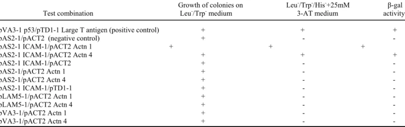
All the clones coding for α-actinins corresponded to the C-terminal two-thirds of these proteins (residues 501-914 for α-actinin 1, and 544-930 for α-actinin 4). The specificity of ICAM-1/ α-actinin 1 and α-actinin 4 interactions were confirmed by two-hybrid mating assays using purified pACT2 α-actinin 1 and 4 in association with pAS2-1 ICAM-1 as positive control, pTD1-1 encoding Large T-antigen, pVA3-1 encoding murine p53 and pLAM5-1 encoding human lamin C as negative controls (Table I).
Specificity of the polyclonal antibody anti α-actinin 4
Rabbit polyclonal antibody LC17 was raised against the NH2-terminal amino-acid sequence of α-actinin 4. The immunoblot in Figure 1A, using full-length α-actinins produced
in vitro, shows that this polyclonal antibody reacted only with the in vitro translation product
of α-actinin 4 cDNA (lane 4) but not with α-actinin 1 (lane 3), thus demonstrating its specificity. MAb (BM75.2) against α-actinin 1 was used as a control. As shown in Figure 1A,
13
this antibody was reactive only with the in vitro translation product of human α-actinin 1 cDNA (lane 1) but not with human α-actinin 4 (lane 2).
Localization of α-actinins in EC and CHO cells
The cellular localization of α-actinins in HUVECs was studied by immunofluorescence microscopy. Alpha-actinin 1 was expressed along actin fibers and at adherens junctions, at the end of stress fibers (Fig 1B panels 1-3), as already described by other groups (22, 28), and was colocalized with ICAM-1 at the border of the cell (Fig 1C panels 1-3), suggesting an interaction between both proteins in vivo in HUVECs. Alpha-actinin 4 was expressed in a punctate fashion along actin fibers, and near the cell membrane (Fig 1B panels 4-6). Faint α-actinin 4 expression was also observed in the nucleus (Fig 1B panel 5, white arrow), as previously reported by Honda et al (22). ICAM-1 was found to be colocalized with α-actinin 4 at the periphery of the cells (Fig 1C 4-6, white arrows), suggesting a possible interaction between both proteins.
We then studied the localization of endogenous α-actinins in CHO cells transfected with ICAM-1-GFP. Both α-actinins were detected in CHO cells, with α-actinin 1 (Fig 1D panel 5) and α-actinin 4 (Fig 1D panel 2) detected by mAb BM-75.2 and polyclonal antibody LC17 respectively. A very high level of colocalization between ICAM-1 and α-actinin 1 was observed (Fig 1D panel 6), with a strong expression of α-actinin 1 at the periphery of the cell and at sites of cell/substrate interaction. The percentage of colocalization was between 60 to 80% as determined by software analysis on confocal slices, with a Pearson’s correlation (PC) of 0.52. Actinin 4 was expressed in these cells with localization similar to the one of ECs, on actin fibers and near the cell surface (Fig 1D panel 2). Some colocalization could be observed with ICAM-1 at the cell surface (Fig 1D panel 3, white arrows). The percentage of colocalization for α-actinin-4 and ICAM-1 was from 30 to 40%, with a PC of 0.1. This
14
experiment showed that CHO cells express α-actinins and that the localization of transfected human ICAM-1 is similar to the one found in HUVECs.
Cell density and TNFα modify the cellular localization of α-actinin-4
Alpha-actinin 4 was first identified in epithelial cells (22), where it was shown to be localized to the cytoplasm and to the nucleus. In HUVECs α-actinin 4 was expressed along actin fibers and near the cell surface, with only a very faint labeling inside the nucleus (Fig 1). We thus tested the effect of cell density and TNFα treatment on α-actinin 4 cellular localization. In either sparse or confluent resting cells, α-actinin 4 was expressed in the cytoplasm or near the cell surface with little, if any, protein expression inside the nucleus. In contrast, after TNFα treatment, α-actinin 4 was also present inside the nucleus, in addition to its localization in the cytoplasm (Fig 2). In resting confluent HUVECs, α-actinin 4 was strongly expressed at cell-cell contact sites (Fig 2, panel C), but after treatment with TNFα, the majority of α-actinin 4 was redistributed to the cytoplasm and the nucleus (Fig 2, panel D). In contrast, the distribution of α-actinin 1 was unchanged under these conditions (data not shown).
α-Actinin 1 and α-actinin 4 interact with ICAM-1 both in vitro and in eukaryotic cell
ICAM-1 interaction with α-actinins was studied in vitro using fusion proteins containing α-actinin sequences isolated in the two hybrid experiment (residues 501-914 for α-actinin 1, and 544-930 for α-actinin 4) with GST-tag. These fusion proteins were bound to glutathione-agarose beads. These beads were incubated with a cell lysate from CHO expressing ICAM-1, the protein complexes were then resolved by SDS-PAGE, and ICAM-1 interacting with GST-actinin was detected by immunoblot. ICAM-1 was found associated with α-actinin 1 (Fig. 3A, lane 3) and with α-actinin 4 (Fig. 3A, lane 4). In contrast, ICAM-1
15
was not precipitated by GST protein alone (Fig. 3A, lane 1). These results indicated that ICAM-1 interacted with α-actinin 1 and with α-actinin 4 in vitro.
To provide evidence that ICAM-1 and actinins associate in vivo, immunoprecipitation experiments were performed. Extracts of HUVECs were prepared from cells induced by TNFα for 6 hours to increase ICAM-1 expression. As shown in Fig 3B, antibodies against α-actinin 1 (lane 4) or α-α-actinin 4 (lane 5) precipitated ICAM-1. The monoclonal antibody 2D5 was also able to coprecipitate α-actinin 1 and α-actinin 4, respectively (Fig 3C and 3D). These results demonstrated that ICAM-1 interacts in vivo with endogenous α-actinin 1 and 4 in eukaryotic cells.
Identification of amino acids on ICAM-1 cytoplasmic domain involved in ICAM-1/ non-muscular α-actinin interactions: Generation of ICAM-1 mutants.
To establish the functional role of these interactions, we first identified amino acids involved in ICAM-1/α-actinin interactions. A five amino acid sequence (480-RKIKK-484) has been previously shown to be involved in ICAM-1 interaction with α-actinin (11) (Fig 4A). We thus generated point mutations by switching to Alanine the four positive charged amino acids of this short sequence (one Arginine and three Lysines). In addition, we mutated Arginine 486, another charged amino acid located near the short peptide 480-484 (Fig 4A). The influence of each mutation was assessed using a quantitative β-galactosidase assay in a yeast two-hybrid system (MATCHMAKER Gal4 Two-Hybrid). This method allowed us to quantify interactions and to compare the relative strength of protein-protein interactions. In this study, we compared the interaction level between ICAM-1, considered as positive control, and each mutant (ICAM-1 R480A, K481A, K483A, K484A and R486A) with both α-actinin 1 and 4. As shown in Figure 4B, among the five amino acids mutated on ICAM-1, only mutations on Arginine 480, 486 and Lysine 481 reduced at least by 2-fold ICAM-1 interaction with α-actinin 1 and 4, while mutations on Arginine 483 and 484 were without
16
effect. These observations indicated that the binding of both α-actinins on ICAM-1 depended upon the same amino acids on ICAM-1 cytoplasmic tail.
Characterization of recombinant CHO cell lines expressing ICAM-1 mutants
The direct binding of ICAM-1 to α-actinins may affect its function. To test this hypothesis, we developed CHO cell lines stably expressing ICAM-1 mutants, and tested these cell lines using the reconstituted mammalian cell expression model already engineered in our laboratory (6). We first characterized the expression level and localization of these mutant proteins. Expression levels of ICAM-1 mutants in the five recombinant cell lines were quantified by FACScan analysis and compared to the CHO ICAM-1 cell line. As shown in Figure 5A, the selected CHO cell lines expressed each ICAM-1 mutant at a level comparable to CHO ICAM-1. This expression level has already been defined to be similar to TNF-α-stimulated HUVECs, a well-known model of ECs usually used in transmigration assay (6). Cellular distribution of ICAM-1 mutants in these cell lines was analyzed by immunofluorescence staining, followed by confocal analysis. Results in Figure 5B clearly show that the cellular localization of the various mutant ICAM-1 proteins was not altered when compared to the wild type protein.
We then used these cell lines to test the interaction of the different ICAM-1 mutants with α-actinins. Full-length α-actinins fused with a GST-tag were bound on glutathione agarose beads. Beads were then incubated with lysate from each recombinant ICAM-1mutant CHO cell line, and ICAM-1 mutant interaction with GST-actinin was analyzed by immunoblot with mAb 2D5. The quantification of immunoblot signals that represents levels of interaction between GST-actinin with the different ICAM-1 mutants is shown in Figure 5C. This showed that the pattern of interaction of the different ICAM-1 mutants expressed on recombinant cells with α-actinins obtained in this assay was similar to the one obtained in the two-hybrid test represented in Figure 4B. Despite the fact that results were less significant for
17
α-actinin 1 than for α-actinin 4, again we found that Arginine 480, Arginine 486 and Lysine 481 were essential for ICAM-1 interaction with both α-actinins. We thus confirmed, using full length α-actinins and ICAM-1 mutants from recombinant CHO cell lines, that Arginine 480, 486 and Lysine 481 are involved in ICAM-1/α-actinin interactions. Based on these results, these recombinant CHO cell lines were used to investigate the role of ICAM-1/actinins interactions in transmigration.
Fg-dependent transmigration requires ICAM-1/non-muscle α-actinin interactions
All the CHO cell lines have been tested for their ability to form a confluent monolayer. The integrity of the monolayers and their function as barriers were studied by a permeability assay using HRP (29). Three days after seeding, resting and TNFα-stimulated HUVECs presented permeability level of 19.4 ± 0.1% and 21.6 ± 0.1% of controls (Fig 6). All the recombinant CHO cell lines expressing ICAM-1, ICAM-1ΔCyt and mutants (R480A, K481A, K483A, K484A and R486A) formed monolayers that were nearly impermeable to HRP, with 12.0 ± 3.9%, 9.1 ± 0.8%, 9.8 ± 1.9%, 10.1 ± 5.6%, 16.4 ± 1.9%, 5.7 ± 0.6% and 12.2 ± 3.3% of controls, respectively; these values are similar to those obtained with WT CHO monolayers (11.9 ± 1.1%) (Fig 6). These results showed that CHO cells are able to form correct, continuous monolayers with a high cohesion of cell-cell contacts, and a permeability level similar to that observed with HUVECs.
The implication of ICAM-1 and its cytoplasmic tail in fg-dependent transmigration have already been defined in our laboratory (6). To assess if ICAM-1 cytoplasmic interaction with α-actinins was involved in this process, we performed an in vitro transmigration assay of neutrophils across the different CHO cell lines monolayers. As shown in Fig 7, when ICAM-1/α-actinin interactions were altered, the number of neutrophils transmigrating across CHO monolayers was reduced to a level similar to CHO WT or CHO ICAM-1ΔCyt (14.22 ± 3.32% for ICAM-1 R 480A, 14.92 ± 5.25% for ICAM-1 K481A and 16.2 ± 3.54% for ICAM-1
18
R486A vs. 13.33 ± 2.17% for CHO WT and 14.94 ± 1.97% for ICAM-1ΔCyt). To confirm that these results were due to an inhibition of ICAM-1/α-actinin interactions, we used an ICAM-1 K484A mutant which does not disturb these interactions. The level of transmigrated PMN across CHO ICAM-1 R484A monolayer was similar to CHO ICAM-1 (38.31 ± 6.04% vs 42.33 ± 3.76%). These experiments indicated that ICAM-1/α-actinin interactions were implicated in fg-dependent PMN transmigration.
α-actinin 4 depletion affects neutrophil diapedesis.
To test which isoform of α-actinin was involved in diapedesis, we used siRNA to specifically silence expression of either α-actinin 1 or α-actinin 4 protein in HUVEC and then assayed these cells for leukocyte diapedesis. SiRNA Actn4pos150 has been shown to be specific
for α-actinin 4 by Yan et al. (26), while siRNA Actn1pos852 has been designed in our
laboratory.
Transfection of siRNAs directed against α-actinin 1 or α-actinin 4 into HUVECs resulted in a strong and specific silencing of protein expression 3 days after transfection (Fig 8). SiRNA Actn1pos852 resultedin 70% inhibition of α-actinin 1 expression (Fig 8A lane
2) without affecting α-actinin 4 expression (Fig 8A lane 3). Similarly, SiRNA Actn4pos150
specifically inhibited by 90% actinin 4 expression (Fig 8A lane 6), but had no effect on α-actinin 1 expression (Fig 8A lane5). The negative control siRNA had no effect on both actinins (Fig 8A lanes 1 and 4).
As shown on Fig 8B, reduced α-actinin 4 expression was accompanied by a reduction of more than 40% of extravasation, while only a small and non significant decrease was observed after α-actinin 1 silencing.
These results indicate that α-actinin 4 interaction with ICAM1 is essential for diapedesis.
19
Discussion
Molecular mechanisms controlling neutrophil migration across vascular endothelium during immune-inflammatory response remain unclear. Several adhesion proteins expressed by ECs and leukocytes have been shown to be involved in this process. ICAM-1, a member of the Immunoglobulin superfamily is expressed by ECs and some leukocytes and has been identified as a ligand for leukocyte integrins LFA-1 and Mac-1. During diapedesis, these interactions allow leukocyte-endothelium adhesion (7, 30, 31). We have previously shown that ICAM-1 plays an important role in neutrophil transmigration, and that this process requires the ICAM-1 cytoplasmic domain and a Rho-dependent signaling pathway (6). These results were recently confirmed by two publications which demonstrated that the cytoplasmic domain of ICAM-1 was essential for lymphocyte-transendothelial migration in brain microvascular ECs, (17, 18). Although ICAM-1 is known to be important in diapedesis, little is known about the precise molecular mechanisms involved in these functions. Many observations point to the involvement of ICAM-1 cytoplasmic tail in transmission of intracellular signaling (reviewed in (19)). In ECs, crosslinking ICAM-1 with antibodies increases intracellular Ca2+ and activates cytoplasmic protein such as p38 MAPK, pp60src, Rho and protein kinase C which are known to be involved in signal transduction (6, 32-34). These proteins act upon several actin associated proteins including heat shock protein 27, cortactin, focal adhesion kinase, paxillin and p130Cas which may in turn influence actin cytoskeleton (33, 35, 36). Furthermore ICAM-1 signaling can also be initiated through ligation by fg (37, 38), a ligand of ICAM-1 which increases neutrophil transmigration across endothelium when associated to ICAM-1 (21, 39). Finally, an original approach using peptidomimetics of ICAM-1 intracellular domain further suggests that ICAM-1 mediates signaling events during lymphocyte migration across brain EC (18).
20
In this paper, in order to develop insight into the molecular mechanisms underlying ICAM-1 functions, we used a two-hybrid system to identify the cytoplasmic partners of ICAM-1. We focused our attention on the two non-muscle isoforms of actinin family, α-actinin 1 and 4. Interaction of ICAM-1 with α-α-actinin 4 was not previously described, and ICAM-1/ α-actinin 1 interaction has only been observed in vitro (11). This protein family contains two others members defined as muscle isoforms, α-actinin 2 and α-actinin 3). In vivo, α-actinins form anti-parallel dimers (22). Each monomer is divided in a N-terminal actin binding domain, a central domain with four spectrin like motifs which allow dimerization and a C-terminal domain containing an EF-hand calcium binding site (40). These proteins cross-link actin filament together and also connect the actin cytoskeleton to the cell membrane; α-actinin 1 has also been shown to be involved in signal transduction pathways (41). Non-muscle α-actinins share a high degree of similarity (87% amino acid identity) and appear to have different subcellular localizations: α-actinin 1 is localized at the end of actin stress fibers and adherens junctions where it plays an important role in stress fiber formation, promoting cell adhesion and regulating cell shape and motility (42-44); Honda et al have reported that α-actinin 4 is colocalized with actin stress fibers and is dispersed in the cytoplasm and the nucleus (22). This isoform has been characterized recently and its function remains unclear. Firstly associated with cell motility and cancer invasion (22), α-actinin 4 was also defined as a tumor suppressor in human neuroblastoma cells (45). Alpha-actinin 4 has been shown to be involved in a form of human focal and segmental glomerulosclerosis (46) and mice deficient in α-actinin 4 present severe glomerular abnormalities and an increase in cell motility as measured by leukocyte chemotaxis assays (47).
In the present study, we demonstrate that ICAM-1 can directly interact with non-muscle α-actinins in vitro and in vivo. In addition, we show that α-actinin 4 is highly expressed in endothelial cells, and that its cellular location is different in confluent monolayers or in sparse
21
cell culture; this pattern was modified after TNFα treatment. In confluent resting monolayers, α-actinin 4 was concentrated at cell-cell contact sites, in addition to its association with actin stress fibers, with almost no detectable level in the nucleus. After TNFα treatment, α-actinin 4 expression at cellular junctions decreased, and a fraction of α-α-actinin 4 was translocated inside the nucleus, with still a strong association with actin fibers. This clearly indicates that the newly synthesized ICAM-1 after TNF treatment is not associated with actinin-4, as there is no increase of α-actinin-4 near the cell surface. A nuclear translocation has already been reported in epithelial cells (22), after inhibition of PI3 kinase by wortmanin, and after treatment with cytochalasin D, which inhibit actin polymerization. This effect of actin depolymerisation suggested that nuclear translocation of α-actinin 4 could be caused by loss of its association with the cytoplasmic actin cytoskeleton. This could explain our results after TNFα treatment, which has been shown to induce a reorganization of the actin cytoskeleton in HUVECs (48). The presence of α-actinin 4 at sites of cell-cell interaction has already been described in keratinocytes and in an epithelial cell line (49), when cell form extensive cell-cell contacts. Consistent with this report, we have observed that α-actinin 4 is concentrated at cell-cell junctions in endothelial cells. Interestingly, TNFα treatment caused a delocalization of actinin 4 away from cell-cell junctions. These results suggest that α-actinin 4 in endothelial cells is an important component of the cell junctions, and that this localization can be down regulated by TNFα, independently of ICAM-1.
We have determined the functional role of these interactions by disturbing ICAM-1/α-actinin interaction. By generating point mutations on the ICAM-1 cytoplasmic sequence, we identified three amino acids (Arginine 480, 486 and Lysine 481) that were critical in ICAM-1 interaction with both α-actinins, suggesting that α-actinins bind to the same amino acids on ICAM-1. This hypothesis is supported by the important amino acid similarity of the two proteins.
22
In order to assess the specific role of ICAM-1/non-muscle α-actinin interactions we have engineered stable CHO cell lines expressing each mutants of ICAM-1. These cell lines led us to demonstrate a direct implication of ICAM-1/α-actinins interactions in fg-dependent leukocyte transmigration. Given the similar effect of ICAM-1 mutants R480A, K481A and R486A on both α-actinin 1 and 4 interaction, we were not able to conclude which isoform of α-actinins was involved in transmigration. One hypothesis would be that both α-actinins play an equivalent role in these phenomena. However, a recent study using mice deficient in α-actinin 4 suggests that α-actinins are not functionally redundant (47). We have addressed this question using specific siRNAs. Silencing of α-actinin 4 resulted in a 40% inhibition of neutrophils diapedesis, while silencing of actinin 1 was without effect, confirming that of α-actinins 1 and 4 have distinct roles inside the cell. These results suggested that the association of ICAM-1 with actinin 4 is essential for the diapedesis process, while the one with α-actinin 1, which was demonstrated by all the other experiments reported in this paper, remains to be identified.
Although ICAM-1 has been reported to bind several intracellular proteins such as α-actinin (11) β tubulin, GAPDH (12) and ERM protein (9), this is the first evidence of a functional role for an intracellular ICAM-1 partner. Ezrin has been considered as the leading candidate. This protein links ICAM-1 to the actin cytoskeleton (9) and ERM proteins have been recently implicated in leukocyte adhesion and transendothelial migration during inflammation (10), but direct interaction between ICAM-1 and ezrin has never been demonstrated in brain microvascular EC (50). In addition the same authors have shown that ERM proteins do not colocalize with ICAM-1 following cross-linking, a phenomenon mimics leukocyte adhesion on EC and triggers signal transduction (50). However, Hamada et al (51) who recently published the crystal structure of Radixin/ICAM-2 complex, have suggested a consensus motif present in ICAM-1 which includes Lysine 481. Moreover, PIP2 which binds
23
to the ICAM-1 cytoplasmic domain, has been involved in the regulation of ICAM-1/ezrin interaction (9). This signaling molecule has also been demonstrated to regulate α-actinin function (52). These observations suggest that PiP2 participates to the regulation of ICAM-1 interaction with ERM proteins and α-actinins.
In conclusion, our data demonstrate that non-muscle α-actinins are required for ICAM-1 function in diapedesis. This finding also provides a new basis for understanding of mechanisms implicated in signal transduction during inflammation.
24
Acknowledgements
We thank Setsuo Hirohashi for the generous gift of anti-α-actinin 4 antibody, Nathalie Chassignolle for excellent technical assistance, Leila Barki for RT-PCR experiments, and Alexeï Grichine for his assistance with the confocal microscope.
25
References
1.
Steeber, D. A., and T. F. Tedder. 2000. Adhesion molecule cascades direct
lymphocyte recirculation and leukocyte migration during inflammation.
Immunol. Res. 22:299-317.
2.
Worthylake, R. A., and K. Burridge. 2001. Leukocyte transendothelial
migration: orchestrating the underlying molecular machinery. Curr. Opin.
Cell Biol. 13:569-577.
3. Johnson-Leger,
C.,
M.
Aurrand-Lions, and B. A. Imhof. 2000. The parting
of the endothelium: miracle, or simply a junctional affair? J. Cell Sci.
113:921-933.
4.
Ronald, J. A., C. V. Ionescu, K. A. Rogers, and M. Sandig. 2001.
Differential regulation of transendothelial migration of THP-1 cells by
ICAM-1/LFA-1 and VCAM-1/VLA-4. J. Leukoc. Biol. 70:601-609.
5.
Muller, W. A., and G. J. Randolph. 1999. Migration of leukocytes across
endothelium and beyond: molecules involved in the transmigration and
fate of monocytes. J. Leukoc. Biol. 66:698-704.
6.
Sans, E., E. Delachanal, and A. Duperray. 2001. Analysis of the roles of
ICAM-1 in neutrophil transmigration using a reconstituted mammalian
cell expression model: implication of ICAM-1 cytoplasmic domain and
Rho-dependent signaling pathway. J. Immunol. 166:544-551.
7.
van de Stolpe, A., and P. T. van der Saag. 1996. Intercellular adhesion
molecule-1. J. Mol. Med. 74:13-33.
8.
Staunton, D. E., S. D. Marlin, C. Stratowa, M. L. Dustin, and T. A.
Springer. 1988. Primary structure of ICAM-1 demonstrates interaction
between members of the immunoglobulin and integrin supergene families.
Cell 52:925-933.
9.
Heiska, L., K. Alfthan, M. Gronholm, P. Vilja, A. Vaheri, and O. Carpen.
1998. Association of ezrin with intercellular adhesion molecule-1 and -2
(ICAM-1 and ICAM-2). Regulation by phosphatidylinositol 4, 5-
bisphosphate. J. Biol. Chem. 273:21893-21900.
10. Barreiro, O., M. Yanez-Mo, J. M. Serrador, M. C. Montoya, M.
Vicente-Manzanares, R. Tejedor, H. Furthmayr, and F. Sanchez-Madrid. 2002.
Dynamic interaction of VCAM-1 and ICAM-1 with moesin and ezrin in a
26
novel endothelial docking structure for adherent leukocytes. J. Cell Biol.
157:1233-1245.
11.
Carpen, O., P. Pallai, D. E. Staunton, and T. A. Springer. 1992.
Association of intercellular adhesion molecule-1 (ICAM-1) with actin-
containing cytoskeleton and alpha-actinin. J. Cell Biol. 118:1223-1234.
12. Federici, C., L. Camoin, M. Hattab, A. D. Strosberg, and P. O. Couraud.
1996. Association of the cytoplasmic domain of intercellular-adhesion
molecule-1 with glyceraldehyde-3-phosphate dehydrogenase and beta-
tubulin. Eur. J. Biochem. 238:173-180.
13. Marlin, S. D., and T. A. Springer. 1987. Purified intercellular adhesion
molecule-1 (ICAM-1) is a ligand for lymphocyte function-associated
antigen 1 (LFA-1). Cell 51:813-819.
14. Diamond, M. S., D. E. Staunton, A. R. de Fougerolles, S. A. Stacker, J.
Garcia-Aguilar, M. L. Hibbs, and T. A. Springer. 1990. ICAM-1 (CD54):
a counter-receptor for Mac-1 (CD11b/CD18). J. Cell Biol. 111:3129-3139.
15. Languino, L. R., J. Plescia, A. Duperray, A. A. Brian, E. F. Plow, J. E.
Geltosky, and D. C. Altieri. 1993. Fibrinogen mediates leukocyte adhesion
to vascular endothelium through an ICAM-1-dependent pathway. Cell
73:1423-1434.
16. Altieri, D. C., R. Bader, P. M. Mannucci, and T. S. Edgington. 1988.
Oligospecificity of the cellular adhesion receptor Mac-1 encompasses an
inducible recognition specificity for fibrinogen. J. Cell Biol.
107:1893-1900.
17. Lyck, R., Y. Reiss, N. Gerwin, J. Greenwood, P. Adamson, and B.
Engelhardt. 2003. T-cell interaction with ICAM-1/ICAM-2
double-deficient brain endothelium in vitro: the cytoplasmic tail of endothelial
ICAM-1 is necessary for transendothelial migration of T cells. Blood
102:3675-3683.
18. Greenwood, J., C. L. Amos, C. E. Walters, P. O. Couraud, R. Lyck, B.
Engelhardt, and P. Adamson. 2003. Intracellular domain of brain
endothelial intercellular adhesion molecule-1 is essential for T
lymphocyte-mediated signaling and migration. J. Immunol.
171:2099-2108.
19. Wang, Q., and C. M. Doerschuk. 2002. The signaling pathways induced
by neutrophil-endothelial cell adhesion. Antioxid Redox Signal 4:39-47.
27
20. Etienne-Manneville,
S.,
and A. Hall. 2002. Rho GTPases in cell biology.
Nature 420:629-635.
21. Languino, L. R., A. Duperray, K. J. Joganic, M. Fornaro, G. B. Thornton,
and D. C. Altieri. 1995. Regulation of leukocyte-endothelium interaction
and leukocyte transendothelial migration by intercellular adhesion
molecule 1- fibrinogen recognition. Proc Natl Acad Sci U S A
92:1505-1509.
22. Honda, K., T. Yamada, R. Endo, Y. Ino, M. Gotoh, H. Tsuda, Y. Yamada,
H. Chiba, and S. Hirohashi. 1998. Actinin-4, a novel actin-bundling
protein associated with cell motility and cancer invasion [published
erratum appears in J Cell Biol 1998 Oct 5;143(1):following 276]. J. Cell
Biol. 140:1383-1393.
23. Guarente, L. 1983. Yeast promoters and lacZ fusions designed to study
expression of cloned genes in yeast. Methods Enzymol. 101:181-191.
24. Jaffe, E. A., R. L. Nachman, C. G. Becker, and C. R. Minick. 1973.
Culture of human endothelial cells derived from umbilical veins.
Identification by morphologic and immunologic criteria. J. Clin. Invest.
52:2745-2756.
25. Altieri, D. C., A. Duperray, J. Plescia, G. B. Thornton, and L. R.
Languino. 1995. Structural recognition of a novel fibrinogen gamma
chain sequence (117- 133) by intercellular adhesion molecule-1 mediates
leukocyte- endothelium interaction. J. Biol. Chem. 270:696-699.
26. Yan, Q., W. Sun, P. Kujala, Y. Lotfi, T. A. Vida, and A. J. Bean. 2005.
CART: an Hrs/actinin-4/BERP/myosin V protein complex required for
efficient receptor recycling. Mol. Biol. Cell 16:2470-2482.
27. Engvall, E., and E. Ruoslahti. 1977. Binding of soluble form of fibroblast
surface protein, fibronectin, to collagen. Int. J. Cancer 20:1-5.
28. Bauer, K., M. Kratzer, M. Otte, K. L. de Quintana, J. Hagmann, G. J.
Arnold, C. Eckerskorn, F. Lottspeich, and W. Siess. 2000. Human CLP36,
a PDZ-domain and LIM-domain protein, binds to alpha-actinin-1 and
associates with actin filaments and stress fibers in activated platelets and
endothelial cells. Blood 96:4236-4245.
29. Lampugnani, M. G., M. Resnati, M. Raiteri, R. Pigott, A. Pisacane, G.
Houen, L. P. Ruco, and E. Dejana. 1992. A novel endothelial-specific
membrane protein is a marker of cell-cell contacts. J. Cell Biol.
118:1511-1522.
28
30. Carlos, T. M., and J. M. Harlan. 1994. Leukocyte-endothelial adhesion
molecules. Blood 84:2068-2101.
31. Albelda, S. M., C. W. Smith, and P. A. Ward. 1994. Adhesion molecules
and inflammatory injury. FASEB J. 8:504-512.
32. Clayton, A., R. A. Evans, E. Pettit, N. Hallett, J. D. Williams, and R.
Steadman. 1998. Cellular activation through the ligation of intercellular
adhesion molecule-1. J. Cell Sci. 111:443-453.
33. Durieu-Trautmann, O., N. Chaverot, S. Cazaubon, A. D. Strosberg, and P.
O. Couraud. 1994. Intercellular adhesion molecule 1 activation induces
tyrosine phosphorylation of the cytoskeleton-associated protein cortactin
in brain microvessel endothelial cells. J. Biol. Chem. 269:12536-12540.
34. Etienne-Manneville,
S.,
J. B. Manneville, P. Adamson, B. Wilbourn, J.
Greenwood, and P. O. Couraud. 2000. ICAM-1-Coupled cytoskeletal
rearrangements and transendothelial lymphocyte migration involve
intracellular calcium signaling in brain endothelial cell lines [In Process
Citation]. J. Immunol. 165:3375-3383.
35. Adamson, P., S. Etienne, P. O. Couraud, V. Calder, and J. Greenwood.
1999. Lymphocyte migration through brain endothelial cell monolayers
involves signaling through endothelial ICAM-1 via a rho-dependent
pathway. J. Immunol. 162:2964-2973.
36. Etienne, S., P. Adamson, J. Greenwood, A. D. Strosberg, S. Cazaubon, and
P. O. Couraud. 1998. ICAM-1 signaling pathways associated with Rho
activation in microvascular brain endothelial cells. J. Immunol.
161:5755-5761.
37. Gardiner, E. E., and S. E. DSouza. 1997. A mitogenic action for
fibrinogen mediated through intercellular adhesion molecule-1. J. Biol.
Chem. 272:15474-15480.
38. Gardiner, E. E., and S. E. D'Souza. 1999. Sequences within fibrinogen
and intercellular adhesion molecule-1 (ICAM- 1) modulate signals
required for mitogenesis. J. Biol. Chem. 274:11930-11936.
39. Roche, Y., D. Pasquier, J. J. Rambeaud, D. Seigneurin, and A. Duperray.
2003. Fibrinogen mediates bladder cancer cell migration in an
ICAM-1-dependent pathway. Thromb. Haemost. 89:1089-1097.
40. Blanchard, A., V. Ohanian, and D. Critchley. 1989. The structure and
function of alpha-actinin. J Muscle Res Cell Motil 10:280-289.
29
41. Fukami, K., T. Endo, M. Imamura, and T. Takenawa. 1994. alpha-Actinin
and vinculin are PIP2-binding proteins involved in signaling by tyrosine
kinase. J Biol Chem 269:1518-1522.
42. Gluck, U., and A. Ben-Ze'ev. 1994. Modulation of alpha-actinin levels
affects cell motility and confers tumorigenicity on 3T3 cells. J Cell Sci
107 ( Pt 7):1773-1782.
43. Otey, C. A., F. M. Pavalko, and K. Burridge. 1990. An interaction between
alpha-actinin and the beta 1 integrin subunit in vitro. J Cell Biol
111:721-729.
44. Knudsen, K. A., A. P. Soler, K. R. Johnson, and M. J. Wheelock. 1995.
Interaction of alpha-actinin with the cadherin/catenin cell-cell adhesion
complex via alpha-catenin. J Cell Biol 130:67-77.
45. Nikolopoulos, S. N., B. A. Spengler, K. Kisselbach, A. E. Evans, J. L.
Biedler, and R. A. Ross. 2000. The human non-muscle alpha-actinin
protein encoded by the ACTN4 gene suppresses tumorigenicity of human
neuroblastoma cells. Oncogene 19:380-386.
46. Kaplan, J. M., S. H. Kim, K. N. North, H. Rennke, L. A. Correia, H. Q.
Tong, B. J. Mathis, J. C. Rodriguez-Perez, P. G. Allen, A. H. Beggs, and
M. R. Pollak. 2000. Mutations in ACTN4, encoding alpha-actinin-4, cause
familial focal segmental glomerulosclerosis. Nat Genet 24:251-256.
47. Kos, C. H., T. C. Le, S. Sinha, J. M. Henderson, S. H. Kim, H. Sugimoto,
R. Kalluri, R. E. Gerszten, and M. R. Pollak. 2003. Mice deficient in
alpha-actinin-4 have severe glomerular disease. J Clin Invest
111:1683-1690.
48. Friedl, J., M. Puhlmann, D. L. Bartlett, S. K. Libutti, E. N. Turner, M. F.
Gnant, and H. R. Alexander. 2002. Induction of permeability across
endothelial cell monolayers by tumor necrosis factor (TNF) occurs via a
tissue factor-dependent mechanism: relationship between the
procoagulant and permeability effects of TNF. Blood 100:1334-1339.
49. Gonzalez, A. M., C. Otey, M. Edlund, and J. C. Jones. 2001. Interactions
of a hemidesmosome component and actinin family members. J. Cell Sci.
114:4197-4206.
50. Romero, I. A., C. L. Amos, J. Greenwood, and P. Adamson. 2002. Ezrin
and moesin co-localise with ICAM-1 in brain endothelial cells but are not
directly associated. Brain Res. Mol. Brain Res. 105:47-59.
30
51. Hamada, K., T. Shimizu, S. Yonemura, S. Tsukita, and T. Hakoshima.
2003. Structural basis of adhesion-molecule recognition by ERM proteins
revealed by the crystal structure of the radixin-ICAM-2 complex. EMBO
J. 22:502-514.
52. Fukami, K., K. Furuhashi, M. Inagaki, T. Endo, S. Hatano, and T.
Takenawa. 1992. Requirement of phosphatidylinositol 4,5-bisphosphate
for alpha-actinin function. Nature 359:150-152.
31
Footnotes
Abbreviations used in this paper: EC, endothelial cell; ERM, ezrin, radixin, moesin; fg, fibrinogen; siRNA, small-interfering RNA; β-Gal, β-galactosidase; CHO, Chinese hamster ovary; PMN, Polymorphonuclear neutrophil; WT, wild type; IP, immunoprecipitation; PC, Pearson’s correlation.
32
Tables
Table I.
Identification of positive clones by yeast two-hybrid
a__________________________________________________________________________________________ __________________________________________________________________________________________
Growth of colonies on Leu-/Trp-/His-+25mM β-gal
Test combination Leu-/Trp- medium 3-AT medium activity
pVA3-1 p53/pTD1-1 Large T antigen (positive control) + + + pAS2-1/pACT2 (negative control) + - - pAS2-1 ICAM-1/pACT2 Actn 1 + + +
pAS2-1 ICAM-1/pACT2 Actn 4 + + + pAS2-1 ICAM-1/pACT2 + - - pAS2-1/pACT2 Actn 1 + - - pAS2-1/pACT2 Actn 4 + - - pAS2-1 ICAM-1/pTD1-1 + - - pLAM5-1/pACT2 Actn 1 + - - pLAM5-1/pACT2 Actn 4 + - - pVA3-1/pACT2 Actn 1 + - - pVA3-1/pACT2 Actn 4 + - - aYeast transfected with the indicated plasmid construct were plated on both Leu-/Trp- to
assess transfection efficiency and Leu-/Trp-/His-+25 mM 3-AT medium to assess protein-interactions. Cultures were allowed to grow for 5 days at 30°C. Colonies that were able to grow (+) on Leu-/Trp-/His-+25 mM 3-AT medium were subjected to β-galactosidase tests. Colonies that turned blue within 5 hours were considered as positive (+), the others as negative (-). All of these combinations were reproduced at least three times.
33
Figure legends
FIGURE 1: A. Reactivity of polyclonal antibody LC17 raised against α-actinin 4 peptide. In
vitro translation products of full length α-actinin 1 (lanes 1 and 3) and α-actinin 4 (lanes 2 and
4) were immunoblotted with mAb BM75.2 specific to α-actinin 1 (lanes 1 and 2) or with polyclonal antibody LC17 raised against α-actinin 4 (lanes 3 and 4). B. Confocal microscopy in HUVECs showing actin and α-actinin localization. Actin was detected by Texas-red conjugated phalloidin (panel 1 and 4 and in red in panel 3 and 6). Alpha-actinin 1 was stained by BM75.2 mAb (panel 2 and in green in panel 3) and α-actinin 4 was detected by polyclonal antibody LC17 (panel 5 and in green in panel 6). Z projections of stacks are shown. Alpha-actinin 1 is localized at the end of actin stress fibers (arrowheads in panel 3) and α-Alpha-actinin 4 colocalized with actin F (panel 6). Alpha-actinin 4 was also detected at low levels in the nucleus (arrowhead in panel 5). C. Localization of ICAM-1 and α-actinins in HUVECs in a confocal section. HUVECs were first treated with 200 U TNFα for 4 hours in order to increase ICAM-1 expression. ICAM-1 expression was detected using Cy3-labeled mAb anti-ICAM-1 (panel 1 and 4 and in red in panel 3 and 6), and actinins were revealed by double immunofluorescence with either α-actinin 1 stained by BM75.2 mAb (panel 2 and in green in panel 3) or α-actinin 4 detected by LC17 antibody (panel 5 and in green in panel 6). actinin 1 colocalized with ICAM-1 particularly at the edge of the cell (panel 3). Alpha-actinin 4 also colocalized with ICAM-1 in structures that look like focal adhesions (arrowheads in panel 6). D. Localization of ICAM-1 and α-actinins in CHO cells expressing ICAM-1-GFP. A confocal section is represented. ICAM-1 expression is illustrated on panels 1, 4, 3 and 6. Alpha-actinin 1 was stained by mAb BM75.2 (panel 5 and in red in panel 6) and α-actinin 4 was detected by polyclonal antibody LC17 (panel 2 and in red in panel 3). In CHO cells, α-actinin 1 showed diffuse cytoplasmic expression (panel 6) while α-actinin 4 was
34
colocalized with ICAM-1 at the edge of the cell membrane (arrowhead in panel 3). Bar: 10 µm.
FIGURE 2: Effect of cell density and TNFα on subcellular localization of α-actinin 4 in
HUVECs. HUVECs at low density (panels A and B) or at confluency (panels C and D) were either treated for 6 hours with 100U/ml TNFα (panels B and D) or left untreated (panels A and C). After fixation and permeabilization, α-actinin 4 was detected by double immunofluorescence. Bar: 10 µm.
FIGURE 3: ICAM-1 interactions with non-muscle α-actinins. A: In vitro pull-down
experiment. Fusion proteins GST-actinin 1 and 4 were generated with sequences identified in two-hybrid screening. GST-actinin 1 (lane 3) or GST-actinin 4 (lane 4) were linked to Glutathione-agarose beads and incubated with an extract of a CHO cell line expressing ICAM-1. Proteins attached to agarose beads were subjected to SDS-PAGE and transferred to nitrocellulose membranes. ICAM-1 was detected with a mAb (2D5) against ICAM-1. Glutathione agarose bound GST was used as negative control (lane 1) and a lysate of a CHO cell line expressing ICAM-1 was used as positive control (lane 2). Lane 5, 6 and 7 are GST load controls corresponding respectively to GST, GST-actinin 1, and GST-actinin 4, revealed with a GST monoclonal antibody.
B, C and D: Biochemical evidences that ICAM-1 and non muscle actinins interact in
endothelial cells forming a complex which can be isolated by both anti ICAM-1 and anti actinins antibodies.
B: Antibodies against ICAM-1 (lane 2, positive control); actinin 1 (lane 4); actinin 4 (lane 5)
were bound to protein A- Sepharose beads and incubated with endothelial cell extracts (HUVEC). Immunoprecipitated proteins were analysed by SDS-PAGE and western blot with
35
monoclonal antibody 2D5 specific for ICAM-1, together with an endothelial cell extract (lane 1). In lane 3, PECAM-1 monoclonal antibody 5F410 (ref 6) was used as negative control.
C: Antibodies against actinin 1 (lane 2, positive control); ICAM-1 (lane 4) bound to
protein-A Sepharose beads were incubated with endothelial cell extracts (HUVEC). Immunoprecipitated proteins were analysed by SDS-PAGE and western blot with monoclonal antibody BM75.2 directed against actinin 1, together with an endothelial cell extract (lane 1). In lane 3, PECAM-1 monoclonal antibody 5F410 was used as negative control.
D: Antibodies against actinin 4 (lane 2, positive control); ICAM-1 (lane 4) bound to
protein-A Sepharose beads were incubated with endothelial cell extracts (HUVEC). Immunoprecipitated proteins were analysed by SDS-PAGE and western blot with a rabbit antiserum against actinin 4, together with an endothelial cell extract (lane 1). In lane 3, PECAM-1 monoclonal antibody 5F410 was used as negative control.
FIGURE 4: A. ICAM-1 cytoplasmic sequence 478-505. Underlined, the sequence identified
by Carpen et al (11) to be relevant in the interaction with α-actinin. The arrows indicate the five point mutations that were generated by site-directed mutagenesis: Two Arginines in position 480 and 486 and three Lysines in position 481, 483 and 484 were mutated into Alanine.
B. Percentage of interaction of ICAM-1 or each five mutant with non-muscle α-actinin 1 and
4. Quantitative β-Galactosidase assay was performed to compare the relative strength of protein-protein interactions observed. White histograms correspond to ICAM-1 interaction with α-actinin 1 and the gray ones with α-actinin 4. As positive control, we considered ICAM-1 interaction with each α-actinin as ICAM-100%. Data represent mean ± SD of three separate experiments. Student t-test: * (p<0.0001).
36
FIGURE 5: Characterization of CHO cell lines expressing ICAM-1 mutants.
A: FACScan analysis of ICAM-1 mutants expression in comparison to ICAM-1 used as
positive control and CHO WT as negative control. Experiments were conducted as described in Materials and Methods.
B: Localization of ICAM-1 investigated by immunofluorescence staining and confocal
imaging on CHO cells expressing ICAM-1 and mutants. Cells were allowed to spread on fibronectin matrix (5µg/ml) before fixation and staining with monoclonal anti ICAM-1 (20µg/ml). Cells were then imaged using confocal microscopy. Upper panels: Z-projections of confocal slices; lower panels: reconstruction in the z-axis to provide representative cross-sectional image. Bar: 10 µm.
C: Interaction of ICAM-1 mutants with non-muscle α-actinins in vitro. Fusion proteins
GST-actinin 1 and 4 were generated with full-length sequences. Equal amount of GST-GST-actinin 1 or 4 were linked to Glutathione-agarose beads and incubated with 300µg of CHO cell lines extracts expressing ICAM-1 or ICAM-1 mutants. Detection of ICAM-1 interaction with GST-actinin was analyzed by immunoblotting using an ICAM-1 specific mAb (2D5). (-): negative control with lysates from CHO cells expressing ICAM-1 incubated with GST protein alone. (+) positive control, GST-actinins interaction with lysates from CHO cells expressing ICAM-1. The level of interaction between GST-actinins with the different ICAM-1 mutants was quantified on immunoblots using Image J software. GST-actinin interaction with native ICAM-1 was considered as 100%.
FIGURE 6: Permeability analysis of CHO monolayers expressing ICAM-1, ICAM-1ΔCyt or
ICAM-1 mutants in comparison with resting or TNF-α stimulated HUVECs. HRP permeability was referred as percentage of control (+), corresponding to fibronectin-coated Transwell without cells. Data are expressed as mean ± SD of three separate experiments.
37
FIGURE 7: Functional role of ICAM-1/non-muscle α-actinin interactions.
Effect of ICAM-1 mutations on PMN transmigration across CHO monolayers. Transmigration assay was performed as described in Materials and Methods. Data represent mean ± SD of three separate experiments in duplicate. Student t-test: * (p<0.0001).
For student t-test, CHO ICAM-1 was compared to CHO WT and each CHO ICAM-1 mutant to CHO ICAM-1.
FIGURE 8: RNA interference specifically depletes α-actinin 1 or α-actinin 4 and inhibits
neutrophil diapedesis.
A: HUVECs were transfected with α-actinin 1 (lanes 2 and 5), α-actinin 4 (lanes 3 and 6) or
control siRNA (lanes 1 and 4) oligonucleotides and analyzed 3 days after transfection by western blot for expression of α-actinin 1 (lanes 1-3) or α-actinin 4 (lanes 4-6). Blots were also probed with an anti-actin antibody to monitor equal loading.
B: after transfection (3 days) with siRNA control, anti-actinin 1 (Actn1-852) or anti-actinin 4
(Actn4-150), HUVECs were assayed for their ability to support neutrophil diapedesis. The means and standard deviations were calculated from quadruplicate wells and compared using Student's t test (* p<0.001).
38
Figure 1
38
Figure 2
39
Figure 3
40
Figure 4
41
Figure 5
42
Figure 6
43
Figure 7
44
Figure 8
45