?%i
Université de Montréal
Modulation of the actin cytoskeleton in the folliculo-stellate celi une TtT/GF
by serum factors.
par
Guifu Zheng
Département de pathologie et biologie cellulaire f aculté de Médecine
Mémoire présenté à la Faculté des études supérieures en vue de l’obtention du grade de M.Sc.
April 2005
4d © Guifu Zheng, 2005
w
‘L
O7
Université
dl1
de Montréal
Direction des bibliothèques
AVIS
L’auteur a autorisé l’Université de Montréal à reproduire et diffuser, en totalité
ou en partie, par quelque moyen que ce soit et sur quelque support que ce soit, et exclusivement à des fins non lucratives d’enseignement et de recherche, des copies de ce mémoire ou de cette thèse.
L’auteur et les coauteurs le cas échéant conservent la propriété du droit d’auteur et des droits moraux qui protègent ce document. Ni la thèse ou le mémoire, ni des extraits substantiels de ce document, ne doivent être imprimés ou autrement reproduits sans l’autorisation de l’auteur.
Afin de se conformer à la Loi canadienne sur la protection des renseignements personnels, quelques formulaires secondaires, coordonnées ou signatures intégrées au texte ont pu être enlevés de ce document. Bien que cela ait pu affecter la pagination, il n’y a aucun contenu manquant.
NOTICE
The author of this thesis or dissertation has granted a nonexclusive license allowing Université de Montréal to reproduce and publish the document, in part or in whole, and in any format, solely for noncommercial educational and research purposes.
The author and co-authors if applicable retain copyright ownership and moral rights in this document. Neither the whole thesis or dissertation, nor substantial extracts from it, may be printed or otherwise reproduced without the author’s permission.
In compliance with the Canadian Privacy Act some supporting forms, contact information or signatures may have been removed from the document. While this may affect the document page count, it does flot represent any loss of content from the document.
11
Identification du jury
Université de Montréal Faculté des études supérieures
Ce mémoire intitulé:
Modulation of the actin cytoskeleton in the folliculo-stellate celi une TtT/GF
by serum factors
présenté par: Guifu Zheng
a été évalué par un jury composé des personnes suivantes:
Dr. Lucian Ghitescu Président-rapporteur
Dr. Maria Leiza Vitale Directeur de recherche
Dr. R.-Marc Pelletier Codirecteur de recherche
Dr. Louis Gaboury Membre du jury
111
Résumé
Nous avons choisi la cellule folliculo-stellaire (f S) de l’hypophyse antérieure comme modèle pour évaluer les mécanismes intracellulaires qui contrôlent la dynamique du cytosquelette d’actine. Les cellules F5 sont localisées entre les cellules sécrétrices d’hormones de l’hypophyse antérieure et sont impliquées dans le contrôle paracrine de cette activité sécrétrice. La morphologie des cellules F5 est affectée par l’activité endocrine de la glande pituitaire (Cardin et coll., 2000). Ces changements impliquent en particulier la disparitionlréapparition des prolongements cellulaires. L e cytosquelette d’actine est le facteur principal qui détermine la forme de la cellule. Pour comprendre comment les facteurs systémiques et nerveux affectent la morphologie des cellules F5, nous avons étudié la dynamique du cytosquelette d’actine dans une lignée de cellules FS, les cellules TtT/GF. Ces cellules ont été largement utilisées comme modèle expérimental des cellules FS.
Dans la présente étude, nous avons démontré que deux protéines de liaison à l’actine, la cortactine et l’Œ-actinine sont impliquées dans la régulation de la dynamique du cytosquelette d’actine des cellules TtT/Gf par des facteurs de croissance via les voies des tyrosines kinases et d’AMPc/PKA. Dans un premier temps, nous avons étudié comment l’absence de sérum affecte la morphologie des cellules TtT/GF, la dynamique du cytosquelette d’actine et la distribution sub-cellulaire de la cortactine et l’Œ-actinine. Deuxièmement, nous avons utilisé un inhibiteur des tyrosine kinases, la genisteine, pour examiner l’impact de la phosphorylation en tyrosine de la cortactine et de 1’ u-actinine dans les changements de morphologie induits par le sérum. Troisièmement, nous avons utilisé la forskoline et le KT5720 (activateur de l’adenylate cyclase et inhibiteur de PKA,
iv
respectivement) pour explorer le rôle de la voie AMPc/PKA dans la régulation de la dynamique du cytosquelette d’actine dans les cellules TtT/Gf par des facteurs sériques.
La privation du sérum était suivie de la perte du phénotype de fibroblaste typique des cellules TtT/Gf cultivées en présence du sérum. En absence de sérum, les cellules présentaient des fibres d’actine plus épaisses (fibres de stress) comparées aux cellules contrôles cultivées en présence de sérum. En outre, en l’absence de sérum, la formation des pseudopodes et des lamellipodes était réduite, indiquant la perte de mobilité. La privation de sérum induisait aussi un déplacement important de la cortactine de la membrane vers le cytosol d’une part et du cytosquelette vers le non-cytosquelette d’autre part. L’absence de sérum réduisait l’association de l’Œ-actinine avec la membrane plasmique et avec les fractions du cytosquelette. Ces résultats étaient confirmés par les études d’irnmunofluorescence qui ont montré que ces deux protéines de liaison à l’actine étaient transférées de la périphérie de la cellule vers le cytoplasme suite à la privation de sérum. La genisteine, la forskoline, et le KT5720 affectaient l’expression et la localisation sub-cellulaire de la cortactine et de l’Œ-actinine, suggérant que le statut de phosphorylation de ces protéines est important dans le mécanisme de régulation de la morphologie des cellules TtT/GF.
Puis dans leur ensemble, nos résultats montrent que le cytosquelette d’actine, dans cellules de TtT/GF, est sensible à l’environnement extracellulaire et que la cortactine et l’a-actinine seraient des effecteurs de ces stimuli extracellulaires.
MOTS CLÉS cellule folliculo-stellaire, cellules TtT/GF, cytosquelette d‘actine,
protéines de liaison à l’actine, cortactine, a-actinine, hypophyse antérieure, phosphorylation en tyrosine, AMPc/PKA, privation de sérum.
V
SUMMARY
To assess the intracellular mechanisms that modulate the actin cytoskeleton dynamics. we choose the anterior pituitary folliculo-stellate celi as model. Folliculo stellate ceils, which lie in between the hormone secreting celis of the anterior pituitary, are involved in the paracrine control of anterior pituitary hormone secretion. It is known that the morphoÏogy of anterior pituitary foiliculo-stellate ceÏÏs is affected by the endocrine activity of the pituitary gland (Cardin et ai, 2000). These changes particularly involve the disappearance!reappearance of cellular processes, thus affecting the stellate morphology of folliculo-stellate celis. Because the actin cytoskeleton is the main factor determining the ccli shape. to understand how systernic and nervous factors affect the morphology of foiliculo-stellate cells, we decided to investigate the dynarnics of the actin cytoskeleton in a folliculo-stellate celi une, the TtT/GF ceils. These ceils are have been widely used as an experimental modeÏ for foïliculo-stellate celis.
In the present study, we demonstrated that two actin-binding proteins, cortactin and Œ-actinin are involved in the regulation ofthe actin cytoskeleton dynamics by serum factors in TtT/Gf cells via the tyrosine kinases and cAMP/PKA pathways. F irst, we studied how serum starvation affected TtT/GF cells’ rnorphoiogy, the dynamics of the actin cytoskeleton, and cortactin and Œ-actinin subcellular distribution. Secondly, we used the tyrosine kinase inhibitor genistein to test the impact oftyrosine phosphorylation of tïiese two actin-binding proteins in the serum-induced changes of TtT/Gf ccli morphology. Thirdly, we appiied forskolin and KT5720 (adenylate cyclase activator and PKA inhibitor, respectively) to explore the role of cAMP/PKA pathway in the implication ofthese two actin-binding proteins in the regulation ofthe actin cytoskeleton dynamics in TtT/GF celis by serum factors.
vi
Serum deprivation was foÏÏowed by the Ïoss of the fibroblast phenotype typical of TtT/GF ceils cultured in the presence of serum. Serum-deprived celis possessed thicker actin stress fibers when cornpared to serum-cultured celis. In addition, in the absence of serum, the formation of membrane ruffles, pseudopodia and lamellipodia was reduced, indicating the loss of the mobile phenotype. We observed that serum starvation induced a rnarked redistribution of cortactin from the membrane into the cytosol and from the cytoskeleton into the non-cytoskeleton fractions. Serum withdrawaÏ reduced x-actinin association with the membrane and the cytoskeleton fractions. These findings were confirmed by the immunofluoresence studies that showed that these two actin-binding proteins migrated from the periphery into the interior of the celis upon serum removal. Genistein, forskolin, and K15 720 affected the expression and the subcellular localization of cortactin and a-actinin, suggesting that the phosphorylation status of the proteins is important inmediating their participationin the serum-induced modulation of the TtT/GF ceil rnorphology.
Collectively, our investigations show that the actin cytoskeleton in TtT/GF celis is sensitive to the extracellular environment and that the actin-binding proteins cortactin and a-actinin are effectors ofthese extracellular stimuli.
KEY WORDS: follicuÏo-stellate ceil, TtT/GF, actin cytoskeleton, actin-binding proteins, cortactin, Œ-actinin, anterior pituitary, tyrosine phosphorylation, cAMP/PKA, serum starvation.
vii TABLE 0F CONTENTS Titie page Identification ofjury ii Résumé iii Summary y
Table of content Vii
List ofschema xiv
List of figures XV
List ofabbreviations xviii
Dedication xx
Acknowledgements xxi
Preface xxii
1 Introduction
1.1 The pituitary gland 1
1.1.1 General 1
1.1.2 The folliculo-stellate celis (FS cells) 2
1.2 The actin cytoskeleton 4
1 .2.1 The dynamics ofthe actin cytoskeleton in non-muscle celis 5 1.2.1.1 Extracellular and intracellular factors affecting the
dynamics ofthe actin cytoskeleton in non-muscle cells...6 1.2.1.2 Regulation of actin cytoskeleton dynamics through
cAMP/PKA pathway 8
1.2.1.3 The roles ofthe actin cytoskeleton in non-muscle cells..1 1
viii
o
1.2.2 Actin-binding proteins .131.2.2.1 Cortactin 13
1.2.2.1.1 Structural organization 14
1.2.2.1.2 The role of cortactin as a dynamic regulator of
the actin cytoskeleton 15
1.2.2.1.3 Interaction of cortactin with other proteins to regulate the cortical actin cyloskeleton
organization 16
1.2.2.1.4 Phosphorylated status of cortactin 19 1.2.2.1.5 Modulation of cortactin activity by
phosphorylation 22
1.2.2.2 u-Actinin 23
1.2.2.2.1 Structural and functional characteristics of Œ
actinin 24
1.2.2.2.2 The roles ofa-actinin 25
1.2.2.2.3 Phosphorylation status of a-actinin 27
1.3. Aim and hypothesis 28
2 Materials and methods 30
2.1 Materials 30
2.1.1 Cellmodel 30
2.1.1.1 Characteristics ofthe TtT/Gf ceil une 30 2.1.1.2 Relationship between the FS celis and TtT/GF ceil
ix
2.1.1.3 Reasons for choosing TtT/GF ceil une as ourmodel .31
2.1.2 Antibodies 31
2.1.3 Other reagents 32
2.2. Methods 33
2.2.1 Ce!! culture 33
2.2.1.1 General 33
2.2.1.2 Treatments with drugs and FB$ 33
2.2.2 Preparation of subcellular fractions 34 2.2.2.1 Preparation of membrane and cytosol fractions 34 2.2.2.2 Preparation of cytoskeletal and non-cytoskeletal
fractions 35
2.2.3 Electrophoresis and Western blot 36
2.2.3.1 Protein measurements 36
2.2.3.2 Preparation ofthe gels 36
2.2.3.3 Electrophoresis and transfer 36
2.2.3.4 Immunoblotting 37
2.2.3.5 Densitometry 3$
2.2.4 Immunofluorescence 38
3 Results 40
3.1 Effect of serum factors on the dynamics of the actin cytoskeleton in TtT/GF celis via the participation ofactin filament-anchoring proteins 40
3.1 .1 Effect of serum starvation on the actin cytoskeleton of TtT/GF
celis 40
X
and myosin in TtT/GF celis cultured cither in the presence or in the
absence ofserum 44
3.1 .3 Expression and subcellular distribution of cortactin, Œ-actinin, vinculin, and myosin light chain in TtT/GF ceils cultured either in
the presence or in the absence of serum 4$
3.2 Participation of cortactin in the remodeling of the actin cytoskeleton hy
serum factors in TtT/GF celis 52 3.2.1 Time course studies on the changes in the distribution of cortactin in TtT/GF cells cultured in the presence and in the absence of
serum 52
3.2.2 Studies on the phospho-tyrosine status of cortactin under serum and
senim-free conditions 55
3.2.2.1 Co-localization of cortactin and phospho-tyrosine in TtT/GF ceils in the presence and in the absence of serum. Treatment with the tyrosine kinase inhibitor genistein...55
3.2.2.2 Studies on the tyrosine-phosphorylation status ofcortactin in ceils cultured either in serum or serum-free
conditions 61
3.2.2.3 Immunofluorescence studies on the co-Iocalization of cortactin and p-cortactin in celis cultured either in the
presence or in the absence ofserum 64
3.2.3 The role ofcAMP/PKApathway in the involvement of cortactin in the actin cytoskeleton dynamics in TtT/GF celis 68
xi
Q
3.2.3.1 Effect of forskolin on the expression and on thesubcellular localization of cortactin and p-cortactin....68
3.2.3.2 Immunofluorescence studies on the localization of cortactin and p-cortactin in TtT/Gf celis treated with
forskolin 74
3.2.3.3 Effect of the PKA inhibitor K15720 on the expression and the subcellular localization of cortactin and p
cortactin 79
3.2.3.4 Immunofluorescence studies on the localization of cortactin and p-cortactin in TtT/GF celis treated with KT5 720 either in the presence or in the absence of
serum $4
3.3 Participation of u-actinin in the modulation of the actin cytoskeleton
dynarnics by serum factors in TtT/GF celis $9
3.3.1 Time course studies on the localization of a-actinin in TtT/Gf ceils cultured in the presence and in the absence of serum 89 3.3.2 Studies on the phospho-tyrosine status of a-actinin in TtT/GF celis
cultured under serum and serum-free conditions 92 3.3.3 Involvement of cAMP/PKA pathway in the implication of a-actinin
in the actin cytoskeleton dynamics in TtT/GF celis 97 3.3.3.1 Effect of forskolin on the expression and the subcellular
localization of u-actinin in TtT/Gf cells cultured in either serum containing or serum free medium 97
xii
3.3.3.2 Immunofluorescence studies on the Iocahzation of u actinin in TtT/GF celis treated with forskolin 100 3.3.3.3 Effect ofthe PKA inhibitor KT5720 on the expression and the subcellular localization of u-actinin in TtT/Gf celis cultured in either serum containing or serum free
medium 105
3.3.3.4 Immunofluorescence studies on the localization of fL actinin in TtT/GF ceils treated with KT5720 in the presence or in the absence ofserum 10$
4 Discussion 113
4.1 Effect of serum factors on the dynamics of the actin cytoskeleton in TtT/GF celis via the participation of distinct actin-binding proteins 114
4.2 Participation ofcortactin in the modulation of actin cytoskeleton dynamics in
TtT/GF ceils by serum factors 115
4.2.1 Tyrosine-phosphorylation of cortactin regulates the actin cytoskeleton dynamics in T tT/GF cells in the presence of serum
factors 117
4.2.2 Cortactin is involved in the modulation of actin cytoskeleton
dynamics via the cAMP/PKA pathway 120
4.2.3 Conclusion 123
4.3 Participation of a-acfinin in the modulation of actin cytoskeleton dynamics
xlii
4.3.1 Tyrosine phosphorylation of u-actinin regulates the actin cytoskeleton dynamics in TtT/GF celis in the presence of serum
factors 126
4.3.2 Œ-Actinin is involved in the actin cytoskeleton dynarnics via
cAMP/PKA pathway 129
4.3.3 Conclusion 131
4.4 General conclusion 131
xiv
LIST 0F SCHEMAS
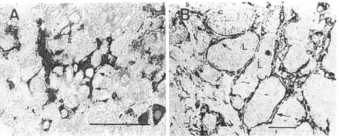
Figure A Immunocytochemistry of GFAP in the anterior pituitary gland (A) and
thyrotropic tumor (B) 3
Scherna 1 Regulated treadmilling model for actin dynamics $ Schema 2 Full-length crtactin is comprised of an N-terminal acidic (NTA) region
followed by 6.5 37-amino acid tandem repeats 15
Schema 3 Cortactin serves to link diverse scaffolding protein complexes to
Arp2/3-driven actin polymerization 17
Schema 4 Diagrammatic representation of cortactin binding proteins and
phosphorylation sites 21
Schema 5 Schematic drawing of an Œ-actinin dimmer 25
Schema 6 Possible interactions between structural and signaling proteins within the cell-substratum focal adhesion—actin complex in ECs 27
xv
LIST 0F FiGURES
Figure 1 Modulation ofthe actin cytoskeleton ofTtT/GF celis by serum factors..42 Figure 2 Effect of serum on the localization of the actin-binding proteins cortactin, cL-actinin, vinculin, and myosin in TtT/GF celis 46 Figure 3 Effect of serum on the expression and on the subcelluÏar localization of
the actin-binding proteins cortactin, u-actinin, vinculin and MLC in
TtT/GF celis 50
Figure 4 Effect of serum on the localization of F-actin and cortactin in TtT/GF
celis 53
Figure 5 Localization of cortactin and phospho-tyrosine in TtT/GF celis cultured in the presence of serum. Effect of the tyrosine kinase inhibitor
genistein 57
Figure 6 Localization of cortactin and phospho-tyrosine in TtT/GF celis cultured in the absence of serum. Effect of the tyrosine kinase inhibitor
genistein 59
Figure 7 Effect of semm on the expression and the subcellular localization of p
cortactin in TtT/GF cells 62
Figure $ A) Effect of serum on the localization of cortactin and p-cortactin in TtT/GF ceils. B). Effect of genistein on serum-induced formation of cdl membrane protrusions and on the localization of cortactin and p
cortactin 65
Figure 9 Effect of forskolin on the expression and the subcellular localization of cortactin and p-cortactin in TtT/GF ceils cultured in the presence of
xvi Figure 10 Effect of forskolin onthe expression and the subcellular localization of p cortactin in TtT/Gf celis cultured in the absence ofserum 72 Figure 11 Effect of forskolin on the localization of cortactin and p-cortactin in
TtT/GF ceils cultured in the presence ofserum 75 Figure 12 Effect of forskolin on the localization of cortactin and p-cortactin in
TtT/GF ceils cultured in the absence ofserum 77
Figure 13 Effectofthe PKA inhibitor KT5720 on the expression and the subceÏÏuÏar localization of cortactin and p-cortactin in TtT/Gf celis cultured in the
presence serum 80
Figure 14 Effect ofthe PKA inhibitor KT5720 on the expression and the subcellular localization of cortactin and p-cortactin in TtT/GF ceils cultured in the
absence ofserum $2
Figure 15 Effect of KT5720 on the localization of cortactin and p-cortactin in TtT/GF celis cultured in the presence ofserum $5
Figure 16 Effect of KT5720 on the localization of cortactin and p-cortactin in
TtT/Gf ceils cultured in the absence ofserum 87
Figure 17 Effect of serum on the localization of F-actin and Œ-actinin in TtT/GF
celis 90
Figure 1$ Effect of genistein on the localization of u-actinin and phospho-tyrosine in TtT/Gf celis cultured in the presence ofserum 93 Figure 19 Effect of genistein on the localization of Œ-actinin and phospho-tyrosine
in TtT/Gf celis cultured in the absence of serum 95 f igure 20 Effect of forskolin on the expression and the subcellular localization ofci
xvii Figure 21 Effect of forskolin on the localization of F-actin and o.-actinin in TtT/GF
celis cultured in the presence ofserum 101
Figure 22 Effect of forskolin on the localization of F-actin and Œ-actinin in TtT/GF
ceils cultured in the absence ofsemm 103
Figure 23 Effect ofthe PKA inhibitor KT5720 on the expression and the subcellular
localization ofŒ-actinin in TtT/GF ceils 106
Figure 24 Effect of KT5720 on the Iocalization of Factin and Œ-actinin in TtT/GF
ceils cultured in the presence ofserum 109
Figure 25 Effect of KT5720 on the localization of F-actin and Œ-actinin in TtT/GF
xviii
c
LIST 0F ARBREVIATIONSABD actin-binding dornain AKAPs A-kinase anchoring proteins Arp actin related proteins
BSA bovine serum alburnin
cAMP cyclic AMP
C cytosol or control
CK cytoskeleton
CH calponin homology
DMEM Dulbecco’s modified Eage medium DMSO dimethyl sulfoxide
ECM extracellular matrix EGF epidermal growth factor F-actin filamentous actin fAK focal adhesion kinase FBS fetal bovine serum
FITC fluorescein isothiocyanate F5 folliculo-stellate
G-actin monomeric actin
GfAP guai fibrillary acidic protein GFP green fluorescent protein
H hour
IL-6 interleukin-6
xix
c
M membraneMLC myosin light chain MLCK myosin light chain kinase
NCK non-cytoskeleton
NTA amino terminal acidic domain P-cortactin phospho-cortactin
PDGF plateÏet-derived growth factor PBS phosphate buffered saline PKA cAMP-dependent protein kinase PMSF phenylmethylsulfonyl fluoride
PM plasma membrane
PRL prolactin
SDS-PAGE dodecyl sulfate-poly-acrylamide gel electrophoresis
SH Src homology
TBS TRIS buffered saline
TRITC tetramethylrhodamine isothiocyanate WASP Wiskott- Aldrich syndrome protein
DEDICATION
To my family and my ftiends who have helped me during my Master studies. xx
xxi
ACKNOWLEDGEMENETS
I wouid like to take this opportunity to thank my research director, Dr. Maria Leiza Vitale, for her encouragement, patience, consideration, and constructive criticisms. I especially appreciated ber great patience and consideration. When I lost my heart, she aiways encouraged me. I value her great encouragement very much. She is aiways ready to help me and aiways provides good advice. She taught me not only the science but also the way to solve the problems.
I would like also to thank my co-director, Dr. R-Marc Pelletier, for bis consultation and assistance. His enthusiasrn and vivid participation are greatly appreciated. I appreciate his kind advice and bis sound judgernent on my work.
I would like to express many thanks to Dr. Lucian Ghitescu for his kind supporting and permission to use instruments in bis lab. I am also grateful to Ms. Anne Guénette and Ms. Manon Moreau for their kind help in printing of my thesis.
I would also like to thank ail the members of our research group, Marie-Eve fortin, Casimir D. Akpovi, Emile Silvas. Li Chen. Sara Solinet, and Méllisa Meilleur, for their cooperation. help, suggestions, support and consideration, which are very important for my work.
I am also grateful to rny following friends. Yu Zhou, for his kind help in the deaÏing with rny figures Pat N. Berger and Tong Lin, for their kind proof reading of my discussion section, Zongjian Jia for bis supporting to my experiments; Carole Ahi f arali and Anaïck Lagana for their kind heip and suggestions.
xxii PREFACE
Zheng, G.F., Pelletier, R .M., and Vitale, M.L. (2005). Tyrosine phosphorylation of cortactïn as a key step in the formation of different membrane-actin cytoskeleton structures in folliculo-stellate celis. (Abstractinpreparation)
Zheng, G.F., Pelletier, R.M., and Vitale, M.L. (2004). Participation de cortactine dans le modulation de la dynamique du cytosquelette d’actine des cellules TtT/GF (poster presentation). 72e Congrès de Ï’ACfAS
Zheng, G.F., Pelletier, R.M., and Vitale, Mi. (2004). The modulation of the cortical actin cytoskeleton by the tyrosine phosphorylation of cortactin in TtT/GF cells (poster presentation). 21e Jourmée Scientifique du Département de Pathotogie et Biologie
Cellulaire, Université de Montréal No. 32
Zlieng, G.F., Pelletier, R.M., and Vitale, M.L. (2003). Effet des facteurs sériques sur la dynamique du cytosquelette d’actine des cellules TtT/GF (poster presentation). Médecine/Sciences 19 (Suppl 2): No.57, P 14
Zheng, G.F., Pelletier, R.M., and Vitale, M.L. (2003). Dynamics of the actin cytoskeleton in TtT/GF folliculo-stellate celis (poster presentation). 20e Jourinée Scientifique du Département de Pathologie et Biologie Cellulaire, Université de Montréal No. 32
1 Introduction 1.1 The pituitary gland 1.1.1 General
ihe pituitary gland or hypophysis is an endocrine gland located in the sella turcica
of the sphenoid, a depression in the bony floor of the cranial cavity. It is cornposed of two distinctive parts: the anterior pituitary and the posterior pituitary (Bali and Baker, 1969; Wingstrand, 1966). The anterior pituitary (adenohypophysis) is a classical gland composed of a mixed population of granular and agranular ceils. The granular ccli group comprises five kinds of endocrine ceils that contain secretory granules in their cytopiasm and synthesize and secrete the pituitary hormones, growth hormone, prolactin, thyroid—stimulating hormone, follicle-stimulating hormone, luteinizing hormone, and adrenocorticotropic hormone which control growth, sexual developrnent and metabolism. The folliculo-stellate ceils (f$ celis) are the major cellular element of the agrantilar celis in the anterior pituitary gland (Matsurnoto. H. et al., 1993). They do not possess secretory granules in their cytoplasrn, thus differing from the glandular celis in their activities and their functions (moue et al., 1992). The posterior pituitary (neurohypophysis) is in fact an extension ofthe hypothalamus. It is composed ÏargeÏy of the axons of hypothalarnic neurons that extend downward as a large bundie behind the anterior pituitary (Grandi and Chicca, 2004). Oxytocin and antidiuretin are the main hormones secreted by the neurohypophysis.
On the one hand, the pituitary gland controls the functions of the other endocrine glands in the body so it is sometimes called the master gland of the endocrine system (Grandi and Chicca, 2004). On the other hand, the secretion of pituitary hormones is modulated by systernic influences and by the hypothalamus. In this late sense, the
pituitary gland is an important link between the nervous system and the peripheral endocrine system.
1.1 .2 The folliculo-stellate ceils (FS ceils)
Ihe FS ceils of the anterior pituitary gland were first reported by Rinhart and
farquhar (1953). FS celis represent about 5 to 10% of the anterior pituitary ceil population (Rinehart & Farquhar 1953). These stellate-shaped ceils cluster together into follicles with cytoplasmic processes extending outwards to connect other F8 celis and endocrine celis, generating an extensive three-dirnensional network within the anterior pituitary (Allaerts et al., 1990; Soji and Herbert. 1989; moue et al., 1999). Immunohistochernical studies at the light and electron microscopic levels revealed that FS celis are localized tbroughout the anterior pituitary, and form 2 to 1 0 celi clusters around a follicular cavity (Lloyd and Mailoux, 1988; Soji and Herbert, 1989). F8 ccli morphological features also include the presence of lysosomes, the expression of the protein S-100 and of the intermediate filament protein GFAP (glial fibrillary acidic protein) (Figure A).
o
The precise functions of the FS ceils in the anterior pituitary gland are flot yet truiy understood. Several functions have been ascribed to FS celis including supportive and trophic effects, a rote in ion transport, phagocytic and catabolic activities (Perryman, 1989), and many paracrine functions. With their long cytoplasmic processes rich in gap junctions extending between other endocrine celi types (Morand et ai, 1996), the F5 ceils are thought to play a role in intercellular communication within the anterior pituitary. There is substantial evidence that F5 ceils modulate pituitary hormone secretion from surrounding endocrine ceils through the release of several bioactive molecules, including follistatin, interleukin-6 (IL-6), nitric oxide, basic fibroblast growth factor, leptin (Allaerts et ai, 1990). A paracrine mode of interaction was also suggested by the observation that FS ceils contain and release the S-100 protein, and that this protein can stimulate PRL release from cultured clonai PRL ceils (Ishikawa et al., 1983) and from normal dissociated anterior pituitary celis (Lioyd and Mailloux, 198$). Moreover, f auquier et al. have demonstrated that the FS ceil network forms an extensive functional intrapituitary circuitry in which information, Ca2 and small diffusible
Figure A. lrnmunocytochernistry of GfAP in the anterior pituitaty gland (A) and thyrotropic turnor (B). FS cells in the mouse anterior pituitary, surrounding neighboring glandular celis, are positively stained for GfAP (A, atiows). The GFAP-imrnunopositive celis are flat and uniformly surround the lobules (L) of parenchymal tissue in the thyrotropic pituitary turnor (B, arrows). Bars are 50 pm(molLeet al., 1992).
4
Q
molecules can be transferred through gap junctions over long distances (Fauquier et al., 2001).Work from our lab has shown that the morphology of anterior pituitary FS celis is affected by the secretory activity of the gland (Cardin et al, 2000). These changes particularly include the disappearance/reappearance of cellular processes. thus affecting the stellate morphology of FS ceils. Because the actin cytoskeleton is the main factor determining the celi shape, to understand how systemic and nervous factors affect the morphology of fS cells, we decide to investigate first the dynamics of the actin cytoskeleton ofF$ ceils.
1.2 The actin cytoskeleton
The cytoskeleton, the ceil skeleton, is composed of three major types of filaments: the microtubules. the microfilaments. and the intermediate filaments. Microfi laments are polymers of actin that together with a large number of actin-binding and associated proteins constitutes the actin cytoskeleton (Stôssel 1993; Botstein et al., 1997; Winsor and Schiebel 1997). Actin exists either in a monorneric (G-actin) or in a polymeric forrn (f-actin). Each actin molecule can bind ATP, which is hydrolyzed to ADP afier incorporation of the actin molecule into the filament. Polymers assemble spontaneously via non-covalent interactions between the monomeric subunits and are highly dynamic structures with subunits turnover at both ends. The rate-lirniting step in actin polyrnerization is nucleation, the assembly of the first subunits to generate a new filament. Actin filaments are structurally polarized and the kinetics of polymerization at each end is different, with the plus end growing more quickly than the minus one (Anja and Micheal 1998). few filament properties are regulated by direct covalent
5
modification of the filament subunits, indeed most of the regulation is perforrned by accessory proteins that bind to either the filaments or their free subunits.
The assembiy and reorganization of actin filaments are controlled by a host of actin-binding proteins under the influence of external and internai stimuli (Aspenstrom, 1999; Cooper, 1991; Hall, 1998; Hatano, 1994; Janmey. 199$; Tapon and Hall, 1997). Actin-binding proteins can bind to actin monomers and/or to actin filaments to form diverse structures and networks that vary in different ceil types and in different parts of the same ceil (Cariier, 1998; Condeeiis, 1993; Furukawa and Fechheimer, 1997; McGough, 1998; Puius et ai., 1998; Smail et al., 1998; Van Troys et al., 1999). A large number of actin-binding proteins control assembly and Iength of actin filament. iink actin filaments into bundies or networks, define the three-dimensionaÏ organization of actin filaments and link filaments to other cytoplasmic and membrane components (Stôssel et al., 1985; Pollard and Cooper. 1986; Stôssel, 1989). bringing the cytoskeietal structure under the control of extracellular and intracellular signais.
1.2.1 The dynamics ofthe actin cytoskeleton in non-muscle cells
In nonmuscle celis, most actin filaments are highly dynamic structures that are being constantly assernbled, disassembied, and reorganized under the influences of intra and/or extra-celiular stimuli as the celi changes its shape, divides, crawls, and adheres to a substratum or to neighboring celis and adapts to the environrnent (Bershadsky and Vasiliev, 1988; Bray, 1992; Mitchison and Cramer, 1996; SrnaÏÏ et aÏ., 1996; 1999; Stôssel, 1993; 1994; Theriot, 1994; Welch et al., 1997).
6
1 .2.1 .1 Extracellular and intracellular factors affecting the dynamics of the actin cytoskeleton in non-muscle celis
A variety of extracellular signais influence the actin cytoskeleton dynarnics. For example, growth factors or extraceliular matrix (ECM) proteins transmit signais through actin cytoskeleton to modulate intraceilular trafficking, ceil morphoiogy. celi migration.
and process extension (Zigmond, 1996). Epidermal growth factor (EGF) exerts its effects in the target celis by binding to the plasma memberane EGf receptor, resulting in the activation of its tyrosine kinase activity and subsequent receptor autophosphorylation, which is essentiai for the interaction of the receptor with its substrates (Boonstra et al.. 1995). The EGF receptor is an actin-binding protein (Den Hartigh et al., 1992) which causes a rapid actin depolymerisation and the formation of membrane ruffles (Rijken et la., 1991). These membrane ruffles function as the prirnary site of signal transduction afler EGF binding, and thus are thought to be signal transduction structures (Boonstra et al.. 1995).
EGF-induced eariy signai transduction causes rapid remodeiing of the actin microfliament system in a variety of ceiis (Rijken et al., 1991; Peppeienbosch et ai., 1993). This is the case for other growth factors, such as nerve growth factor (Paves et ai., 1990) and plateiet-derived growth factor (PDGF) (Arvidsson et al., 1992; Kundra et al., 1994). The receptors for various growth factors, including PDGf (frackelton et al., 1984; Ek and Heldin, 1984), insulin (Kasuga et al., 1982) and ïnsulin-like growth factor
I (Sasaki et al., 1985) are ail protein tyrosine kinases.
In addition, a number of key intraceiiuiar components had been found essentiai for control of actin polymerization and depolymerization including profihin, Arp2/3, WASp/Scar/WAVE, actin capping proteins, actin severing proteins, and Rho farnily
7
GTPases (Pollard et al., 2000; Bear et al., 2001). It is now clear that the Arp2/3 compiex regulates the assembly of new actin filament networks at the leading edges of ceils. Proteins ofthe WASP (Wiskott - Aldrich syndrome protein) farnily bind directly to the
Arp2/3 complex and stirnulate its ability to promote the nucleation of new actin filaments. Upstream of WASP-family proteins, receptor tyrosine kinases, the Rho family GTPase Cdc42, and likely G protein-coupled receptors, receive and transmit the signais leading to WASP-Arp2/3 complex-mediated actin nucleation. Together, these data suggest that signaling pathways from outside the celi induce actin polymerization with the resultant polyrnerization-driven celi motility. Scar proteins might be involved in pathways through serpentine receptors and heterotrimeric G-proteins (Machesky and Insall, 1999).
Cdc42 and PIP2 also cooperate with N-WASP to activate Arp2/3 complex-induced acfin nucleation activity (Rohatgi et al., 1999). This indicates a signaling pathway from inositol phospholipids to Cdc42 and WA$P/N-WASP. In addition to Arp2/3 complex mediated actin nucleation, both mammalian celis and Listeria also use EnaJVASP proteins to promote actin filament assembly (Machesky and Insail, 1999).
A regulated treadmiliing model was suggested for actin dynarnics in rnotility. In this model, the Arp2/3 complex becomes activated through one of the WASP farnily proteins depending on the nature of the signal for the actin polyrnerization. Activated Arp2/3 complex binds to the sides of actin filaments and nucleates new branches with ftee barbed ends. Once these dendritic structures assemble, their eiongation are controlled by capping proteins, which dynamically associate and dissociate with the barbed ends of the filaments, with the relative rates of capping and uncapping being controlled by signaling intermediates such as plasma membrane phosphoinositides.
8
Additionally, the rate at which new filaments elongate is accelerated by regulated association with EnaJVASP proteins and profihin. Established filaments are severed and depolymerized by cofihin (Maciver, 199$). Finally, when the filament depolymerises, the Arp2/3 complex falis off and may be recycled in new filaments (Schema 1).
1.Lo nd A,,tivtior 2. Ain Nu,Ietir. d Brrhing
R] Sopontro I_______
___
WASP SCAR ArpZ’
- Actin Monomers
(s2s3) ÇArP2I3)
in5tr,o Act•r, Firrt,,t
3. Ftgotd Trodr,,iUing 4. DopcIyrner.ztion to Brrhpont 3nd
Rolooso of Arp213 Cp!os PIP2
-———4XX)
Ap2/3
Ccliii r
Schema 1. Reguiated treadmiliing model for actin dynamics. (1) Varlous signais trigger localization and/or activation of WASP/Scar proteins, activating and localizing the Arp2/3 complex to nucleate actin filaments. (2) Arp2/3 compiex binds to the sides of actin filaments and nucieates branches. (3) The growing barbed ends ofthe filaments are reguiated by capping protein and ge]solin. Ena! VASP proteins cata]yze the e]ongation of newly nucieated filaments. The pointed end of the filaments is depolymerizing with the help of cofilin. (4) 1f the filaments depolymerize down to a branchpoint, Arp2/3 complex may fali off and is recycled to participate in nucleation (Macheskey and Insaii, 1999).
1.2.1.2 Regulation of actin cytoskeleton dynamics through cAMP/PKA pathway Cyclic AN’W (cAMP) is a key second messenger mediating the biological effects of several hormones which activate the adenylate cyclase catalyzing the production of intracellular cAlvW. The mode of action ofcAIVW ïs via the activation of the cAIvW dependent protein kinase (PKÀ or A-kinase) by promoting the dissociation of the inactive holozyme into reguÏatoIy subunits and active catalytic subunits (Glass and
9
Krebs, 1980), which catalyze the phosphorylation of certain proteins which serve to regulate their subsequent biological activity (Cohen. 1982 and 1985; Glass and Krebs, 1980; Krebs and Beavo, 1980; Shackter et al., 1984). PKA is implicated in mediating a number of intracellular events including the regulation of cytoskeletal structure (Albertini and Herman, 1984; Porter et al., 1974).
A variety of signaling substances utilize the PKA pathway to regulate actin cytoskeletal dynarnics and ceil migration (Howe, 2004). While some of these intracellular events require PKA activity, others are inhibited by it. Also, celi migration and invasion can be impeded by either inhibition or hyper-activation of PKA. In addition, a number of A-kinase anchoring proteins (AKAPs) serve to associate PKA with various components of the actin cytoskeleton, thereby enhancing and/or specifying cAMP/PKA signaling inthose regions (Howe, 2004).
Agonists of cAMP/PKA signaling cause significant changes in cellular architecture, such as dissolution of stress fibers and induction of stellate morphology in
neurons and other celis (Dong et al., 1998; Ramakers and Moolenaar, 199$; Edwards et al.. 1993). Negative effects of PKA on migration were reported for endothelial celi migration on vitronectin (Kim et al., 2000). Also, matrix-specific down-regulation of cAMP/PKA signaling is required for collagen-induced f-actin synthesis and stress fiber formation in endothelial ceils (Whelan and Senger. 2003). Conversely. eÏevation of cAMP and activation of PKA have been shown to be required for efficient celi migration in several systems. These include: formation of filopodia and lamellipodia in response to follicle stimulating hormone (Grieshaber et aI., 2000), mammary epithelial ceil migration on laminin (Plopper et al., 2000), microfilament assernbly (Whittard and Akiyama. 2001), and activation of Cdc42 and Rac (Feoktistov et al., 2000; O’Connor
10
O
and Mercurio, 2001).cAMP/PKA signaling exerting a negative or positive effect on the cytoskeleton. Rather,It is clear that in most ceil types, it is not simply a matter of a balance of cAMP/PKA activity in extent. space, and time is crucial for successful cytoskeletal organization (Edin et al., 2001; Ydrenius, et al., 199L O’Connor and Mercurio, 2001).The control of nonmuscle microfilament contraction via phosphorylation of myosin light chain kinase (MLCK) by PKA has been suggested (Conti and Adeistein, 19$ 1). In contrast to skeletal muscle, actin-rnyosine interaction in nonmuscle celis is controlled by the phosphorylation of the regulatory myosin light chain (Sellers and Adelstein, 1987), which is catalyzed at least partly, by the enzyme myosin light chain kinase (MLCK), the activitv ofwhich is dependent upon calcium and calmodulin (Conti and Adeistein, 1981). The activity of MLCK can be inhibited in vitro and in vivo by phosphorylation of the enzyme catalyzed by PKA (Conti and Adeistein, 1981; Silver and DiSalvo. 1979). This phosphorylation interferes with Ca2/calmodulin binding to MLCK and thereby results in the inhibition of MLCK activity in vitro (Adeistein, 1982). In nonmuscle ceils PKA regulates microfilament structure tbrough the phosphorylation and inhibition ofMLCK activity (Lamb et al., 198$).
It is shown that PKA direct!)’ phosphorylates monorneric actin at serine residues. a modification that significantly decreases monomer polyrnerizibi1ity’ in vitro (Ohta et al., 1987). Although this event is not yet well elucidated, at least the data indicate a pathway for regulation of actin dynarnics by PKA (Howe, 2004).
11
1.2.1.3 The roles ofthe actin cytoskeleton in non-muscle celis
The actin cytoskeleton of an eukaryotic celi is central to locomotion, phagocytosis, contractility, shape changes, cytokinesis, maintenance of polarity, exocytosis and endocytosis. Actin filament nucleation most frequently occurs at the plasma membrane, which is regulated by external signais, allowing the celi to change its shape and stiffness rapidly in response to changes in its external environrnent. Therefore. the highest density of actin filaments in most ceils is at the periphery. These actin filaments in the layer underlying the plasma membrane, called the cell cortex, determine the shape and movement of the cell surface. Actin structures can forrn many different types of celi surface projections, including microvilli or filopodia, flat protrusions called larnellipodia that help move cells over solid substrates. Regulation of cell morphology is essential for cell division and ceIl ftmction in organisms. Srnall GîPbinding protein Rho famiÏy plays a key role in such regulations by controlling the actin cytoskeleton (Hall, 1998; Etienne-Maimeville and Hall, 2002).
Research in fibroblasts on the molecuÏar mechanisms mediating morphological transformations has identified a signaling cascade via smail GTPases that link membrane receptors to the cytoskeleton (Ridley and Hall, 1992). RhoA is activated by extracellular signais such as lysophosphatic acid (LPA) leading to the assembly of stress fibers and focal adhesions (Hall et al.. 1993; Ridley and Hall, 1994; Hall, 1998). Assembly of microfilarnents is facilitated by the localization of RhoA and Rad in caveolae (Michaely et al., 1999), plasma membrane domains associated with actin-rich regions. Rad indcices membrane rufiing and lameilipodium formation. and Cdc42 induces the formation of microspikes and filopodia. ail of which are dependent on filamentous actin (F-actin) organization (Ridley, 2000 and 2001; Ridley and Hall, 1992; Nobes and Hall,
12
1995; Johnson and Pringle. 1990). The morphological transformations require only subcellular redistribution of cytoskeletal proteins instead of synthesis or degradation of the respective proteins (Safavi-Abbasi et al., 2001).
Experimental evidence suggests that the cortical cytoskeleton regulates exocytosis (Trifaro and Vitale, 1993). Disruption of F-actin enhances stimulated secretion in pancreatic f3-cells (Orci et al., 1972), cultured chrornaffin cells (Leikes et al., 1986), perrneabilized rnast ceils (Koffer et al., 1990), and lactotrope cells (Carbajal and Vitale, 1997). F-actin disassembly takes place in discrete zones of the cell cortex during hormone and neurotransmitter secretion and the areas of exocytosis correspond to cortical areas devoid ofF-actin (Vitale et al., 1991; Nakata and Hirokawa, 1992).
The participation of actin-regulatory proteins during the actin reorganization that take place during secretion is suggested by the finding that actin-binding protein such as fodrin (Perrin and Aunis, 1985), scinderin (Vitale et al., 1991) talin and Œ-actinin (Nguyen et al., 1999) redistribute with cortical f-actin during stimulated secretion. Actin cytoskeleton is crucial for transport of endocytosed molecules. The initial internalization step requires actin cytoskeleton (Larnaze et al., 1997) and the later trafficking of endocytosed molecules is affected by disruption of the actin cytoskeleton (Durrbach et al., 1996). Endocytosed molecules are in general exocytosed at sites of plasma membrane protrusions (Bretscher and Aguado-Velasco, 1998).
1.2.1.4 Actin dynamics in FS ceils
As the dynarnics of the actin cytoskeleton in non-muscle cells, the actin dynarnics in FS celis is also affected by a variety of factors including extra- and intra-cellular
I,-,
L)
factors, such as serum, hormones and related actin-binding proteins. In the rnink anterior pituitary, Cardin et al (2000) found the levels of sorne hormones influence the morphology of the FS celis via the cytoskeleton reorganization. However, the precise mechanisms underlying the actin dynamic regulation in FS celis are not yet fully understood.
1.2.2 Actin-binding proteins
Due to the involvement of actin-binding proteins in the control of actin cytoskeleton dynarnics, we decided to study the participation ofthese regulatory proteins
in the modulation of the actin cytoskeleton organization in fS ceils under different experimental conditions. Our previous resuits have shown that in these celis the reorganizaion of the cell’s shape implicated the formation of cellular processes. Therefore, among the several actin-binding proteins, we focused our studies on the role of two actin-binding proteins that mediate the interaction between actin filaments and the plasma membreane: cortactin and cL-actinin.
1.2.2.1 Cortactin
Vertebrate celis exhibit a cortical cytoskeletal network that resides beneath, and is associated with, the inner surface of the plasma membrane. This cortical cytoskeleton is comprised of a dense network of actin filaments and associated actin-binding proteins (Srnall et al., 1981; Stôssel et al., 1981). Cortatin is an actin-binding protein that is enriched in cortical structures such as membrane ruffles and lamellipodia and plays an important role in the dynamic regulation of cortical actin cytoskeleton. The name
14
cortactin just reflects the cortical subcellular localization and its actin-binding activity (Wu and Parsons, 1993).
1.2.2.1.1 StructuraI organization
Cortactin, an important actin-binding protein, was initially identified as a tyrosine phosphorylated protein in v-$rc infected chicken embryo flbroblasts (Kanner et ai., 1990). Cortactin has a unique structure (Wu et ai., 1991), comprising several domains (Weed and Parsons, 2001) ($chema 1). The first $4-94 amino-terminal residues are largely unstructured, containing a large number of acidic residues between arnino acids 15 and 35 and is referred to as the amino terminai acidic domain (NTA; Weed et al., 2000). The NTA region is followed by a series of six complete 37 amino acid tandemly repeating segments and one incompiete segment of 20 residues in length. The compiete repeats form a helix-turn-heiix structure (Wu et al., 1991) and are terrned cortactin repeats (Sparks et al., 1996a) since they show no sequence sirnilarity to other known repeats except in HS1, a cortactin-like protein. The repeats’ region is followed by an ct helical dornain, a proÏine-rich dornain abundant in tyrosine, serine and tbreonine residues, and a Src homoiogy (SH) 3 domain at the distal carboxyl terminus. The cortactin 5H3 domain shares significant homology to SH3 domains from Src-family kinases, various adapter and cytoskeletai proteins (Wu et al., 1991; Sparks et al., 1996a). Cortactin genes have now been cloned from a number of diverse organisrns. Ail cortactins are structurally similar to one another. with the SH3 domain showing the greatest amount of evoiutionary conservation whiie the proline-rich domain the least (Schema 2). Only one cortactin gene has been identified to date, and in most celi types. it encodes a single cortactin protein product that ofien migrates as two separate bands in
15
SDS-PAGE wïth relative molecular weights of 80 and 85 kDa (Wu et al., 1991). C.
Northem blot analysis indicates that cortactin mRNA is expressed in nearly ail mammalian tissues (Miglarese et al., 1994; Du et al., 199$).
NTA Repeats al-I
FL Cortactin
NT4CT
Schema 2. Full-length cortactin is comprised of an N-terminal acidic (NTA) region followed by 6.5 37-amino acid tandem repeats. A predicted Œ-helical and proline-rich region are followed by an SH3 domain (Kinley et al., 2003).
1.2.2.1.2 The role of cortactin as a dynamic regulator of the actin cytoskeleton Localization and biochemical studies show that cortactin plays an important role in regulating cortical actin assembly and organization tWeed and Parsons, 2001). In most celi types, cortactin localizes in cytopiasmic punctate structures of unknown composition concentrated at the perinuclear region, and also with F-actin at sites of dynarnic peripheral membrane activity tWeed and Parsons, 2001). Cortactin transiocates from the cytoplasm to the periphery in response to many of the stimuli that induce its tyrosine phsphorylation, including growth factor treatrnent, integrin activation and bacterial entry (Ozawa et ai., 1995; Weed et ai., 199$; Cantarelli et al., 2000). These events also iead to activation of Rad (Hartwig et al., 1995; Clark et ai., 199$; Mounier et al., 1999), which together with Cdc42 are responsible for controiling the formation of cortical actin networks (Ridley et ai., 1992; Kozma et ai., 1995). Rad activation is required for cortactin translocation to the penphery ofthe celi tWeed et al.,
16
the F-actin disrupting drug cytochaÏasin D (Wu and Parsons, 1993). Taken together, cortical localization of cortactin is closely associated with Racl-mediated actin assembly tWeed and Parsons, 2001).
Cortactin interacts directly with F-actin through sequences in the repeats’ domain (Wu and Parsons, 1993). Deletion mapping analysis lias determined that the fourth cortactin repeat is responsible for f-actin binding and cortical localization (Weed et al.. 2000). Cortactin binds to the sides of actin filaments as determined by negative stained electron microscopy of recombinant cortactinlF-actin mixtures and by competition binding experiments with myosin subfragrnent 1 (Ohoka and Takai, 199$). Recombinant cortactin is found to crosslink F-actin (Huang et aÏ.. 1997). Because ofits cortical localization and the fact that it is tyrosine phosphorylated in response to ligand stimulation as well as in cellular transformation, cortactin is considered an interesting candidate for a signal-transducing molecule or an effector molecule in the integration of extracellular stimulation with intracellular changes (Grinstein et al., 1989; Erpel and Courtneidge, 1995: Wu and Parsons, 1993).
1.2.2.1.3 Interaction of cortactin with other proteins to regulate the cortical actin cytoskeleton organization
Cortactin serves to link diverse scaffolding protein complexes to Arp2/3 (Weed and Parsons, 2001). These multiprotein scaffolds are associated to the actin cytoskeleton through sequences within the cortactin NTA and repeat domains (Weed and Parsons, 2001) (Schema 3).
17
I’,,,t-Synptic Dnity
A great deal of factors involved in the de novo assembly of actin-rich structures have been identified. Among these factors, the Arp2/3 complex has emerged as a key regulator of actin-filament nucleation (Weaver et ai., 2001) that is conserved from yeast to humans (Pollard et al., 2000). The Arp 2/3 complex is composed of seven subunits, including two actin-reiated proteins, Arp2 and Arp3, and five other subunits. It is concentrated in dynamic actin structures such as the leading edges of motile cells (Weich et ai, 1997). Cortactin binds to the Arp2/3 complex through its N-terminai domain and stimulates its actin-nucleation activity. Association of cortactin with the Arp2/3 complex is essential and sufficient for cortactin to localize within actin-rich patches. Furthermore, the F-actin-binding activity of cortactin is essential for cortactin to stimulate the Arp2/3 complex (Uruno et al., 2001), suggesting the important relationship between them. Cortactin aiso inhibits debranching of aged Arp2/3 networks
UIRII NMDAR IP3R mGIoK — -EVH iIoN Tight Junction (Irn.din OccIdIn
:i’i
- WNABAhmnSchema 3 Cortactin serves to link diverse scaffolding protein complexes to Arp2/3-driven actin polymerization. Schematic representation of the proposed molecular interactions within the neuronal post-synaptic density (top), epithelial tight junction (bottom lefi) and in receptor mediated endocytosis (bottom right) (Weed and Parsons, 2001).
1$
between Arp2/3 complex and mother/daughter filament networks tWeed and Parsons, 2001). The association of cortactin with mobile cortical actin structures shows a functional role for cortactin in regulating Arp2/3 activity (Dai et al., 2000; Kaksonen et al., 2000). Localization of cortactin to the ce!! periphery coupled with its ability to stimulate and stabilize Arp2/3-dependent actin polymerization in vitro suggests a role for cortactin in the regulation of actin cytoskeletal dynamics (Weed and Parsons, 2001).
Arp2/3 complex in celis is inactive until cellular signals result in its activation (Goode et al., 2001). In higher eukaryotes, the only known activators of Arp2/3 complex are the Wiskott-Aldrich syndrome family of proteins (WA$ps) (Goode et al., 2001), which contain a terminal acidic dornain that binds the Arp2/3 complex. The C-terminal region of WASp also contains a verprolin homology and connecting (VC) domain, which binds monomeric actin (G-actin) (Goode et al., 2001). Cortactin can function cooperatively with WASp proteins in addition to independently activating Arp2/3 complex tWeed and Parsons, 2001). The ability of cortactin to bind and avtivate Arp2/3 complex indicates a role for cortactin in actin-based motility events tWeed and Parsons, 2001). Enhanced ccl! migration in NIR 3T3 and endothelial cells overexpressing cortactin supports this notion (Patel et al., 199$; Huang et al., 199$).
Cortactin has another role in recruiting other components to the periphery, including perhaps the small GlPases themselves. Weed et al showed that short-terrn expression of active Rac 1 induces cortactin accumulation under the membrane without an obvious accumulation ofRaci itself(Weed et al., 199$). This indicates that Rad can “send” cortactin to the periphery of the cdl without being translocated itself. During longer-term expression, however, Di Ciano et al. found strong colocalization of active Rad and cortactin in membrane ruffles (Di Ciano et al., 2002). Taken together, F-actin
19
binding activity of cortactin, its cortical localization, and its tyrosine phosphorylation
e
indicate that cortactin plays a role in mediating microfliament-membrane interactions inresponse to signais propagated by receptor and/or nonreceptor protein tyrosine kinases (Wu and Parsons, 1993).
1.2.2.1.4 Phosphorylated status of cortactin
Cortactin is a target for both tyrosine and serine/threonine protein kinases tWeed and Parsons, 2001) and is normally phosphorylated on serine and threonine, but it becomes tyrosine phosphorylated in response to growth factor stimulation and by activated Src (Wu et al., 1991; Maa et ai., 1992; Zhan et al., 1993).
In nontransformed ceHs, cortactin transientiy associates with c-Src in response to thrombin activation in plateiets (Wong et ai., 1992) and in response to fGF-1 in fibrobiasts (Zhan et al.. 1994). A weaith of data indicates that cortactin tyrosine phosphorylation is closeiy correlated with Src activity in a number of signaiing pathways and the strong correlation between Src activation and cortactin tyrosine phosphorylation in these studies suggest that cortactin is phosphoryiated directly by Src tWeed and Parsons, 2001). Src directiy phosphoryiates cortactin in vitro (Huang et ai., 1997) on three tyrosine residues in murine cortactin (tyrosine-421, -466. and -482) located within the proline-rich dornain (Huang et ai., 199$). Cortactin tyrosine phosphoryiation requires Rac 1 -induced cortactin targeting to cortical actin networks, where it is tyrosine phosphorylated at positions 42i and 466 in a hierarchicai manner, that is, with tyrosine 421 phosphorylation being required for phosphoryiation of tyrosine 466 (Head et al., 2003). Expression of a cortactin construct where tyrosines 421, 466, and 482 were mutated to phenylaianine dramaticaiiy reduced cortactin tyrosine
20
and 482 were mutated to phenylalanine dramatically reduced cortactin tyrosine phosphorylation following expression in v-.$rc transformed 313 fibroblasts, indicating that these residues are targeted by Src in vivo (Huang et al., 1998).
Fer is a second nonreceptor tyrosine kinase involved in growth factor-mediated cortactin tyrosine phosphorylation (Weed and Parsons, 2001). NIH 313 fibroblasts treated with PDGF lead to Fer activation and cortactin tyrosine phosphorviation (Downing and Reynolds. 1991; Kim and Wong, 199$) that is markedly reduced in fibroblasts derived from Fer mice (Craig et aÏ., 2001). Fer immunoprecipitates with cortactin and the f er $H2 domain interacts with cortactin in celi extracts, suggesting that Fer directly interacts with and phosphorylates cortactin tWeed and Parsons, 2001).
In addition to tyrosine phosphorylation, serine and threonine phosphorylation may regulate cortactin function tWeed and Parsons, 2001). The EMSI gene in NIH 3T3 ceils encodes the human homologue of cortactin, an $0-$5-kDa multidomain actin-binding protein that is a target of both growth factor- and adhesion-regulated signaling pathways (Wu et aI., 1991; Schuuring et al., 1993). EMS1 is phosphorylated on serine and threonine residues in untransformed chick embryo fibroblast celis (Wu et al., 1991) and in two human squamous carcinorna celi unes (Van Damme et al., 1997).
As mentioned above, two cortactin species of $0 and $5 kDa are present in many celi types (Wu et al 1991; Schuuring et al.. 1993; Katsube et al., 199$; Van Damme et al., 1997). In ceils amplified at the 11q13 locus, EGF, serum, or vanadate treatrnent caused an increase in serine and threonine phosphorylation of EMS 1, resulting in an electrophoretic mobility shifi from 80 to $5 kDa. EGF- or vanadate-induced increase in serine and threonine phosphorylation ofEMS1 also resulted in a change in localization from the cytoplasm to the cell-substratum junctions (Campbell et al., 1999). Several
21
Schema 4. Diagrammatic representation of cortactin binding proteins and phosphorylation sites. Binding of Arp2/3 complex occurs through the three amino acid ± DDW -motif within the NTA domain. The actin-binding domain is Iocated
within the repeats region, requiring the fourth repeat and possibly adjacent sequences. Tyrosine phosphorylation by Src and fer occurs within the proline-rich dornain at Y421, Y466 and Y482. Putative serine phosphorylation by ERK1/2 occurs at S405 and S41$. The SH3 domain interacts with numerous PDZ-containing scaffolding proteins as weli with dynamin and CBP9O tWeed and Parsons, 2001).
o
unes of evidence suggest that the serine/threonine phosphorylation of EMS1 play an independent role in regulation of EMS 1 function to tyrosine phosphorylation. Firstly, a wide variety of celi types display both the 80- and 85-kDa forms of the EMS1 protein, and the presence of at least two forms of the protein is conserved in human, mouse, chicken, and Drosophila (Wu et al., 1991; Katsube et al., 199$; Van Damme t al.,1997). In addition, serine and tbreonine phosphorylation can occur independently of tyrosine phosphorylation, and only serine and threonine phosphorylation correlates with the EMS1 mobility shifi (Van Damme et al., 1997) (Schema 4).
Src Fer Fyn? SH2 ?‘ ____j
10
001
Yi YY2J”
Si.* ________________Q Q
___I
Q
some aspects of actin organization and ceil motility. the full significance of Src mediatedWhile phosphorylation of cortactin by Src is thought to be functionally linked to cortactin tyrosine phosphorylation is not yet clear (Weed and Parsons, 2001). The functional significance of fer-mediated cortactin tyrosine phosphorylation is also unknown tWeed and Parsons, 2001). In normal celis. cortactin does flot contain detectable phosphotyrosine, and the majority ofthe protein partitions with the detergent soluble fraction. that is to say with the ‘non-cytoskeleton’ fraction. In celis expressing wlld type v-Src or an SH3 Src deletion, cortactin becomes tyrosine-phosphorylated, and a sigmficant increase in cortactin association to the Triton-insoluble cytoskeletal fraction can be observed (Heidi and Maril n. 1995).The tyrosine phosphorylation of cortactin is increased in response to diverse stimuli including receptor tyrosine kinase activation (Maa et al., 1992; Zhan et al., 1994), integrin-rnediated ceil adhesion (Vuori and Ruoslabti. 1995). oncogenic transformation (Wu et al., 1991; Kaimer, et al., 1990), and platelet aggregation (Wong et al., 1992; Fox et al., 1993) and is strongly linked to activation of Src-family kinases. Many of these processes resuit in extensive cytoskeletal rearrangement, suggesting that tyrosine-phosphorylated cortactin is involved in this reorganization of the actin cytoskeleton (Campbell et al, 1999). In addition to cytoskeletal reorganization induced by stimuli resulting in EMS I tyrosine phosphorylation, the subcellular localization of EMS1 is also controÏÏed by the smaÏÏ GlPases Ras and Rad. In untransformed NIH
3T3 ceils, EMS 1 is found complexed with myosin II and actin, and this complex is disrupted afier Ras transformation (He et al., 199$). Afier growth factor-induced Rad activation in Swiss 3T3 cells, cortactin localizes to membrane ruffles, a process that can also be induced by activated Rad tWeed et al., 199$). These data indicat that the
Li
subcellular localization of cortactin may be also regulated by mechanisrns that are independent of cortactin tyrosine phosphorylation (Campbell et al., 1 999).
According to Di Ciano et al (2002), tyrosine phosphorylation of cortactin can be a compensatory process that facilitates the disassembly of the Arp2/3-actin-cortactin complex. Such disassernbly. enhanced by Src kinases, can be important for the dynamic recycling of the molecule during ceil movement; as the leading edge is propelled forward, cortactin at the base of the lamellipodium may becorne phosphorylated and detach from actin. Afler dephosphorylation, cortactin may be rebuilt into the new front (Di Ciano et al., 2002). One role for serine/threonine phosphorylation of cortactin is perhaps in the regulation ofEM$1 subcellular localization (Van Damme et al., 1997).
1.2.2.2 c-Actinin
Œ-Actinin, an ubiquitous actin filament cross-linker belongs to a large family of actin-binding proteins that includes fimbrin, dystrophin, and spectrin (Hammings et al.,
1995). It has been identified in most eukaryotic organisms. from hurnan (Beggs et al., 1992; Milis et al., 2001) and mouse (Miils et al., 2001) to fly (Drosophila meÏanogaster) and worm (Caenorhabditis elegans) (Fyrberg et al., 1990; Barstead et al., 1991). Ail family members share a common 27-kDa F-actin binding domain (Puius et ai., 199$). At least four human Œ-actinin genes have been described. One gene (aac I hurnan) gives rise to two alternative spliced isoforms, the srnooth muscle and the cytoskeletal/non muscle isoforms (Waites et ai., 1992). The two isoforms differ in a region that spans the tau of the first EF-hand calcium-binding motif; 27 amino acid residues in the non-muscle isoform were replaced by a distinct stretch of 22 amino acids in the smooth muscle isoform (Waites et ai., 1992). As a result of this substitution, the binding of the
24
O
non-muscle isoform to actin is inhibited by calcium (Tang et ai, 2001), whereas actin interaction with the smooth muscle isoform is calcium-insensitive (Beggs et ai, 1992; Waites et al.. 1992). Two genes (aac2 and aac3j encode several a-actinin skeletal muscle isoforrns (Beggs et al., 1992). An additional hurnan a-actinin isoform (aac4j that exhibits 80% sequence identity to the smooth muscle and non-muscle isoforms was recentÏy cÏoned from a tumor celi une (Honda et al., 1998). The muscle isoforms, c actinin 2 and a-acfinin 3 are localized to the Z-disc of the sarcomeres (Blanchard et al.. 1989; Milis et al.. 2001). a-Actinin 1 and Œ-actinin 4 can be found at the leading edge of motile cells, at ceil adhesion sites and focal contacts, and along actin stress fibers in migrating celis (Barstead et ai., 1991).1 .2.2.2.1 Structural and functional characteristics of Œ-actmin
The whole length Œ-actinin has a molecular mass of 93 to 103 kDa (Blanchard et al., 1989). Œ-Actinin exists as an anti-parallel homodimer, which orients the actin binding domains at opposite ends. allowing each dimmer to bind two actin filaments. leading to the formation of bundies of actin filaments (Tang, et al., 2001). The N-terminal actin-binding domain of Œ-actinin consists of two calponin homology (CH) dornains. both of which interact directly with the actin filament (Keep et al., 1999; Norwood et al., 2000). At the C-terminus, there are two Ef-hands, where the second Ef-hand is crucial for calcium binding (Witke et al., 1993; Janssen et al., 1996). The rod domain, which is important for dirnerization of a-actinin, is formed by triple-helical repeats (spectrin repeats) and connects the N-and C-terminus. (Djinovic-Carugo et al., 1999; Ylaime et al., 2001). There are usually four spectrin repeats in the rod domain
25
1999; Ylanne et al., 2001). There are usually four spectrin repeats in the rod domain (Schema 5), but Œ-actinin from some organisms lias only one or two repeats (Virel ami
Backman, 2004).
1.2.2.2.2 The roles ofa-actïnin
a-Actinin fiinctions as an actin cross-linking protein abundant at focal adhesions (Maruyama and Ebashi, 1965; Lazarides and Burridge, 1975; Podlubnaya et al., 1975), and has been suggested to play important roles in nascent focal adhesion assembly ami stress fibre extensions from the integrin-based cell—substrate adhesion complex. Analyses of green fluorescent proteins (GFP)-Œ-actinin dynamics showed that once the interaction between the tips of protrusions and extracellular matrix stabilizes, Œ-actinin begins to localize in small foci at the leading edge, whicb then grow in size and extend small fibre-like structures toward the celi body (Ediund et al., 2001, Laukaitis et al.,
Schema 5. Schematic drawing of an u-actimn dimer. Fach molecule
consïsts of an N-terminal actin-binding domain (ABD), a central region with four Œ-helical spectrin-like repeats fR1—R4) and a C-terminal calmodulin-like fCaM) domain. The molecules in a dimer are aligned in antiparallel fashion (Otey andCarpen,2004)
26
2001). On the other hand, Œ-actinin was demonstrated to be critical for the correct positioning of zyxin at focal adhesions, which is suggested to induce actin polymerization at focal adhesions independent of the Arp2/3 complex by forming a complex with a mammalian member ofthe EnaJVASP famiiy, Mena (Drees et al., 1999; Reinhard et al., 1999; fradelizi et ai., 2001).
The localization of a-actinin in focal adhesion plaques suggestes that it might serve to anchor the network of actin filaments to the plasma membrane (Izaguirre et al., 2001). This is substantiated by the finding that Œ-actinin associates with the cytoplasmic tail of members of several adhesions receptor families inciuding integrins (Otey et al., 1990; Sampath et al., 1998), cadherins (Knudsen et al., 1995; Nieset et al., 1997), and intercellular adhesion molecules (Carpen et al., 1992; Heiska et ai., 1996).
u-Actinin plays an important role in the regulation of ccli adhesion by linking actin filaments directiy to integrin receptors (Pavaiko et al., 1991). Œ-Actinin constitutes a direct link between the actin cytoskeleton and the cytoplasmic domains of several ccli surface receptors. This cytoskeletal connection is implicated in receptor function and anchorage, cytoskeletal reorganization, and concomitant signaling events (Yarnada and Geiger, 1997; Pavaiko and LaRoche, 1993; Wyszynski et ai., 1997). Integrins bind to a number of cytoskeletal proteins through their cytopiasmic domains, including talin, a actinin, and filamin, ah of which bind F-actin (Liu et al., 2000). These integrin-binding proteins bind to another cytoskeletal or scaffold protein, and these hierarchical multiple protein complexes are considered to mediate the attacbment of integrins to actin filaments (Miyamoto et al., 1995) (Schema 6).
27
Œ-Actinrn was found to interact with synthetic peptides that correspond to the cytoplasmic tau of
f3,
f33 (Otey et al., 1990 adn 1993) and f32 (Sampath et al., 1998). Integrin receptor aggregates molecules with which u-actinin interacts indicates that in addition to its role as an actin cross-linking protein. Œ-actinin also functions as a scaffold to promote protein-protein interactions (Izaguirre et al., 2001). The reported interactions of Œ-actinin with actin. integrins, vinculin (Wachsstock et al., 1987), zyxin (Crawford, 1992), the p85 subunit of P13-kinase (Shibasaki et al., 1994), PKN ta fatty acid) and Rho-activated serinlthreonine protein kinase (Mukai et al., 1997) are consistent with this possibility.1.2.2.2.3 Phosphorylation status of u-actinin
ct-Actinin us tyrosine-phosphorylated in activated platelets and activated T-cells (Izaguirre et al., 1999; Egerton et al., 1996). Using the recombinant protein, a-actinin is found to he phosphorylated in non-hematopoietic celis and the tyrosine residue at
Schema 6. Possible interactions between structural and signaling proteins within the cell—substratum focal adhesion—actin complex in ECs. Plasma membrane (PM), extracellular matrix (ECM) (Lee and Gotlieb, 2003).