HAL Id: hal-02625841
https://hal.inrae.fr/hal-02625841
Submitted on 26 May 2020
HAL is a multi-disciplinary open access archive for the deposit and dissemination of sci-entific research documents, whether they are pub-lished or not. The documents may come from teaching and research institutions in France or abroad, or from public or private research centers.
L’archive ouverte pluridisciplinaire HAL, est destinée au dépôt et à la diffusion de documents scientifiques de niveau recherche, publiés ou non, émanant des établissements d’enseignement et de recherche français ou étrangers, des laboratoires publics ou privés.
Distributed under a Creative Commons Attribution - NonCommercial - ShareAlike| 4.0
Ontogeny of human mucosal-associated invariant T cells
and related T cell subsets.
Ghada Ben Youssef, Marie Tourret, Marion Salou, Liana Ghazarian,
Véronique Houdouin, Stanislas Mondot, Yvonne Mburu, Marion Lambert,
Saba Azarnoush, Jean-Sebastien Diana, et al.
To cite this version:
Ghada Ben Youssef, Marie Tourret, Marion Salou, Liana Ghazarian, Véronique Houdouin, et al.. Ontogeny of human mucosal-associated invariant T cells and related T cell subsets.. Journal of Ex-perimental Medicine, Rockefeller University Press, 2018, 215 (2), pp.459-479. �10.1084/jem.20171739�. �hal-02625841�
Ar ticle
The Rockefeller University Press
IntroductIon
Mucosal-associated invariant T (MAIT) cells are noncon-ventional CD3+ CD4− CD161high T lymphocytes, which
ex-press a semi-invariant TCR (Vα7.2-Jα33/20/12 in humans,
Vα19-Jα33 in mice, combined with a restricted set of Vβ
chains; Tilloy et al., 1999; Treiner et al., 2003; Reantragoon et al., 2013; Lepore et al., 2014). MAIT TCRs recognize mi-crobial-derived riboflavin (vitamin B2) biosynthesis inter-mediate derivatives, such as 5-(2-oxopropylideneamino)-6-d-ribitylaminouracil (5-OP-RU), presented by the mono-morphic MHC class I-related molecule (MR1; Treiner et al., 2003; Kjer-Nielsen et al., 2012; Corbett et al., 2014). MAIT cells are preferentially localized in mucosal tissues, including gut and lung, and the liver and represent the most abundant innate-like T cell population in human peripheral blood, comprising up to 10% of the entire T cell population (Martin et al., 2009; Dusseaux et al., 2011). This compares with just 0.1% for natural killer T (NKT) cells, another pop-ulation of semi-invariant innate-like T cells recognizing gly-colipids presented by CD1d. Upon recognition of microbial
antigens, MAIT cells display immediate effector responses by secreting inflammatory cytokines and mediating cyto-toxicity against bacterially infected cells (Gold et al., 2010; Dusseaux et al., 2011; Le Bourhis et al., 2013; Kurioka et al., 2015; Dias et al., 2017). Thus, MAIT cells have emerged as potentially crucial for antimicrobial defense (Le Bourhis et al., 2010; Georgel et al., 2011; Meierovics et al., 2013; Leung et al., 2014; Smith et al., 2014; Booth et al., 2015; Mei-erovics and Cowley, 2016; Chen et al., 2017). In addition to microbial products derived from vitamin B2 synthesis, other MR1-binding ligands have been identified, includ-ing the nonstimulatory folic acid derivative 6-formyl-pterin (6-FP; Kjer-Nielsen et al., 2012), and various activating and nonactivating drugs and drug-like molecules (Keller et al., 2017b), but the clinical relevance of these ligands is yet to be elucidated. Finally, MAIT cells can respond to a combination of cytokines, such as IL-12 and IL-18, in an MR1-independent fashion (Ussher et al., 2014; Slichter et al., 2016), further extending their potential participation in
Mucosal-associated invariant t (MAIt) cells are semi-invariant Vα7.2+ cd161highcd4− t cells that recognize microbial riboflavin
precursor derivatives such as 5-oP-ru presented by Mr1. Human MAIt cells are abundant in adult blood, but there are very few in cord blood. We longitudinally studied Vα7.2+ cd161high t cell and related subset levels in infancy and after cord blood
transplantation. We show that Vα7.2+ and Vα7.2− cd161high t cells are generated early during gestation and likely share a
common prenatal developmental program. Among cord blood Vα7.2+ cd161high t cells, the minority recognizing Mr1 :5 -oP -ru
display a trAV/trBV repertoire very similar to adult MAIt cells. Within a few weeks of life, only the Mr1 :5 -oP -ru reactive Vα7.2+ cd161high t cells acquire a memory phenotype. only these cells expand to form the adult MAIt pool, diluting out other
Vα7.2+ cd161high and Vα7.2− cd161high populations, in a process requiring at least 6 years to reach adult levels. thus, the high
clonal size of adult MAIt cells is antigen-driven and likely due to the fine specificity of the tcrαβ chains recognizing Mr1-restricted microbial antigens.
Ontogeny of human mucosal-associated invariant T cells
and related T cell subsets
Ghada Ben Youssef,
1* Marie Tourret,
1* Marion Salou,
2* Liana Ghazarian,
1* Véronique Houdouin,
1,3Stanislas Mondot,
2Yvonne Mburu,
2Marion Lambert,
1Saba Azarnoush,
1Jean-Sébastien Diana,
1Anne-Laure Virlouvet,
4Michel Peuchmaur,
1,5Thomas Schmitz,
6Jean-Hugues Dalle,
1,7**
Olivier Lantz,
2,8,9,10** Valérie Biran,
4** and Sophie Caillat-Zucman
1,111Institut national de recherche médicale (INS ERM) UMR1149, Center for Research on Inflammation, Paris Diderot University, Paris, France 2Institut Curie, PSL Research University, INS ERM U932, Paris, France
3Service de Gastroentérologie et Pneumologie Pédiatrique, 4Service de Pédiatrie et Réanimation Néonatale, 5Service de Pathologie Pédiatrique, 6Service
d’Obstétrique, and 7Service d’Hématologie Pédiatrique, Hôpital Robert Debré, Assistance Publique-Hôpitaux de Paris, Paris, France 8Centre d’Investigations Cliniques CIC-BT1428 IGR/Curie, Paris, France
9Equipe labellisée de la Ligue de Lutte contre le Cancer and 10Département de Biopathologie, Institut Curie, Paris, France 11Laboratoire d’Immunologie, Hôpital Saint-Louis, Assistance Publique-Hôpitaux de Paris, Paris, France
© 2018 Ben Youssef et al. This article is distributed under the terms of an Attribution–Noncommercial– Share Alike–No Mirror Sites license for the first six months after the publication date (see http ://www .rupress .org /terms /). After six months it is available under a Creative Commons License (Attribution– Noncommercial–Share Alike 4.0 International license, as described at https ://creativecommons .org / licenses /by -nc -sa /4 .0 /).
*G. Ben Youssef, M. Tourret, M. Salou, and L. Ghazarian contributed equally to this paper.
**J.-H. Dalle, O. Lantz, and V. Biran contributed equally to this paper. Correspondence to Sophie Caillat-Zucman: sophie.caillat@inserm.fr
Jour
nal of Exper
imental Medicine
on July 17, 2019 jem.rupress.org Downloaded from http://doi.org/10.1084/jem.20171739 Published Online: 16 January, 2018 | Supp Info:a wide array of inflammatory conditions (Loh et al., 2016; van Wilgenburg et al., 2016).
At birth, adaptive immunity is naive in the absence of in utero exposure to antigens. Maturation of the immune re-sponse occurs gradually after birth in rere-sponse to antigenic stimulation from the environment (Adkins et al., 2004; Levy, 2007). In the absence of a fully developed adaptive immunity, newborns are heavily dependent on innate immunity for the control and prevention of infections during the first months of life (Kollmann et al., 2017). Preterm neonates suffer a high frequency and severity of microbial infections, many of them occurring spontaneously across epithelial barriers because of the immaturity of the immune system. Because MAIT cells represent a large pool of T cells able to rapidly respond to a wide range of microorganisms, they might be crucial for newborn immunity before the maturation of the specific and long-term memory adaptive immunity.
How and when human MAIT cells develop and differentiate after birth remains, however, little explored. MAIT cells represent only a very small fraction of cord blood T cells but, in contrast, are predominant in adult blood (Martin et al., 2009; Dusseaux et al., 2011; Walker et al., 2012), indicating that thymopoiesis is complemented by an important postnatal peripheral expansion. Using MR1 :5 -OP -RU tetramers, Koay et al. (2016) recently delineated a three-stage developmental pathway for mouse and human MAIT populations. Immature stage 1 and stage 2 MAIT cells (tetramerpos Vα7.2+ CD161− in humans) predominate in
the thymus but represent minor subsets in periphery, where mature stage 3 MAIT cells (tetramerpos Vα7.2+ CD161high) are
largely predominant. In mice, MAIT cell maturation requires the promyelocytic leukemia zinc finger (PLZF) transcription factor and commensal microbiota (Martin et al., 2009; Koay et al., 2016). However, studies in mice are not really contributive to understand the mechanisms driving postnatal MAIT development in the human, because of fundamental differences regarding the maturity of the immune system at birth and the frequency of MAIT cells (Cui et al., 2015; Rahimpour et al., 2015). Owing to the difficulty of performing longitudinal studies in infants, only a couple cross-sectional studies have been conducted on small cohorts, showing that MAIT frequency gradually increased with age in the peripheral blood (Dusseaux et al., 2011; Koay et al., 2016). However, comprehensive analysis across the ages is lacking. In addition, it can be discussed whether MAIT cells are defined as Vα7.2+ CD161high cells selected by the restricting MR1
molecule expressed by CD4+CD8+ thymocytes leading to
PLZF expression or only as T cells binding to the MR1 :5 -OP -RU tetramer (Keller et al., 2017a; Legoux et al., 2017).
Here, we analyzed the postnatal expansion and maturation of Vα7.2+ CD161high CD4− T cells in large series
of newborns, according to gestational age and environmental factors. We also studied their recovery in children after unrelated cord blood transplantation, another clinical situation that heavily depends on innate immunity for the control and
prevention of infections. In both clinical settings, we show that the extrathymic development of Vα7.2+ CD161high T
cells follows a stepwise program, with an early maturation step contrasting with a very slowly progressive expansion. This postnatal developmental scheme is not observed in the Vα7.2+ CD161high MR1 :5 -OP -RU tetramerneg and Vα7.2−
CD161high populations, which are both much more abundant
in the cord blood than Vα7.2+ CD161high MR1 :5 -OP -RU
tetramerpos T cells. Strikingly, MR1 :5 -OP -RU tetramerpos
cells very rapidly predominate among Vα7.2+ CD161high T
cells after birth, becoming the vast majority by 6 mo of age. Molecular analysis of the TCR repertoire suggests that only a few clones found in cord blood naive Vα7.2+ CD161high
T cells expand after birth to reach high clonal size in adult blood, probably because of their unique ability to recognize MR1-restricted microbial antigens.
results
Vα7.2+ cd161high t cell frequency in the blood at birth is
low and inversely correlated with gestational age
In humans, until MR1 :5 -OP -RU tetramers were available, MAIT cells were identified as T cells expressing the Vα7.2
TCRα segment and high level of CD161 among CD3+
γδ− CD4− cells. In adult blood, this population fully overlaps
with the population labeled by MR1 :5 -OP -RU tetramers (Reantragoon et al., 2013). In cord blood however, the frequency of tetramerpos Vα7.2+ CD161high T cells is much
lower than in adult blood (Koay et al., 2016), but the exact correspondence between Vα7.2+ CD161high cells and MR1 :5
-OP -RU tetramerpos cells remains unclear. As MR1 tetramers
were not available when starting the present study, we analyzed
the presence of the Vα7.2-Jα33 rearrangement (TRAV1-2
TRAJ33) by quantitative PCR (qPCR) in sorted Vα7.2+
CD161high and Vα7.2+ CD161− populations from cord blood
and adult blood CD4− T cells (Fig. 1 A). The Vα7.2-Jα33
rearrangement was 3.4 times less represented in the Vα7.2+
CD161high fraction from cord blood compared with adult
blood. Of note, this rearrangement was still overrepresented
(3.6 times) in the cord blood Vα7.2+ CD161high subset
compared with the CD161− one. Thus, as in adult blood, the
Vα7.2+ CD161high population from cord blood, but not the
CD161− one, encompasses T cells bearing the Vα7.2-Jα33
rearrangement characteristic of MAIT cells.
These results were confirmed and extended by using deep sequencing of the TRAV1-2 gene and quantification of the canonical MAIT TCR (TRAV1-2 TRAJ33/20/12 and 12–amino acid–long CDR3) in sorted Vα7.2+ CD161high
and Vα7.2+ CD161− populations from cord blood and adult
blood (Fig. 1 B, left). Although canonical MAIT TCRs were 5.7 times less frequent in the Vα7.2+ CD161high fraction
from cord blood compared with adult blood (mean ±16.6% and 95.7%, respectively), they were still identified at a much higher frequency than in the Vα7.2+ CD161− population,
both in cord blood and adult blood. Moreover, we found that the cord blood Vα7.2+ CD161high population was not
en-riched for another recently described conserved Vα7.2+ T cell
population, the CD1b-reactive germline-encoded mycolyl lipid-reactive (GEM) T cells characterized by a TRAV1-2 TRAJ9 and 13–amino acid–long CDR3 TCR (Van Rhijn et al., 2013; Fig. 1 B, right).
Altogether, these results indicate that the Vα7.2 and CD161 staining allows identification of circulating stage 3 MAIT cells at birth. However, the Vα7.2+ CD161high
frac-tion in cord blood may also encompass other T cells proba-bly sharing a common developmental pathway (as discussed in Legoux et al. [2017]).
We determined the frequency and absolute numbers of peripheral blood Vα7.2+ CD161high CD3+ CD4− T cells in a
cohort of 153 neonates (born between 24 and 42 wk postcon-ception) admitted at birth in the Neonatology Department. In line with studies in cord blood, the frequency of Vα7.2+
CD161high T cells in newborns was 30 times lower than in
healthy adults (mean proportion among CD3+ cells 0.07%
and 2.16%, respectively; Fig. 1, C and D). Of note, signifi-cant differences were observed when newborns were divided according to gestational age, with extremely preterm babies displaying higher Vα7.2+ CD161high T cell frequencies than
term babies (mean in group 1: 0.10%; group 2: 0.08%; group 3: 0.07%; group 4: 0.04%; P for trend = 0.0002; Fig. 1 E). This inverse relationship with gestational age over the 24-to
42-wk span was not observed for conventional αβ T cells
(both CD4 and CD8; Fig. 1 E, bottom). The frequency of Vα7.2+ CD161high T cells in neonates was not correlated to
that of their mother on the day of delivery (Fig. 1 F). CD161 expression in CD8 T cells is a marker of type-17 differentiation program and is associated with specific tissue homing properties (Billerbeck et al., 2010; Maggi et al., 2010). Although the CD161high CD8 T cell population is dominated
by Vα7.2+ MAIT cells in adult blood, the opposite has been
observed in cord blood where Vα7.2− cells are predominant
(Walker et al., 2012). In neonates, Vα7.2− CD161high T cells
were 10 times more abundant than Vα7.2+ CD161high T cells,
and their frequency was inversely correlated with gestational age (Fig. 1 E). Moreover, Vα7.2+ and Vα7.2− CD161high T cell
frequencies were strongly correlated (r = 0.80, P < 0.0001), supporting the hypothesis that these two populations have a common prenatal developmental program (Walker et al., 2012; Legoux et al., 2017; Fig. 1 G). NKT cell frequencies were also negatively related to gestational age, but their cor-relation with Vα7.2+ and Vα7.2− CD161high T cell frequencies
was much weaker (Fig. 1, E and G).
In 21 twin pairs the percentages of Vα7.2+ CD161high
T cells were highly correlated between monozygotic, and strikingly also dizygotic twins, at variance with the large variability observed among unrelated neonates. The same correlation was observed for Vα7.2− CD161high T cells and
NKT cells (Fig. 1 H). This suggests that common prenatal environmental factors may control the development of in-nate-like T cell populations.
Collectively, our results indicate that fetal Vα7.2+
CD161high T cells undergo an early wave of thymic
devel-opment, in agreement with their presence in the thymus of aborted second-trimester fetuses (Leeansyah et al., 2014). Their decline with gestational age, also observed for Vα7.2−
CD161high and NKT cells, may be related to their recruitment
to mucosal tissues at the end of gestation, although there was no correlation between gestational age and Vα7.2+ CD161high
T cell frequency in the fetal tissues examined in a previous study (Leeansyah et al., 2014). More likely, this decreased fre-quency is due to the dilution of innate-like T cells by con-ventional T cells seeding the periphery, as suggested by the concomitant increase of conventional CD4 and CD8 T lym-phocyte absolute numbers (Fig. S1 A).
Postnatal expansion and maturation of Vα7.2+ cd161high t cells
The low frequency of MAIT cells at birth may be related to the absence of microbial-derived MR1-ligands because of the sheltered intrauterine life. The acute transition from the normally sterile intrauterine environment to the postbirth antigen-rich environment (85% of the bacteria colonizing the gut synthesize riboflavin; Mondot et al., 2016), should lead to an expansion of MAIT cells and acquisition of
mem-Figure 1. Vα7.2+ cd161high t cell frequencies in neonates. (A) Quantification of the Vα7.2-Jα33 rearrangement (TRAV1-2 TRAJ33) by qPCR. Vα7.2-Jα33 expression was normalized to Cα expression in FACS-sorted Vα7.2+ CD161high and Vα7.2+ CD161− cells from three cord blood and two adult blood samples.
The median from adult blood Vα7.2+ CD161high populations was arbitrarily set to 100%. (B) Quantification of the canonical MAIT TCRα reads (TRAV1-2
TRAJ33/20/12 and 12–amino acid–long CDR3; left) and GEM TCRα reads (TRAV1-2 TRAJ9 and 13–amino acid–long CDR3; right) in sorted Vα7.2+ CD161high
and Vα7.2+ CD161− populations from three cord blood and three adult blood samples. (c) Representative Vα7.2+ CD161high cell staining in a neonate and
a healthy adult control. Numbers indicate the percentage of Vα7.2+ and Vα7.2− CD161high cells among the CD3+ CD4− γδ− T cell gate. (d) Statistical dot
plots showing the percentage of Vα7.2+ CD161high cells among CD3+ T lymphocytes in 153 neonates and 36 healthy adult controls. Geometric means
(horizontal bars) and statistical significance (Mann–Whitney test) are indicated. (e) Individual values and geometric means (horizontal bars) of frequencies
of Vα7.2+ CD161high, Vα7.2− CD161high, NKT, and conventional CD4 and CD8 T cells in each group of neonates (GA 24–27 wk, n = 28; GA 28–31 wk, n =
48; GA 32–36 wk, n = 41; >37 wk, n = 36). Differences between means were analyzed by one-way ANO VA with posttest for linear trend. (F) Correlation
between the frequencies of Vα7.2+ CD161high cells in 115 neonates and their mothers at birth. (G) Correlation between the frequencies of Vα7.2+ CD161high,
Vα7.2− CD161high, and NKT cells in 153 neonates at birth. The percentage of cells was transformed by using log to base 10 to perform statistical analysis.
Spearman rank correlation coefficients (r) are indicated. (H) Correlation of the frequencies of Vα7.2+ CD161high cells (left), Vα7.2− CD161high cells (medium),
and NKT cells (right) between 21 twin pairs at birth (4 monozygotic and 17 dizygotic pairs, indicated by asterisks and circles, respectively). *, P < 0.01; **, P < 0.001; ***, P < 0.0001.
ory features in the first weeks of life. Because the number of infants still hospitalized after 1 mo of life was relatively small, we extended our study to 132 additional neonates with ges-tational age distribution similar to that of the first cohort. As Vα7.2+ CD161high T cell frequencies in this replication
hort entirely confirmed those from the first one, the two co-horts were combined hereafter (Fig. S1 B). Vα7.2+ CD161high
T cell numbers only slightly increased during the first 2 mo of life, remaining much scarcer than in adult blood (Fig. 2 A). Vα7.2− CD161high T and NKT cells did not significantly
in-crease over time, as expected because their numbers in adults are quite similar to those in neonates.
Upon delivery, neonates are rapidly exposed to and colonized by maternal microorganisms whose composition is determined by the type of delivery (vaginal or C-section; Dominguez-Bello et al., 2010). Because microbial coloniza-tion plays a major role in MAIT expansion in mice (Martin et al., 2009), we wondered if the mode of delivery had an impact on Vα7.2+ CD161high T cell numbers 1 mo after birth
(Fig. 2 B). There was no difference in Vα7.2+ CD161high (as
well as Vα7.2− CD161high and NKT) levels between vaginally
and C-section–delivered infants, indicating that the type of microbiota colonizing the baby at birth does not influence early postnatal development of these T cells.
Nonmicrobial environmental factors, such as prolonged exposure to corticosteroid (CS) or vitamin D levels, were previously described as influencing MAIT cell frequencies in adults (Hinks et al., 2015, 2016). Because antenatal CS ther-apy is recommended in women at risk of imminent preterm birth, we compared Vα7.2+ CD161high T cell frequencies in
neonates and in their mothers between those receiving or not CS at least 48 h before delivery. There was no effect of such short-term CS treatment either in neonates or in their mothers (Fig. 2 C). Because serum vitamin D levels were not available, we sought to identify whether there was a seasonal pattern in Vα7.2+ CD161high T cell frequencies, given the
known seasonal variation in vitamin D levels. There was no association between Vα7.2+ CD161high T cell frequencies and
the month in which neonates were born, suggesting the lack of relation with sunlight exposure of the mother (Fig. 2 D).
In cord blood, MAIT cells are naive (CD45RA+/RO−)
and express the CD8αβ heterodimer, whereas they are mostly
memory and express the CD8αα homodimer in adults (Martin
et al., 2009; Dusseaux et al., 2011; Walker et al., 2012; Koay et al., 2016). As expected, the vast majority of Vα7.2+ CD161high
T cells in neonates displayed a naive phenotype and expressed the CD8β chain, regardless of gestational age. Surprisingly, even though Vα7.2+ CD161high T cells did not expand much
in the early postnatal life, they very rapidly acquired a memory phenotype (Fig. 2 E). As early as 2 mo of age, almost 40% of Vα7.2+ CD161high T cells were already CD45RO or CD8αα/
αβlow, in sharp contrast to conventional T cells, which were
nearly all still naive (mean 5.2% CD45RO+; P < 0.0001).
Because we could not follow up the development of Vα7.2+ CD161high T cells in neonates after their
dis-charge (i.e., at a maximum of 2 mo of life), we performed a cross-sectional analysis in 79 healthy children aged 10 mo to 17 yr and 50 adults aged 18–50 yr. Vα7.2+ CD161high T
cell frequencies slowly increased during infancy to reach a plateau around 6 yr of age, extending previous studies in smaller cohorts (Dusseaux et al., 2011; Koay et al., 2016). In contrast, Vα7.2− CD161high T cell percentages remained
sta-ble over time (Fig. 2 F).
Collectively, these results indicate that maturation of Vα7.2+ CD161high T cells rapidly occurs after birth, whereas
their gradual expansion with age is a very slow process. Vα7.2+ cd161high t cell recovery after
cord blood transplantation
The progressive maturation of the neonatal immune system after birth shares some similarities with the progressive recov-ery of the immune cell subsets after allogeneic hematopoietic stem cell transplantation (HSCT) in children. These two clin-ical settings are characterized by a delayed development of the long-term protective adaptive immunity, associated with high predisposition to severe microbial infections. To study Vα7.2+
CD161high T cell recovery after HSCT, we took advantage
of a longitudinal study in 17 children who received unre-lated cord blood (UCB) transplantation. Vα7.2+ CD161high T
cells were almost undetectable in the circulation 1 mo after transplantation, indicating that the host’s MAIT cells were not resistant to the myeloablative conditioning regimen given be-fore HSCT, despite their expression of the multidrug efflux
protein ABCB1 (Dusseaux et al., 2011). Moreover, Vα7.2+
CD161high T cell numbers remained extremely low during the
12-mo study period, whereas Vα7.2− CD161high cells,
con-ventional T cells, and B cells progressively increased to reach normal values 9–12 mo after transplantation (Fig. 3 A). Of note, 80% of circulating Vα7.2+ CD161high T cells displayed
a naive phenotype 1 mo after HSCT, supporting that they represented donor (cord blood)-derived cells transferred with the graft and not host’s residual cells. Thereafter, they rapidly acquired memory characteristics, as observed after birth. A rebound in naive Vα7.2+ CD161high T cells occurred between
6 and 12 mo after HSCT (mean 42.5% vs. 75.6% CD45RO−;
P = 0.06), suggesting the emergence of new HSC-derived naive cells recently exiting the thymus (Fig. 3 B). Importantly, the reconstitution profile of Vα7.2+ CD161high T cells was not
influenced by the occurrence of acute (<100 d after trans-plant) graft-versus-host disease (aGVHD) or severe micro-bial infection (Fig. 3 C).
To check whether the lack of recovery of circulating Vα7.2+ CD161high T cells could be related to their attraction
to inflammatory tissues early after HSCT, we performed im-munohistochemical analysis on biopsy samples. In the absence of available MR1 tetramers for in situ staining, we relied on Vα7.2 staining (which also detects other Vα7.2+ conventional
T cells). In control samples, few Vα7.2+ cells were observed
in the normal lamina propria, mostly at the bases of the villi and more occasionally at the villus tip and within the
ep-Figure 2. Kinetics of Vα7.2+ cd161high t cell expansion and maturation after birth. (A) Comparison of Vα7.2+ CD161high, Vα7.2− CD161high,
and NKT cell expansion after birth in the whole neonate cohort. Results show the mean ± SEM of frequencies (left) and absolute numbers (right) in the indicated populations during the first 2 mo of life. Note that a varying number of subjects was analyzed at the different time points (n = 286 at day 0, n = 186 at day 3, n = 118 at day 30, and n = 49 at day 60) because of drop out (most subjects from groups 3 and 4 were discharged at day 30). Dashed lines represent the mean values in the corresponding population as measured for healthy adults. (B) Absence of relationship between
percentages of Vα7.2+ CD161high, NKT, and Vα7.2− CD161high cells at 1 mo of life and delivery type (vaginal or C-section). Differences between means
were analyzed by using the Mann–Whitney unpaired test. (c) Effect of antenatal CS therapy on Vα7.2+ CD161high T cell frequencies in neonates
analyzed at birth and 1 mo of age (left) and in their mothers on the day of delivery (right). Results are depicted as box and whisker plots in the presence (+CS) or absence (-CS) of antenatal CS therapy (two 12-mg intramuscular doses of betamethasone 24 h apart at least 48 h before deliv-ery). (d) Absence of variation in neonatal Vα7.2+ CD161high T cell frequencies (mean ± SEM) by month of birth. (e) Parallel changes in the CD45RO
ithelium, as reported (Reantragoon et al., 2013; Fig. 3 D). Vα7.2+ T cells were not found in intestinal lymphoid
struc-tures (mesenteric lymph nodes, appendix), except for rare cells in the mantle region of intestinal lymphoid follicles. We next analyzed intestinal biopsies taken for diagnostic purpose (aGVHD vs. viral infection occurring within 3 mo after trans-plantation). Intestinal aGVHD was assessed by the presence of apoptotic epithelial cells (determined by caspase 3 staining; not depicted). Although large CD3 and CD8 T cell infiltrates were evidenced, no Vα7.2+ lymphocytes were detected in all
aGVHD samples examined, whatever the anatomical location (stomach, ileum, colon, rectum) or the intensity/severity of the immune process. Vα7.2+ cells were also undetectable in
biopsies from HSCT recipients with documented viral in-fection or reactivation (CMV, adenovirus, HHV6; Fig. 3 E). Thus, the low recovery of circulating Vα7.2+ CD161high T
cells after HSCT is not related to their rapid trafficking to mucosal tissues in response to local inflammatory signals.
To extend our findings beyond 1 yr after transplant, we performed a cross-sectional analysis in 20 additional UCB transplant recipients sampled at the time of a rou-tine hospital visit 1–6 yr after HSCT. Similar to what we observed during infancy, Vα7.2+ CD161high T cell
frequen-cies and absolute numbers progressively increased to reach values observed in healthy, age-matched controls around 5 yr after HSCT (Fig. 3 F).
Collectively, these results indicate that, although effec-tive thymic output may ensure an export of some new naive Vα7.2+ CD161high T cells after UCB transplantation, it does
not contribute much to their peripheral expansion, which requires other factors. In mice, the presence of B cells was shown to be necessary for MAIT cell expansion (Martin et al., 2009). As the B cell compartment fully recovered as early as 3 mo after UCB transplantation, it is unlikely that the lack of B cells is responsible for the absence of MAIT expansion in the periphery in this setting.
relationships between Vα7.2+ cd161high t cells
and infections in neonates
Infection is particularly prevalent in early preterm births: cho-rioamnionitis is implicated in >85% of deliveries occurring before 28 wk of gestation, but the initiating infection may be subclinical (Rubens et al., 2014). We analyzed the potential re-lationship between microbial infection and peripheral Vα7.2+
CD161high T cell numbers in 51 neonates with early-onset
in-fection. Of note, when infection was microbiologically doc-umented (85% of cases), it usually corresponded to known riboflavin-producing pathogens (Escherichia coli, Candida albicans, Pseudomonas aeruginosa, group B Streptococcus). Although relative frequencies of Vα7.2+ CD161high T cells at
birth were slightly lower in newborns with early-onset infec-tion than in those without infecinfec-tion, their absolute numbers were not different (Fig. 4 A). Moreover, Vα7.2+ CD161high
T cell frequencies showed a similar inverse relationship with gestational age in uninfected and infected neonates (Fig. 4 B) and a similar increase during the first 2 mo of life (Fig. 4 C). There was no difference in the Vα7.2+ CD161high T cell
mat-uration profile (CD45RO or CD8β) according to the
pres-ence or abspres-ence of infection (not depicted).
Extremely preterm neonates in the intensive care unit (ICU) are at high risk of developing infections related to invasive aspects of intensive care. Adult patients with severe nonstreptococcal bacterial infections display an early decrease in MAIT cell count, and the persistence of MAIT deple-tion is associated with further development of ICU-acquired infections (Grimaldi et al., 2014). We thus analyzed Vα7.2+
CD161high T cell numbers according to the presence or
ab-sence of infection acquired during the first 3 wk of life but found no difference (mean absolute number ± SEM at day 60: 3.1 ± 0.8/µl and 2.9 ± 1.5/µl, respectively; NS).
Collectively, these results indicate that neonatal infec-tion is not associated with any changes in peripheral Vα7.2+
CD161high T cell levels or phenotype. In particular, early-onset
infection with riboflavin-producing pathogens is not suffi-cient to induce significant expansion of circulating Vα7.2+
CD161high T cells in the postnatal period.
In fetal and adult tissues, a large proportion of T cells (either Vα7.2+ or Vα7.2−) express high levels of CD161
(Leeansyah et al., 2014; Fergusson et al., 2016), making the identification of MAIT cells difficult in tissues without RT-qPCR or MR1-tetramer staining. Still, the dynamic of Vα7.2+ CD161high T cell accumulation in tissues after birth
is not known. We analyzed the frequencies of Vα7.2+ and
Vα7.2− CD161high T cells among mononuclear cells isolated
from surgically resected intestinal tissues of different ana-tomical locations (ileum, colon, rectum) in 16 children aged 1 mo to 16 yr (Fig. 4 D). Whatever the child’s age was, Vα7.2+ CD161high T cells were relatively rare within normal
intestine (mean percentage of CD3 T cells ± SEM: 0.27 ± 0.06%; n = 8 patients), i.e., at a proportion similar to those reported in fetuses and adults (Leeansyah et al., 2014; Serriari et al., 2014; Sundström et al., 2015; Fergusson et al., 2016). They were barely detectable within appendixes (0.07 ± 0.03%; n = 5 patients). We also analyzed intestinal samples from three neonates with necrotizing enterocolitis, a devastating hyperinflammatory disease that mostly affects preterm newborns and likely results from inappropriate re-sponse to gut microbiota (Neu and Walker, 2011). Even in this inflammatory setting, frequencies of Vα7.2+ CD161high
T cells were very low. In all intestinal samples, Vα7.2−
CD45RO− and CD8β+ cells at the different time points. (F) Relationship between log10-based transformed Vα7.2+ CD161high cell percentages and age.
Results show individual values of Vα7.2+ CD161high and Vα7.2− CD161high cell frequencies in 79 healthy children aged 10 mo to 17 yr and 50 healthy
Figure 3. Vα7.2+ cd161high t cell
re-covery and maturation after unrelated cord blood transplantation in children. (A) Recovery dynamics of Vα7.2+ CD161high,
Vα7.2− CD161high, and conventional T and B
cells during the first 12 mo after UCB trans-plantation (n = 17 children). Data are depicted as the mean absolute values ± SEM. Dashed lines represent the mean values in the corre-sponding population as measured for healthy, age-matched children (n = 75). (B) Parallel
changes in the CD45RO and CD8β Vα7.2+
CD161high cell phenotype during the first 12
mo after UCB transplantation. Results show the mean frequencies ± SEM of CD45RO− and
CD8β+ cells at the different time points. The
rebound in CD45RO− MAIT numbers around 9
mo after HSCT (P = 0.06 compared with val-ues at 6 mo) coincides with the emergence of newly thymus-derived naive cells. (c) Recovery
dynamics of Vα7.2+ CD161high T cells according
to the presence or absence of aGVHD (left) or severe microbial infection (right). (d)
Immu-nohistochemical staining of intestinal tissue sections from normal small intestine samples showing the presence of CD8+ (panels a and
c) and Vα7.2+ (panels b and d–f) cells.
Stain-ing is detected with AP- (panels a–d) or HRP (panels e and f)-antibody conjugate. Bars, 50 µm. (e) Immunohistochemical staining of
in-testinal biopsies taken for diagnostic purpose of aGVHD (panels a–d) or CMV infection (pan-els e and f) in HSCT children recipients, show-ing the presence of CD3+ (panel a) and CD8+
(panels c and e) cells but the absence of Vα7.2+
(panels b, d, and f) cells. Staining is detected with HRP-antibody conjugate. Bars, 50 µm.
(F) Relationship between Vα7.2+ CD161high cell
percentages (left) or absolute numbers (right) and time from transplantation in 36 cord blood transplant children recipients. Dashed lines represent the 95% confidence interval as measured for healthy, age-matched controls.
CD161high T cells were 10 times more abundant than Vα7.2+
CD161high T cells (Fig. 4 D).
In contrast with thymic and cord blood cells, Vα7.2+
CD161high T cells express a memory (CD45RO+)
pheno-type in fetal mucosae (Leeansyah et al., 2014). Similarly, in intestinal samples, most Vα7.2+ CD161high T cells expressed
CD45RO (mean 91% CD45RO+) and high levels of CD69, a
marker of tissue-resident memory T cells (Sathaliyawala et al., 2013; Thome et al., 2014; 70–100% being CD69+; Fig. 4 E).
Conversely, only a fraction of Vα7.2− CD161high and
conven-tional T cells expressed a memory phenotype, in agreement with recent data in mucosal tissues from pediatric organ do-nors (Thome et al., 2016).
Phenotype and functional capacities of peripheral Vα7.2+ cd161high t cells
The very low expansion of Vα7.2+ CD161high T cells after birth
or cord blood transplantation may be related to cell intrinsic characteristics or to limited availability of microbial-derived MR1-ligands. We first examined if the expansion of peripheral neonatal Vα7.2+ CD161high T cells was restricted by selective
cytokine responsiveness. We found similar prominent expres-sion of IL-18Rα and IL-7Rα in cord blood and adult Vα7.2+
CD161high T cells. Expression of IL-12Rβ2 and IL-23R was
much lower but again comparable in cord blood and adult cells
(Fig. 5 A). This suggests that the low expansion of naive Vα7.2+
CD161high T cells after birth or after HSCT is not related to
hyporesponsiveness to innate or homeostatic cytokines. The PLZF transcription factor is considered a master regulator of innate-like T cell development and maturation, including NKT and γδ T cells (Kovalovsky et al., 2008; Sav-age et al., 2008). A gradual acquisition of PLZF expression occurs between stage 2 and stage 3 MAIT cells in the thymus (Koay et al., 2016) and continues during postthymic matura-tion of MAIT cells in peripheral blood (Cui et al., 2015; Ra-himpour et al., 2015). We found that naive cord blood Vα7.2+
CD161high T cells expressed significantly lower levels of PLZF
than adult ones (Fig. 5 B), suggesting that final maturation of cord blood Vα7.2+ CD161high T cells requires an early
acti-vation signal after birth. Notably, a significant proportion of naive cord blood Vα7.2+ CD161high T cells expressed
measur-able level of CD25 (Fig. 5 B), and PLZF levels were signifi-cantly higher in CD25-positive cells than in CD25-negative cells (Fig. 5 C). This may be related to a low level of stimu-lation by microbial antigen already available at birth, as also suggested by the higher proportion of cycling cells in cord blood Vα7.2+ CD161high T cells in comparison with adult
ones (mean Ki67 expression: 4% vs. 0.65%; Fig. 5 D). Adult MAIT cells are hypoproliferative in vitro com-pared with other T cell subsets (Dusseaux et al., 2011; Turtle et
Figure 4. relationships between Vα7.2+ cd161high t cells and early-onset infection in neonates. (A) Individual values and means (horizontal bars) of Vα7.2+ CD161high T cell frequencies (left) and absolute numbers (right) at birth in extremely preterm neonates with (+) and without (−) early-onset
infection. Differences between means were analyzed by using Mann–Whitney unpaired test. (B) Relationship between log10-based transformed Vα7.2+
CD161high cell percentages at birth and gestational age over the 24- to 42-wk span in the absence (left) or presence (right) of early-onset infection.
Spear-man rank correlation coefficients (r) and P values are indicated. (c) Kinetics of Vα7.2+ CD161high cell expansion during the first 2 mo of life in neonates with
and without early-onset infection. Results show the mean percentage ± SEM of Vα7.2+ CD161high cells over time and P value (two-way ANO VA). A varying
number of subjects was analyzed at the different time points (n = 285 at day 0, n = 285 at day 3, n = 118 at day 30, and n = 49 at day 60) because of drop out. (d) Frequencies of Vα7.2+ CD161high (left) and Vα7.2− CD161high (right) cells among CD3 T cells isolated from surgically resected normal intestine (n
= 8), necrotizing enterocolitis (n = 3), and appendix (n = 5) samples. (e) Representative histograms of CD45RO and CD69 expression in Vα7.2+ CD161high
cells (shaded gray), Vα7.2− CD161high cells (dotted line), and conventional T cells (black line) isolated from a normal intestinal sample in a 2-mo-old child.
al., 2011). To assess the intrinsic proliferative capacity of cord blood Vα7.2+ CD161high T cells, we analyzed their in vitro
proliferation in response to mitogen. As indicated by the high proportion of CFSElow cells, cord blood Vα7.2+ CD161high T
cells strongly proliferated after 6-d PHA stimulation, similarly to conventional CD8 T cells. Adult Vα7.2+ CD161high T cells
proliferated much less efficiently (Fig. 5 E).
Finally, we investigated the ability of cord blood Vα7.2+ CD161high T cells to respond to short-term
stimula-tion with microbial MR1 ligands. Magnetic bead-enriched
CD4− T cells were incubated overnight in the presence of
MR1-expressing THP1 cells loaded with
paraformalde-hyde-fixed E. coli, and IFNγ and Granzyme B (GrB)
pro-duction was assessed in Vα7.2+ CD161hi cells. E. coli clearly
induced intracellular accumulation of IFNγ and GrB in
adult Vα7.2+ CD161high T cells, but not in cord blood cells
(Fig. 5 F), in line with their naive phenotype. Longer E. coli stimulation (up to 5 d) induced a high mortality of cord blood Vα7.2+ CD161high T cells. Overnight stimulation by
IL-12 and IL-18 combination was also unable to stimulate cord blood cells (not depicted). Thus, in contrast to mature
MAIT cells in adult blood, cord blood Vα7.2+ CD161high
T cells are not able to display immediate effector functions toward bacterially infected cells. These data indicate that, al-though they have intrinsic ability to proliferate, cord blood Vα7.2+ CD161high T cells need functional maturation and/
or expansion after birth to acquire detectable effector activ-ities after microbe-derived antigen recognition.
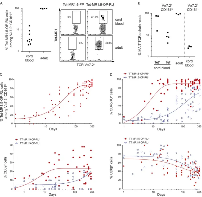
tcr repertoire of Vα7.2+ cd161high Mr1 :5 -oP -ru
tetramerpos/neg t cells in cord blood
As the MR1 :5 -OP -RU tetramer became available during the course of this study (National Institutes of Health [NIH]
tetramer core facility), we determined how the Vα7.2+
CD161high and MR1 :5 -OP -RU tetramerpos/neg populations
overlap. More than 92% of Vα7.2+ CD161high cells were
stained with the MR1 :5 -OP -RU tetramer in adult blood, in contrast with only 2–15% in cord blood (Fig. 6 A), in line with previous results (Eckle et al., 2014; Koay et al., 2016).
We further characterized the α chain of these populations
by deep sequencing and found that the Vα7.2+ CD161high
tetramerpos subset in cord blood was highly enriched in the
canonical MAIT TCR α chain compared with the tetramerneg
subset (Fig. 6 B). Yet, the canonical MAIT TCR α chains
were more frequent in the Vα7.2+ CD161high tetramerneg
than in the Vα7.2+ CD161− population. These results fully
support our initial analysis of the TCR α chain in cord blood subsets (Fig. 1 A). Altogether, they suggest that the CD161high
phenotype in cord blood Vα7.2+ T cells delineates a subset
of cells enriched in the canonical MAIT TCR α chain, with
only a part reactive to 5-OP-RU presented by MR1, certainly
according to the fine specificity imparted by the TCR β
chain. It is likely that these few clones then expand to form the adult MAIT pool because almost all Vα7.2+ CD161high
cells in adult blood express the canonical MAIT TCR and are stained by the MR1 :5 -OP -RU tetramer.
We therefore studied peripheral blood samples from 83 healthy infants aged from 1 d to 12 mo and found that the proportion of MR1 :5 -OP -RU tetramerpos cells increased
very rapidly, representing already more than 50% of Vα7.2+
CD161high cells at 1 mo of life and the vast majority at 6
mo (Fig. 6 C). Conversely, MR1 :5 -OP -RU tetramerneg
cells tended to disappear (Fig. S2). Moreover at 1 mo, more
than 80% of tetramerpos cells already expressed a mature
phenotype, whereas maturation of Vα7.2+ CD161high
tetramerneg cells was delayed (20% at 1 mo, 80% at 7 mo).
Reduction in CD8β expression was concomitant to the
acquisition of CD45RO. In addition, from 3–4 wk of age, an important proportion of Vα7.2+ CD161high tetramerpos (but
not tetramerneg) cells expressed CD69, suggesting a recent
activation in vivo (Fig. 6 D).
Altogether, these results indicate that expansion and maturation of the tetramerpos cells within the Vα7.2+
CD161high population occurs in the first days after birth,
sug-gesting the presence of some 5-OP-RU ligand providing an early maturation signal. This signal is likely not quantitatively sufficient to drive significant expansion of peripheral blood MAIT cells, which remain in very low amounts at this stage.
Figure 5. Phenotype and functional characteristics of neonatal Vα7.2+ cd161high t cells. Lymphocytes were isolated from 6–10 cord blood samples from full-term healthy neonates and the same number of healthy adult donors. (A) Expression levels of the indicated cytokine receptors. Box and whisker
plot shows median, interquartile range, and the 10th and the 90th percentiles of geometric mean fluorescence intensity (MFI; Mann-Whitney test). (B)
Left: Representative histograms of PLZF and CD25 expression by cord blood and adult blood Vα7.2+ CD161high T cells. Right: Mean values ± SEM of PLZF
and CD25 expression by Vα7.2+ CD161high cells and conventional T cells from cord blood and adult blood (Mann–Whitney test). (c) Left: Representative
histogram of PLZF expression in CD25+ and CD25− cord blood Vα7.2+ CD161high T cells and adult blood cells. Right: Mean values ± SEM of PLZF expression
by CD25+ and CD25− Vα7.2+ CD161high cord blood cells (paired t test) and adult blood Vα7.2+ CD161high cells. Data were not acquired on the same flow
cytometer as those in B, so MFI values cannot be compared. (d) Representative Ki67 intracellular staining in freshly isolated Vα7.2+ CD161high cells from
cord blood (shaded gray) and adult blood (black) samples. The percentage of cycling Vα7.2+ CD161high cells, estimated by the fraction of Ki67+ cells, in the
cord blood sample is indicated. (e) Vα7.2+ CD161high cell proliferating capacity. Left: Representative CFSE staining gated on Vα7.2+ CD161high T cells (shaded
gray) and conventional CD8 T cells (black line) from cord blood and adult blood after 6-d culture with PHA. Dashed histogram shows the basal CFSE staining at day 0. Right: Individual values and means (horizontal bars) of proliferation index in response to PHA in cord blood and adult blood Vα7.2+ CD161high
cells. P-value (Mann–Whitney test) is indicated. (F) Vα7.2+ CD161high T cell responses to microbial MR1 ligands. Freshly isolated CD4-negative T cells were
cultured overnight in the presence or absence of THP-1/E. coli or THP-1 alone as negative control, at a 1:1 THP1/CD8 T cell ratio. Intracellular accumulation of IFNγ (left) and GrB (right) in Vα7.2+ CD161high cells from cord blood or healthy adult control was evaluated by flow cytometry. *, P < 0.01; ***, P < 0.0001.
To verify our hypothesis that the fine specificity of
the TCR conferred by the β chain must be important for
5-OP-RU recognition and cell expansion, we studied TCR β
chain repertoire of adult and cord blood subsets. Adult MAIT cells display oligoclonal expansions with high clonal size and
biased TRBV usage (Tilloy et al., 1999; Lepore et al., 2014). Although cord blood MAIT cells display a diverse repertoire as studied with anti-Vβ antibodies (Walker et al., 2012), no extensive molecular characterization of their TRBV reper-toire has been performed so far. Using 5′ rapid
amplifica-Figure 6. Postnatal expansion of Vα7.2+ cd161high Mr1 :5 -oP -ru tetramerpos and tetramerneg cells. (A) Identification of MR1 :5 -OP -RU reactive cells. Left: Dot plots showing the percentages of MR1 :5 -OP -RU tetramerpos cells among CD3+ CD4− Vα7.2+ CD161high cells in nine cord blood and four
healthy adult blood samples. Right: Representative staining with MR1 :6 -FP tetramer (negative control, left quadrants) or MR1 :5 -OP -RU tetramer (right quadrants) in a cord blood and a healthy adult control. Numbers indicate the percentage of tetramerpos cells among the CD3+ CD4− Vα7.2+ CD161high T
cell gate. (B) Percentage of canonical MAIT TCRα chain (as in Fig. 1 B) reads among FACS-sorted MR1 :5 -OP -RU tetramerpos, tetramerneg, or total CD3+
CD4− Vα7.2+ CD161high cells from cord blood and a healthy adult and Vα7.2+ CD161− from cord blood (negative control). (c) The proportion of MR1 :5 -OP
-RU tetramerpos cells among CD3+ CD4− Vα7.2+ CD161high cells rapidly increases with age. Results show individual values in 79 healthy children aged 1 to
360 d. (d) Maturation (CD45RO and CD8β) and activation (CD69) phenotype of MR1 :5 -OP -RU tetramerpos (red dots) and tetramerneg (blue dots) cells among
tion of cDNA ends (RACE) technique followed by deep sequencing, we characterized the repertoire of FACS-sorted cord blood Vα7.2+ CD161high T cells in comparison to other
cord blood CD8 T cell subsets (Vα7.2− CD161high, Vα7.2+
CD161−, Vα7.2− CD161−) and to the same subsets in adult
blood. Importantly, memory (CD45RO+) subsets were sorted
in adult blood, to allow a meaningful comparison with adult MAIT cells, which are all memory. The rarefaction curves of different subsets, which represent cumulative frequency of individual clonotypes ranked by decreasing frequency, were compared (Fig. 7 A). For cord blood subsets, the curves were straight and overlapping, indicating similar high diversity be-tween the subsets. From cell dilution experiments, we
esti-mated the 5′RACE template exchange efficacy during the
reverse transcription step at ∼1–2% (not depicted). Because only 3,000 cells from cord blood were studied, the 30–60 clones making up 80–90% of the reads indicate that all the T cell expressed a different TCR. Identical results were obtained for four cord blood donors (Fig. S3). In contrast, although 10 times as many cells (35,000) were studied, adult MAIT cells encompassed oligoclonal expansions with less than 10 clones making up to 80% of the reads. Albeit less strikingly than in the donor depicted in Fig. 7 A, adult MAIT cells harbored similar oligoclonal expansions in the 13 donors studied (Fig. S3), in line with previous data (Tilloy et al., 1999; Lepore et al., 2014). In many cases, the oligoclonality of MAIT cells was similar to that of the other subsets, certainly because only
memory (CD45RO+) CD8+ T cells were included in our
sorting, contrary to a previous study (Lepore et al., 2014), and memory CD8 T cells are known to include oligoclonal expansions (Hingorani et al., 1993).
We then assessed whether cord blood and adult blood Vα7.2+ CD161high T cells displayed similar biases in the TCR
β chain repertoire. TRBV and TRBJ biases were conserved between donors, excluding a stochastic process. We observed preferential usage of TRBV 4.3, 6.4, 15, and 20.1 and TRBJ 2.6 and lower usage of TRBV 4.1 and 5.1 and TRBJ 1.2 in adult MAIT cells in comparison with both other memory adult CD8 T cell subsets (not depicted) and cord blood Vα7.2+ CD161high (Fig. S4, summarized in Fig. 7 B, left panel
for TRBV, right panel for TRBJ). Importantly, no such TRBV
or TRBJ usage bias was observed for cord blood Vα7.2+
CD161high in comparison with other cord blood CD8 subsets
(Fig. S5), suggesting that the TRBV bias observed in adult MAIT cells is not related to a pairing problem. However, when comparing cord blood Vα7.2+ CD161high MR1 :5 -OP
-RU tetramerpos and tetramerneg subsets (Fig. 7 C), similar to
adult MAIT cells, we observed that cord blood MR1 :5 -OP -RU tetramerpos population tended to express less TRBV4.1,
TRBV5.1, and TRBJ1.2 as well as more TRBV6.4 compared with the MR1 :5 -OP -RU tetramerneg population. No similar
biases between cord blood MR1 :5 -OP -RU tetramerpos and
adult MAIT cells were observed for the other segments, but this is probably related to a lack of experimental power because of the very low number of cells studied for the
tetramer-positive subset (50 cells). Altogether, these data indicate that the fine specificity of the TCRβ chain is involved in MR1 :5 -OP -RU complex recognition in accordance with structural data (Eckle et al., 2014).
dIscussIon
Here, by combining flow cytometry, deep sequencing, and further MR1 :5 -OP -RU tetramer staining, we decipher the postnatal development of human MAIT cells and other Vα7.2+
and Vα7.2− CD161high subsets. We longitudinally characterize
the maturation and expansion of these populations in infancy and after cord blood transplantation, two clinical settings sharing some similarities for establishment of mature protective immunity. The remarkably superimposable results observed in these two situations allow us to provide a new view of MAIT cell ontogeny in the human.
We characterize three distinct human CD3+ CD4−
CD161high populations present at birth: (1) a subset of Vα7.2+
cells enriched in the canonical MAIT TCR α chain, among
which a fraction is reactive to 5-OP-RU presented by MR1 (possibly as a result of the β chain usage), is the naive counterpart of mature MAIT cells found in adult blood and corresponds to the recently described stage 3 MAIT cells that just exit the thymus (Koay et al., 2016); (2) a subset of Vα7.2+
cells not reactive to 5-OP-RU presented by MR1, which predominate at birth but are rapidly outnumbered by MR1 :5 -OP -RU reactive cells; (3) an abundant subset of Vα7.2− cells,
the proportion of which remain stable over time. These three populations are already present at midgestation as observed in the cord blood of high-preterm neonates, in line with their presence in the thymus of aborted second-trimester fetuses (Leeansyah et al., 2014). These Vα7.2+ and Vα7.2− CD161high
populations likely share a common prenatal developmental program, as suggested by the strong correlation we observe between their frequencies at birth. Moreover, their percentages are highly similar in both monozygotic or dizygotic twin pairs, suggesting that prenatal environmental factors may also control their development. Although it is highly probable that
CD161high cells expressing the canonical MAIT TCR are
selected on MR1 expressed by double-positive thymocytes, the cell type and molecule selecting CD161high cells that do
not express the canonical MAIT TCR remain elusive.
At birth, Vα7.2+ CD161high cells exhibit a naive
phenotype and intermediate PLZF levels. Accordingly, they are unable to rapidly produce cytokines or cytotoxic molecules in response to bacterial ligands, in contrast with mature adult MAIT cells. Neither do they respond to stimulation by exogenous IL-12 and IL-18, despite high expression of the receptors for these cytokines. Strikingly,
although they represent a minor proportion of Vα7.2+
CD161high cells at birth, MR1 :5 -OP -RU tetramerpos cells
very rapidly predominate over tetramerneg cells. By the age of
6 mo, almost all Vα7.2+ CD161high T cells are MR1 :5 -OP -RU
-reactive MAIT cells. Therefore, the term MAIT cells can be confidently assigned to Vα7.2+ CD161high CD4− T cells from
the age of 6 mo after birth (or after HSCT), a crucial point for previous or further human studies conducted in the absence of MR1 tetramer identification.
Importantly, this complete shift in the proportion of tetramerpos MAIT cells within the Vα7.2+ CD161high pool is
concomitant with their activation (CD69+) and maturation
Figure 7. tcrβ repertoire analysis of cord blood and
adult blood MAIt cells. (A) The analysis was performed by
5′RACE-PCR of sorted T cell subsets. Examples of rarefaction curves from the indicated subsets in cord blood (left) and adult (right) samples. The cumulative frequency of productive TCRβ rearrangement is plotted for individual clonotypes ranked according to decreasing frequency. In adult blood, only memory (CD45RO+) subsets were studied to allow a fair
comparison. (B) Comparison of the TRBV and TRBJ fragments
usage in sorted CD3+ CD4− Vα7.2+ CD161high cells from 4
cord blood (blue) and 14 adult blood (red) samples. Only the Vβ (left) and Jβ (right) genes in which differential expression between neonates and adult samples was found are plotted. The whole dataset is plotted in Fig. S3. (c) Comparison of the
TRBV (left) and TRBJ (right) gene usage (same segments as in B) in sorted Vα7.2+ CD161high MR1 :5 -OP -RU tetramerpos
and tetramerneg cells from four cord blood samples. The
analysis was performed by multiplex PCR followed by high-throughput sequencing.
(CD45RO+). It is likely that colonization by the commensal
microbiota provides a key early activation signal, as supported by the impaired development and maturation of MAIT cells in germ-free mice compared with specific pathogen–free mice (Martin et al., 2009; Koay et al., 2016). Whether MAIT cell maturation in the early postnatal period is correlated with the gut microbiota diversity or function (availability of riboflavin metabolites) is currently under investigation in our laboratory. However, although microbe-mediated expansion of peripheral MAIT cells was demonstrated in mouse infection models (Meierovics et al., 2013; Chen et al., 2017), early- or late-onset neonatal microbial infection is not sufficient to induce a significant expansion in circulating MAIT cells. Indeed, MAIT cells very slowly populate the
periphery during infancy, taking ∼5–6 yr to achieve adult
levels. This gradual process, observed both in childhood and after cord blood transplantation, is not related to a MAIT-cell intrinsic proliferative defect. Rather, it may reflect specific requirements to allow for continuous proliferation, such as repeated exposure to pathogenic bacteria that provide more than single TCR/MR1 :ligand interactions. In mice, administration of synthetic 5-OP-RU alone causes CD69 up-regulation but does not result in MAIT proliferation, whereas the addition of Toll-like receptor agonists causes high levels of activation and proliferation of the MAIT cell pool (Chen et al., 2017). It is possible that some pathogens provide higher amounts of MAIT-activating ligands or an inflammatory/cytokine context allowing larger cell expansion than other pathogens, as shown by the tremendous MAIT expansion in mice during Francisella tularensis infection (Meierovics et al., 2013). Several lines of evidence indicate that MAIT cells can adapt their proliferative and effector responses depending on the microbial and inflammatory signals present (López-Sagaseta et al., 2013; Gold et al., 2014; Slichter et al., 2016; Dias et al., 2017).
Our repertoire analysis of Vα7.2+ CD161high cord blood
and adult subsets indicates that the fine specificity of the TCR
conferred by the β chain must be important for 5-OP-RU
recognition and expansion of Vα7.2+ CD161high T cells
expressing the canonical MAIT TCR α chain. These results
are consistent with structural data (Eckle et al., 2014). Only the few MR1 :5 -OP -RU reactive Vα7.2+ CD161high T cells that
display a TRAV and TRBV repertoire very similar to adult MAIT cells expand in periphery to form the adult MAIT pool, diluting both the Vα7.2− CD161hi and Vα7.2+ CD161hi MR1
:5 -OP -RU tetramerneg T cells. Collectively, these results suggest
that expansion of these few clones already present at birth is driven by repeated interactions with riboflavin-expressing pathogens encountered during the first years of life. Notably, MAIT cell levels exhibit very large interindividual variability (over one log range) in child and adult blood. It is tempting to speculate that microbial infection history contributes to the variable extent of MAIT cell expansion. Only a careful longitudinal analysis of MAIT levels in several populations of various ages in various environments will answer this question.
Although accurate identification of MAIT cells in tis-sues is limited in the absence of tetramer staining, Vα7.2+
CD161high cells have been found in fetal tissues (Leeansyah et
al., 2014), and we detected them in the intestine of 1-mo-old babies. Of note, these cells are tissue-resident memory cells, unable to recirculate, as indicated by the entirely naive phe-notype of circulating Vα7.2+ CD161high T cells at birth. Even
in the absence of live microbes in the placenta, maternal gestational microbes might be a source of microbial-derived metabolites reaching fetal tissues, as recently demonstrated in a model of transient microbial colonization in pregnant mice (Gomez de Agüero et al., 2016). However, in con-trast with their gradual expansion in peripheral blood after birth or cord blood transplantation, we found that Vα7.2+
CD161high cells did not accumulate much in intestinal
tis-sues during childhood. The lack of any detectable Vα7.2+
CD161high cells in the intestine of HSCT recipients, in
par-ticular during the course of aGVHD, is quite surprising. Mu-cosal disruption, microbial translocation, and inflammation contribute to aGVHD pathogenesis and should allow at least a few graft-derived Vα7.2+ CD161high cells to rapidly home
to mucosal sites. MAIT cells are highly sensitive to apoptotic stimuli (Gérart et al., 2013). One hypothesis is that microbial translocation triggers activation-induced apoptosis of newly generated MAIT cells within mucosal tissues or that post-transplant CS therapy exacerbates apoptotic sensitivity and prevents MAIT survival in tissues.
In addition to providing a more complete view of the physiological MAIT cell development, our results have im-portant implications for appreciating the impact of MAIT cell loss in other situations. Thus, in line with our observa-tion after cord blood transplantaobserva-tion, MAIT cells remained undetectable up to 2 yr after autologous HSCT in patients with multiple sclerosis (Abrahamsson et al., 2013). MAIT loss was correlated with good clinical response, supporting MAIT involvement in the disease pathology. Whether the benefit is still maintained at a time MAIT cells recover, i.e., 5–6 yr after transplantation, will deserve investigation. Our data may also explain the lack of circulating MAIT recovery in otherwise successfully treated HIV-infected patients (Cosgrove et al., 2013; Leeansyah et al., 2013). Rather than evoking an irre-versible MAIT loss in a context of HIV infection, it is more likely that complete MAIT recovery requires a minimum of 5–6 yr after initiation of antiretroviral treatment, as indicated by our preliminary results on long-term treated HIV patients (M. Lambert and S. Caillat-Zucman, personal data).
Finally, our results indicate that the early expansion of MAIT cells after birth dilutes Vα7.2+ CD161high cells not
reactive to 5-OP-RU presented by MR1. Part of these MR1 :5 -OP -RU tetramerneg express the canonical MAIT TCR α
chain and are thus likely selected on MR1 in the thymus. Because a formal demonstration of their MR1 restriction is not feasible in humans, single-cell TCR sequencing followed by TCR reexpression would be necessary to formally characterize the MR1 :5 -OP -RU tetramerneg cells that express