HAL Id: inserm-01857808
https://www.hal.inserm.fr/inserm-01857808
Submitted on 17 Aug 2018
HAL is a multi-disciplinary open access
archive for the deposit and dissemination of
sci-entific research documents, whether they are
pub-lished or not. The documents may come from
teaching and research institutions in France or
abroad, or from public or private research centers.
L’archive ouverte pluridisciplinaire HAL, est
destinée au dépôt et à la diffusion de documents
scientifiques de niveau recherche, publiés ou non,
émanant des établissements d’enseignement et de
recherche français ou étrangers, des laboratoires
publics ou privés.
The clinical implications of G1-G6 transcriptomic
signature and 5-gene score in Korean patients with
hepatocellular carcinoma
Sung-Min Ahn, Farhan Haq, Inkeun Park, Jean-Charles Nault, Jessica
Zucman-Rossi, Eunsil Yu
To cite this version:
Sung-Min Ahn, Farhan Haq, Inkeun Park, Jean-Charles Nault, Jessica Zucman-Rossi, et al.. The
clinical implications of G1-G6 transcriptomic signature and 5-gene score in Korean patients with
hepatocellular carcinoma. BMC Cancer, BioMed Central, 2018, 18 (1), pp.571.
�10.1186/s12885-018-4192-1�. �inserm-01857808�
R E S E A R C H A R T I C L E
Open Access
The clinical implications of G1-G6
transcriptomic signature and 5-gene score
in Korean patients with hepatocellular
carcinoma
Sung-Min Ahn
1,2†, Farhan Haq
3†, Inkeun Park
1, Jean-Charles Nault
4,5,6,7, Jessica Zucman-Rossi
4,5,6,7*and Eunsil Yu
8*Abstract
Background: Efforts have been made to classify Hepatocellular Carcinoma (HCC) at surgically curable stages because molecular classification, which is prognostically informative, can accurately identify patients in need of additional early therapeutic interventions. Recently, HCC classification based French studies on the expression of 16 genes and 5 genes were proposed. In 16-gene classification, transcriptomic signatures (G1-G6) were used to classify HCC patients into clinical, genomic and pathway-specific subgroups. In 5-gene score classification, the good or poor prognosis of HCC patients was predicted. The patient’s cohort in these studies was mainly from Caucasian and African populations. Here, we aimed to validate G1-G6 and 5-gene score signatures in 205 Korean HCC patients since genomic profiles of Korean patients are distinct from other regions.
Methods: Integrated analyses using whole-exome sequencing, copy number variation and clinical data was performed against these two signatures to find statistical correlations. Kaplan-Meier, univariate and multivariate COX regression analysis were performed for Disease-Specific Survival (DSS) and Recurrence-Free Survival (RFS).
Results: The G2 and G3 subgroups of transcriptomic signature were significantly associated withTP53 mutations while G5 and G6 subgroups were significantly associated withCTNNB1 mutations which is in concordance with original French studies. Similarly, the poor prognosis group of 5-gene score showed shorter DSS (p = 0.045) and early RFS (p = 0.023) as well as a significant association with microvascular invasion, tumor size (> 5 cm), elevated AFP levels, andRB1 mutations. However, the 5-gene score was not an independent prognostic factor for survival.
Conclusion: The G1-G6 and 5-gene signatures showed significant concordance between genetic profiles of Korean HCC patients and patients in original French studies. Thus, G1-G6 and 5-gene score signatures can be targeted as potential therapeutic biomarkers against HCC patients worldwide.
Keywords: 5-gene score, G1-G6 subgroups, Survival, Prognosis
* Correspondence:jessica.zucman-rossi@inserm.fr;esyu@amc.seoul.kr
†Sung-Min Ahn and Farhan Haq contributed equally to this work. 4Inserm, UMR-1162, Génomique Fonctionnelle des Tumeurs Solides, Équipe
Labellisée Ligue Contre le Cancer, 27 rue Juliette Dodu, F-75010 Paris, France
8Department of Pathology, University of Ulsan College of Medicine, Asan
Medical Center, 88, OLYMPIC-RO 43-GIL, SONGPA-GU, SEOUL 138-736, South Korea
Full list of author information is available at the end of the article
© The Author(s). 2018 Open Access This article is distributed under the terms of the Creative Commons Attribution 4.0 International License (http://creativecommons.org/licenses/by/4.0/), which permits unrestricted use, distribution, and reproduction in any medium, provided you give appropriate credit to the original author(s) and the source, provide a link to the Creative Commons license, and indicate if changes were made. The Creative Commons Public Domain Dedication waiver (http://creativecommons.org/publicdomain/zero/1.0/) applies to the data made available in this article, unless otherwise stated.
Background
Hepatocellular carcinoma is the most common type of liver cancer worldwide [1]. Liver resection is one of the most viable treatment option for HCC patients, but as-sociated with high risk of recurrence [2,3]. In advanced HCC, no clinical trial studies have convincingly improved survival, except for the sorafenib trial [4]. The failure of these trials is partly due to the lack of effective molecu-lar markers or the minimal validation of known mo-lecular markers in diverse multi-ethnic populations. Efforts have been made to classify HCC at surgically curable stages because molecular classification, which is prognostically informative, can accurately identify patients in need of additional early therapeutic inter-ventions [5–9].
In HCC, several molecular classification-based micro-array studies have been reported. In the first two studies, HBV-positive HCC patients from Belgium and China were classified into good or poor prognosis groups [10, 11]. The expression of hepatoblast-related genes was significantly associated with poor prognosis of HCC patents [11]. In another HCC study using a Caucasian population of HCC patients, a gene signa-ture of 186 genes was identified in non-tumor liver, out of which 113 showed good prognosis and 73 showed poor prognosis [12].
Recently, HCC classification methods based on the expression of 16 genes and 5 genes were proposed [13, 14]. In the 16-gene based classification, transcrip-tomic signatures (G1-G6) were used to classify HCC patients into clinical, genomic and pathway-specific G1-G6 subgroups [13]. In the 5-gene score-based classification, the good or poor prognosis of HCC patients was predicted [14]. The study showed the ef-ficacy of 5-gene score as a potential biomarker in American, Caucasian and Chinese HCC patients [14].
In this study, we aimed to validate the clinical rele-vance of the 16-gene and 5-gene score methods in a Korean population. One of the key motivations of this study was that the genomic profiles of HCC patients were distinct between Western and Korean populations. For example, the TP53 mutation rate is very high in Korean populations, but unlike African or Chinese populations, the TP53 R249S mutation rate is almost zero and has no association with tumor recurrence and survival [5, 8, 15, 16]. In addition, genetic aberra-tions in RB1 are associated with poor prognosis in Korean HCC patients, which has not been observed in Western populations [5, 8]. In this study, we gen-erated the transcriptomic signatures using the 16-gene and 5-gene score methods, and analyzed them with the genomic profiles obtained by whole-exome se-quencing and clinicopathological features of the HCC cases used.
Methods
Study design and clinical samples
Two hundred five Fresh frozen tissues of HCC patients at a surgically curable stage were used in this study. The surgically curable stage was defined by Milan criteria which clearly excludes extra hepatic metastasis and macrovascular invasion. Patients with either one of them cannot have surgical resection. The institutional review board of ASAN, South Korea, approved all of the samples, along with documented consent from all pa-tients who participated in the study (2012–0389). The estimated tumor cellularity of each sample was more than 70%. Clinical features include tumor size, micro-vascular invasion, recurrence, the Edmondson-Steiner histological grade, the fibrosis stage of the non-neoplastic liver tissue, viral infection, tumor nodules (monofocal vs multifocal) and the serum alpha-fetoprotein levels (Table 1).
Table 1 Patients and tumor characteristics
Characteristics Frequencies Age at Surgery (Median, Range) 55 (26–80)
< 60 years 137 (66.83%) > 60 years 68 (33.17%) Etiology of the liver disease
HBVa 148 (72.20%)
HCVb 20 (9.76%)
NBNCc 37 (18.05%)
Tumor size (median, range) 3.8 (1.2–16) < 5 cm 140 (68.29%) > 5 cm 68 (33.17%) Microvascular invasion
Yes 59 (28.78%) No 146 (71.22%) Serum AFP level (median, range) 42.7 (0.76–472,000)
< 20 91 (44.39%) > 20 114 (55.61%) Hepatic fibrosis stage
1,2,3 109 (53.17%) 4 96 (46.83%) Edmondson-Steiner grade 1,2 137 (66.83%) 3,4 68 (33.17%) Tumor nodules Single 193 (94.15%) Multiple 12 (5.85%) a HBV: hepatitis B virus. b HCV: hepatitis C virus. c NBNC: non-hepatitis B, non-hepatitis C
Whole exome sequencing and copy number variation (CNV) analysis
As mentioned in our previous study, DNeasy Blood and Tissue kit (Qiagen) were used for DNA extraction from the tumor tissues. Exome sequencing was done using Illumina HiSeq 2000 platform [5]. The sequenced reads were aligned to the UCSC hg19 release of the human gen-ome. Somatic mutations inTP53, CTNNB1 and RB1 were identified using MuTect [17]. CNV analysis was per-formed using the Affymetrix Cytoscan HD platform. CNV data were analysed with the Nexus Copy Number software (BioDiscovery, CA, ver. 6.1). Furthermore, LOH events were identified using PSCBS algorithm [18].
G1-G6 classification and 5 gene score prediction
The detailed information about the development of G1-G6 and 5-gene score method was described in previous HCC studies [13,14]. Briefly, the G1-G6 classification was done using robust unsupervised hierarchical clustering using 6712 probes of Affymetrix HG-U133A GeneChip™ [13]. The stability and reproducibility of the clusters were carefully evaluated. The mean reproducibility of all groups was more than 90%. On the basis of clusters, tumors were classified into G1-G6 subgroups. Furthermore, using quan-titative RT-PCR data and applying 5 prediction algorithms (including SVM, PAM, kNN, DQDA, DLDA) on 103 genes associated with the prognosis and diagnosis of cancer, 16 genes were identified which properly classified HCC patients into G1-G6 subgroups. Similarly, univariate Cox model was generated for each of the 103 genes against prognosis and survival of HCC patients. 31 signifi-cant genes were further optimized to only 5 genes using multivariate COX model [14]. In this study, we evaluated the performance of G1-G6 and 5-gene scores in Korean population using high quality RNA data 205 of HCC patients.
Statistical analysis
We performed several statistical analyses against these two molecular markers to establish their clinical and patho-logical relevance in a Korean population. IBM SPSS Ver-sion 20 was used for all statistical analyses. Fisher’s exact test (P < 0.05) was used to calculate any association be-tween the genomic data and the G1-G6 and 5-gene score. Kaplan-Meier survival analysis was performed using DSF and RFS data. Previously, early recurrence was defined as recurrence before 24 months [14]. However, in our study recurrence before 12 months was considered as early recur-rence. Univariate and multivariate Cox regression survival analyses were performed to validate the prognostic associ-ation between clinical data, genomic data and G1-G6 and 5-gene score.
Results and discussion
Out of 231 cases of HCC used in previous study, we were able to extract high-quality RNA of 205 HCC cases [5]. We then found the association of G1-G6 subgroups and 5-gene score with mutations, CNVs and clinicopath-ological features (see Table1for clinical features). Validation of the G1-G6 classification in Korean population
Out of the 205 cases analyzed, 16 (7.81%) fell into G1, 23 (11.22%) into G2, 16 (7.81%) into G3, 110 (53.65%) into G4, 27 (13.17%) into G5 and 13 (6.34%) into G6. The occurrence of the G1-G3 and G5-G6 subgroups in a pre-vious French study were slightly higher compared to our dataset, except for the G4 subgroup, which accounted for the largest proportion in both studies (Fig.1) [13,14]. Of note, there is no significant difference between G1-G6 dis-tribution in both cohorts (Mann–Whitney test, P = 0.297). In addition, the two cohorts were strongly correlated (Spearman Correlation = 0.89,P = 0.033).
Molecular and clinicopathologic characteristics correlated with the G1-G6 classification
Then, we analyzed the G1-G6 subgroups with genetic aberrations and the clinical features of the 205 cases of HCC. G2 and G3 were significantly associated withTP53 mutations (P < 0.005) and G5 and G6 with CTNNB1 mu-tations (P < 0.005). Of note, almost all of the G6 subgroup cases (12/13) harbored somaticCTNNB1 mutations. The associations between G2-G3 andTP53 mutations and be-tween G5-G6 and CTNNB1 mutations in this study were consistent with the original observations made in the French study [13].
As for CNVs, G2 was significantly associated with 13q LOH (P < 0.05); G2-G3 with 17p LOH (P < 0.05); and G1-G3 with 4q (P < 0.001), 5q (P < 0.001) and 16p LOH (P < 0.001). All of these LOH events were associated with G1-G3, which are collectively recognized as HCC subgroups with chromosomal instability [13]. Again, the
observations in this study were consistent with the ori-ginal observations made in the French study [13].
As for clinicopathological features, G1-G3 groups were significantly associated with AFP > 100 IU/ml (P < 0.001), whereas, G4-G6 were significantly associated with AFP < 100 IU/ml (P < 0.001). G5 was significantly associ-ated with tumor size (> 5 cm) (P < 0.05), which was not observed in the previous study. When we performed a survival analysis, the G1-G6 subgroups did not show any significant difference in either DSS or RFS (i.e., recurrence before 12 months), which was consistent with the French study [13].
5-gene score and its role in prognostication
To validate the 5-gene score, we classified patients into a good prognosis group (81 cases, 40%) and a poor progno-sis group (124 cases, 60%) according to the 5-gene score and evaluated the difference in DSS and RFS. As demon-strated in Fig. 2, the poor prognosis group showed a shorter median DSS (P < 0.05) and RFS (P < 0.05).
Molecular and clinicopathologic features correlated with the 5-gene score
We found that the poor prognosis group, as predicted by the 5-gene score, was significantly associated with TP53 mutations (P < 0.005). This association needs to be interpreted with caution. In our previous study, we re-ported that TP53 mutations were not associated with poor survival in a Korean population; however, in other studies,TP53 mutations, especially the R249S mutation resulting from aflatoxin B1 exposure, were associated with poor survival in HCC patients. In the 205 cases of HCC that we used in this study, no case harbored the TP53 R249S mutation. However, in the French study, 12 out 62 patients showed R249S mutations, all in migrants
from Africa or Asia, but did not show any association with either survival or poor prognosis as predicted by the 5-gene score [14].
In addition, in our recent HCC study, we reported that RB1 aberrations (Homozygous Deletions and Inactivat-ing Mutations) were associated with the poor prognosis of HCC patients after resection. Consistent with previ-ous observations, the poor prognosis predicted by the 5-gene score also showed association withRB1 aberrations (i.e., 13 out of 18 (72%) cases fell into the poor prognosis group). In addition, the poor prognosis group was also significantly associated with a loss of heterozygosity (LOH) events at the 4q, 5q, 16p, 17p and 22q chromo-somal arms, which was also consistent with the French study (P < 0.05) [13].
As for clinical features, the poor prognosis group was significantly associated with microvascular invasion (P < 0.005), tumor size (> 5 cm) (P < 0.05), and high AFP levels (> 20 ng/ml) P < 0.005). In addition, we found that the poor prognosis group was significantly associated with G1-G3 and the good prognosis group with G4-G6 (P < 0.005).
Univariate and multivariate analysis for disease-specific survival (DSS)
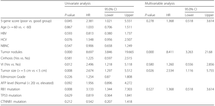
With a median follow-up period of 53.3 months (range 5.5–88.9 months), 30 patients died due to HCC. The two- and five-year DSS rates were 94.5% and 79.9%, re-spectively. In the univariate analysis, DSS was signifi-cantly associated with the 5-gene score (hazard ratio (HR) 2.381, 95% confidence interval (CI) 1.021–5.56, P = 0.045), tumor nodules (HR 8.69, 95% CI 3.846– 19.6653, P < 0.001), microvascular invasion (HR 2.496, 95% CI 1.218–5.118, P = 0.012), tumor size (HR 2.674, 95% CI 1.297–5.512, P = 0.008) and RB1 aberrations
Fig. 2 The good and poor prognosis groups, predicted by 5-gene score, show a significant difference in both DSS and early RFS
(HR 3.133, 95% CI 1.344–7.303, P = 0.008) (Table 2). In the multivariable analysis, the 5-gene score was not vali-dated as a significant prognostic factor; tumor nodules (HR 8.411, CI 3.263–21.681, P < 0.001) and tumor size (HR 2.534, CI 1.116–5.755, P = 0.026) were the signifi-cant prognostic factors for the survival of HCC patients after resection (Table2).
Univariate and multivariable analysis for early recurrence-free survival (RFS)
During follow-up, 49 out of 205 patients had disease re-currence within 12 months. In the univariate analysis, the early RFS was significantly associated with the 5-gene score (HR 2.087, 95% CI 1.106–3.936, P = 0.023), tumor nodules (HR 5.308, 95% CI 2.474–11.390, P < 0.001), Table 2 Cox regression analysis for DSS in 205 patients with resectable HCC
Univariate analysis Multivariable analysis
95.0% CI 95.0% CI
P-value HR Lower Upper P-value HR Lower Upper 5-gene score (poor vs. good group) 0.045 2.381 1.021 5.551 0.278 1.368 0.518 3.614 Age (> = 60 vs. < 60) 0.867 1.033 0.706 1.511
HBV 0.593 0.813 0.380 1.737 HCV 0.076 1.548 0.956 2.507 NBNC 0.547 0.906 0.658 1.249
Tumor nodules 0.000 8.697 3.846 19.665 0.000 8.411 3.263 21.68 Cirrhosis (Yes vs. No) 0.581 1.225 0.597 2.515
VI (Yes vs. No) 0.012 2.496 1.218 5.118 0.580 1.260 0.556 2.856 Tumor size (> = 5 cm vs < 5 cm) 0.008 2.674 1.297 5.512 0.026 2.534 1.116 5.755 Edmonson Grade 0.226 1.254 0.87 1.808
AFP level (Normal (< 20) vs. elevated) 0.092 1.956 0.896 4.272
RB1 mutation 0.008 3.133 1.344 7.303 0.527 1.368 0.518 3.614 TP53 mutation 0.629 0.819 0.364 1.841
CTNNB1 mutation 0.212 0.542 0.207 1.418
*VI Microvascular Invasion
Table 3 Cox regression analysis for eary RFS in 205 patients with resectable HCC
Univariate analysis Multivariable analysis
95.0% CI 95.0% CI
P-value HR Lower Upper P-value HR Lower Upper 5-gene score (poor vs. good group) 0.023 2.087 1.106 3.936 0.361 1.373 0.696 2.708 Age (> = 60 vs. < 60) 0.469 1.113 0.833 1.488
HBV 0.814 1.079 0.572 2.035 HCV 0.044 1.476 1.010 2.156 NBNC 0.100 0.772 0.567 1.051
Tumor Nodules 0.000 5.308 2.474 11.390 0.000 5.956 2.554 13.892 Cirrhosis (Yes vs. No) 0.223 1.418 0.809 2.485
VI (Yes vs. No) 0.009 2.131 1.21 3.754 0.904 0.959 0.483 1.901 Tumor size (> = 5 cm vs < 5 cm) 0.012 2.053 1.169 3.604 0.058 1.885 0.978 3.634 Edmonson Grade 0.985 0.997 0.74 1.344
AFP level (Normal (< 20) vs. elevated) 0.001 3.192 1.631 6.248 0.004 2.823 1.387 5.745 RB1 mutation 0.009 2.62 1.27 5.413 0.488 1.318 0.604 2.873 TP53 mutation 0.769 1.094 0.602 1.987
CTNNB1 mutation 0.338 0.702 0.341 1.447
HCV (HR 1.476, CI 1.010–2.156, P = 0.044), vascular inva-sion (HR 2.131, 95% CI 1.210–3.754, P = 0.009), AFP 20 ng/ml (HR 3.192, CI 1.631–6.248, P = 0.001), tumor size (HR 2.053, 95% CI 1.169–3.604, P = 0.012) and RB1 aberrations (HR 2.62, 95% CI 1.302–5.228, P = 0.009) (Table 3). In the multivariate analysis, the 5-gene score was not validated as a significant prognostic factor; AFP20ng/ml (HR 2.918, CI 1.423–5.984, P = .003), tumor nodules (HR 6.818, CI 2.873–16.184, P < 0.001), HCV (HR 1.718, CI 1.159–2.546, P = .007) and tumor size (HR 1.909, CI 0.984–3.705, P = .056) were found as the signifi-cant prognostic factors for early RFS of HCC patients after resection (Table3).
In this study, we aimed to validate the association of the G1-G6 signature and the prognostic value of the 5-gene score in Korean HCC patients. These two molecu-lar signatures showed remarkable concordance between CNV and the mutation profiles of Korean HCC patients and the patients in French studies [13, 14], except for minor discrepancies. For example, G5 and G6 rates are lower in our cohort than in the original cohort, which seems to be related to the lower rate ofCTNNB1 muta-tion in our cohort. According to the 5-gene score, the poor prognosis group showed shorter disease-specific survival and early recurrence-free survival as well as a significant association with microvascular invasion, tumor size, high AFP levels, andTP53 mutations.
However, the 5-gene score was not an independent prognostic factor for the survival of HCC patients. This may be due to the low event rate [only 30 patients out of 205 (14.6%) died during follow-up, in contrast to French data, in which 106/314 (33.8%) died during follow-up], which may have resulted in different multi-variable outcomes.
Conclusions
Thus, our analysis suggests that G1-G6 and 5-gene sig-natures are in concordance between genetic profiles of Korean HCC patients and patients in original French studies. Therefore, in the future, by combining all of these cohorts, we may be able to assertively establish the clinical and pathological relevance of the 5-gene score and de-velop therapeutic strategies for HCC patients worldwide. Abbreviations
CNV:Copy Number Variation; DSS: Disease-Specific Survival; HCC: Hepatocellular Carcinoma; LOH: Loss Of Heterozygosity; RFS: Recurrence-Free Survival Acknowledgements
The bio specimen and data used in this study was provided by Asan Bio-Resource Center, Korea, Korea Biobank Network (2012-8 (51)). Institut National de la Santé et de la Recherche Médicale (INSERM) generated transcriptomic and 5-gene score signatures.
Funding
This research was supported by a grant of the Korea Health Technology R&D Project through the Korea Health Industry Development Institute, funded by
the Ministry of Health & Welfare (HI16C1985) and by Basic Science Research Program through the National Research Foundation of Korea funded by the Ministry of Science, ICT and future Planning(NRF-2015R1A2A2A04004063). Availability of data and materials
The whole exome sequencing data was uploaded to cbioportal (http:// www.cbioportal.org/study?id=lihc_amc_prv) as described in our previous study [5]. CytoscanHD data was uploaded to Gene Expression Omnibus database (accession number: GSE54504).
Authors’ contributions
SMA, JZR, EY contributed to conception and design of the study, and revised the manuscript. FH contributed to analysis, interpretation and writing of the manuscript. IP and JCN contributed in the analysis and revising manuscript. All authors of the study have read and approved the manuscript. Ethics approval and consent to participate
The institutional review board of ASAN, South Korea, approved all samples, along with documented consent from all patients who participated in the study (2012–0389).
Consent for publication Not applicable Competing interests
The authors declare that they have no competing interests.
Publisher’s Note
Springer Nature remains neutral with regard to jurisdictional claims in published maps and institutional affiliations.
Author details
1
Department of Genome Medicine and Science, College of Medicine, Gachon University, Seongnam, South Korea.2Department of
Hematology-Oncology, Gachon University Gil Hospital, Incheon, South Korea.
3Department of Biosciences, Cancer Genetics and Epigenetics Lab, COMSATS
Institute of Information Technology, Islamabad, Pakistan.4Inserm, UMR-1162, Génomique Fonctionnelle des Tumeurs Solides, Équipe Labellisée Ligue Contre le Cancer, 27 rue Juliette Dodu, F-75010 Paris, France.5Labex Immuno-Oncology, Sorbonne Paris Cité, Faculté de Médecine, Université Paris Descartes, Paris, France.6Sorbonne Paris Cité, UFR SMBH, Université Paris 13, F-93000 Bobigny, France.7Université Paris Diderot, F-75013 Paris,
France.8Department of Pathology, University of Ulsan College of Medicine, Asan Medical Center, 88, OLYMPIC-RO 43-GIL, SONGPA-GU, SEOUL 138-736, South Korea.
Received: 29 January 2017 Accepted: 6 March 2018
References
1. McGlynn KA, London WT. The global epidemiology of hepatocellular carcinoma, present and future. Clin. Liver Dis. 2011;15(2): 223–x. 2. Forner A, Gilabert M, Bruix J, Raoul J-L. Treatment of intermediate-stage
hepatocellular carcinoma. Nat Rev Clin Oncol. 2014;11(9):525–35. 3. Llovet JM, Burroughs A, Bruix J. Hepatocellular carcinoma. Lancet. 2003;
362(9399):1907–17.
4. Llovet JM, et al. Sorafenib in advanced hepatocellular carcinoma. N Engl J Med. 2008;359(4):378–90.
5. Ahn S-M, et al. Genomic portrait of resectable hepatocellular carcinomas: implications of RB1 and FGF19 aberrations for patient stratification. Hepatology. 2014;60(6):1972–82.
6. Dong H, Qian Z, Zhang L, Chen Y, Ren Z, Ji Q. Genomic and transcriptome profiling identified both human and HBV genetic variations and their interactions in Chinese hepatocellular carcinoma. Genomics Data. 2015;6:1–3. 7. Fujimoto A, et al. Whole-genome sequencing of liver cancers identifies
etiological influences on mutation patterns and recurrent mutations in chromatin regulators. Nat Genet. 2012;44(7):760–4.
8. Guichard C, et al. Integrated analysis of somatic mutations and focal copy-number changes identifies key genes and pathways in hepatocellular carcinoma. Nat Genet. 2012;44(6):694–8.
9. Kan Z, et al. Whole-genome sequencing identifies recurrent mutations in hepatocellular carcinoma. Genome Res. 2013;23(9):1422–33.
10. Lee J-S, et al. Classification and prediction of survival in hepatocellular carcinoma by gene expression profiling. Hepatol Baltim Md. 2004;40(3):667–76. 11. Lee J-S, et al. A novel prognostic subtype of human hepatocellular
carcinoma derived from hepatic progenitor cells. Nat Med. 2006;12(4):410–6. 12. Hoshida Y, et al. Gene expression in fixed tissues and outcome in
hepatocellular carcinoma. N Engl J Med. 2008;359(19):1995–2004. 13. Boyault S, et al. Transcriptome classification of HCC is related to gene
alterations and to new therapeutic targets. Hepatology. 2007;45(1):42–52. 14. Nault J, et al. A hepatocellular carcinoma 5-gene score associated with
survival of patients after liver resection. Gastroenterology. 2013;145(1):176–87. 15. Woo HG, et al. Association of TP53 mutations with stem cell-like gene
expression and survival of patients with hepatocellular carcinoma. Gastroenterology. 2011;140(3):1063–1070.e8.
16. Schulze K, et al. Exome sequencing of hepatocellular carcinomas identifies new mutational signatures and potential therapeutic targets. Nat Genet. 2015;47(5):505–11.
17. Cibulskis K, et al. Sensitive detection of somatic point mutations in impure and heterogeneous cancer samples. Nat Biotechnol. 2013;31(3):213–9. 18. Olshen AB, Bengtsson H, Neuvial P, Spellman PT, Olshen RA, Seshan VE.
Parent-specific copy number in paired tumor-normal studies using circular binary segmentation. Bioinforma Oxf Engl. 2011;27(15):2038–46.
• We accept pre-submission inquiries
• Our selector tool helps you to find the most relevant journal • We provide round the clock customer support
• Convenient online submission • Thorough peer review
• Inclusion in PubMed and all major indexing services • Maximum visibility for your research
Submit your manuscript at www.biomedcentral.com/submit