HAL Id: tel-01715600
https://tel.archives-ouvertes.fr/tel-01715600
Submitted on 22 Feb 2018HAL is a multi-disciplinary open access archive for the deposit and dissemination of sci-entific research documents, whether they are pub-lished or not. The documents may come from teaching and research institutions in France or abroad, or from public or private research centers.
L’archive ouverte pluridisciplinaire HAL, est destinée au dépôt et à la diffusion de documents scientifiques de niveau recherche, publiés ou non, émanant des établissements d’enseignement et de recherche français ou étrangers, des laboratoires publics ou privés.
Characterization of the Purkinje cell to nuclear cell
connections in mice cerebellum
Orçun Orkan Özcan
To cite this version:
Orçun Orkan Özcan. Characterization of the Purkinje cell to nuclear cell connections in mice cerebel-lum. Neurobiology. Université de Strasbourg, 2017. English. �NNT : 2017STRAJ085�. �tel-01715600�
Ecole Doctorale des Sciences
Albert-Ludwigs-Universität
de la Vie et de la Santé
Freiburg im Breisgau
THÈSE
Présentée par:
Orçun Orkan ÖZCAN
En vue d’obtenir le grade de docteur de l’université de Strasbourg et Freiburg
Spécialité: Neurosciences
Soutenue le: 20 Mars 2017 THÈSE dirigée par:
Dr. Philippe ISOPE INCI, Université de Strasbourg
Prof. Dr. Arvind KUMAR KTH Royal Institute of Technology, Stockholm Prof. Dr. Ad AERTSEN Albert Ludwig University of Freiburg
RAPPORTEURS:
Prof. Dr. Chris I. DE ZEEUW Erasmus Medical Center, Rotterdam Dr. Yann HUMEAU IINS, Université de Bordeaux AUTRES MEMBRES DE JURY :
Dr. Romain GOUTAGNY LNCA, Université de Strasbourg
Characterization of the Purkinje cell to nuclear cell connections in
mice cerebellum
Caractérisation des connexions cellules de Purkinje - cellule des noyaux
profonds dans le cervelet de souris
This study was carried out as a joint project of the following research institutes: Physiology of Neural Networks (group of Dr. Philippe Isope)
Institut des Neurosciences Cellulaires et Intégratives, UPR 3212 CNRS Université de Strasbourg
5 rue Blaise Pascal, 67084 Strasbourg, France and
Bernstein Center Freiburg (group of Prof. Dr. Ad Aertsen) Albert-Ludwigs Universität
Hansastrasse 9a, 79104 Freiburg, Germany
TABLE OF CONTENTS
Acknowledgements………....………7
Summary in French (Résumé)………....………8
Summary………...………....……….9
Abbreviations…..………....……….11
Table of Figures and Tables………..………...13
1
INTRODUCTION ... 15
1.1
A short history of cerebellar physiology and function ... 15
1.2
Functions of the cerebellum ... 17
1.3
Anatomical organization of the cerebellum ... 19
1.3.1 Gross anatomy of the cerebellum ... 19
1.3.2 Gross functional divisions of the cerebellum ... 20
1.3.3 Cerebellar inputs ... 21
1.3.4 Cerebellar outputs ... 22
1.4
The cerebellar cortex ... 22
1.4.1 Information flow in the cerebellar cortex ... 22
1.4.2 Cellular structure of the cerebellar cortex ... 25
1.4.2.1 The Purkinje cell ... 25
1.4.2.2 The granule cell ... 26
1.4.2.3 The Golgi cell ... 27
1.4.2.4 Molecular layer interneurons ... 27
1.4.3 Functional microzones in the cerebellar cortex ... 28
1.4.4 Functional role of the cerebellar cortex and Purkinje cells ... 30
1.5
The deep cerebellar nuclei (DCN) ... 30
1.5.1 Anatomy of the DCN ... 30
1.5.2 Functional role of the DCN ... 31
1.5.3 Cellular structure of the DCN ... 33
1.5.3.1 The principal projection cell ... 33
1.5.3.2 The inhibitory nucleo-cortical cell ... 35
1.5.3.2.1 The local collaterals of inhibitory nucleo-cortical cell ... 36
1.5.3.3 The non-inhibitory interneurons ... 37
1.5.3.4 The Nucleo-olivary projection cell ... 38 3
1.5.3.5 The glycinergic premotor projection cell ... 38
1.5.3.6 The distribution of cell types among different nuclei ... 39
1.5.4 Functional connectivity of the cerebellar cortex to the DCN... 39
1.5.4.1 Projections of the cerebellar cortex to the DCN ... 40
1.5.4.2 Convergence and divergence of Purkinje cells in the DCN ... 42
1.5.5 Purkinje cell inhibition on different cells of the DCN ... 43
1.5.5.1 Purkinje cell innervation on the principal cells ... 44
1.5.5.2 Effects of synchronized Purkinje cell inputs ... 45
1.5.5.3 Purkinje cell innervation on the nucleo-olivary cells ... 46
1.5.5.4 Purkinje cell innervation on the inhibitory nucleo-cortical cells ... 47
1.5.5.5 Purkinje cell innervation on other cells in the DCN ... 47
1.5.5.6 Plasticity of PC connections in the DCN ... 47
1.5.6 Information processing in the DCN ... 48
1.5.6.1 Mossy fiber innervation and plasticity in the DCN ... 49
1.5.6.2 Climbing fiber innervation in the DCN ... 52
1.5.6.3 Integration in the inhibitory nucleo-cortical cells ... 53
1.5.6.4 Integration in the principal cells ... 53
1.5.6.5 Receptive fields in the DCN ... 54
1.5.6.6 PC inhibition vs local inhibition in the DCN ... 56
2
OBJECTIVES OF THE STUDY ... 57
3
RESULTS ... 58
3.1
Multiple cells from connected regions in the cerebellar cortex and in the DCN are
simultaneously recorded under optogenetic stimulation in vivo ... 58
3.1.1 Tracing PC projections with virus and dye injections... 58
3.1.2 Determining recording areas after the experiments ... 60
3.1.3 Simultaneous multielectrode recordings from the two areas ... 60
3.2
Two groups of DCN cells are identified using their waveforms ... 63
3.2.1 Discrimination of cells based on waveform parameters ... 63
3.2.2 Properties of the two groups of cells ... 66
3.3
Low and high frequency oscillations in the DCN have different durations and phase
locking ratios ... 68
3.3.1 Detection of LFP in the DCN ... 68
3.3.2 Phase locking to detected oscillations in the DCN ... 70
3.4
Specific responses of the two groups of DCN cells to optogenetic PC activation .... 72
3.4.1 Functional connectivity of PCs to DCN cell groups ... 72
3.4.2 DCN cells from group 1 are time-locked to PC stimulation ... 75
3.4.3 Distinct Dynamic Ranges in DCN cell groups ... 78
3.4.4 Spiking relation between simultaneously recorded cell pairs from the two groups ... 81
4
DISCUSSION ... 82
4.1
Revisiting research questions and outlook ... 82
4.2
Discrimination of DCN cells in vivo ... 83
4.2.1 Comparison of extracellular and whole-cell recording techniques ... 84
4.3
Changes in oscillations and phase locking under anesthesia ... 85
4.4
Manipulation of optogenetic stimulation intensity in vivo ... 86
4.5
Rate and temporal coding in DCN ... 87
4.6
Integration of excitatory and inhibitory afferents in the DCN ... 89
4.6.1 Efficacy of local inhibition in vivo ... 91
5
MATERIALS AND METHODS ... 94
5.1
Virus / Dye injections and tracing experiments ... 94
5.2
In-vivo experimentation ... 95
5.2.1 Mouse model and optogenetic stimulation ... 95
5.2.2 Surgery ... 95
5.2.2.1 Effects of anesthetic agent urethane ... 95
5.2.3 In vivo multielectrode recordings ... 96
5.3
Data Analysis ... 96
5.3.1 Detection of local field potentials ... 96
5.3.2 Spike sorting ... 97
5.3.3 Obtaining waveform parameters ... 99
5.3.4 Hierarchical clustering on principal components ... 100
5.3.5 Statistical analyses ... 100
6
REFERENCES ... 102
7
Résumé de thèse en français ... 122
Acknowledgements
I want to thank my primary thesis supervisor, Dr. Philippe Isope gratefully. Since I changed my field, the whole journey of this study was very easy for me and I was lucky to work with Philippe all this time. His support during all the phases of this study made it possible for me to complete it. He was both a good supervisor and a good teacher. He never hesitated to deal with details whenever I needed, from welcoming arrangements to his very valuable reviews on the thesis manuscript.
I want to thank my supervisors, Prof. Dr. Arvind Kumar and Prof. Dr. Ad Aertsen for their important contributions to this study. Their advice and input helped a lot to improve the project. Arvind welcomed me many times in Freiburg and in Stockholm, our meetings always gave new ideas for the analyses. Ad’s supervision was also essential to conclude on the findings of this study.
I want to thank my current and previous colleagues who helped me tremendously and made it easier to work for long hours. I was lucky to be in the same team with Kevin, Ludovic, Alvaro, Joseph, Antoine, Anais, Francesca, Elise and Fernando. I will always remember this study together with the times we spent together. Especially, I want to thank Kevin and Ludovic for their support.
I also want to thank Dr. Didier De Saint Jan, Dr. Romain Goutagny and Prof. Dr. Bernard Poulain for their valuable comments on my work during several occasions.
I want to thank my friends Tina, Madalina, Ola, Esra, Gulebru, Semih, Verda and Seher for their support. We shared good and bad times together and their friendship was very important for my life in Strasbourg.
I want to thank the organizers of the NeuroTime program, Dr. Paul Pevet and Dr. Domitille Boudard, who made the necessary funding available for this study.
Summary in French (Résumé)
Le cervelet intègre les commandes motrices ainsi que les informations sensorielles vestibulaires, visuelles et auditives qui interviennent dans les fonctions d’apprentissage et de coordination motrice. L’étape finale de cette intégration s’effectue au sein des noyaux cérébelleux profonds (DCN). Ces derniers reçoivent des projections neuronales des cellules de Purkinje (PC), mais également des fibres moussues et grimpantes. Nous avons étudié les connexions entre les PCs et les différents types cellulaires des DCN par une approche optogénétique in vivo, en stimulant le cortex cérébelleux de souris transgéniques L7-ChR2. Cette approche a été couplée à des enregistrements en multi électrode dans deux régions connectées; le lobule IV/V du cortex cérébelleux et le noyau DCN médian. Les cellules enregistrées dans le DCN in vivo ont été classées en fonction de la forme et la fréquence de décharge de leurs potentiels d’action, celles-ci correspondant aux propriétés des cellules GABAergiques et non-GABAergiques précédemment décrites par d’autres études in vitro. Des potentiels de champ locaux de différentes durées ont été observés. Cependant, les cellules des DCN sont synchronisées uniquement au sein des bandes de fréquence bêta, gamma et hautes fréquences. La stimulation optogénétique a conduit à l’excitation des PCs et à l’inhibition des cellules des DCN (codage en fréquence). D’un point de vue temporel, seul la décharge des cellules non-GABAergiques était affectée par la stimulation. De plus, nous avions prédit l’inhibition locale de la part des cellules GABAergiques, mais ce phénomène n’a pas pu être observée lors des mes expériences in vivo. Au final, synchroniser les PCs entraine un codage temporel au sein des réseaux de neurones en sortie du cervelet. La circuiterie neuronale propre au DCN favorise ce phénomène car les cellules GABAergiques responsables de l’inhibition locale n’affectent pas le codage temporel des neurones en sortie des DCN.
Mots clés: Cervelet, optogénétique, Noyaux cérébelleux profonds, cellules de Purkinje, inhibition locale, inter neurones GABAergiques, Cellules de projection glutamatergiques, électrophysiologie in
vivo, codage temporel, codage de fréquence, souris transgénique L7-ChR2
Summary
The cerebellum integrates motor commands with somatosensory, vestibular, visual and auditory information for motor learning and coordination functions. The final step of this integration is done in the deep cerebellar nuclei (DCN) that process inputs from Purkinje cells (PC), mossy and climbing fibers, the two main excitatory inputs of the cerebellar cortex. We investigated the properties of PC connections to the different DCN cell types using optogenetic stimulation in L7-ChR2 mice combined with in vivo multi electrode extracellular recordings in lobule IV/V of the cerebellar cortex and in the medial nuclei. We identified two groups of DCN cells with significant differences in their action potential waveforms and firing rates which matched with the properties of GABAergic and non-GABAergic cells discriminated in vitro. The discharge of the DCN cells was correlated to the local field potentials and we found that DCN cells were phase locked to oscillations in the beta, gamma and high frequency bands. Although optogenetic stimulation of PCs resulted in the inhibition of the two groups of DCN cells (rate coding), spike times were controlled only for non-GABAergic cells. Moreover, local inhibition onto non-GABAergic cells was not observed in our in vivo experiments. Our results suggest that synchronized PC inputs drive the output of cerebellum by temporally controlling only non-GABAergic cells. Also, the internal DCN circuitry supports this phenomenon since GABAergic cells are not temporally controlled and the local inhibition they provide does not alter the time-locked output of the DCN.
Keywords: Cerebellum, optogenetic stimulation, deep cerebellar nuclei, Purkinje cells, local inhibition, GABAergic interneurons, Glutamatergic projection cells, in vivo electrophysiology, rate coding, temporal coding, L7-ChR2 mice
Abbreviations
-A-AMPA: 2-amino-3-(3-hydroxy-5-methylisoxazol-4-yl)
propanoic acid AP: Action potential -B- BC: Basket cell -C- CF: Climbing fiber ChR2: Channelrhodopsin-2 -D-
DAO: Dorsal Accessory Olive DCN: Deep Cerebellar Nuclei
dIH: Dorsolateral hump of anterior interposed nuclei
-E-
EPSCs: Excitatory Postsynaptic Currents EPSPs: Excitatory Postsynaptic Potentials -G-
GABA: gamma-Aminobutyric acid GABAAR: GABA A-type receptor
GAD67: glutamic acid decarboxylase GFP: Green fluorescent protein GlyT2: Glycine transporter 2
-I-
IFF: Instantaneous Firing Frequency IntP: Posterior Interposed Nuclei IntA: Anterior Interposed Nuclei IPSCs: Inhibitory Postsynaptic Currents IPSPs: Inhibitory Postsynaptic Potentials ISI: Inter spike interval
-L-
Lat: Lateral Nuclei
LFP: Local Field Potentials LTD: Long term depression LTP: Long term potentiation -M-
MAO: Medial Accessory Olive Med: Medial Nuclei
MedDL: Medial Nuclei Dorsolateral protuberance -N- NMDA: N-Methyl-D-aspartate -P- PBS: phosphate-buffered saline PC: Purkinje cell
PO: Principal Olive -S-
SC: Stellate cell
Table of Figures and Tables
Figure 1: Early somatotopic maps in the cerebellum………...….………...…16
Figure 2: Anatomical planes of the cerebellum. ……….….……….…18
Figure 3: General anatomy and functional divisions of the cerebellar cortex……….………..…20
Figure 4: Information flow in the cerebellar cortex……….….………….…24
Figure 5: PC and its complex spike..……….………...…26
Figure 6: Olivo-cortico-nuclear loop in the cerebellum. ………...…29
Figure 7: Anatomical structure of DCN…..………...…...………….31
Figure 8 DCN cell responses to passive and active movement………...…32
Figure 9: Afferent and efferent connections of the cerebellar cortex………..…………..…40
Figure 10: Aldolase C-positive and -negative compartments for the cerebellar cortex and the DCN..41
Figure 11: The effect of synchronized PC IPSPs in whole-cell condition…..……….……….45
Figure 12: Short-term depression characteristics of PC to DCN cell connection…..….…….……….48
Figure 13: Integration of inputs in a DCN cell in vivo…..………...………….50
Figure 14: Different responses evoked by mossy fiber in PCs and in DCNs cell…..…….….……….51
Figure 15: Long term potentiation of mossy fiber EPSCs in the DCN…..……….……...……..…….52
Figure 16: Climbing fiber induced IPSPs in a DCN cell recorded in vivo.……….……….…….53
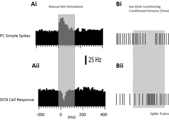
Figure 17: Sensory modulation of a DCN cell in an anesthetized rat.……….………...…….54
Figure 18: Stimulation of skin areas evoke very different responses in a DCN cell in vivo.……..…..55
Figure 19: PC projections to medial DCN obtained with two different injections.…….………...….59
Figure 20: Recording locations obtained after histological analysis.……….……….…………..60
Figure 21: Four channels tetrode recordings in vivo.……….………...…….61
Figure 22: Simultaneous Recording of a PC and a DCN in vivo.……….………....…….62
Figure 23: Hierarchical clustering tree…..……….………...……….64
Figure 24: Distribution of cells with respect to 1st and 2nd principal components...…...…….65
Figure 25: Contribution of waveform parameters to principal components…...…..…………. 65
Figure 26: Average waveforms of the two groups with standard deviations. …...………66 12
Figure 27. Comparison of Group1 / Group 2 DCN cells in vivo and non-GABAergic (GAD-) /
GABAergic (GAD+) DCN cells (Uusisaari et al., 2007) in vitro. …...……….67
Figure 28: Detection of oscillations in DCN…...………..………69
Figure 29: Phase locking to detected oscillations in DCN…...………..71
Figure 30: Protocols applied for optogenetic stimulation of PCs…...………...………72
Figure 31: Effect of PC stimulation on firing rate in DCN cells (Group1: red, Group 2: blue)…...….74
Figure 32: Time-locked spiking of DCN cells to fixed frequency stimulation of PCs (Cell A-B)…...76
Figure 33: Average power density at stimulation frequencies for Group 1 and Group 2….………….77
Figure 34. Rayleigh test for uniform distribution was rejected for time-locked cells………..….78
Figure 35: Different firing frequency ranges in DCN cell………...…………..80
Figure 36: Cross-correlation graphs for a Group1 cell- Group2 cell pair………..81
Table 1: Comparison of DCN cell groups obtained in vivo (present study) and in vitro (Uusisaari at al., 2007) ………..……….83
Figure 37: Effects of asynchronous and synchronous PC inputs in DCN……….88
Figure 38: Converging of several input in cerebellar nuclei………...……..….91
Figure 39: Unitary conductances for AMPA, NMDA and GABAA inputs………...………..….91
Figure 40: A speculative feed forward inhibition pathway in DCN………..93
Figure 41: Silicon probe structure with 16 channels………...………..….96
Figure 42: Spike Sorting with OpenElectrophy software………...…..….98
Figure 43: Parameters obtained from the average waveform in an example cell…….……….99
1 INTRODUCTION
1.1 A short history of cerebellar physiology and function
The first sources distinguishing the cerebellum from the rest of the brain date back to Aristotle (384-322 B.C.) and Erasistratus (304-250 B.C.). They used the name paracephalon (meaning “similar to brain”) without giving functional details (Malomo et al., 2006). Later, structure and role of the cerebellum was defined by Galen (129-201), stating that it is positioned behind the brain and gives rise to the cranial nerves and the spinal cord. In accordance with the dominant Greek theory of the time, Galen proposed that the cerebellum regulates the animal spirit which flows through the body. Due to the influence of this holistic approach, progresses in neuroanatomical investigations were delayed.
New methodological approaches were introduced by Andreas Vesalius and the structure of human cerebellum was presented in his book in 1543. Further advancements followed his work and, soon after, Costanzo Varolio introduced more details describing connections of the cerebellum to the brain stem. First, the cerebellar cortex and its possible links to spinal cord through white matter was noted by Marcello Malpighi (1665). Later, the deep cerebellar nuclei (DCN) were observed by Raymond de Vieussens as an ash gray area. Detailed descriptions of the lobules of the cerebellar cortex was given by Vincenzo Malacarne in 1776 in his book devoted to the cerebellum. With later contributions from his contemporaries, the gross anatomical description of the cerebellum was completed in the 19th century.
Hypothesis on the role of the cerebellum based on experimental findings also came in the 19th century. Luigi Rolando (1809, 1823) and Jean Pierre Flourens (1924) made successive observations following the ablation of the cerebellum: movements were still possible in general but they were not regular or coordinated, and sensory and cognitive functions were not affected. Later, Luigi Luciani (1891) compiled his observations of several symptoms due to cerebellar lesions and classified them as muscular weakness, lack of muscular tone and unstable muscular contraction. This classification was further developed by Joseph Babinski (1902) by identifying related impairments in rapid and complex
movements. With the reports of Gordon Holmes (1917, 1922) from many patients, the general implications of the cerebellum in human pathology were confirmed.
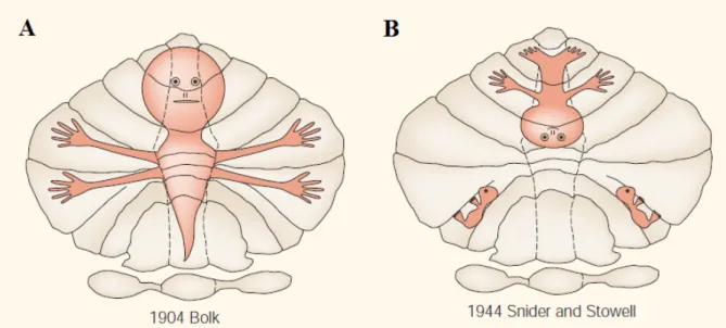
Figure 1: Early somatotopic maps in the cerebellum. Very simplified representations of body parts were given by Bolk (A), and Snider and Stowell (B), the first approaches within the historical context (adapted from Manni and Petrosini, 2004)
Further progress in deriving anatomical definitions of the cerebellum during this period was a different line of research. Lodewijk Bolk compared several mammals and described four regions: anterior and posterior vermis and two hemispheres having foliations. Together with Olof Larsell, they introduced most of the nomenclature that we use today. Bolk also proposed the first single somatotopic map on the surface of the cerebellum which represented different parts of the body (Figure 1 A) while the following studies challenged this idea with different maps. Snider and Stowell suggested an inverted mapping of gross body parts (Figure 1 B) and also bilateral afferents. Further information regarding historical development of functional localization in the cerebellum can be found in Manni & Pertosini (2004).
A clearer picture of the cerebellar macroscopic architecture was revealed through precise drawings of the cells and their connections by Santiago Ramon y Cajal (1911). With later advancements in microscopy techniques, Palay and Chan-Palay (1974) provided further information. Similarly, electrophysiological techniques were developed after the first electrical recording of cerebellar activity by Adrian (1935), and a functional architecture of cerebellar network was described by
Eccles, Ito and Szentagothai (1967) in their seminal book “The cerebellum as a Neuronal Machine”. A very influential model of cerebellar cortex was given by David Marr in 1969. He described the input layer of the cerebellar cortex (i.e. granular cell layer which will be described later) as a pattern discriminator where discriminated patterns are then learned by the Purkinje cell (PC) layer (also described later) as evidenced by long term plasticity. This model had been further improved by James Albus (1971) and soon after it was partially validated by Ito and Kano (1982) that discovered long term depression at the granule cell to PC connection. It was suggested that cerebellar computations are carried out in the DCN that integrate all the input from the cerebellar cortex into a relatively small volume (Ito, 1984). Up to now, our knowledge about cerebellar somatotopy and cellular populations has improved a lot, however, we are still missing an accurate description of computations carried out by the cerebellum. Notably, rules governing the integration of information in the DCN are poorly known, and this area of research will be my focus in following chapters of my thesis.
1.2 Functions of the cerebellum
The cerebellum collects sensory and motor information from the body via the brainstem and the spinal cord, and from the cerebral cortex. With this collection of information, the cerebellum coordinates and adjusts movements, maintains posture and balance. It also functions as an adaptive filter involved in the gain of some reflexes (for a review, see Dean et al., 2010). Similar computational mechanisms are employed in the cerebellum during voluntary and involuntary tasks for accurate timing of actions (for a review, see De Zeeuw et al., 2011). In the case of a voluntary movement, the cerebellum receives a copy of the efferent motor command (Von Holst, 1954; Wolpert et al., 1998). As the movement proceeds, the updated state of the whole body is continuously delivered from somatosensory, vestibular, visual and auditory systems to the cerebellum where they are integrated, and a prediction about the next sensory state during the movement is generated. The difference between predicted and actual sensory feedback is determined and motor errors are detected (Wolpert et al., 1998; Bastian, 2006). Such a “prediction – error control” mechanism sequentially checks whether the intended movement is correctly executed (Thach et al., 1992; Jueptner and Weiller, 1998).
Figure 2: Anatomical planes of the cerebellum. Representation of a mouse brain in anteroposterior, ventrodorsal, and mediolateral axes with the cerebellum (in blue) (A). Midline is given in dotted lines. Sagittal (B), Horizontal (C) and Coronal (D) planes are given with the cerebellar cortex (in blue) and the DCN (in gray). Medial (Med), Anterior Interposed (IntA), Posterior Interposed (IntP) and Lateral (Lat) nuclei are shown. Slicing orientations shown in the upper right corners.
There is also evidence suggesting that the cerebellum takes part in cognitive tasks as well, which previously were attributed only to cerebral cortex (Ivry et al., 1988; Ito, 2008). Consequently, it is proposed that the cerebellum functions as a co-processor, working in parallel with the rest of the brain (D’Angelo and Casali, 2012). According to this theory, the cerebellum can take part in many different
tasks, operating in loops with specific parts of the brain related not only to sensory-motor processing but also to cognitive functions.
1.3 Anatomical organization of the cerebellum
1.3.1 Gross anatomy of the cerebellum
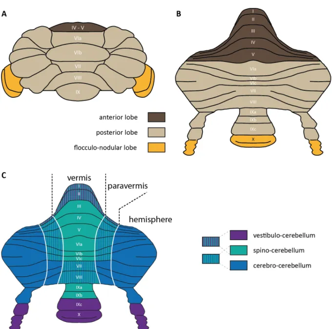
The cerebellum consists of a foliated cortex, with inner part containing mostly white matter that carries axons from/to the cortex, and three nuclei (DCN) that lie below the cortex inside the white matter bilaterally (Figure 2). The cerebellar cortex is divided into different numbers of lobules across species. In mammals, including humans, there are always 10 lobules that vary in size (Larsell, 1952). Lobules lie parallel to each other, extending from the vermis to the sides of the hemispheres (Figure 3 A). Lobules are separated from each other by superficial fissures, and two deep fissures group lobules into 3 lobes: anterior and posterior lobes are separated by the primary fissure while the secondary (posterolateral) fissure divides the flocculonodular lobe from the posterior lobe. Anterior lobe includes lobules I – V, posterior lobe includes lobules VI – IX, and flocculonodular lobe includes last lobule X (Figure 3 B).
The cerebellum has a symmetric structure along the midline: medial part of its cortex is called the vermis and the two lateral parts connected to the vermis are called the hemispheres (Figure 3 C). The hemispheres are also divided into two parts: the lateral hemisphere, and a more medial part, the paravermis (Figure 3 C). The DCN in rodents have 3 parts: medial, interposed and lateral nucleus. The cerebellum is connected to the brainstem with three pairs of cerebellar peduncles: namely, inferior, medial and superior cerebellar peduncles. The inferior cerebellar peduncle carries cerebellar afferents arriving from the spinal cord and medulla oblongata. The medial cerebellar peduncle, which lies rostrolateral to the inferior one, carries afferents arriving from the pontine nuclei. The last cerebellar peduncle, the superior one, lies medial to the inferior peduncle and carries cerebellar efferents from the DCN rostrally. Uncinate tract and bulbar connections transfer cerebellar efferents to the brainstem (Carleton and Carpenter, 1983).
Figure 3: General anatomy and functional divisions of the cerebellar cortex (A) Lobes and lobules in the anteroposterior segmentation (B) Cerebellar cortex unfolded in anteroposterior axis, showing lobule numberings (C) Mediolateral segmentation into vermis, paravermis and the hemisphere, together with functional divisions given in colors (Figure modified from J. Chaumont)
1.3.2 Gross functional divisions of the cerebellum
The architecture of the cerebellar cortex was considered isotropic across lobules. Same groups of cells are repeatedly located at their respective places in different layers of the cortex. However, some differences in cell structure and patterned molecular expressions have been recently reported (for a
review, see Cerminara et al., 2015). These differences highlight smaller regions within the cerebellar cortex that will be explained in the following parts of the thesis.
Inputs from different parts of the body divide the cerebellar cortex into bigger functional regions (Brodal, 2004) and this topographical organization of cerebellar inputs determines the functional divisions of the cerebellum. First, the flocculonodular lobe receives mostly primary vestibular afferents and projects to extracerebellar vestibular nuclei. This is the most primitive part of the cerebellum that controls eye movement and balance in higher vertebrates. Due to its function, it is called vestibulocerebellum (Figure 3 C, purple).
In the mediolateral classification, the vermis and the paravermis receive somatosensory inputs from the spinal cord and are therefore together functionally called spinocerebellum (Figure 3 C, green). Spinocerebellum also receives visual, auditory and vestibular inputs for the integration. Vermis receives input from the head and proximal parts of the body and controls posture, locomotion and gaze. Paravermis receives input from more distal parts of the body, the limbs and controls movements of these areas. The vermis and the paravermis mainly project to the medial and the interposed nuclei, respectively. The lateral parts of the cerebellar cortex, the hemispheres, are functionally called cerebrocerebellum since they receive information mainly from the cerebral cortex (Figure 3 C, blue). Cerebrocerebellum, which is more developed in humans and apes, is associated with the planning of motor actions. It projects back to the cerebral cortex via the lateral nuclei and the thalamus.
1.3.3 Cerebellar inputs
Proprioceptive and exteroceptive information together with the efferent copy of the motor commands are delivered to the cerebellum through two input pathways: 1) the climbing fibers and 2) the mossy fibers. Climbing fibers arrive solely from inferior olive, which is the largest nucleus situated in the medulla of the hindbrain. Mossy fibers, originate from several nuclei of the brainstem and the spinal cord which are commonly called precerebellar nuclei.
The inferior olive integrates sensory motor information from different parts of the brain like cerebral cortex, spinal cord and precerebellar nuclei, and generates climbing fibers that connects to the cerebellar cortex and to the DCN. It also receives feedback from the output of the cerebellum. Inferior olive is divided into three subnuclei: namely, principal olive, dorsal accessory olive, medial accessory olive, and four smaller subnuclei. The function of inferior olive is not clearly known, but current theories suggest that it takes part in movement error detection and movement timing control (De Zeeuw et al., 1998; Llinás et al., 2004; Mathy & Clark, 2013).
Numerous precerebellar nuclei target parts of the cerebellar cortex via the mossy fibers. Briefly flocculus receives inputs from the nucleus intercalatus, the medial vestibular nucleus, supragenual nucleus, nucleus prepositus hypoglossi, nucleus reticularis tegmenti pontis, and lateral reticular nucleus. Vermis and hemispheres receive connections from the pontine nucleus, lateral reticular nucleus, the nucleus intercalatus, nucleus reticularis tegmenti pontis, and external cuneate nucleus. There are also direct inputs that arrive from spinal cord that carry sensory information to cerebellar cortex without any relays.
1.3.4 Cerebellar outputs
The output gate of the cerebellum is the DCN. All processed information from the cerebellar cortex is sent to the DCN where it is integrated with collaterals of cerebellar inputs, mossy and climbing fibers. In addition to the DCN, a fourth nuclei, the vestibular nuclei, lie in the brainstem can be included in the output of the cerebellum. The vestibular nuclei seem to lie anatomically outside of the cerebellum, however, they receive similar inputs and generate an integrated output like the other cerebellar nuclei. The cerebellum projects to brain regions like the brainstem, diencephalon and the spinal cord. More specifically, the medial nuclei project mainly to the vestibular nuclei and also send projections to spinal cord, midbrain and thalamus. The interposed nuclei have a different main target, the red nucleus of the midbrain, while they also project to the vestibular nuclei and thalamus. Lastly, lateral nuclei predominantly project to thalamus by crossing the superior cerebellar peduncle. This connection takes part in the cerebellar thalamic cortical loop involved in error based control of complex movements. The cerebellum also sends feedback connections to inferior olive (for a review, see Brodal, 2004).
1.4 The cerebellar cortex
1.4.1 Information flow in the cerebellar cortex
The cerebellar cortex has a surprisingly uniform structure within each lobule in terms of cell structure and organization, as briefly mentioned earlier. It has been separated into three layers. The most inferior part is the granular layer which accommodates the most numerous cells of the brain, the granule cells. Within the granular layer there are also Golgi cells and other interneurons, namely, unipolar brush cells and Lugaro cells, all of which are much less numerous compared to granule cells. Mossy fibers terminate in this layer in a special structure called the glomerulus (Jakab and Hámori,
1988). Here, mossy fiber terminals, granule cell dendrites and the Golgi cell axon terminals make a ~10 μm spherical junction shielded by a glial sheet. Granule cell dendrites can be excited by mossy fiber terminals and inhibited by the Golgi cell axons (Figure 4 Ai).
The activation of Golgi cells, on the other hand, can happen through two direct synapses: 1) those from different mossy fibers (Kanichay and Silver, 2008) and/or 2) those from granule cells (Midtgaard, 1992; Dieudonne, 1998; Cesana et al., 2013). Consequently, Golgi cells can evoke feedforward and/or feedback inhibition onto granule cells; this inhibitory control shapes the excitatory input on granule cells in a glomerulus (Hamann et al., 2002).
Granule cells send their axons to the uppermost layer, the molecular layer, where they form parallel fibers and excite the PCs (Figure 4 Aii). A large information transfer happens in the molecular layer where a PC can receive inputs coming from about 175000 granule cells fibers (Napper and Harvey, 1988). In addition, there are around 10 molecular layer interneurons contacting each PC, and they provide feedforward inhibition on them (Eccles et al., 1967).
The second cerebellar input, the climbing fibers, directly target PCs and introduce a very strong excitation (Figure 4 B). PCs generate a complex spike which is formed of a spike and several spikelets. This input also affects interneurons through glutamate spillover in the molecular layer (Szapiro and Barbour, 2007). Therefore, the two excitatory inputs from mossy fibers and climbing fibers, and inhibitory inputs from molecular layer interneurons converge on the PCs and strongly modify ongoing PC activity according to their timings with respect to each other.
When climbing fiber and parallel fiber inputs arrive to a PC at the same time, parallel fiber-to-PC synapses are durably depressed (long term depression, LTD, Ito and Kano, 1982). The same synapses can go through a potentiation (long term potentiation, LTP) when parallel fiber inputs arrive without a climbing fiber input (Lev-Ram et al., 2002 and Coesmans et al., 2004). Consequently, there are LTD and LTP mechanisms mutually working at the granule cells to PC connections. Finally, PCs integrate excitation and inhibition that they receive, produce the sole output of the cerebellar cortex and send projections to the DCN.
The PC layer lies between the two layers described above, where PC somas position in one plane and define the layer. In total, there are seven types of neurons that I will briefly describe in the following paragraphs: the granule cells, Golgi cells, Lugaro cells and unipolar brush cells (in the granular layer), PCs (in PC layer), basket and stellate cells (in molecular layer).
Figure 4: Information flow in the cerebellar cortex (Ai) Mossy fiber inputs (yellow arrow) excite granule cells (GrC) and Golgi cells (GoC) within a glomerulus (black dashed area). Golgi cells provide feedforward and/or feedback inhibition to granule cells in this structure (Aii) Granule cell axons give parallel fibers (PF) to excite Purkinje cell (PC) dendrites which also receive feedforward inhibition from molecular layer interneurons, namely stellate cells (SC) and basket cells (BC) (B) Climbing fibers (CF, blue arrow) induce strong excitation in PCs and also modify plasticities of in the cerebellar cortex. PC output (red arrow) is generated by the combination of these inputs. (Figure modified from A. Valera)
1.4.2 Cellular structure of the cerebellar cortex
1.4.2.1 The Purkinje cell
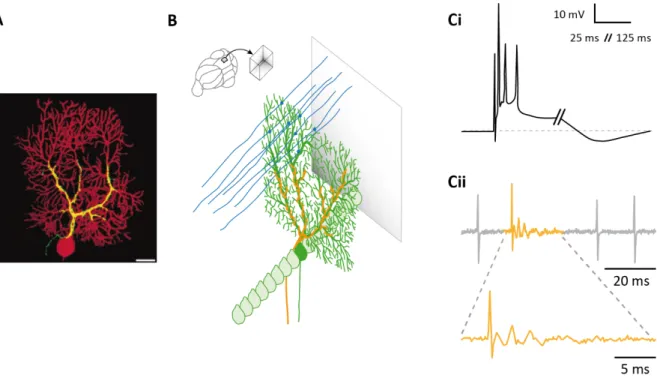
The Purkinje cell (PC) is the principal cell of the cerebellar cortex and it has been described by the physiologist Johannes Purkinje in 1837. PC dendrites make a planar arborisation in the molecular layer, where a single PC receives around 175000 excitatory synapses from parallel fibers (Napper and Harvey, 1988). Conversely, a single adult PC receives only one unique input from climber fibers. PCs’ big planar dendritic arborisation lays in a sagittal plane orthogonal to the plane of the parallel fibers (Figure 5 A-B). This dendritic arborisation generally has one primary trunk that forms secondary and tertiary dendrites with thinner branchlets, resembling in a tree structure. Parallel fibers contact PCs on dendritic spines located on branchlets, and each dendritic spine receives only one parallel fiber connection most of the time (Napper and Harvey, 1988; O’Brien and Unwin, 2006). Differently to a single contact made by many parallel fibers, climbing fibers make a single connection to each PC with hundreds of release sites that cover PC’s dendritic trunk (Palay and Chan-Palay, 1974) (Figure 5 B-yellow). This exceptionally strong input results in complex spikes formed by one spike and several spikelets (Figure 5 C).
PCs send their projections to the DCN and make GABAergic inhibition in a projection area containing several different cell types in the DCN. Projection areas of different PCs overlap (Trott et al. 1998a, 1998b; Panto et al. 2001) and, consequently, each nuclear cell receives inhibition from several PCs. This connection has high divergence and convergence ratios and will be further explained in the next chapter. PC axons also make collaterals, and can contact different cell types in the cerebellar cortex: other PCs (Palay and Chan-Palay, 1974; Bornschein et al., 2013), Lugaro cells (Palay and Palay, 1974; Hirono et al., 2012), molecular layer interneurons (Palay and Chan-Palay, 1974; O’Donoghue et al., 1989) and also Golgi cells (Palay and Chan-Chan-Palay, 1974; Hirono et al., 2012). However, the distribution of these collaterals has not been extensively described. Recent studies suggest that they are distributed mostly in the parasagittal bands and a few of them cross short distances laterally (Hawkes and Leclerc, 1989; Sugihara et al., 2009).
Figure 5: PC and its complex spike (A) Composite image of a climbing fiber (yellow) and a PC (red). Scale bar, 20 μm (adapted from Brenowitz and Regehr, 2012) (B) Dendritic arborisation of a PC (in green) in the sagittal plane, innervation of the parallel fibers (in blue) and the climbing fibers (in yellow) (Ci) Complex spike recorded in vitro current clamp mode (adapted from De Zeeuw et al., 2011) (Cii) Juxtacellular recording of simple spikes (gray) and a complex spike (yellow) of a PC (recorded by J. Chaumont)
1.4.2.2 The granule cell
The granule cells are the biggest number of cells in the brain, estimated roughly 100 billion in humans (Andersen et al., 1992). They fill up the volume between PC layer and the white matter in the cerebellar cortex. Granule cells are very small in somatic size, 4.82 μm on average (Harvey and Napper, 1991). This makes a very high density of granule cells in the most inferior layer, the input stage, of the cerebellar cortex (Harvey and Napper, 1988).
Granule cells have a few short dendrites that end as a dendritic claw, where they receive excitatory input from mossy fibers (Llinas et al., 2004) in the glomerulus. Unlike their dendrites, granule cells have long axons. They first ascend into the molecular layer of the cerebellar cortex and then bifurcate to give parallel fibers, expanding in both directions in the mediolateral axis. The ascending part and the parallel fibers both make synapses with PCs.
The ascending axon of the granule cells makes less synapses than the parallel fiber part of the same axon which is due to the difference in the length of these compartments. However, synapses are more densely found in the ascending axon than the parallel fibers. The percentage of the ascending axon
inputs per total granule cell inputs on PCs is estimated to be 7-24 % (Gundappa-Sulur et al., 1999), which actually makes more than 10000 synapses.
1.4.2.3 The Golgi cell
The Golgi cell is the second important cell in the granular layer. It receives excitation from mossy fibers and granule cells, and then it inhibits the granule cells in the glomerulus as described in the information flow.
The Golgi cell is named after the inventor of the silver nitrate method of staining, Camillo Golgi (1873). He described the morphology of the cells which was further extended by Ramon y Cajal (1911). Marr (1967) suggested that Golgi cells were driven by parallel fibers and mossy fibers. More detailed descriptions from Palay and Chan-Palay (1974) postulated that Golgi cells receive inputs not only from granule cells and mossy fibers but also from climbing fibers, PCs and molecular layer interneurons. The axonal part of the Golgi cell makes a big plexus, which extends in the sagittal plane and mediolateral plane. It sends projections to several glomeruli in its plexus. Between Golgi cells there are also gap junction connections that synchronize their activity (D’Angelo and De Zeeuw, 2009; Vervaeke et al., 2012; Szoboszlay et al., 2016) and impose a network inhibition on the excitatory inputs to the cerebellar cortex.
The last two cell types in the granular layer are Lugaro cells and unipolar brush cells are, and their properties are not extensively described (for more information, see Palay and Chan-Palay, 1974; Altman and Bayer, 1977; Geurts et al., 2001; Lainé and Axelrad, 2002; Mugnaini et al., 2011; Hirono et al., 2012).
1.4.2.4 Molecular layer interneurons
The upper layer of the cerebellar cortex is mostly filled with a huge dendritic tree structure arising from PCs in the parasagittal plane, and with long parallel fibers arising from granule cells that travel orthogonal to PCs’ dendrites. In between these structures, there are also molecular layer interneurons: stellate and basket cells. They locally inhibit PCs and other molecular interneurons in the same layer. Molecular layer interneurons are contacted and excited by parallel fibers, and they provide feedforward inhibition to PCs. They have dendrites in the parasagittal plane, like PC dendrites, but it is known that they can also inhibit neighboring PCs laterally that do not receive the same beam of parallel fibers (Eccles et al., 1967; Llinas and Sugimori, 1980; Cohen and Yarom, 2000).
The distinction between the two types of molecular layer interneurons, stellate and basket cells, is not totally clear and a smooth transition between the cell types was suggested by several studies (Sultan and Bower, 1998; Mittmann and Häusser, 2007; Schilling et al., 2008). However, differences in the short-term dynamics of parallel fiber connections to stellate and basket cells have been shown in Bao et al. (2010). In response to 50 Hz train stimulation of parallel fibers, synapses to stellate cells facilitate while synapses to basket cells were just slightly depressed. The two cell types differed in their respective locations as well: stellate cells were found to be located mostly on the superior part of the molecular layer, while basket cells were found mostly on the lower part. Basket cells have a special axonal extension which is found to cover the soma and initial part of PC axons. This special structure affects the spike initiation zone of PCs and it was hypothesized that it delivers electric field potentials to inhibit PC firing (Korn and Axelrad, 1980); this was further investigated and defined as ephaptic inhibition (Blot and Barbour, 2014).
1.4.3 Functional microzones in the cerebellar cortex
While the gross functional divisions of the cerebellum are determined by the coarse topography of cerebellar inputs, the precise synaptic arrangements in the cerebellum lead to a more specific functional organization similar to modular structures observed in the visual cortex (Callaway and Katz, 1993).
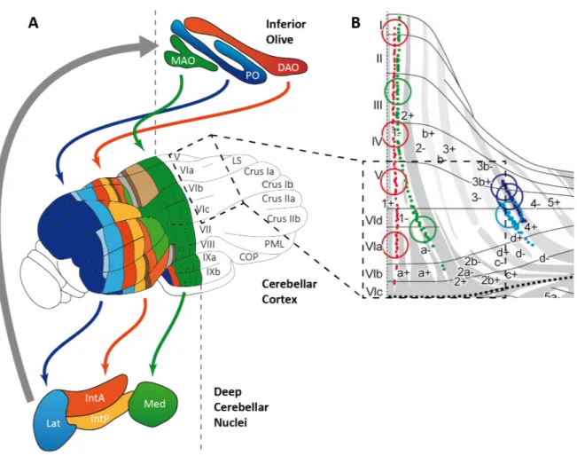
Modules in the cerebellum are distributed across the inferior olive, the cerebellar cortex and the DCN, and they function through the cooperation among these three regions. Boundaries of modules are determined by the projections between these regions, notably the climbing fibers contact PCs in precise parasagittal bands of cerebellar cortex matching with aldolase C compartments (Figure 6 B, small dots in color).
Since climbing fiber inputs are somatotopically organized, PCs in parasagittal bands share the same receptive fields. These bands can also be distinguished from each other by the expression of molecular marker zebrin II in PCs (Brochu et al., 1990) which represents the metabolic enzyme aldolase C (Figure 6 B white and gray areas, Ahn, 1994). PCs then project to specific regions in the DCN that generate the output of the cerebellum and also send feedback connections to the inferior olive. (Oscarsson, 1979; Sugihara and Shinoda, 2004; Voogd and Ruigrok, 2004; De Zeeuw et al., 2011). Consequently, a cerebellar module operates in an olivo-cortico-nuclear loop (Figure 6 A, Ruigrok, 2011), and its cortical element is called a microzone (for a review, see Apps and Hawkes, 2009).
Figure 6: Olivo-cortico-nuclear loop in the cerebellum. (A) An overview of connected regions between the inferior olive, the cerebellar cortex and the DCN. Regions in the inferior olive: DAO (Dorsal accessory olive), PO (Principal olive), MAO (Medial accessory olive) and regions in the DCN Med (Medial Nuclei), IntP (Interposed Nuclei – posterior), IntA (Interposed Nuclei – anterior) and Lat (Lateral Nuclei). (adapted from Apps and Hawkes, 2009) (B) Climbing fiber projections in an unfolded cerebellar cortex within the loop operation. Red, green, blue and violet dots each show climbing fibers distributions from tracing experiments of small injections of BDA (Biotinylated dextran amine) to different subnuclei of the inferior olive. (adapted from Sugihara and Shinoda 2004). In this network, mossy fiber and climbing fiber inputs that originate in the same body part partially match in microzones of the cerebellar cortex (Garwicz et al., 1998; Pijpers et al., 2006; Voogd et al., 2003) which suggests that microzones control a specific group of muscles (Apps and Garwicz, 2005; Thach et al., 1992). Mossy fiber inputs are relayed to local PCs through granule cells in the cortex (Ekerot and Jorntell, 2001; Isope and Barbour, 2002; Valera et al., 2016), however PCs of distant microzones are also targeted by the parallel fibers (Ekerot and Jorntell, 2003; Dean et al., 2010;
Valera et al., 2016). Consequently, local and/or distal granule cell inputs can be precisely selected in a PC through LTP and LTD mechanisms (Jörntell and Hansel, 2006; Gao, 2012b; Valera et al., 2016). Moreover, the recent study of our laboratory (Valera et al, 2016) focusing on lobules controlling locomotion discovered that the functional synaptic organization between microzones was conserved across animal. We therefore postulate that intermodular communication underlies the coordination between motor units.
1.4.4 Functional role of the cerebellar cortex and Purkinje cells
As mentioned in the very beginning, David Marr (1969) described the cerebellar cortex as a pattern discriminator, where PCs can learn the patterns of mossy fiber inputs that relay on granule cells. This theory was further improved by Albus (1971), Ito and Kano (1982). Now, I would like to introduce what information PCs transmit and how it is encoded. PCs discharge in correlation with different motor behaviors. For optokinetic reflex and vestibulo-ocular reflex in mice (De Zeeuw et al., 1995; Van Alphen et al., 2001; Katoh et al., 2015), and for reaching movements in monkey (Pasalar et al., 2006), detailed correlations of PC discharge with the motor activities were found. Similarly, the beginning of smooth pursuit eye movements were highly correlated with PC activity (Medina and Lisberger, 2007), and PCs, as a population, were found to encode temporal properties of saccade movements (Their et al., 2000; for a review see Ebner et al., 2011). In a recent study from Chen et al. (2015), it has been shown that specific groups of PCs represented the movements of whiskers in mice: a group of PCs increased their firing rate with whisker movements while another group of PCs decreased, and these changes were linear with the whisker position. Another encoding mechanism was also observed which was underlain by a synchronization of the activity of a group of PCs (De Zeeuw et al., 1997; Heck et al., 2007) under the control of climbing fiber afferents. Such examples show that PCs can encode several aspects of motor activities through modification of firing rates (rate coding) and/or specific discharge timings (temporal coding) together with possible synchrony in a group of PCs (Witter et al. 2011). The extent to which this information coding is preserved in the DCN depends on the details of the corticonuclear projections.
1.5 The deep cerebellar nuclei (DCN)
1.5.1 Anatomy of the DCN
The DCN lie in the inferior part of the cerebellum surrounded by the cerebellar cortex. As mentioned above, there are three nuclei: medial, interposed (anterior and posterior) and lateral nuclei, all having
bilateral pairs. They have a different nomenclature in humans: fastigial, emboliform, globose and dentate. The nuclei are located in the ventral part of the cerebellum where they are connected to the brainstem. PCs of the vestibulocerebellum project to another pair of nuclei outside of the cerebellum, the vestibular nuclei which is out of the scope of this study.
Anatomical properties of the DCN have been defined for different species. The medial nucleus (Med) is located next to the midline (Figure 7 A). A dorsolateral protuberance of medial nucleus (MedDL) is observed in rodents (Goodman et al. 1963). Interposed nucleus is separated in anterior (IntA) and posterior (IntP) parts. A similar dorsolateral hump of anterior interposed (dIH) nucleus is present next to lateral nucleus (Lat) in rodents (Voogd et al., 2013). In primates, lateral nucleus have a curved shape that extends in dorsomedial and ventrolateral directions. In humans, these two extensions are further developed with foliations and are called microgyric and macrogyric parts, respectively (Demole, 1927). In monkey and cat cerebellum, another group is recognized as Y nucleus which is the lateral extension of the vestibular nucleus and it was not defined in humans (Yamamoto et al. 1986, Stanton 1980). A group of cells, observed between medial and posterior interposed nuclei, can be grouped into the fifth nucleus defined as interstitial cell groups (iCG) which is present in rats (Buisseret-Delmas and Angaut, 1993) and other mammals (Voogd et al., 2013).
Figure 7: Anatomical structure of the DCN. (A) Division of cerebellar nuclei and (B) their three dimensional structure (adapted from Sugihara & Shinoda, 2007). Details are given in the text (adapted from J.Chaumont).
1.5.2 Functional role of the DCN
The final stage of cerebellar computation is carried out in the DCN. Excitatory connections of mossy fibers and climbing fibers plus inhibitory connections from a large number of PCs target the DCN (van der Want and Voogd, 1987; De Zeeuw et al., 1997, Ruigrok et al., 2015). Such a diverse input collection from the brainstem, the inferior olive, the spinal cord and the cerebellar cortex is combined and processed in the DCN. A specific study from Brooks and Cullen (2013) showed that the DCN
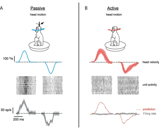
cells can differentiate an unexpected movement from a self-generated voluntary one. For an unexpected movement applied externally, the recorded DCN cell reliably represented the actual head velocity using sensory information (Figure 8 A, raster plots for ten trails and average firing rate). However, if the same movement was executed voluntarily, the incoming sensory information about the head velocity was cancelled with precision, and the same DCN cell did not respond at all (Figure 8 B, raster plots for ten trails and average firing rate). This precise cancellation shows the critical integration capabilities of the principal cells in the DCN where sensorimotor information is delicately processed. Distinguishing between unexpected and voluntary movements is one of the examples of the specific integration capabilities by the DCN.
Figure 8: DCN cell responses to passive and active movement. In vivo recordings from medial nucleus in monkeys revealed different responses to the same movement (head motion). (A) Passive movement (externally applied, unexpected) was reliably represented by firing rate of a DCN cell. Raster plots illustrating spiking activity in ten trials and average change in the firing rate is given. (B) When the same movement was active (self-generated, voluntary), head velocity was not represented in raster plots and firing rate of the DCN cell didn’t change. Shaded areas gives standard deviation between trials (adapted from Brooks and Cullen, 2013).
Previously, the DCN were thought to be a relay station, but two lines of research instigated a need to establish the functional role of the DCN. Firstly, an increasing body of studies describes the interplay between different inputs in the DCN (Ouardouz and Sastry, 2000; Gauck and Jaeger, 2003; Ohyama et al., 2003; Pugh and Raman, 2006; Person and Raman, 2010). Similarly to the example above, the DCN were shown to store some motor memories within their intrinsic connections (Chen et al., 1999; Wada et al., 2007). These studies suggested that information processing in the cerebellum is carried out not only in the cerebellar cortex but also in the DCN.
The second line of research that points to the information processing function of the DCN is a set of studies that describe different cell types. In a study from 2001, Czubayko and colleagues have characterized two types of the DCN neurons with respect to their intrinsic physiological properties. Later, studies by Uusisaari and colleagues (Uusisaari, et al., 2007; Uusisaari and Knöpfel, 2008; Uusisaari and Knöpfel, 2010; Uusisaari and Knöpfel, 2011), Bagnall and collegues (2009) characterized six different cells types in the DCN. After a recent study (Ankri et al., 2015), two of the proposed cell types have been joined, giving the result of five different DCN cell types described in
vitro.
These five cell types are the following: 1) large glutamatergic projection cells, 2) GABAergic and glycinergic nucleo-cortical cells with local collaterals, 3) non-inhibitory interneurons, 4) GABAergic nucleo-olivary cells, and 5) glycinergic premotor projection cells. Discussions are still open about cell types, since the specifications of different cells were given by in vitro studies mostly and not all of the cell types were identified in vivo. However, the observed varying intrinsic properties of these cells may have particular importance for the computations in the DCN. Especially, local inhibition in the DCN (Wassef et al., 1986; Husson et al., 2014) is very likely to modulate the output; this topic will be further explained.
A recent in vivo whole cell recording study from Canto et al. (2016) also compared DCN cells and I will elaborate on it in the discussion.
1.5.3 Cellular structure of the DCN
1.5.3.1 The principal projection cell
The principal projection cells are the biggest cells of the nuclei and are the best described amongst DCN cells. Being the principal cells of the nuclei, they received most of the attention. Making use of their big size, most of the in vitro studies had targeted specifically them over the other DCN cell
types. (Telgkamp and Raman, 2002; Pedroarena and Schwarz, 2003; Person and Raman, 2011). The principal projection cells have a special importance since they generate and convey the main output of the cerebellum to the brainstem, the spinal cord, several premotor areas and cortical areas via the thalamus in the brain (Asanuma et al., 1980; Sultan et al., 2012; Ruigrok and Teune, 2014).
The principal cells are glutamatergic projection cells with soma diameter of 20-35 µm. Uusisaari and Knöpfel (2013) suggested that they vary considerably in terms of morphology, and that further subtypes could be described. However, distinctive physiological properties have not yet been found to define the subgroups. These cells have big dendritic structure which extends to a big portion of each nucleus (Chan-Palay, 1973). Dendritic surface area was shown to be around 25000 – 50000 µm2, which accommodates a large number of synapses: estimated to be 850 – 1700 (Matsushita and Iwahori, 1971c; Bengtsson et al., 2011). It has been found that axon terminals from PCs and collaterals of climbing and mossy fibers converge onto these cells (Matsushita and Iwahori, 1971c), covering as high as 50 % of cell surface with synaptic buttons (Angaut and Sotelo, 1973). Uusisaari et al. (2011) suggested that cell bodies of the principal cells are mostly covered by GABAergic presynaptic terminals of PC axons. The convergence of mossy and climbing fiber inputs was shown solely for the principal cells, but their relative input strength is unknown (Uusisaari et al., 2011). A fourth afferent to the principal projection cell comes from another DCN cell: collaterals of the inhibitory nucleo-cortical cells (Ankri et al., 2015), with fewer connections to proximal and distal dendrites. It was shown that collaterals of the inhibitory nucleo-cortical cells had significant local inhibitory effect on the principal cells with mixed GABA/glycinergic transmission in vitro (Husson et al., 2014). Such intra-nuclear interactions are likely to have critical importance, especially for the principal projection cells that generate the output of the cerebellum (Wassef et al., 1986; Husson et al., 2014). Properties of local inhibition in DCN will be further investigated in the following chapters. In terms of output, the principal cells of the DCN were also found to project collaterals to the cerebellar cortex (Houck and Person, 2015). In a recent study, this circuitry was shown to take part in motor learning tasks in the cerebellum (Gao et al., 2016) by transmitting an efference copy of the cerebellar output back to the cerebellar cortex, which supports and enhances cerebellar learning. The principal projection cells of the DCN were shown to have short action-potential half-widths (Uusisaari et al. 2007: Large GAD - cells). A baseline firing rate of 29.9 ± 5.8 Hz (Uusisaari et al. 2007) was observed when recording in vitro at 24 °C, and much higher rates of 91.59 ± 7.4 Hz when recording at physiological temperature in vitro at 36 °C (Person and Raman, 2011a). These cells can keep their spontaneous firing even under high convergence of PC inhibitory axons, however
experiments performed at lower temperatures masked their firing capabilities (Person and Raman, 2011a). In vivo recordings show that the principal projection cells discharge around 60 Hz (Rowland and Jaeger 2005), and that this value can go up to 160 Hz for short periods during cerebellar behaviors (Thach, 1968). These principal projection cells were also observed to exhibit a weak frequency adaptation in response to direct current injections so that they can keep spiking at different frequencies sustainably and transfer information reliably (Uusisaari et al., 2013). Their intrinsic tendency to preserve spontaneous firing enables these neurons to respond to excitatory or inhibitory inputs by increase or decrease in their firing rates without being strongly affected by any of the inputs continuously. Considering together with the patterns in the spontaneous firing of PCs (Shin et al., 2007), the cerebellum can use temporal signatures to encode activity. Consequently, the output of the cerebellum is given by spontaneously active principal projection cells which delicately integrate inputs from four different sources, namely PCs, inhibitory local collaterals, climbing and mossy fibers.
1.5.3.2 The inhibitory nucleo-cortical cell
The inhibitory nucleo-cortical cells of the cerebellar nuclei were initially thought to be small in number (Matsushita and Iwahori 1971a, b), however now they are considered to be as numerous as the principal projection cells (Husson et al., 2014). They are spread out together with the other DCN cell types, with their small soma size (10-20 µm) and dendritic extensions of around 150 µm (Uusisaari and Knöpfel, 2013). They project to the cerebellar cortex and also have collaterals providing local inhibition (Matsushita and Iwahori, 1971c, Chan-Palay, 1973a). The inhibitory nucleo-cortical cells release both GABA and glycine (Husson et al., 2014).
Compared to the principal cells of the DCN, the inhibitory nucleo-cortical cells have significantly different action-potential waveforms. They have longer action-potential half-widths, ranging between 0.7 and 1.2 ms, while principal cells have half-widths <0.6 ms at room temperature (Uusisaari and Knöpfel, 2013). These two cell groups also have significantly different firing rates: the principal cells fire around 30 Hz, while the nucleo-cortical cells fire slower than 12 Hz in vitro at 24 °C (Uusisaari and Knöpfel, 2013). The observed lower firing rate is probably due to the intrinsic membrane properties: inhibitory nucleo-cortical cells have less Kv3.1b and Kv3.3 potassium channels at their somatodendritic membrane segments (Raman et al., 2000; Alonso-Espinaco et al., 2008) compared to the principal cells. This leads to weaker afterhyperpolarization (Uusisaari at al., 2007) and probably decreased recruitment of depolarizing currents. Difference in this mechanism probably have important contribution to the difference in the firing rates between these two DCN cell types.
Another observed difference between these two DCN cell types was the spiking in response to injected current. The principal cells can spike up to 200 Hz with increasing injected current steps, while inhibitory nucleo-cortical cells spike up to 50 Hz only. Their spiking frequency accommodation was also found to be high compared to the principal cells. When a step depolarization evoking 40 Hz spiking was applied to the two groups of cells, the principal cells kept spiking at 70 % of this frequency but inhibitory nucleo-cortical cells could only keep spiking at around 40 % of this frequency (Uusisaari and Knöpfel, 2011). Consequently, the inhibitory nucleo-cortical cells appear to have limited capabilities to spike sustainably above their baseline frequency, and they operate in a smaller range of frequencies compared to principal cells. This difference between the groups is likely to be important for the computation within the DCN through the local inhibition. Since inhibitory nucleo-cortical cells with local collaterals cannot keep spiking at high frequencies, local inhibition that they provide would also be limited and it would have a restricted effect on their targets, the principal cells of the DCN.
1.5.3.2.1 The local collaterals of inhibitory nucleo-cortical cell
In a study from Uusisaari et al. (2010), glycinergic neurons were targeted in the lateral DCN using transgenic mice that expressed a fluorescent protein under the control of the glycine transporter (GlyT) 2 gene (Zeilhofer et al., 2005). It was found that a portion of glycinergic cells were not spontaneously active (Glycinergic inactive (Gly-I): Uusisaari and Knöpfel, 2010), they have long axons that leave the DCN in slices. This portion of cells was observed to spike bursts of action potentials only, and cannot keep their spiking regularly. Being purely glycinergic, they were characterized as a separate population in the mouse lateral cerebellar nucleus. These purely glycinergic cells were reported to have shorter action potentials compared to mixed GABAergic/glycinergic type nuclear cells (Uusisaari and Knöpfel, 2010).
However, this distinction was challenged in a recent study were both GABAergic and glycinergic markers were complementarily used (Ankri et al., 2015). It has been found that the nucleo-cortical projections actually arrived from the mixed-type inhibitory interneurons (Husson et al. 2014) that were thought to be responsible for only local inhibition. Finally, two types of cells, which were defined differently as Gly-I (Uusisaari and Knöpfel, 2010) and the mixed GABA-glycinergic neurons (Husson et al., 2014), are now proposed to belong to the same type according to the latest study (Ankri et al., 2015). These cells were also found to target a specific subset of Golgi cells in the cerebellar cortex in the same study, therefore they were called inhibitory nucleo-cortical cells with the local collaterals.
1.5.3.3 The non-inhibitory interneurons
In addition to the inhibitory interneurons described above, the DCN are likely to contain some non-inhibitory interneurons that were classified as such because of their small soma size (15-25 µm) and lack of GABAergic transmission. Using the glutamate decarboxylase (GAD) 67-green fluorescent protein (GFP) knock-in mouse model, Uusisaari and colleagues (2007) defined them as GADnS (GAD negative Small) neurons, and findings regarding this group of neurons are restricted only to this study to the best of my knowledge.
The non-inhibitory interneurons (GADnS) were defined to be putatively glutamatergic and were distinguished from the principal projection cells (defined as large GAD negative cells (GADnL), Uusisaari at al., 2007) by having the membrane capacitance <100 pF. This distinction is somewhat arbitrary since the reason for choosing a 100 pF threshold is not clear. Uusisaari et al. used other electrophysiological, but not morphological, measures for this classification, especially for cells with a capacitance of approximately 100 pF. Such a post-hoc classification might be biased since the electrophysiological criteria were chosen arbitrarily. Consequently, the existence of a distinct group of GADnS, the non-inhibitory interneurons, should be evaluated more carefully.
GAD negative population (GADnL & GADnS, Uusisaari at al., 2007) showed two clusters for the distribution of membrane capacitance versus action-potential width, however this does not exclude the possibility of two different types of glutamatergic projection neurons that send axons to different extracerebellar targets. This possibility should be tested with tracing studies before establishing GADnS group as interneurons, since we still lack information about their axonal extensions, which might not be observed from cell fillings in slices.
They exhibit similar morphological properties with the inhibitory interneurons with small soma size and limited dendritic extensions. However their electrophysiological properties in vitro were distinct from the two groups described so far. They had medium duration action potential (AP) widths of 1.15 ± 0.18 ms which was between the short AP of the principal cells and the long AP of inhibitory interneurons. Their AP peak amplitude was the biggest among the three with 27.1 ± 1.1 mV where the inhibitory interneurons were the second with 21.0 ± 2.3 mV, and the principal cells had the smallest peak amplitude of 9.0 ± 4.4 mV. These two properties were found to be significantly different among the three groups in-vitro by Uusisaari at al. (2007), however they were not observed by another study so far.