HAL Id: tel-00119552
https://tel.archives-ouvertes.fr/tel-00119552
Submitted on 11 Dec 2006
HAL is a multi-disciplinary open access archive for the deposit and dissemination of sci-entific research documents, whether they are pub-lished or not. The documents may come from teaching and research institutions in France or abroad, or from public or private research centers.
L’archive ouverte pluridisciplinaire HAL, est destinée au dépôt et à la diffusion de documents scientifiques de niveau recherche, publiés ou non, émanant des établissements d’enseignement et de recherche français ou étrangers, des laboratoires publics ou privés.
Etude des interactions du photosensibilisant
méta-tétra(hydroxyphényl)chlorine avec les protéines de
plasma et les cellules
Siarhei Sasnouski
To cite this version:
Siarhei Sasnouski. Etude des interactions du photosensibilisant méta-tétra(hydroxyphényl)chlorine avec les protéines de plasma et les cellules. Pharmaceutical sciences. Université Henri Poincaré -Nancy I, 2006. English. �tel-00119552�
UNIVERSITE HENRI POINCARE-NANCY I
FACULTE DE MEDECINE
THESE
pour obtenir le grade de
DOCTEUR DE L’UNIVERSITE HENRI POINCARE-NANCY I
Discipline : Bio ingénierie
Présentée et soutenue publiquement
par Siarhei Sasnouski
Le 23 octobre 2006
Etude des interactions du photosensibilisant méta-tétra(hydroxyphényl)chlorine
avec les protéines de plasma et les cellules
---
Directeurs de Thèse : Dr. L. Bezdetnaya-Bolotine
Dr. V. Zorin
---
JURY
Jury :
Dr. L. Bezdetnaya-Bolotine (CAV CRAN UMR CNRS 7039)
Dr. V. Zorin (BSU, Minsk)
Pr. A. Fedulov (BSMU, Minsk)
Pr. F. Guillemin (CAV CRAN UMR CNRS 7039)
Rapporteurs :
Dr. S. MacRobert (UCL, Londres)
Dr. D. Brault (BIOMOCETI, UMR CNRS 7033, Paris)
SUMMARY
I GENERAL INTRODUCTION………...……….………...….5
II INTRODUCTION…..………..6
II.1. HISTORY AND CLINICAL APPLICATIONS OF PHOTODYNAMIC THERAPY…..6
II.2. PHOTOSENSITIZATION MECHANISMS………..9
2. 1. Pathways of molecular excitation and deactivation. ………..…9
2. 2. Mechanisms of photosensitized reactions. ………...…11
2.2.1. Types I photosensitization mechanism. ………..12
2.2.2. Types II photosensitization mechanism. ………13
2. 3. The properties of an ideal photosensitizer. ………...…17
2. 4. Tetraphenylchlorin series photosensitizers. ……….18
2. 4. 1. The 5,10,15,20-tetrakis(m-hydroxyphenyl)chlorin. ………….………20
2. 5. Cells and tissue damage effects of PDT. ………..…22
2. 5. 1. Vascular shutdown and inflammation. ……….23
2. 5. 2. Direct cell destruction. ………..23
II.3. PHOTOPHYSICAL AND PHOTOCHEMICAL PROPERTIES OF SENSITIZERS….24 3. 1. Photobleaching of sensitizers. ………..24
3. 1. 1. Parameters effecting photobleaching: aggregation state, pH, ionic strength and oxygen concentration. ………..28
3. 2. Effect of aggregation state on photophysical and photochemical properties of sensitizers. ………31
3. 3. Photophysical properties of porphyrinoid sensitizer non-covalently bound to proteins. ………32
3. 4. Electronic structure of porphyrinoid photosensitizers. ………35
II.4. PHOTOSENSITIZERS INTERACTIONS WITH PLASMA PROTEINS…………..…36
4. 1. Pharmacokinetics of sensitizers. ………..…37
4. 2. Kinetic and equilibrium characteristics of sensitizers interactions with proteins.39 4. 2. 1. Mechanisms of sensitizers redistribution between plasma proteins… .40 4. 2. 1. 1. Collision mechanism. ………....…40
4. 2. 1. 2. Redistribution through the aqueous phase………..41
4. 2. 2. Thermodynamics of sensitizers redistribution between plasma proteins. Eyring theory. ………..……….44
5. 1. Techniques to study sensitizer intracellular localisation and aggregation state…47
5. 1. 1. Confocal laser scanning fluorescence microscopy. ………..…48
5. 1. 2. Fluorescence lifetime imaging microscopy (FLIM). ………49
5. 2. Sub-cellular localisation and dynamics of sensitizers during PDT. ……….51
5. 2. 1. Sites of sub-cellular localization of hydrophilic and hydrophobic sensitizers. ………52
5. 2. 2. Relocalisation of sensitizers upon irradiation. ………..54
III OBJECTIVES ………...…………..……….55
IV RESULTS………..…….………...………57
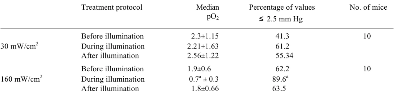
IV. 1. MTHPC-based photodynamic treatment in vivo. ………..57
IV. 2. Investigation of mTHPC interactions with plasma proteins. ……….65
IV. 3. Redistribution of mTHPC from plasma proteins to model membranes………...75
IV.4. Calculation of quantum yield of MCF-7 cells inactivation by mTHPC-PDT: influence of incubation time and sensitizers localization. ………....85
IV.5. Theoretical and experimental study of the effects of solvent on the electronic structure of tetrapyrrole compounds: application for the determination of the structure of aggregates.……….……….………106
V GENERAL DISCUSSION.……….……….120
VI CONCLUSION AND PERSPECTIVES………...128
REFERENCES………...………...130 ANNEXES……….….148 French summary………..148 Abbreviations………..161 Scientific works………...162 ACKNOWLEDGMENTS………....164
I GENERAL INTRODUCTION
Photodynamic therapy (PDT) has been developed as a treatment modality for a number of malignant and non-malignant disorders. PDT treatment is based on the presence of a drug with photosensitising and tumour localizing properties combined with visible light and oxygen. Separately, these three components are harmless, but in combination they may destroy tissue and inactivate cells.
It was shown that direct injection of sensitizers into the tumor is ineffective. Hence, the transport of photosensitizers (PSs) in the blood after intravenous injection seems to influence the photodynamic efficiency. In aqueous media like blood plasma, most of the tetrapyrrolic PSs form dimers and higher aggregates and as such are ineffective in producing singlet oxygen, thus resulting in a drop of their photosensitizing efficiency. Both aggregation and disaggregation of porphyrins occur in the blood circulation, and the competition between these processes could affect the in vivo PDT efficacy.
The PSs accumulation in cells can be realized by passive diffusion through plasmatic membrane or by various types of endocytosis. During interactions with plasma proteins hydrophobic sensitizers dissociate from an aggregate and bind to protein molecules. The type of protein-carrier governs the delivery of sensitizer to the tumor. In vivo transport of hydrophobic porphyrinoid derivatives is carried out by lipoproteins. Serum albumin serves as a carrier for amphiphilic and hydrophilic photosensitizers. The nature of the carrier protein also affects the drug localisation in the tumor with albumin primarily delivering bound drugs to the vascular stroma, while lipoproteins internalize sensitizers in malignant cells. Plasma proteins binding affinity for various photosensitizers can play an important role in drug distribution and photodynamic efficacy.
Accurate dosimetry is necessary to ensure complete treatment and to allow for consistent and reproducible patient outcome. It is accepted that the phototherapeutic effect of PDT is, in most cases, a result of singlet oxygen generation during activation of photosensitizer by light. The objective of PDT is to deliver a cytotoxic species dose that is sufficient to kill the malignant cells in a tumour. Dynamic variations and interrelationship of several parameters of PDT treatment, such as photosensitizer concentration, localization, photo-stability and aggregation state, optical properties of the tissue, characteristics of irradiation, make the treatment very complex. Therefore understanding of the influence of
these parameters on photodynamic toxicity may provide valuable information for optimization of the PDT treatment protocols.
Meta-tetra(hydroxyphenyl)chlorin (mTHPC) or Foscan® is a second-generation photosensitizer and is one of the most effective sensitizers studied to date. mTHPC has been granted European approval for palliative treatment of patients with advanced head and neck cancers and undergoes clinical open-label multicenter studies for the treatment of early squamous cell carcinoma. It is about two orders of magnitude more active compared to Photofrin.
The first objective of the present work was the study of the correlation between mTHPC-PDT efficiency and its biodistribution as a function of time. In a second part, we examined influence of the aggregation state of the photosensitizer on its interactions with plasma proteins. In a third part, we studied the kinetic characteristics and mechanism of sensitizer redistribution from the complexes with plasma proteins. The fourth part of the work consists of the assessment of mTHPC-PDT dosimetry and phototoxicity in vitro. The fifth part of the work was the study of electronic properties of sensitizer using Huckel-based quantum mechanical model of Van der Waals interactions and determination of mTHPC aggregates structure.
II INTRODUCTION
II.1. History and clinical applications of Photodynamic Therapy
Light has been employed in the treatment of disease since antiquity. Phototherapy has been applied by humans for 3000 years and was known by the Egyptians, the Indians and the Chinese (Spikes 1985). Herodotus (6C BC) is recorded as noticing the beneficial effect of sunlight on bone growth, and the eminent Hippocrates (460-375 BC) recommended the use of heliotherapy for various human diseases. But the first relevant “modern” scientist in the field of phototherapy was Niels Rydberg Finsen. From 1895 until 1903 he performed phototherapy on 800 patients, and in 1903 he was awarded the Nobel Prize for Physiology-Medicine for his work on the use of light from the carbon arc in the treatment of lupus vulgaris (skin tuberculosis) (Szeimies 2001). The concept of cell death being induced by the interaction of light and chemicals has first been reported by a German medical student Oscar Raab. In the winter semester of 1897-1898 he started an investigation on the toxicity of acridine to paramecia. This work was carried out under the direction of Professor Dr. Hermann von Tappeiner. Initially, Raab found that the apparent toxicity of low concentrations of acridine
varied significantly from day to day. However, he soon noted that the toxicity depended on the intensity of sunlight in the laboratory. He was then able to show that low concentration of acridine and some other colored dyes such as eosin, that had no effect in the dark, provoked the rapid killing of paramecia in the presence of light (Raab 1900). In 1902, C. Ledoux-Lebards observed that eosin killed paramecia more efficiently in open flask than in a closed bottle (Ledoux-Lebards 1902), and he postulated that the presence of oxygen is essential for photoinactivation. It is in 1904 that von Tappeiner and Jodlbauer coined the term “photodynamische Wirkung“ (von Tappeiner and Jodlbauer 1904) which we translate as “photodynamic action” for oxygen-requiring photosensitized reactions in biological systems. Although the mechanism of action was still unknown, it did not take long until this new therapeutic approach was tried out on patients. The first paper reporting a clinical trial was published in November 1903 by von Tappeiner and Jesionek (von Tappeiner and Jesionek 1903). The photosensitizers used so far were dyes like chinidine, acridine and eosin, and further studies were devoted to develop new clinically relevant photosensitizers.
In 1911, Walter Hausmann injected 2 mg hematoporphyrin subcutaneously in mice, which were exposed to sunlight and he observed edema, erythema and skin necrosis (Haussman 1911). The first report on the use of hematoporphyrin in humans was done by Meyer-Betz who injected himself with 200 mg hematoporphyrin and became extremely photosensitive during more than two months (Meyer-Betz 1913). Accumulation and retention of hematoporphyrin in human neoplastic tissue was evidenced by Auler and Banzer in 1942 (Auler and Banzer 1942). Interrupted by the Second World War clinical studies on photodynamic treatment were not performed in a major organized way until the middle 70’s, largely through the efforts of Dougherty.
Photodynamic therapy uses the combination of a photosensitizing drug and light to cause selective damage to the target tissue. Firstly, the sensitizer is injected into the bloodstream and it begins to redistribute to cells throughout the body. After certain period, when sensitizer retention in the tumor becomes greater than in normal tissue, the tumor region is illuminated with a light source with appropriate emission wavelength. Absorption of this light by tumor-localized sensitizer leads to generation of toxic free radicals and finally to destruction of malignant tissue (Henderson and Dougherty 1992). Tumor destruction can be realized both by direct cells killing or by photodamage of the tumor vasculature resulting in local hypoxia and indirect cells killing (Dougherty et al. 1998). Within a few hours after PDT tumor tissue exhibits extensive regions of necrosis and apoptosis. During the first 24 h the treated area shows evidence of swelling, infiltration of inflammatory cells and tissue
breakdown (Dougherty et al. 1998). After PDT treatment a large number of cytokines and inflammatory mediators are released (Gollnick et al. 1997). The enhanced immune response in the tumor area is necessary for complete elimination of the tumor tissue (Korbelik and Dougherty 1999).
Some advantages of PDT over other techniques include some degree of selectivity of PS binding to tumor tissue, the absence of systemic toxicity of the drug alone, the ability to focus the light on the tumor region. Moreover, the treatment can be repeated multiple times safely and can be used after surgery, chemotherapy or radiotherapy. PDT can induce a long-term anti-tumor immunity, a relatively unique response among anticancer therapies (MacDonald et al. 1999). As most of PSs are fluorescent the imaging and detection strategies can be applied in PDT protocols, known as photodetection or photodiagnosis. They may be used to detect otherwise hidden disease such as dysplasia, to delineate tumor borders, or to visualize disease in inaccessible areas such as the esophagus, bronchus or colon. Another application of fluorescent imaging and quantification is its ability to improve PDT dosimetry by measuring the amount of PS in the lesion before applying the appropriate illumination parameters. Among disadvantages of PDT are the prolonged skin photosensitivity, limited depth of light penetration (< 1 cm) and the possibility to treat only localized superficial tumors. The improved understanding of the tissue and cellular factors that control PDT and increased experience in the clinic has led to much larger, better-controlled clinical trials and the approval of drugs makes PDT a clinical reality.
Photofrin® was the first approved in 1993 in Canada, now approved in more than 40 countries (1995 approval in USA, Canada, Japan and Europe) for advanced and early lung cancer, superficial gastric cancer, oesophageal adenocarcinoma, cervical cancer, and bladder cancer. Then the Levulan® got the FDA approval in 1999 for actinic keratosis, followed in 2001 by mTHPC (approved for advanced head and neck cancer, Europe, Norway and Iceland). Currently, two derivatives of 5-ALA, methylaminolevulinate (MAL) and hexylaminolevulinate (HAL), gained marketing authorization from the regulatory offices in Europe and Australia. MAL is marketed under the trade name Metvix® for the treatment of actinic keratosis (AK) and difficult-to-treat basal cell carcinoma (BCC), HAL has recently been launched under the trade name Hexvix® for the improved diagnosis of superficial bladder cancer in Europe. PDT has also indications for non-oncological diseases, such as wet age related macular degeneration using benzoporphyrin derivative (Visudyne®, FDA and European approval in 2000). Also a number of other conditions have also been treated
including psoriasis, rheumatoid arthritis, menorrhagia and benign prostatic hyperplasia. In addition, PDT-mediated immune-modulation, bone marrow purging and PDT of certain bacterial, fungal and viral infections are being evaluated.
II.2. Photosensitization mechanisms
2.1. Pathway of molecular excitation and deactivation
The absorption of light by a chromophore is the initial step in all photophysical and photochemical reactions. The energy of the absorbed light promotes molecules from their ground state to states of higher energy (excited states). At room temperature, almost all the molecules are in their ground state S0, which is the electronic state associated with the lowest energy and a configuration where all electrons are orbitally paired. During an electronic transition one of the electrons is excited from an initially occupied orbital of low energy to a previously unoccupied orbital of higher energy. The molecule undergoes transition from its ground state S0 to an excited state S1.
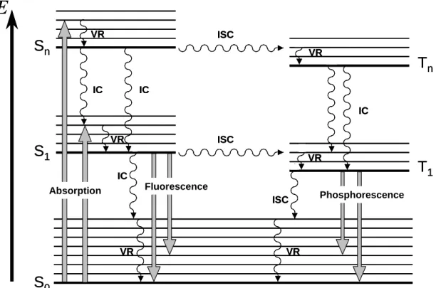
The excited state S1 is energetically less preferable than S0. Several physical pathways, leading to deactivation of excited state can be followed, represented in the Jablonski diagram (Fig. 2.1). A molecule in a high vibrational level of the excited electronic state Sn quickly falls to the lowest vibrational level (Vibrational Relaxation VR). Also, a molecule in a higher excited state Sn can fall to the first excited singlet state S1 (Internal Conversion IC). Then, the singlet state S1 can rapidly return to the ground state level S0 by two mechanisms: a radiative process emitting a quantum of fluorescence or a non-radiative IC with dissipating the excitation energy into the heat (Table 2.1). Owing to IC and VR procceses, photons of fluorescence are generally emitted from the lowest vibrational sublevel of the excited singlet state (S1) level. This implies that the form of fluorescence spectrum does not depend on the excitation wavelength (Vavilov’s rule). Emitted photons have lower energy than absorbed photons, so fluorescence emission maximum is red-shifted as compared to the absorption maximum (λemission > λabsorption, Stokes-Lommel’s law).
Figure 2.1 : Jablonski diagram, where IC stands for internal conversion, ICS for intersystem
crossing and VR, for vibrational relaxation.
S
0S
1S
nT
1T
nE
Absorption IC Fluorescence Phosphorescence IC IC IC ISC ISC VR VR VR VR VR VR ISCS
0S
1S
nT
1T
nE
Absorption IC Fluorescence Phosphorescence IC IC IC ISC ISC VR VR VR VR VR VR ISCIn addition to radiationless and radiative process, sensitizer molecule from the first exited singlet state can undergo a transition to a triplet state T1 via intersystem crossing (ISC). The lifetime of the triplet state is much longer (τ ~10-3 - 10-7 s) than the lifetime of the singlet state (τ ~10-10 s), thus increasing dramatically the probability of interactions of neighbouring molecule with sensitizer in its triplet state. There are several pathways for the triplet state T1 to return to a ground state S0. De-excitation can occur with the emission of a photon, called phosphorescence, but at room temperature and due to Vavilov’s rule phosphorescence intensity is weak and difficult to detect. The excited triplet state T1 can be alternatively deactivated by undergoing intersystem crossing followed by vibrational relaxation (Fig. 2.1).
For most of the organic molecules, only the singlet state S1 and triplet state T1 of lowest energy can be considered as likely candidates for the initiation of photochemical and photophysical reactions. This is due to the fact that higher order electronic state (n ≥ 2) undergos very rapidly internal conversion from Sn to S1 and from Tn to T1. This generalization (which was used here in the description of the Jablonski diagram Fig. 2.1) is known as Kasha’s rule.
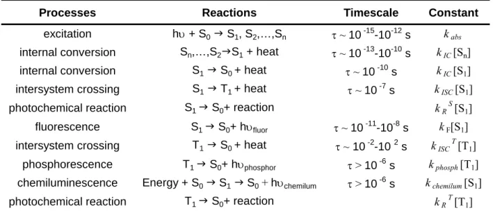
Table 2.1. : Photochemical processes involved in the activation and deactivation pathway of the
photosensitizers and some of their characteristics
Processes Reactions Timescale Constant
excitation hυ + S0 J S1, S2,…,Sn τ ~ 10 -15-10-12 s kabs internal conversion Sn,…,S2JS1 + heat τ ~ 10 -13-10-10 s kIC[Sn]
internal conversion S1 J S0 + heat τ ~ 10 -10
s kIC[S1]
intersystem crossing S1 J T1 + heat τ ~ 10 -7 s kISC[S1]
photochemical reaction S1 J S0+ reaction kRS[S1]
fluorescence S1 J S0+ hυfluor τ ~ 10 -11-10-8 s kF[S1]
intersystem crossing T1 J S0 + heat τ ~ 10 -2
-10 2 s kISCT[T1]
phosphorescence T1 J S0+ hυphosphor τ > 10 -6 s kphosph[T1]
chemiluminescence Energy + S0 J S1 J S0 + hυchemilum τ > 10 -6 s kchemilum[S1]
photochemical reaction T1 J S0+ reaction kRT[T1]
2.2 Mechanism of photosensitized reactions
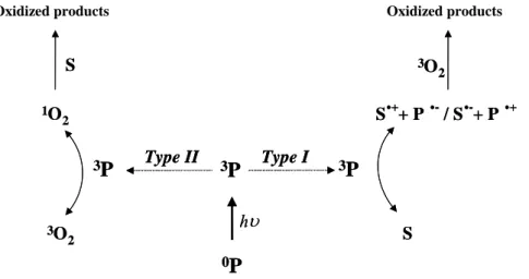
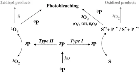
Photosensitized reaction can be defined as a process in which light activation of a chromophore induces chemical changes in another molecule than chromophore. The initial step of the reaction is the absorption of a photon by the photosensitizer, leading to the generation of molecules in excited triplet states (3P*). The reaction can follow two competing pathways called Type I and Type II reactions (Sharman et al. 2000). According to the definition established by Foote (Foote 1991) and as shown in Fig. 2.2, a Type I mechanism involves the direct interaction of 3P* with a substrate (S), whereas in a type II process, 3P* reacts first with molecular oxygen to produce highly reactive oxygen intermediate that easily initiates further reactions.
3P 1O 2 3O 2 S S.++ P .-/ S.-+ P .+ hυ 3O 2 Type I Type II 0P 3P 3P S 3P 1O 2 3O 2 S S.++ P .-/ S.-+ P .+ hυ 3O 2 Type I Type II 0P 3P 3P S Oxidized products
Figure 2.2. : Diagram of photosensitizations mechanisms occurring after absorption
of a photon by photosensitizer.
Oxidized products
2.2.1. Type I photosensitization processes
In a type I photochemical reaction, the exited triplet state of the photosensitizer (3P*) interacts directly with the substrate molecule (S) and leads to the formation of pairs of neutral radicals or radical ions following an electron or hydrogen transfer as shown in the Eqs 1 and 2 :
Both the excited photosensitizer and the ground state substrate can act as hydrogen donor or acceptor (Eq. 3-4). 3P* + S P·- + S·+ (1) 3P* + S P·+ + S·- (2) 3PH* + S P· + SH· (3) 3P* + SH PH· + S· (4)
The resulting radical species from Type I primary processes can subsequently participate in different kinds of reactions. In the presence of oxygen, for example, oxidized forms of the sensitizer or of the substrate readily react with O2 to give peroxyl radicals, thus initiating a radical chain auto-oxidation (Eqs 5 and 6).
S·+ O2 SOO· (5) SOO·+ SH S· + SOOH (6)
Semireduced forms of the photosensitizer or of the substrate also interact efficiently with oxygen and the electron transfer, which takes place between reactants, generate superoxide radical anion (eq 7).
S·-+ O2 S + O2·-
(7) P·-+ O2 P + O2·-
Any reaction that generates O2·- will also produce hydroperoxide H2O2 by spontaneous dismutation (eq 8) or one-electron reduction (eq 9).
O2·-+ O2·-+ 2H+ O2 + H2O2 (8) O2·-+ 2H+ + e- H2O2 (9)
Hydroperoxide is a moderate oxidant, but when it accumulates, it can react with superoxide radical anion (eq 10) or undergo ferrous ion catalysed reduction to give rise to an extremely reactive hydroxyl radical (Haber-Weiss reaction)(eqs 11 and 12).
O2·-+ H2O2 O2 + OH- + ·OH (10) O2·-+ Fe3+ O2 + Fe2+ (11)
Haber-Weiss reaction
2.2.2. Type II photosensitization processes
H2O2 + Fe2+ OH- + ·OH + Fe3+ (12)
This type of reaction requires the presence of molecular oxygen. In most cases, the reaction proceeds via non-radiative energy transfer from the excited triplet state
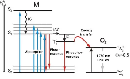
photosensitizer to the oxygen molecule in its triplet state. Singlet oxygen can only be generated by photosensitizer that has an energy gap between the ground state and the excited triplet state higher than the energy ΔE needed to excite oxygen into its singlet state (Fig. 2.3).
ΔE being very low (94 kJ mol-1 (van Lier and Spikes 1989), almost all the tetrapyrrolic
photosensitizers can mediate generation of singlet oxygen.
1270 nm 0.98 eV Energy transfer Phosphor-escence Fluor-escence Absorption 1270 nm 0.98 eV Energy transfer Phosphor-escence Fluor-escence Absorption
Due to the higher lifetime of triplet compared to the singlet state of porphyrin-like photose
(13)
wo forms of singlet oxygen with different excited state energies are generated: 1O2 (1∆g , E =
of xanthene dyes even in oxygen-saturated solutions (Gollnick et al. 1992). Figure 2.3. : Simplified Jablonski diagram, showing the
activation and deactivation pathways during a Type II reaction.
nsitizers, photochemical reactions most likely occur with sensitizer in its triplet state. Energy transfer from the excited triplet state of the sensitizer to the ground state (triplet) oxygen is a spin allowed process and the molecule of oxygen undergoes transition from its ground triplet into excited (singlet) state:
h 3 1
2 2
P
⎯⎯→
νP + O
→
P + O
T
94 kJ mol-1) and 1O2 (1∑g , E = 157 kJ mol-1) (Lang et al. 1998). But 1O2 (1∑g) form is rapidly transformed into 1O2 (1∆g) with almost unit efficiency. It was shown through a theoretical estimation based on oxygen diffusion in aqueous solution, that the lifetime of the intermediate to be attacked by oxygen must be at least 10-6 s (Imamura and Koizumi 1955). More recently, it was found that oxygen exerted no measurable effect on the short-lived excited singlet state
Quantum yield of singlet oxygen formation is defined as the ratio of triplet state deactivation rate, that leads to energy transfer to oxygen molecule and the sum of all deactivation rates, leading to triplet state deactivation, like phosphorescence and ISC. For pure Type II reaction, the quantum yield of singlet oxygen formation can be defined as (for constant description see Table 2.1.) :
T R 1 T T
k [T ] [S]
=
k
[T ] + k
ΔΦ
phosph 1 ISC[T ] + k [T ] [S]
1 R 1 (14)Data on 1O2 generation quantum yields in different medi in
Table 2.2.
yields of 1O2 (1∆g) generation (ФΔ) by sensitizers.
Reference
a for some sensitizers are given
Table 2.2. Quantum
Photosensitizer ФΔ Medium Method
HP 0.53 methanol 1270 nm luminescence (Chacon et al. 1988) PPIX 0.56 PB/TX100 lysozyme sensitization (Fernandez et al. 1997) BPD-MA 0.77 benzene 1270 nm luminescence (Aveline et al. 1994) AlPcS4 0.38 PB/TX100 lysozyme sensitization (Fernandez et al. 1997) mTHPP 0.46 air-saturated methanol 1270 nm luminescence (Bonnett et al. 2001) mTHPC 0.43 air-saturated methanol 1270 nm luminescence (Bonnett et al. 2001) mTHPBC 0.43 air-saturated methanol 1270 nm luminescence (Bonnett et al. 2001)
In the pres of o que iple
ang et al. 1998):
where is the sum of all triplet states deactivation rate constants in the absence of oxygen,
is the bimolecular rate constant of triplet states quenching by oxygen, [O2] is the
value of is sensitive to small amounts of oxygen in the system and can be used as a
o:
ence xygen the observed nching rate of PS tr t states is given by
(L T
k
+ k [O ]
(15) ob decay q 2k =
T decay k q kconcentration of oxygen. As diffusion controlled value of is about 109 – 1010 M-1s-1 the
T decay
direct measure of the oxygen.
The quantum yield of singlet oxygen formation Ф
q
k k
Δ depends on the quantum yield of the triplet states ФT according t
T
=
S S
q Δ ΔΦ
Φ
Δ 1O2 and is given by (16)where S is the fraction of triplet molecules quenched by oxygen and yielding
et
k
=
qS
k
Δ (17)where ket the rate constant of energy transfer leading to the formation of 1O2 and Sq is the action of oxygen dependent triplet deactivations
fr
[ ]
[ ]
q 2 T decay q 2k O
S
k
+ k O
Δ=
(18)The denominator represents all pathways of triplet deact or many porphyrins the
value of SΔ is about 0.75 (Keene et al. 1986).
otoxicity (Aveline et al. 1998). The authors obser
concentratio 15). The values of = 6.6 x 10 and = 9 x 10 (corresponding
.
ng.
06 M-1s-1 (Ave
ivations. F
Laser flash photolysis studies in vitro showed that oxygen and local PS concentrations influence the reaction mechanism and phot
ved the reduction of fluorescence and IC yields on increasing of the photosensitizer concentration. This phenomenon was explained as PS association leading to self-quenching of the triplet state. To obtain the values of T
decay
k and kq constants the observed
rate constant k of PS triplet states quenching is measured as a function of oxygen ob
T decay
8
q
3
to triplet state lifetime 110 µs) were observer for deuteroporphyrin (DP) in L1210 cells (Aveline et al. 1998). The comparable value of kT = 5 5
x 10
n (eq. k M-1s-1 k
decay
8 M-1s-1 was reported for zinc phthalocyanine in vitro (Firey et al. 1988). These values are significantly less than that normally found for oxygen quenching of such triplet states in aqueous solution kT = 1.85
x
10
decay
9 M-1s-1 (Reddi et al. 1983). This can be explained by lower oxygen content in membrane compared to solution, by lowered diffusion rate of reactants and PS protein bindi
A plot of initial triplet state decay rate constant as a function of sensitizer concentration gave the value of rate constant of triplet state quenching by ground state DP k = 1S
line et al. 1998). Low value of triplet self-quenching constant k compared to oxygen S
quenching constant T means that the self-quenching can compete with the quenching of
decay
triplet state by oxygen at high local PS concentration and such competition can exist in vitro as local PS content in lipid bilayer can reach an order of mM.
Moreover, the good correlation between calculated SΔ values (eq. 18) and cellular photo
ground state
. 3. The properties of an ideal sensitizer
) has been for a very long time the only photose
photodynamic activity is acceptable, it is still modest. The selectivity for the tumour versus toxicity with different oxygen levels was observed for DP proving that cell killing is due to singlet oxygen formation. Study of photophysical parameters of PSs in biosystems can give valuable information about reaction mechanism and sensitizer state and environment.
Singlet oxygen is a very reactive molecule. It is much more electrophilic than its
and can rapidly oxidize biomolecules. It is a metastable species with a lifetime varying from about 4 µs in water to 25-100 µs in non polar organic solutions (Kohen et al. 1995). The life time of singlet oxygen decreases in biological environment due to the presence of various quenchers, and is calculated to be about 170-330 ns (Baker and Kanofsky 1992). According to Moan and coworkers, this short lifetime allows the diffusion of singlet oxygen to a maximum distance of 50 nm at the sub-cellular level (Moan and Boye 1981; Moan 1990; Moan and Berg 1991). Singlet oxygen can be either deactivated by returning to the ground state, or react with electron-rich regions of biomolecules to give oxidized species. It should be mentioned that as emission of fluorescence and ISC are competitive processes there is an inverse negative relationship between the quantum yield of PS fluorescence and the quantum yield of triplet states formation. This implies that the more strong the fluorescence of PS makes is less efficient triplet states producer (Bonnett et al. 1989). As fluorescence is used for detection of PS in tissues the ratio of fluorescence and triplet states quantum yields Φ Φf / ISC should be optimized.
2
Haematoporphyrin derivative (HpD
nsitizer used in clinical PDT. It belongs to the so called first generation photosensitizers. It was the first photosensitizer to receive regulatory approval from the Canada in 1993, and it is now approved in more than 40 countries. Many clinical trials have been realized with this drug, so that there is now a very large experience and the benefit of hindsight. Despite these advantages HpD presents several major drawbacks. It is a complex mixture and its exact composition is rather difficult to reproduce. The absorption maximum of HpD in the red is at 630 nm, which is located at the start of the “therapeutic window” (Fig. 2.4), and the molar extinction coefficient is rather low (about 1170 M-1cm-1). Although its
the healthy tissue is low, therefore inducing side effect such as skin sensitisation remaining for several weeks.
During the 80’s it has becomes evident that HpD was not a perfect photosensitizer and several requirements for an ideal photosensitizer were established consequently (Bonnett et al. 198
the “optical window” of the visible spectrum, where absorption of tissue chromophores is minimal (Fig. 2.4)
ygen
•
icity
le with rapid clearance from the body to prevent skin
• position, and preferably a pure chemical substance
9; Allison et al. 2004):
• Strong absorption in
• High quantum yield of triplet states formation, with a triplet energy greater than 94 kJmol-1, the excitation energy for Δg singlet ox
• High singlet oxygen quantum yield
Lack of dark toxicity
• Absence of mutagenicity/carcinogen
• Pharmacokinetic profi
photosensitization
• High selectivity for the tumour tissue versus healthy tissue Uniform stable com
Figure 2.4. Optical window in tissue. Absorption spectra of important tissue chromophores such as water, oxy- and deoxyhemoglobin and melanin are plotted on a logarithmic scale.
2. 4. Tetraphenylchlorin series photosensitizers
een developed so far as possible in agreement with the above requirements of the ideal photosensitizer. They are pure chemical
naphthalocyanines, benzoporphyrins, purpurins, chlorines and porphycenes) and natural porphyr
he discovery and the chemical synthesis pathway of these com
Second generation photosensitizers have b substances with synthetic (Phthalocyanines,
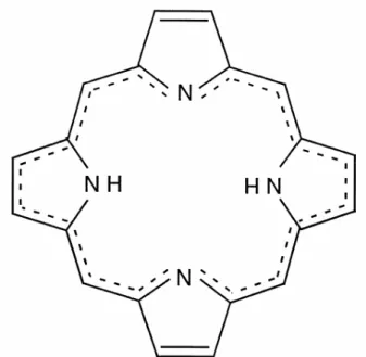
inoids (pheophorbides, bacteriochlorins, bacterio-pheophorbides) origin. Most of the second generation photosensitizers are tetrapyrrolic compounds with side chains added so as to stabilize and improve the absorption in the red. Phthalocyanines are tetrapyrrolic compounds where pyrrole groups are condensed with a benzenic group and where a nitrogenous bridge replaces a methene one, thus enhancing the molar absorption coefficient of these molecules and with λmax of absorption around 700 nm. Texaphyrins are also synthetic relatives of porphyrins, due to their side chains these molecules are water-soluble, and rapidly cleared from the circulation with a wide absorption band centred at 732 nm. However, one of the most active photosensitising agent appears to be 5,10,15,20-tetrakis(m-hydroxyphenyl)chlorin (mTHPC) (Fig. 2.5). This sensitizer requires
very low drug (0.15 mg/kg) and light (20 J/cm2) doses and strongly absorbs in the
“therapeutic window” at 652 nm (Bonnett et al. 1989). Unfortunately 2nd generation
sensitizers generally do not manifest a large tumour localizing selectivity. Therefore research
has been focused on developing third generation photosensitizers. The 2nd generation
photosensitizers are introduced in a vehicle (e.g. liposomes) which will drive the molecule until the desired target. Another method is to graft amino-acids, proteins, polymers, carbohydrates or anti-body on an existent photosensitizer (Moser 1998).
The photosensitizers of tetraphenylchlorin series derive from the meso-tetra(hydroxyphenyl)porphyrins, they are namely the meso-tetra(hydroxyphenyl)chlorin and the meso-tetra(hydroxyphenyl)-bacteriochlorin (mTHPBC) (Fig. 2.5). T
pounds was done by Bonnett et al. (Berenbaum et al. 1986; Bonnett et al. 1989). The meta isomer mTHPP was found to be the most active isomer in the in vivo assay (Berenbaum et al. 1986). The same meta isomer of the chlorin mTHPC was identified as the most active chlorin isomer (Bonnett et al. 1989).
N NH N NH OH OH OH O H m-THPP N NH N NH OH OH OH O H m-THPP N NH N NH OH OH OH O H m-THPC N NH N NH OH OH OH O H m-THPC N NH N NH OH OH OH O H m-THPBC N NH N NH OH OH OH O H m-THPBC
Figure 2.5. : Molecular structures of mTHPP, mTHPC and mTHPBC.
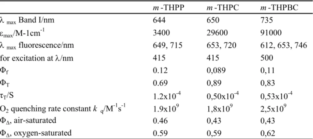
The attractive properties of this series are the strong absorption in the far red region. Where the molar extinction coefficient in ethanol is 1170 M-1cm-1 for Photofrin® at 630 nm, it is 3400 M-1cm-1 at 644 nm for m-THPP, 29600 M-1cm-1 at 650 nm for mTHPC and 91000 M-1cm-1 at 735 nm for m-THPBC (Table 2.3). They have a high triplet states quantum yield formation ranging between 0.69-0.89 and a high quantum yield of singlet oxygen formation (0.43-0.45).
Owing to their photophysical properties these photosensitizers were expected to be valuable compounds for PDT. Actually it has been shown that mTHPP was 25-30 times as potent as haematoporphyrin derivative in sensitising tumours (Berenbaum et al. 1986), and mTHPC considering global photodynamic doses (light dose x photosensitizer dose) was found to be 100 to 200 times as potent as haematoporphyrin derivative (Savary et al. 1997; Savary et al. 1998).
Table 2.3. : Some photophysical properties of m-THPP, m-THPC and m-THPBC in
methanol from (Bonnett, charlesworth et al. 1999).
m -THPP m -THPC m -THPBC
λ max Band I/nm 644 650 735
εmax/M-1cm-1 3400 29600 91000 λ max fluorescence/nm 649, 715 653, 720 612, 653, 746 for excitation at λ/nm 415 415 500 Φf 0.12 0,089 0,11 ΦT 0.69 0,89 0,83 τT/S 1.2x10⁻⁴ 0,50x10⁻⁴ 0,53x10⁻⁴ -1 -1 9 9 9
2. 4. 1. The 5,10,15,20-meta-tetra(hydroxyphenyl)chlorin
mTHPC is a second-generation photosensitizer (Bonnett et al. 1989) and is one of the most effective sensitizers studied to date (Dougherty et al. 1998). It mediates cell photodamage, principally through singlet oxygen formation (Melnikova, Bezdetnaya et al. 1999) and its efficacy is sensitive to oxygenation conditions (Coutier et al. 2002). mTHPC has been granted European approval for palliative treatment of patients with advanced head and neck cancers and undergoes clinical open-label multicenter studies for the treatment of early squamous cell carcinoma (Copper et al. 2003; Hopper et al. 2004).
mTHPC is introduced to patients intravenously. mTHPC’s hydrophobic nature defines its affinity to plasma proteins. Hence, the interactions with plasma components and blood cells can play an important role in mTHPC-PDT efficacy. Studies on mTHPC interaction with plasma protein fractions are sparse (Michael-Titus et al. 1995; Hopkinson et al. 1999; Kessel 1999). mTHPC displays some unusual properties in vitro and in vivo compared with many other sensitizers. Gradient-density ultracentrifugation demonstrated the presence of weakly fluorescing aggregated mTHPC species in the regions of albumin or HDL/albumin (Hopkinson et al. 1999; Kessel and E. Sykes 1999). mTHPC forms large-scale aggregates in aqueous media that monomerize upon interaction with plasma proteins (Bonnett et al. 2001). This sensitizer is rigidly fixed in model membranes and strongly retained in cells in vitro (Ball et al. 1999; Bombelli et al. 2005). mTHPC displays an unusual pharmacokinetic behaviour in human and rabbit plasma with a secondary peak at about 10 and 6 h after in intravenous injection, respectively (Ronn et al. 1997; Glanzmann et al. 1998). These phenomena were supposed to be explained by initial retention of PS in the liver or sensitizer aggregates in the vasculature. Similar pharmacokinetic profile was reported only for hexyl-ether derivative of pyropheophorbide-a in mice (Bellnier et al. 1993). MTHPC has small initial volume of distribution with high retention in the vasculature together with two peaks of PDT efficacy (2h and 24h) in mice (Jones et al. 2003).
It has been demonstrated that the Golgi apparatus and endoplasmic reticulum (ER) are preferential sites of mTHPC accumulation in MCF-7 human adenocarcinoma cells after 3h of incubation (Teiten, Bezdetnaya et al. 2003). Golgi apparatus and ER were shown to be the primary PDT-induced damage sites as measured by enzymes photoinactivation technique (Teiten, Bezdetnaya et al. 2003; Teiten, Marchal et al. 2003). Damage to Golgi apparatus was confirmed by fluence-dependent alterations of Golgi apparatus and mitochondria morphology
(Melnikova, Bezdetnaya, Bour et al. 1999). Both apoptotic and necrotic pathway are implicated in mTHPC-mediated HT29 cell photoinactivation that is governed by mitochondrial membrane photodamage manifested by cytochrome C release and dissipation of mitochondrial membrane potential (Marchal et al. 2005).
During irradiation at 650 nm the absorption spectra of mTHPC in organic, PBS and PBS containing 10% FCS the major absorption bands at 380-450 and 650 nm decreased (Hadjur et al. 1998). A new absorption band was observed at 320 nm, attributed to the formation of a photoproduct. The spectra of mTHPC fluorescence also decreased upon irradiation but no fluorescent photoproducts were detected. A strong dependence of the photodegradation on oxygen concentration and the formation of photoproducts have been reported (Hadjur et al. 1998). Hadjur et al. determined the quantum yields of photobleaching ФPb in aqueous solution containing 10 % FCS to be 1.54 x 10⁻⁵ for air saturated conditions and 1.8 x 10⁻⁶ after N2 bubling. In aerobic conditions the photodegradation, as well as the formation of photoproducts, have been competitively inhibited by singlet oxygen quenchers. On the basis of photobleaching experiments Handjur et. al. also determined the quantum yield of singlet oxygen production (ФΔ) by mTHPC, which appeared to be 0.3 in ethanol and 0.01 in PBS suggesting that mTHPC is highly aggregated in aqueous media (Hadjur et al. 1998). Products of mTHPC oxidation irradiated in methanol have been separated and identified by high-performance liquid chromatography (HPLC). The major compound of oxygenation process has been described as β-hydroxy-mTHPC with an absorption band around 423 nm (Jones et al. 1996). MTHPC has been reported to be a moderately photolabile compound. A comparative study of mTHPBC and mTHPC in methanol–water (3:2, v/v) solution demonstrated a 90 fold greater mTHPBC photobleaching rate compared to mTHPC (Bonnett, Djelal et al. 1999). Rovers et al. in an in vivo study on Colo 26 tumour bearing mice showed that the rate of bleaching of mTHPBC was approximately 20 times greater than that of
mTHPC (Rovers, de Jode, Rezzoug et al. 2000). The ФPb value for mTHPC in PBS with 10 %
FCS solution is an order of magnitude lower compared to BPD-MA (ФPb = 2.07 x 10⁻⁴)
(Aveline et al. 1994).
mTHPC has a strong absorbance in the red region (650 nm) with high molar extinction coefficient (Table 2.2) (Bonnett, Djelal et al. 1999). This offers promising therapeutic perspectives for PDT of deep tumours and pigmented tissues. Pre-clinical studies have demonstrated that in female BALB/c mice bearing PC6 tumour cells the depth of necrosis was 3.79 ± 0.28 mm for mTHPC dose of administered photosensitizer 0.375 µmol/kg for mTHPC (Bonnett et al. 1989). Another in vivo study demonstrated that area of necrosis after
irradiation of mTHPC-sensitised liver is 26 ± 4 mm2 (Rovers, de Jode and Grahn 2000). The absence of correlation between PS concentration in tumor and PDT efficiency was observed in vivo (Veenhuizen et al. 1997; Ris et al. 1998).
2. 5. Cells and tissue damage effects of PDT
PDT induces both direct and indirect antitumor effects (Castano et al. 2005). It can directly destroy tumor cells that undergo apoptosis and necrosis accompanied by induction of the inflammatory response and a slowly developing adaptive immunity that can potentiate local antitumor effects and might possibly induce systemic immunity. PDT together with inflammatory response can also damage tumor vasculature leading to the early vascular shutdown and ischemia-related cell death.
2. 5. 1. Vascular Shutdown and Inflammation
The alteration of endothelial cells during PDT treatment seems to be the origin of modifications observed in vasculature (Fingar et al. 2000). PDT provokes modifications of organisation of the proteins of cytoskeleton of endothelial human cells with consecutive induction of calcium influx in cells (Foster et al. 1991). The modifications of cytoskeletal proteins induce the changes of cells form and the loss of intracellular communications (Fingar et al. 2000). Such changes serve as a signal to the platelets and neutrophils activation which adhere on the vessel wall, roll toward the constriction and aggregate, at which point they migrate into the surrounding tissues following chemokine gradients (Steele et al. 1985). After adhesion platelets release a great quantity of vasoactive molecules such as thromboxan which amplify platelets aggregation being powerful vasoconstrictor (Fingar et al. 1992). In the region of injury cascades of eicosinoids lead to vessel constrictions. The formation of space between endothelial cells contribute to the reduction of tumoral perfusion and vascular permeability (McMahon et al. 1994; Zilberstein et al. 2001). It was demonstrated that vascular destruction occurred to a greater extent in vivo (Henderson et al. 1984). This cause the blood stasis and tumor cells starvation of oxygen and nutrients and reduce the survivability of cells
in vivo (Henderson et al. 1985; Henderson and Fingar 1987; Fingar et al. 1992). It was
reported that vascular destruction after PDT is accompanied by inflammatory response like after tissue injury (Korbelik 1996).
Different photosensitizers do not produce the same type of vascular response: NPe6-PDT produce blood stasis mainly due to platelets aggregated on the artery walls while SnEt2 produces an inflammatory response without vessel constriction or platelet aggregation (McMahon et al. 1994).Vascular destruction is generally considered to be one of the major effects contributing to tumor destruction.
2. 5. 2. Direct cell destruction
One of the first who provided the evidence that cells may undergo two distinct types of cell death was Kerr (Kerr et al. 1972). The first type is known as necrosis, a violent and quick form of death affecting extensive cell populations, characterized by cytoplasm swelling, destruction of organelles and disruption of the plasma membrane, leading to the release of intracellular contents and inflammation. Necrosis has been referred to as accidental cell death, caused by physical or chemical damage and has generally been considered an unprogrammed process. During necrosis decomposition of cell is principally mediated by proteolytic activity (Castano et al. 2005).
Several types of cell death were termed apoptosis or programmed cell death (Agostinis et al. 2004; Almeida et al. 2004). They are identified in single cells usually surrounded by healthy-looking neighbors, and characterized by cell shrinkage, blebbing of the plasma membrane, the organelles and plasma membrane retain their integrity for quite a long period. As a rule the apoptotic program initiated by PDT is the rapid release of mitochondrial cytochrome C into the cytosol followed by activation of the apoptosome and procaspase 3. In vitro, apoptotic cells are ultimately fragmented into multiple membrane-enclosed spherical vesicles. In vivo, these apoptotic bodies are scavenged by phagocytes, inflammation is prevented. Apoptosis, requires transcriptional activation of specific genes, include the activation of endonucleases, consequent DNA degradation into oligonucleosomal fragments, and activation of caspases. Some alternative modes of cell death have bee described: mitotic cell death (Castedo et al. 2004), programmed necrosis (Bizik et al. 2004), cathepsin-mediated lysosomal death pathway (Leist and Jaattela 2001) and autophagic cell death (Yu et al. 2004).
Photosensitizers that localize in cellular organelles such as endoplasmic reticulum or mitochondry can induce apoptosis via photodamage of Bcl-2 and Bcl-xl proteins (Kessel and Luo 1999). With PS localized in the plasma membrane, the photosensitization process can be switched to the necrotic cell death likely due to loss of plasma membrane integrity and rapid depletion of intracellular ATP (Kessel and Poretz 2000; Agostinis et al. 2004). It is believed
that lower dose PDT leads to more apoptosis, while higher doses provoke more necrosis (Plaetzer et al. 2002). Cells sufficiently damaged by PDT are killed, regardless of the mechanism involved. This means that inhibition of apoptosis reorients cells to necrotic pathway, but cannot increase cell survival (Thibaut et al. 2002).
II. 3. Photophysical and photochemical properties of sensitizers
3. 1. Photobleaching of sensitizers
During the photodynamic treatment in addition to the reaction of PS with biological substrate, self-photosensitization occurs, the reactive oxygen intermediates can interact with the photosensitizer, leading to its transformation and/or destruction. This phenomenon is called photobleaching. Photobleaching is relevant to a variety of fields, from laser technology to photomedicine. The first observation of photobleaching in the photodynamic therapy field was made in 1986 by Moan (Moan 1986). Photobleaching is usually observed as lowering of the optical density or the fluorescence intensity of the solution during irradiation (Spikes 1992; Rotomskis et al. 1996). Two types of photobleaching can be considered (Bonnett and Martínez 2001):
- photomodification, where loss of absorbance or fluorescence during
irradiation leads only to PS transformation into modified form.
- “true photobleaching”, where chemical change is profound and results in
small fragments that do not absorb in the visible spectral region.
3P 1O 2 3O 2 S S.++ P .-/ S.-+ P .+ hυ 3O 2 Type I Type II 0P 3P 3P S
Oxidized products Oxidized products
Photobleaching 3O 2 (O2 . -, .OH, H2O2) 0P 0P
Figure 2.6. : Diagram of photobleaching mechanisms occurring after absorption of
The main reactions leading to photobleaching are presented in Fig. 2.6. Irradiation of medium containing photosensitizer leads to the production of reactive oxygen species. These oxygen radical species react with the neighbouring molecules, including the photosensitizers, leading to their destruction. Photobleaching can occur via two pathways, the Type I way involving reactive oxygen species and Type II way involving singlet oxygen.
The sensitivity of PS to photodegradation by light is determined by its photobleaching quantum yield. The photobleaching quantum yield (ФPb) at time t of irradiation is determined as the number of moles of PS photobleached (nPS) devided by the number of moles of photons absorbed (nPh) during the same time. For the case of irradiation of PS solution in the cuvette the ФPb is expressed as (Aveline et al. 1994):
0 t S Pb Ph 0 0 A exp( )
(A - A )V
=
lN
(1 10
)
t ktdt
ε
− −Φ
−
∫
, where 0 Ph I N A N hc λ = (19)where A0 and A1 are optical densities of the PS before and after irradiation during time t, VS is the volume of the sample (in liters), ε is the molar absorption coefficient (in M-1cm-1) at irradiation wavelength, l is the optical pathlength (in cm), k is photobleaching constant (s-1), NPh is the photon flux at irradiation wavelength λ (in mol photons s-1), NA is Avogadro’s number, h is Plank’s number and c is the velocity of light.
There are large differences in the ФPb of photosensitizers (Table 2.4). These
differences are attributed to oxidation potential, lipophilicity, presence of a metallic ion, kind of reactions involved (Type I or II).
The differencies in ФPb of photosensitizers can be explained in the basis of their redox potentials. In organic solvents sensitizers with the lowest redox potential show the most rapid photobleaching (Bonnett and Martínez 2001). The relative photobleaching rates of PSs are proportional to the values of their redox potentials (Chang et al. 1981). Thus the rates of oxygen-mediated photobleaching of sensitizers can be predicted on the basis of their redox potentials values.
Table 2.4. Photobleaching quantum yield of some photosensitizers in PBS
photosensitizer Concentration (M) Photobleaching
quantum yield References MACE (Mono-L-aspartylchlorin
e6) 5 x 10
-6
8.2 x 10⁻⁴
(Spikes and Bommer 1993) Sn aspartyl chlorin e6 5 x 10⁻⁶ 5.7 x 10⁻⁶ (Spikes and Bommer 1993) Zn aspartyl chlorin e6 5 x 10⁻⁶ 1.9 x 10⁻² (Spikes and Bommer 1993) Chlorin e6 5 x 10⁻⁶ 1.9 x 10⁻³ (Spikes and Bommer 1993) Chlorin e6 10-4 74.7 x 10⁻³ (Rotomskis et al. 1997) Sn chlorin e6 5 x 10⁻⁶ 1.3 x 10⁻⁵ (Spikes and Bommer 1993) Zn chlorin e6 5 x 10⁻⁶ 1.8 x 10⁻² (Spikes and Bommer 1993) Hematoporphyrin 10-4 1.05 x 10⁻³ (Rotomskis et al. 1997) Hematoporphyrin 5 x 10⁻⁶ 4.7 x 10⁻⁵ (Spikes 1992)
Photofrin® 10-4 9 x 10⁻⁵ (Rotomskis et al. 1997) Photofrin® 5 x 10⁻⁶ 5.4 x 10⁻⁵ (Spikes 1992) TSPP4 10 -4 2 x 10⁻⁴ (Rotomskis et al. 1997) TSPP4 5 x 10 -6 9.8 x 10⁻⁶ (Spikes 1992) Uroporphyrin I 5 x 10⁻⁶ 2.8 x 10⁻⁵ (Spikes 1992) BPD-MA 2.8 x 10⁻⁵ (Aveline et al. 1994)
Kinetic parameters of photobleaching are mainly derived from spectroscopic measurements assessed by UV-Vis or fluorescence spectroscopy. Several important mechanistic issues of photobleaching were obtained from the detailed analysis of spectroscopic modifications. In the earlier studies on photobleaching of PSs the kinetic decay of photosensitizer was considered to be mono-exponential decay e-αD, where α stands for the photobleaching constant (s-1 or J-1 x cm2) and D stands for the fluence of irradiation (J x cm-²).
As became clear later, the photobleaching kinetic is a complex phenomenon which cannot be described by a single exponential decrease (Sørensen et al. 1998; Moan et al. 2000). Several parameters can influence the kinetic decay such as the oxygen depletion during PDT and different types of binding sites for the sensitizer PS in cells and tissues. For some photosensitizers the decay rates have been shown to be practically independent of the concentration of the dye during illumination (Moan 1986; Mang et al. 1987; Sørensen et al. 1998); and thus exhibit a first order decay. However, for the majority of dyes the photobleaching decay is highly dependent on the initial concentration of the photosensitizer
(Moan et al. 1988), meaning that the photoproducts from the PS can cause the destruction of a neighboring molecules (Moan et al. 1997). For example, the values of ФPb for the different concentrations of Ce6 are very different as for other dyes (PF, hematoporphyrin and TSPP4) (Table 2.3). The deviation from the first-order photobleaching kinetics can be due to oxygen depletion during PDT, photochemical modifications of sensitizer, different types of sensitizer binding sites in tissues and relocalization of sensitizer during light exposure (Moan et al. 1997; Sorensen et al. 1998).
Photobleaching leads to important consequences for light dosimetry in PDT (Potter et al. 1987). Photodynamic dosimetry, based on calculation of the therapeutic dose, was first introduced by Potter et al in 1987 and modified by Robinson et. al. (Robinson et al. 1998). This model includes such parameters as sensitizer and oxygen concentration, illumination fluence rate and several photophysical constants of PS. Dysart et. al. have proposed an implicit approach to assessing PDT efficacy where changes of PS fluorescence during treatment are used to predict treatment outcome (Dysart et al. 2005). The starting point of the authors is the statement that if the biological response to PDT and photobleaching are both mediated by singlet oxygen, hence, photobleaching should yield information about the biological outcome of the treatment.
The photobleaching kinetics for ground-state PS undergoing singlet oxygen–mediated bleaching can be described by the differential equation that is based on homogenous distribution of sensitizer and oxygen (Georgakoudi et al. 1997):
1 0 os 0 2
d[S ]
= -k [S ][ O ]
dt
(20)where [S0] and [1O2] are concentrations of ground state PS and singlet oxygen, respectively; kos is the bimolecular rate constant of 1O2 reaction with ground state sensitizer S0. In reality, if the concentration of PS is low enough, the only PS molecule with which the singlet oxygen can react is the parent PS molecule. For these low PS concentrations, the rate of photobleaching will depend only on the rate of singlet oxygen generation because the volume through which each singlet oxygen molecule can diffuse before reacting will contain exactly one PS molecule, independent of PS concentration. Taking into account the short lifetime and diffusion distance of singlet oxygen in biological media the eq. 20 can be modified by the addition of a constant, δ: 1 0 os 0 2
d[S ]
= -k ([S ] + )[ O ]
dt
δ
(21)δ is is effective minimum concentration of PS. It is determined by the distance of singlet oxygen diffusion befor reaction with adjacent PS molecule and is given by:
3/ 2 A
1
N (6D )
δ
τ
Δ=
(22)where NA is Avogadri’s number, D is the diffusion coefficient of singlet oxygen in cells,
τ
Δ is singlet oxygen lifetime. At constant oxygenetion during treatment the PDT dose(total amount of 1O2 molecules generated) at time T may be expressed:
T 0 os 0
CS
Dose =
(t)[S ](t)dt
k
τ
Δ Δ∫
Φ
(23)where Ф(t) is the fluence rate of excitation light, C is a constant, SΔ is the fraction of
triplet molecules quenched by oxygen (eq. 17). If Ф(t) remaines constant during the treatment the singlet oxygen dose can be estimated directly from PS photobleaching curve [S0](t).
Using this model, Dysart et. al. have determined important photophysical and photobiological parameters of mTHPC in MLL cells (Dysart et al. 2005). The estimation of values SΔ= 0.96 ± 0.01, δ = 33 ± 6 µM,
τ
Δ=0.03 – 0.018 µs, kos = (7.8 – 11.1) x 106 M-1s-1 were obtained. Moreover, it was estimated that number of singlet oxygen molecules per cell required to reduce survival by 1/e is in the range N1/e = (7.6 – 11.1) × 108 for MLL cells with mTHPC. The proposed model explains the dependence of bleaching kinetics on PS concentration and shows the possibility of singlet oxygen concentration estimation on the basis of PS photobleaching kinetics without the need for measurements of ground-state oxygen concentrations or treatment fluence rate. Other authors have reported the values of N1/e to be 3.9 × 10⁷ with ALA-induced PpIX in AML5 leukemia cells (Niedre et al. 2003) and 1.2 × 10⁸ for TA-3 cells with HpD (Dougherty et al. 1976).3. 1. 1. Parameters effecting photobleaching. Aggregation state, pH, ionic strength and oxygen concentration
Previous work in our laboratory demonstrated a different photosensitivity of monomeric and aggregated forms. In a first study Bezdetnaya et al. (Bezdetnaya et al. 1996) demonstrated that for HpD and PpIX quantum yield of photobleaching obtained by matching fluorescence where higher than that obtained by matching absorbance (10 and 11 times for HpD and PpIX respectively). The authors concluded that this difference reflected the
preferential photobleaching of photolabile monomeric forms compared to aggregates. In another study they confirmed this assumption using mTHPC (Belitchenko et al. 1998).
Several studies of Rotomskis and co-workers demonstrated that photobleaching efficiency of haematoporphyrin-like sensitizers seems to be consistent with their aggregation state and the presence of covalently linked structures. Dimethoxyhaematoporphyrin (DMHp) and Hp are present in an equilibrium of monomeric and aggregated forms in aqueous solution (Streckyte and Rotomskis 1993). Their absorption bleaching rate constants are two to four times higher than that of HpD, a sensitizer containing mostly linear structures of porphyrins linked by ether, ester and/or carbon-carbon bonds (Dougherty et al. 1984), and 10 to 20 times higher than that of Photofrin® (PF), which contains covalently linked ”sandwich” type structure (Streckyte and Rotomskis 1993). In HpD, some of the side chains are involved in ether and ester linkages, and therefore this compound is more photostable than DMHp and Hp. In PF and Photosan-3 (PS) (highly aggregated “sandwich” type structure (Streckyte and Rotomskis 1993), almost all side chains are involved in covalently linked structures, probably accounting for the high photostability of these sensitizers. The presence of a certain amount of protoporphyrin in PS is probably responsible for its lower photostability compared to PF.
Lowering the pH value of a photosensitizer solution results in a shift of both the absorption and the fluorescence spectrum as well as in a decrease of the fluorescence intensity, indicating an aggregation at low pH values (pH < 5) (Cunderlikova et al. 1999). Reddi et al. (Reddi and Jori 1988) also demonstrated an aggregation of hematoporphyrin and Photofrin® when decreasing the pH from 7.4 to 5.0 and they also demonstrated the decrease of the photobleaching quantum yield to 70 % for hematoporphyrin and 30 % for Photofrin®, thus suggesting a resistance toward photobleaching of aggregated species.
Changing the ionic strength by varying the buffer concentration can affect the aggregation state of a sensitizer. An increase of the buffer concentration of a TPPS4 solution increases the aggregation of the sensitizer and reduces the photobleaching quantum yield by 50 % (Davila and Harriman 1990). Thus, it follows from all this studies that the quantum yield of photobleaching is inversely proportional to the aggregation state of the photosensitizers.
It was observed that the quantum yield of photobleaching of several porphyrins in phosphate buffer is reduced with the lack of oxygen (using nitrogen bubbling) (Spikes 1992). Same observation was made for endogenously formed porphyrins in bacteria (Konig et al. 1993). An observation of the involvement of oxygen in vivo has been realised by Robinson and co-workers (Robinson et al. 1998). During a photobleaching experiment with
ALA-induced PpIX the mice died and they observed a slowdown of the photobleaching. They correlated this bleaching decrease to the oxygen decline in the skin, due to the death of the animal.
Several studies from the laboratory of T. H. Foster documented the oxygen depletion during PDT. Oxygen consumption model was refined by Georgakoudi and co-workers (Georgakoudi et al. 1997; Georgakoudi and Foster 1998) by taking into account the parameter of photobleaching of Photofrin in EMT6 spheroids. This improvement considerably changed the kinetic profile of the oxygen aspects of Photofrin-PDT. The authors observed a rapid decrease in oxygen concentration during irradiation followed by a progressive return to the values measured before the irradiation. The first phase is due to the photochemical oxygen consumption which is faster than the diffusion of the oxygen through the spheroid. The second phase, corresponding to the comeback of oxygen to the initial value, is due to a slowdown of the photochemical consumption of the oxygen explained by the decrease in photosensitizer concentration (photobleaching), together with the diffusion. This was in agreement with the mathematical model assuming that the photobleaching was based on a reaction between singlet oxygen and photosensitizer.
In their further studies Foster and co-workers investigated the impact of irradiance on photobleaching (Finlay et al. 2001; Finlay et al. 2002). In a study reporting the photobleaching of ALA-induced Protoporphyrin IX (Pp IX) in normal rat skin (Finlay et al. 2001) it was demonstrated that the photobleaching kinetics were different with the change of the irradiance. High irradiance led to rapid oxygen consumption and a slow down of the photobleaching. In a second study, Finlay et al. (Finlay et al. 2002) showed that photobleaching kinetics of m-THPC on normal rat skin exhibits two distinct phases. The first phase was shown to be irradiance independent, whereas the second phase revealed an irradiance dependency consistent with an oxygen-dependant reaction process. Using mathematical model of photobleaching based on selfsensitized singlet oxygen reactions the fluence rate dependence of the cell survival and of mTHPC photobleaching was due to photochemical oxygen consumption and a predominantly singlet oxygen-mediated mechanism of mTHPC photobleaching (Coutier et al. 2001). It was demonstrated that high fluence rates lead to rapid photochemical oxygen consumption in mTHPC-PDT, where at lower fluence rates intratumor oxygen content was maintaines at levels comparable to those measured before illumination (Coutier et al. 2002). The authors proposed that improved tumor destruction could be expected by reducing the rate and the extent of oxygen depletion during mTHPC photodynamic therapy using low fluence rates.
3. 2. Effect of aggregation state on photophysical and photochemical properties of sensitizers.
Hydrophobic PSs with high value of octanol-water partition coefficient form dimers and higher micelle-like aggregates in aqueous media and their physical and chemical properties differ noticeably from those of the monomeric sensitizer (Brown et al. 1976). The aggregated PSs are generally have much lower fluorescence and triplet states quantum yields that leads to lowering of the quantum yield of singlet oxygen production (Redmond et al. 1985; Tanielian et al. 2001) and drop of photosensitizing efficiency (Ma et al. 1994; Ball et al. 1998; Theodossiou et al. 2004). Action spectra have significantly greater resemblance to the fluorescence excitation spectra than to the absorption spectra of the HpD in cells (Moan and Sommer 1984) indicating that fluorescent monomeric species of sensitizer are more photodynamically active compared to aggregates.
The explanation of reduced singlet oxygen production by aggregates can be done taking into account that competition between the type I and II photosensitization mechanisms is substantially altered as a consequence of protein binding and dye aggregation, favoring type I mechanism by protection of triplet species against collisional oxygen quenching (Bartlett and Indig 1999). The limited access of oxygen to interact with PSs is due to stabilization of aggregated species by hydrophobic forces, hydrogen bonds and π-π interactions of the aromatic rings (Lang et al. 1998; Bonnett et al. 2001). When strong electronic coupling exists among PS molecules in an aggregate the resonance light scattering (RLS) can be detected from the solution of such aggregates. RLS effect is observed as increased scattering intensity at or very near the wavelength of absorption maximum of aggregated molecular species (Pasternack and Collings 1995; Collings et al. 1999). The intensity of scattering depends on the square of the volume of the aggregate and increases as a consequence of aggregation.
Hydrophobicity of PSs influences not only their aggregation state but also accumulation in cells. The strong linear correlation between PSs cell uptake and octanol-water partition coefficient was observed (Oenbrink et al. 1988). Aggregated PS species are assumed to internalize in cells via endocytotic pathway, whereas sensitizers in monomeric state can be transported by passive diffusion through plasmatic membrane or internalized in complexes with plasma proteins. After endocytosis aggregated PSs are believed to localize in lysosomes (Berg et al. 1993; MacDonald et al. 1999).
During interactions with plasma proteins the value ot the fluorescence yield of hydrophobic sensitizers augment with time (Belitchenko et al. 1998) that can be explained by