RESEARCH OUTPUTS / RÉSULTATS DE RECHERCHE
Author(s) - Auteur(s) :
Publication date - Date de publication :
Permanent link - Permalien :
Rights / License - Licence de droit d’auteur :
Bibliothèque Universitaire Moretus Plantin
Institutional Repository - Research Portal
Dépôt Institutionnel - Portail de la Recherche
researchportal.unamur.be
University of Namur
The activation of cultured keratinocytes by cholesterol depletion during reconstruction
of a human epidermis is reminiscent of monolayer cultures
De Vuyst, Évelyne; Giltaire, Séverine; Lambert De Rouvroit, Catherine; Chrétien, Aline;
Salmon, Michel; Poumay, Yves
Published in:
Archives of Dermatological Research
DOI:
10.1007/s00403-015-1537-3 Publication date:
2015
Document Version
Early version, also known as pre-print
Link to publication
Citation for pulished version (HARVARD):
De Vuyst, É, Giltaire, S, Lambert De Rouvroit, C, Chrétien, A, Salmon, M & Poumay, Y 2015, 'The activation of cultured keratinocytes by cholesterol depletion during reconstruction of a human epidermis is reminiscent of monolayer cultures', Archives of Dermatological Research, vol. 307, pp. 309-318.
https://doi.org/10.1007/s00403-015-1537-3
General rights
Copyright and moral rights for the publications made accessible in the public portal are retained by the authors and/or other copyright owners and it is a condition of accessing publications that users recognise and abide by the legal requirements associated with these rights. • Users may download and print one copy of any publication from the public portal for the purpose of private study or research. • You may not further distribute the material or use it for any profit-making activity or commercial gain
• You may freely distribute the URL identifying the publication in the public portal ? Take down policy
If you believe that this document breaches copyright please contact us providing details, and we will remove access to the work immediately and investigate your claim.
1
The activation of cultured keratinocytes by cholesterol depletion during reconstruction of a human epidermis is reminiscent of monolayer cultures
ÉVELYNE DE VUYST1, SÉVERINE GILTAIRE1, CATHERINE LAMBERT DE ROUVROIT1, ALINE
CHRÉTIEN2, MICHEL SALMON2 and YVES POUMAY1
1 Cell and Tissue Laboratory-URPHYM-Narilis, University of Namur, Namur, Belgium.
2 StratiCell, Les Isnes, Belgium.
Keywords
Reconstructed human epidermis; methyl-beta-cyclodextrin; cholesterol, atopic dermatitis; cell signaling; epidermal barrier
Contract grant sponsor: Région Wallonne; Contract grant number: 1217660.
Contract grant sponsor: FNRS; Contract grant number: 1.5.033.06F. Contract grant sponsor: FRFC; Contract grant number: 2.4.522.10F. *Correspondence to:
Yves Poumay, Cell and Tissue Laboratory, URPHYM, NARILIS, University of Namur, 61, rue de Bruxelles, B-5000 Namur, Belgium. E-mail: yves.poumay@unamur.be
2
Abstract
Transient cholesterol depletion from plasma membranes of human keratinocytes has been shown to reversibly activate signalling pathways in monolayer cultures. Consecutive changes in gene expression have been characterized in such conditions and were interestingly found to be similar to transcriptional changes observed in keratinocytes of atopic dermatitis (AD) patients. As an inflammatory skin disease, AD notably results in altered histology of the epidermis associated with a defective epidermal barrier. To further investigate whether the activation of keratinocytes obtained by cholesterol depletion could be responsible for some epidermal alterations reported in AD, this study was undertaken to analyze cholesterol depletion in stratified cultures of keratinocytes, i.e. a reconstructed human epidermis (RHE). RHE contain heterogeneous populations of keratinocytes, either proliferating or progressively differentiating and stratifying towards the creation of a cornified barrier. Cholesterol depletion induced in this model was found reversible and resulted in activation of signalling pathways similar to those previously identified in monolayers. In addition, selected changes in the expression of several genes suggested that keratinocytes in RHE respond to cholesterol depletion as monolayers. However, preserved histology and barrier function indicate that some additional activation, likely from the immune system, is required to obtain epidermal alterations such as the ones found in AD.
Introduction
A major function of human skin, at the epidermal level, is the production and maintenance of an effective barrier between the organism and its environment that protects against pathological invaders and external substances, but also impedes water loss from the body. This barrier function resides within the most superficial epidermal layers of the skin [23]. Therefore keratinocytes, the main cell type in the epidermis, organize into stratified layers. They proliferate in the deepest basal layer of the tissue, then undergo a complex differentiation program through the suprabasal layers, finally creating squamous terminally keratinized dead cells inside the cornified layer [6]. A physiological balance between keratinocyte proliferation, terminal differentiation and superficial desquamation is observed under normal conditions as a consequence of tightly regulated and coordinated signalling pathways in keratinocytes that control their epidermal phenotypes.
Several previous studies have shown the critical involvement of membrane cholesterol in regulating keratinocytes through important signalling pathways [9,11,15-17,24,25]. In plasma membranes, cholesterol accumulates in detergent-resistant micro-domains named lipid rafts. Such domains exhibit elevated affinities for actors like the epidermal growth factor receptor (EGFR) involved in precise signalling pathways, suggesting that cholesterol is required in plasma membranes to organize specialized micro-domains able to regulate signalling [3,12,17,24]. Keratinocytes in culture contain cholesterol in plasma membranes and its depletion can be induced by incubation of cells with methyl--cyclodextrin (MCD), a molecule containing a hydrophobic cavity able to extract cholesterol from membranes [14]. This depletion is followed by repletion and cell recovery within a few hours [9,11,15,18]. After plasma membrane cholesterol deprivation, keratinocytes exhibit dimerization and tyrosine phosphorylation of EGFR, as well as phosphorylation of extracellular signal-regulated kinase (ERK) [16]. Interestingly, mitogen-activated protein kinase p38 (MAPKp38) is also activated and induces the expression of the differentiation marker involucrin (IVL) [11], as well as the expression of the heparin-binding EGF-like growth factor (HB-EGF) [9,18], but p38 activation does not depend on the tyrosine kinase activity of EGFR, indicating a role for an additional activation pathway to modulate the keratinocyte phenotype after cholesterol depletion [11]. While trying to analyse in a more comprehensive fashion the alterations induced by cholesterol depletion in keratinocytes, a transcriptomic analysis confirmed and revealed multiple regulations in the expression of various genes, either implicated in epidermal proliferation (HB-EGF), differentiation (IVL, transglutaminase-1 (TGM-1)), barrier formation (filaggrin (FLG), loricrin (LOR)) or inflammation (interleukin-8 (IL-8)) for instance [19]. Interestingly, signalling pathway analysis of those data suggested that cholesterol-depleted keratinocytes mimic the transcriptional profile observed in keratinocytes from lesions of atopic dermatitis (AD) patients [19]. AD is characterised by a defective epidermal barrier and skin inflammation [4] and is an increasingly common skin condition that affects 15-30% of children in industrialized countries.
Those previous results were obtained through analysis of keratinocytes cultured as monolayers wherein cells exhibit an incomplete keratinization process and cannot establish any epidermal barrier. Therefore, the present study has been performed in order to transpose cholesterol-depletion conditions into keratinocytes involved in the reconstruction of a keratinized tissue, a context much closer to the in vivo environment of this cell type [8,22]. Reconstructed human epidermis (RHE) were thus analysed for alterations already identified in monolayer cultures of keratinocytes during tissue reconstruction. Signalling pathways and specific gene expression were characterized in RHE after cholesterol depletion, as well as morphology and functionality of the epidermal barrier.
3
Materials and methodsCell culture and the production and analysis of reconstructed human epidermis
Normal adult human skin samples were obtained from abdominoplasties after informed consent and superficially sliced using a dermatome. Epidermal keratinocytes were isolated and cultured as described earlier to first expand the cell population, and then produce RHE over polycarbonate filters (pore diameters: 0.4 µm) at the air-liquid interface [5,8,22]. Morphology, RNA and protein extraction of RHE was realised according to De Vuyst and collaborators [5].
Chemicals and Antibodies
MCD suitable for use in cell culture was obtained from Sigma-Aldrich (Diegem, Belgium). MCD was used at a concentration of 1% (wt/vol), corresponding to approximately 7.5mM. This concentration has been chosen in accordance with previous studies performed on monolayer cultures of keratinocytes [9,15,18,19] and showing that one hour of treatment was sufficient to significantly decrease the concentration of cholesterol without affecting cell viability [9,11]). Rabbit anti-EGFR, rabbit anti-p38, rabbit anti-phospho-p38, rabbit anti-ERK1/2, mouse phospho-ERK antibodies were purchased from Cell Signaling (Leiden, Netherlands). Rabbit anti-phospho-EGFR (Tyr1173) antibody was obtained from Invitrogen (Ghent, Belgium). Anti-rabbit and anti-mouse HRP-antibodies were purchased from Cell Signaling (Leiden, The Netherlands).
Cholesterol extraction and quantification
RHE were lysed in chloroform. Proteins and lipids were then solubilised and separated in chloroform:methanol (2:1). The organic phase was washed twice in 0.05M NaCl, then twice in 0.36M CaCl2:methanol (1:1). Triton X-100/acetone were then added and samples were evaporated under air flow with
SpeedVac SC100 before being solubilised in demineralised water.
Cholesterol was quantified using the Amplex Red Cholesterol Assay kit from Molecular probes (Ghent, Belgium) according to the manufacturer’s protocol.
Filipin staining
Frozen sections of the epidermis were fixed for 30 min with 4% paraformaldehyde then washed with PBS and incubated with 50 µg/ml of filipin III from Sigma-Aldrich (Diegem, Belgium) for 30 min. After a washing step, sections were mounted in Mowiol (Molecular Probes, Ghent, Belgium) and analysed by epifluorescence using Olympus AX70 microscope with a UV filter and processed by Canon EOS1100D sotfware. Pictures were taken using uniform conditions (quick exposure time to avoid the rapid bleaching of filipin).
Western blotting
RHE were lysed according to De Vuyst et al., 2013 [5]. Protein concentrations of the cell lysates were measured using the Pierce kit (Thermo scientific, Rockford, USA). The extracted proteins were separated by SDS-polyacrylamide gel electrophoresis and blotted onto polyvinylidene difluoride (PVDF) membranes (GE Healthcare Bio-Sciences, Diegem, Belgium). Membranes were blocked in PBS containing 0.1% Tween 20 and containing 5% powdered milk (blocking buffer) before being incubated with specific primary antibodies diluted in blocking buffer and horseradish peroxidase-conjugated secondary antibodies. Finally, detection was performed using the BM Chemiluminescence Blotting Substrate (Roche Diagnostic, Mannheim, Germany). Densitometric values of Western blot data were measured using ImageJ software.
Reverse transcription and quantitative RT-PCR
RNA was reverse transcribed into cDNA using the Super Script II RNase H-reverse transcriptase kit (Invitrogen, Merelbeke, Belgium) and qRT-PCR was performed with the Power SYBR Green Master Mix (Applied Biosystems, Lennik, Belgium). Genes were normalized to the housekeeping gene RPLP0 shown to be stable in conditions of keratinocyte differentiation [21]. Sequences of the primers that were used are available as supplementary data (Table 1).
4
Trans-epithelial electrical resistance (TEER) measurements and permeability to lucifer yellow
TEER measurements were performed using Millicel electrical resistance system from Millipore (Billerica, MA, USA) as described by Frankart and collaborators [8].
Lucifer yellow obtained from Sigma-Aldrich (Diegem, Belgium) was applied topically at 1 mM concentration on the epidermis for 6 hours at 37°C. At the end of the incubation, the medium beneath the epidermis was collected for measurement of the fluorescence corresponding to dye (ex: 485nm; em: 535nm) and the RHE were fixed and
embedded in paraffin. Sections were visualised under fluorescence microscopy.
Results
During in vitro reconstruction of the epidermal tissue, proliferation and differentiation of keratinocytes lead to progressive morphogenesis of the characteristic stratified layers of the epidermal tissue so that, after 11 days of culture at the air-liquid interface, the morphology of the tissue is representative of the in vivo normal human epidermis [8], although it is exclusively composed of keratinocytes. Because cholesterol depletion in keratinocytes had been solely studied in monolayer cultures, despite data indicating altered gene expression of several actors of epidermal keratinization and barrier formation [11,19], the effects of cholesterol depletion were here monitored in RHE treated on the fifth day of reconstruction. This time-point was chosen because, at this stage, an efficient epidermal barrier begins to form [8]. Indeed, granular and cornified layers appear at this stage simultaneously with the increase in TEER [8]. We assumed that an already established barrier such as the one present after 11 days of reconstruction of the RHE should be less sensitive to the alterations produced by cholesterol depletion than the barrier developing from the fifth day of reconstruction. In the present study, RHE were therefore incubated on the fifth day of reconstruction for one hour in the presence of MCD, then left to recover for different periods in normal culture medium.
Cholesterol can be depleted from the deepest layers of RHE by MCD
Cholesterol content was assessed using an enzymatic assay on lipid extracts from RHE. Data from three independent experiments reveal that the amount of cholesterol is decreased at the end of the treatment with MCD, although the reduction is found statistically significant only one hour later, i.e. after 1 hour of recovery (Figure 1A). Later on, RHE recovered cholesterol contents similar to initial control conditions. This illustrates that MCD is able to extract cholesterol from RHE, but also that cholesterol is synthesized by keratinocytes. Of course, these results represent total cellular cholesterol content measured in each RHE and likely underestimate the actual variations of plasma membrane cholesterol of keratinocytes from the lowest layers.
Since this procedure removes cholesterol from the bottom of RHE, cholesterol was localized in RHE sections using a filipin-based staining. Filipin is a naturally fluorescent dye that binds to non-esterified cholesterol also in cell membranes, allowing its localization in cells and tissues [20]. Presented data are representative of three independent experiments. In the control epidermis, a blue staining is observed in the whole tissue with the highest concentration in plasma membranes and in the upper granular and cornified layers. After incubation with MCD, cholesterol is depleted from the lower living layers of keratinocytes, while it remains inside the superficial outermost layers. After 4 hours of recovery in normal culture medium, the reappearance of filipin staining around keratinocytes of the lowest layers suggests slow cholesterol re-synthesis in RHE. After 24 hours of recovery, the cholesterol content and localisation were close to the control conditions (Figure 1B). Interestingly, the MCD present in culture medium does not reach the cornified layer of RHE since the cholesterol content was never modified there (Figure 1B), an observation that is in agreement with the absence of cholesterol extraction during topical application of MCD on the apical surface of RHE (data not shown).
Cholesterol depletion in RHE results in activated cell signalling
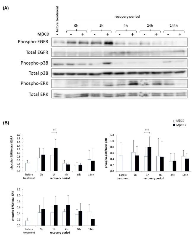
As a possible consequence of cholesterol depletion, activation of the cell signalling was then investigated in a time-course experiment. This was also realised in RHE produced and analysed in three independent experiments. Indeed, tyrosine phosphorylation of the EGF receptor (EGFR) as well as serine/threonine phosphorylation of the p38 mitogen-activated protein kinase (p38 MAPK) and of the extracellular signal-regulated kinase (ERK) were detected at different time-point following the incubation of RHE with MCD (Figure 2). This is reminiscent of the activated cell signalling reported after cholesterol depletion in keratinocyte monolayers [9,15]. Phosphorylation of these proteins was significantly increased after one hour of recovery and lasted for at least 24 hours during the recovery period after cholesterol depletion, however it was no more detectable after six days (144h) of recovery, on the eleventh day of tissue reconstruction, when morphogenesis of the RHE is usually considered as complete.
5
Activation of gene expression by cholesterol depletion in RHE
Characterization of consequences induced by cholesterol depletion on gene expression in RHE was then undertaken for genes already found regulated by this treatment in keratinocyte monolayers [19]. Quantitative RT-PCR was used to determine relative mRNA expression levels of target genes in RHE produced and analysed in three independent experiments. The mRNA levels were measured before the treatment of RHE with MCD, after the treatment, as well as during different recovery periods (Figure 3). The heparin-binding EGF-like growth factor (HB-EGF) and the plasminogen activator urokinase receptor (PLAUR) are mainly expressed after 1 and 4 hours of recovery period. Differentiation markers involucrin (IVL) and transglutaminase-1 (TGM-1) are maximally induced after 4 and 8 hours of recovery period. Maximal expression of the interleukin 8 (IL-8) was observed between 1 and 8 hours of recovery period followed by a decrease during the next hours. These results illustrate responses similar to the one observed in monolayer cultures [19]. However certain significant downregulations reported for the expression of keratin 10 (KRT10), loricrin (LOR), and filaggrin (FLG) in keratinocyte monolayers were not confirmed in RHE after cholesterol depletion.
Thus results show several similarities between monolayer cultures on one hand [11,19] and in RHE on the other hand regarding the cell signalling observations and gene expression studies. In other words, keratinocytes, analysed while anchored to plastic culture dishes or embedded together inside a more complex stratified tissue displaying heterogeneous epidermal cell phenotypes, behave in comparable ways after cholesterol depletion. Cholesterol depletion does alter neither the histology of RHE nor their barrier
Because transcriptional profiling of cholesterol-depleted keratinocytes has identified AD as the disease that exhibits the keratinocyte’s phenotype most closely associated with cholesterol depletion [19], the histology of RHE and their barrier properties were analysed after cholesterol depletion.
The histology of RHE (Figure 4) was compared from the fifth day of tissue reconstruction, when cholesterol was depleted by treatment with MCD, until the eleventh day of RHE culture, when the epidermal morphogenesis is achieved [8]. Results shown are representative of RHE produced and analysed in three independent experiments. Comparison using classical HE staining reveals identical histology between RHE cultured after cholesterol depletion for 1 hour, or not (Figure 4). The epidermal morphogenesis is identical in both conditions, especially characterized by simultaneous appearance of granular and cornified layers at the surface of RHE. These observations were confirmed by immunolocalization of markers of epidermal differentiation (data not shown).
The three-dimensional RHE cultured at the air-liquid interface resembles the in vivo epidermis and is therefore also suitable to study alterations of the barrier’s function. Two different procedures were used to analyse the barrier: measurement of trans-epithelial electrical resistance (TEER) and permeability of RHE to the fluorescent dye lucifer yellow. The TEER measured through the epidermal tissue increases during the reconstruction, especially after the fifth day of culture, indicating the development of a functional barrier [8]. When comparisons of TEER were made between RHE from three independent experiments treated with MCD versus control RHE without this treatment, no significant differences could be observed during the recovery period pursued until the eleventh day of reconstruction (Figure 5A). Similarly, the permeability of RHE towards the fluorescent dye lucifer yellow applied topically was also unchanged after cholesterol depletion. Indeed, when tested by fluorescent microscopy of histological sections of RHE (Figure 5B), or by measurement of lucifer yellow-associated fluorescence in culture medium under RHE (Figure 5C), the permeability remained unchanged in both conditions.
Discussion
Previous works on cultures of keratinocytes have shown that treatment of monolayers with MCD depletes cholesterol from plasma membranes [11]. Moreover, cholesterol depletion in keratinocytes was shown to result in a kind of cell activation associated with EGFR phosphorylation and internalization [11,15,16], associated with release of extracellular ATP, activation of P2 purinergic receptors, as well as expression and release of HB-EGF [9]. Further it was associated with a larger number of genes whose expression was altered, including genes involved in inflammatory response and reminiscent of genes altered in AD lesions [19]. Here, data collected with keratinocytes embedded in RHE treated or not with MCD at day 5 of reconstruction in order to deplete cholesterol from their plasma membranes reveal that a similar profile of cellular activation is obtained. Indeed, phosphorylation of EGFR, but also of MAP kinases p38 and ERK are observed simultaneously with transiently enhanced gene expression of HB-EGF, IVL, IL-8, TGM-1 and PLAUR. However, despite good similarities between keratinocyte responses after cholesterol depletion in monolayers and RHE, other genes do not seem to be
6
identically regulated in both contexts. For instance, the expression levels of FLG and LOR were affected in cholesterol-depleted keratinocyte monolayers [19], whereas they did not vary in RHE. It is important to note that in monolayer cultures of keratinocytes all cells are immersed and thus affected by the treatment, whereas in a three-dimensional reconstructed epidermis the basal layer is the only one to be in direct contact with the culture medium containing molecules responsible for the treatment. However MCD is able to penetrate, between cells of the basal layer, into the suprabasal layers (Figure 1B) what probably creates a diffusion gradient of the molecule across the RHE and could explain the difference of sensitivity between the two culture models. Interestingly, the differently regulated genes, FLG and LOR, that are expressed in the granular and cornified layers and participate in the formation of the epidermal barrier, do not seem affected by the cholesterol depletion in the present study while they were significantly altered in the monolayer cultures [19]. The presence of cholesterol in the upper layers of RHE, as evidenced by filipin staining (Figure 1B), could explain why the expression of these genes is not affected by the treatment. In addition to the diffusion gradient across the RHE, the water-soluble properties of MCD can also impede this molecule to reach the hydrophobic cornified layer. This could also explain why the morphology (Figure 4) and the epidermal barrier (Figure 5) are not affected by cholesterol depletion in RHE. In monolayer cultures, keratinocytes show morphological alterations such as slender and stretcher cells with larger intercellular spaces (data not shown). However, in a stratified tissue an effective barrier forms during reconstruction, thanks to functional junctions between cells. These junctions are probably stronger in a tissue and thus less affected by treatments than in monolayer cultures. This could explain the absence of enlargement of the intercellular spaces between keratinocytes in RHE.
Keratinocytes activated by incubation with MCD and thereby deprived from cholesterol exhibit phenotypic characteristics of AD in monolayers. Our approach of cholesterol depletion in RHE was thus undertaken to mimic some pathological situation such as AD in an in vitro model of the human epidermis, which is closer to the in vivo situation. After collecting encouraging observations of keratinocyte activation in terms of cell signalling and specific gene expression in RHE, the subsequent deceptive conclusions drawn in regard of histology and barrier function of the activated tissues led us to hypothesize a more complex situation to understand epidermal alterations in AD. Indeed, activation of the immune system is part of AD disease and has been shown able to induce a weakening of the epidermal barrier, notably by the release of specific interleukins that result in decreased expression of FLG [1,10]. In consequence, since the hereby presented model is made of keratinocytes only, it should therefore be envisaged to add immune components such as AD-linked interleukins in the culture medium [2,10,13] or to set-up a co-culture model including activated lymphocytes for instance [7,26] .
Taken together, our data demonstrate that cholesterol depletion elicits keratinocyte responses in a complex stratified tissue made of heterogeneous cell phenotypes. These responses include activation of signalling pathways and altered gene expression, two observations that suggest to combine cholesterol depletion with alterations induced by the immune system in order to mimic more closely characteristics of AD in pathological RHE.
Acknowledgments
The authors gratefully thank J. Malaisse for help in statistical analysis, B. Balau, V. De Glas, K. De Swert and D. Van Vlaender for technical help as well as Dr B. Bienfait (Clinique St Luc, Bouge) for providing skin samples. This research received financial support, including research fellowships for EDV and SG, from the Région Wallonne (convention n°1217660-SPW-FUNDP-StratiCELL SA). Additional financial support was provided by FRFC 2.4.522.10F and FNRS 1.5.003.06F grants to YP
Conflict of interest
None to declare.
References
1. Boguniewicz M, Leung DY (2011) Atopic dermatitis: a disease of altered skin barrier and immune dysregulation. Immunological reviews 242 (1):233-246
2. Brandt EB, Sivaprasad U (2011) Th2 Cytokines and Atopic Dermatitis. Journal of clinical & cellular immunology 2 (3):110
7
3. Calder PC, Yaqoob P (2007) Lipid rafts--composition, characterization, and controversies. The Journal of nutrition 137 (3):545-547
4. Cork MJ, Danby SG, Vasilopoulos Y, Hadgraft J, Lane ME, Moustafa M, Guy RH, Macgowan AL, Tazi-Ahnini R, Ward SJ (2009) Epidermal barrier dysfunction in atopic dermatitis. J Invest Dermatol 129 (8):1892-1908
5. De Vuyst E, Charlier, C, Giltaire, S, De Glas, V, Lambert de Rouvroit, C, Poumay, Y. (2014) Reconstruction of Normal and Pathological Human Epidermis on Polycarbonate Filter. In: Turksen K (ed) Epidermal Cells - Methods and Protocols. Methods in Molecular Biology, 3rd edn. Humana Press, Ottawa, pp 191-201
6. Eckhart L, Lippens S, Tschachler E, Declercq W (2013) Cell death by cornification. Biochim Biophys Acta 1833 (12):3471-3480
7. Engelhart K, El Hindi T, Biesalski HK, Pfitzner I (2005) In vitro reproduction of clinical hallmarks of eczematous dermatitis in organotypic skin models. Arch Dermatol Res 297 (1):1-9
8. Frankart A, Malaisse J, De Vuyst E, Minner F, de Rouvroit CL, Poumay Y (2012) Epidermal morphogenesis during progressive in vitro 3D reconstruction at the air-liquid interface. Exp Dermatol 21 (11):871-875 9. Giltaire S, Lambert S, Poumay Y (2011) HB-EGF synthesis and release induced by cholesterol depletion of
human epidermal keratinocytes is controlled by extracellular ATP and involves both p38 and ERK1/2 signaling pathways. J Cell Physiol 226 (6):1651-1659
10. Howell MD, Kim BE, Gao P, Grant AV, Boguniewicz M, DeBenedetto A, Schneider L, Beck LA, Barnes KC, Leung DY (2009) Cytokine modulation of atopic dermatitis filaggrin skin expression. The Journal of allergy and clinical immunology 124 (3 Suppl 2):R7-R12
11. Jans R, Atanasova G, Jadot M, Poumay Y (2004) Cholesterol depletion upregulates involucrin expression in epidermal keratinocytes through activation of p38. J Invest Dermatol 123 (3):564-573
12. Kabouridis PS, Janzen J, Magee AL, Ley SC (2000) Cholesterol depletion disrupts lipid rafts and modulates the activity of multiple signaling pathways in T lymphocytes. European journal of immunology 30 (3):954-963
13. Kamsteeg M, Bergers M, de Boer R, Zeeuwen PL, Hato SV, Schalkwijk J, Tjabringa GS (2011) Type 2 helper T-cell cytokines induce morphologic and molecular characteristics of atopic dermatitis in human skin equivalent. Am J Pathol 178 (5):2091-2099
14. Keller P, Simons K (1998) Cholesterol is required for surface transport of influenza virus hemagglutinin. J Cell Biol 140 (6):1357-1367
15. Lambert S, Ameels H, Gniadecki R, Herin M, Poumay Y (2008) Internalization of EGF receptor following lipid rafts disruption in keratinocytes is delayed and dependent on p38 MAPK activation. J Cell Physiol 217 (3):834-845
16. Lambert S, Vind-Kezunovic D, Karvinen S, Gniadecki R (2006) Ligand-independent activation of the EGFR by lipid raft disruption. J Invest Dermatol 126 (5):954-962
17. Lingwood D, Simons K (2010) Lipid rafts as a membrane-organizing principle. Science 327 (5961):46-50 18. Mathay C, Giltaire S, Minner F, Bera E, Herin M, Poumay Y (2008) Heparin-binding EGF-like growth factor
is induced by disruption of lipid rafts and oxidative stress in keratinocytes and participates in the epidermal response to cutaneous wounds. J Invest Dermatol 128 (3):717-727
19. Mathay C, Pierre M, Pittelkow MR, Depiereux E, Nikkels AF, Colige A, Poumay Y (2011) Transcriptional profiling after lipid raft disruption in keratinocytes identifies critical mediators of atopic dermatitis pathways. J Invest Dermatol 131 (1):46-58
20. Maxfield FR, Wustner D (2012) Analysis of cholesterol trafficking with fluorescent probes. Methods in cell biology 108:367-393
21. Minner F, Poumay Y (2009) Candidate housekeeping genes require evaluation before their selection for studies of human epidermal keratinocytes. J Invest Dermatol 129 (3):770-773
22. Poumay Y, Dupont F, Marcoux S, Leclercq-Smekens M, Herin M, Coquette A (2004) A simple reconstructed human epidermis: preparation of the culture model and utilization in in vitro studies. Arch Dermatol Res 296 (5):203-211
23. Proksch E, Brandner JM, Jensen JM (2008) The skin: an indispensable barrier. Exp Dermatol 17 (12):1063-1072
24. Simons K, Toomre D (2000) Lipid rafts and signal transduction. Nat Rev Mol Cell Biol 1 (1):31-39
25. Sonnino S, Prinetti A (2013) Membrane domains and the "lipid raft" concept. Current medicinal chemistry 20 (1):4-21
26. van den Bogaard EH, Tjabringa GS, Joosten I, Vonk-Bergers M, van Rijssen E, Tijssen HJ, Erkens M, Schalkwijk J, Koenen HJ (2013) Crosstalk between Keratinocytes and T Cells in a 3D Microenvironment: A Model to Study Inflammatory Skin Diseases. J Invest Dermatol 134 ((3)):719-727
8
Figures legends
Fig. 1 Effect of incubation with MCD on the concentration and localisation of cholesterol in RHE analysed at day 5 of tissue reconstruction. (A) Cholesterol amount was quantified in RHE left untreated (before treatment and ctrl 24h) or in RHE incubated with 7.5mM MCD for one hour before different recovery periods (0h, 1h, 4h and 24h). Data are presented as means SEM of three independent experiments; Anova1RM; Fischer-LSD test; * = p ≤ 0.05. (B) Frozen sections of RHE were stained with filipin and observed under a fluorescent microscope using UV wavelength as excitation light (DAPI filters). RHE were left untreated (MCD -) or treated with 7.5mM MCD for one hour (MCD +) followed by different recovery periods (0h, 1h, 4h and 24h). White dotted lines delineate the polycarbonate filter (scale bar = 50 µm; n=3 independent experiments).
9
Fig. 2 EGFR, p38 and ERK signalling are induced by cholesterol depletion in RHE. RHE were either left untreated
(before treatment or MCD-) or incubated for 1 hour with 7.5mM MCD on the fifth day of tissue reconstruction (MCD+), before being cultured in normal medium for different recovery periods (0h, 1h, 4h, 24h or 144h) in order to analyse them until the eleventh day of tissue reconstruction. (A) Cell lysates were prepared from the different RHE for protein analysis through Western blotting, using antibodies to recognize the total forms of EGFR, p38 or ERK, as well as particular phosphorylated forms (phospho-EGFR (Tyr 1173), phospho-p38 and phospho-ERK). Results are from three different independent experiments of which a representative one is shown. (B) Quantification of Western blot signals specific to phosphorylated forms of EGFR, p38 and ERK and analysed over time upon incubation or not with MCD. Results were normalized to total EGFR, p38 or ERK protein signals
10
used as loading controls. N=3 independent experiments and error bars represent the standard deviation (anova2RM; ** = p ≤ 0.01, *** = p ≤ 0.001)
Fig. 3 Analysis of gene expression after cholesterol depletion (7.5mM MCD for 1h on day 5 of tissue reconstruction). Total RNA were extracted before treatment with MCD or after the treatment, at different time-points during the recovery period. RNA was reverse transcribed into cDNA and analysed by real-time PCR. Relative mRNA expression levels were obtained after normalization in regard of the RPLP0 reference gene and compared to the level measured for each gene in RHE analysed before treatment. This level was arbitrarily fixed at 1 for comparison. Error bars represent the 95% confidence interval. N=3 independent experiments (anova2RM; * = p ≤ 0.05, ** = p ≤ 0.01, *** = p ≤ 0.001)
11
Fig. 4 Histological analysis of cholesterol depleted reconstructed epidermis (MCD+) versus their respective untreated controls (MCD-). RHE were treated at day 5 of reconstruction with 7.5mM of MCD for 1 hour before different recovery periods, until eleventh day of tissue reconstruction, (0h, 1h, 4h, 8h, 24h, 96h and 144 hours in normal culture medium. After fixation, RHE were embedded in paraffin before staining with haematoxylin and eosin. Scale bar = 50 µm. Data are representative of three independent experiments
13
Fig. 5 Evaluation of the barrier function of cholesterol depleted epidermis. (A) Trans-epithelial electrical resistance
measurements were performed on RHE left untreated (MCD-) or incubated in the presence of MCD for 1 hour at day 5 of epidermis reconstruction (MCD +) before different recovery periods (0h, 1h, 4h, 24h and 144h). Data are presented as means SEM of three independent experiments. (B) Permeability to the fluorescent dye lucifer yellow of cholesterol depleted RHE (MCD +) was analysed versus untreated RHE (MCD -), just after the one hour treatment with MCD. Sections of paraffin-embedded RHE were visualised using fluorescence microscopy. White dotted lines delineate the polycarbonate filter (scale bar = 100 µm; data are representative of three independent experiments). (C) Lucifer yellow dye passage through the epidermis was measured as fluorescence in the medium below the epidermis after the incubation with MCD (MCD +) or in the medium under the untreated epidermis (MCD -). Data are presented as means SD of three independent experiments


![Stabilisation de la chaîne d'acquisition analogique du scanner LabPET[indice supérieur TM] II](data:image/gif;base64,R0lGODlhAQABAIAAAP///wAAACH5BAEAAAAALAAAAAABAAEAAAICRAEAOw==)