Airway Mechanics in Asthma: Computational Modeling and Molecular Responses to Stress
by
Barbara G. H. Ressler B.S. Biomedical Engineering Northwestern University, 1993
S.M. Mechanical Engineering
Massachusetts Institute of Technology, 1995
Submitted to the Department of Mechanical Engineering in partial fulfillment of the requirements for the degree of
DOCTOR OF PHILOSOPHY IN MECHANICAL ENGINEERING at the
MASSACHUSETTS INSTITUTE OF TECHNOLOGY June 1999
© Massachusetts Institute of Technology, 1999. All rights reserved.
Author
Barbara G. H. Ressler Department of Mechanical Engineering April 30, 1999
Certified by
Accepted by
I
Roger D. Kamm Professor of Mechanical Engineering Thesis Supervisor
ProgoMechanicl Engineering Chairman, Departmental Committee on Graduate Studies
L1R99
it
a
Airway Mechanics in Asthma: Computational Modeling and Molecular Responses to Stress
by
Barbara G. H. Ressler
Submitted to the Department of Mechanical Engineering on April 30, 1999 in Partial Fulfillment of the Requirements for the Degree of Doctor of Philosophy in
Mechanical Engineering
ABSTRACT
The airway wall remodels extensively in asthma, with a thickening of all wall components. It is generally believed that this remodeling is in response to the chronic inflammation present even in mild asthma. However, remodeling also occurs in tissues that experience alterations in their mechanical stress environment. This thesis explored the hypothesis that airway epithelial cells respond to elevated compressive stresses that occur during asthmatic smooth muscle constriction by signaling for tissue remodeling.
When the smooth muscle surrounding the pulmonary airways constricts, the airways are observed to buckle with multiple folds. Computational modeling of the inner airway wall revealed that wall remodeling, particularly thickening of the collagen layer located beneath the epithelium, strongly affected the buckling process by modifying the number of folds and the resistance to collapse (Hrousis, 1998). In this thesis, the computational model of Hrousis was modified to include a structure representing the epithelial cell layer. Representative normal and asthmatic airway models were selected to explore how remodeling and the epithelial layer affected the mechanics of airway constriction. The models were also used to estimate the magnitude of compressive stresses within the folds of a highly constricted airway.
An in vitro model was developed to study the effects of compressive stresses on airway epithelial cells. Cultures of rat tracheal epithelial cells were subjected to static transmembrane pressure, and changes in gene expression and protein production in response to the stress were examined. The products of the genes selected for study are known to stimulate collagen production and proliferation of fibroblasts, and therefore could contribute to the thickening of the subepithelial collagen layer in asthma. Early growth response-1 (Egr-1), endothelin-1, and transforming growth factor-1 mRNAs were differentially upregulated by transmembrane pressure in a magnitude- and time-dependent manner. Elevated levels of Egr-1 protein were also detected. The response to compressive stress in these experiments was rapid and very pronounced; therefore, these results suggest that the compressive stresses on the epithelium during smooth muscle constriction in asthma may induce wall remodeling and fibrosis. Finally, experiments were performed to elucidate the transduction mechanism of transmembrane pressure.
Thesis Supervisor: Roger D. Kamm
Acknowledgments
I would like to thank the people at MIT who made my 5 1/2 years there a truly enjoyable experience. The first groups to thank are those who funded my research: the Whitaker Foundation, the NIH, and the Freeman Foundation.
I could not have asked for a better adviser, Prof. Roger Kamm. I am grateful for the opportunity to work with him and for his guidance throughout my graduate education. I am certain I will miss his excellent management style once I enter the "real world".
Thanks also go to my supervisors at Brigham and Women's hospital, Dr. Jeff Drazen and Dr. Rich Lee. Without them I would not have learned the skills required or had the facilities available to do the work in this thesis.
I thank Prof. Forbes Dewey and Prof. Doug Lauffenburger, along with Dr. Drazen and Dr. Lee, for serving on my thesis committee. Their suggestions were extremely helpful in completing this work.
I would like to thank Prof. Scott Randell and Prof. Ray Pickles at the University of North Carolina for training me in the cell culture procedure and for sharing their knowledge and advice.
An important factor in my decision to attend MIT was the excellent people I met in the Fluid Mechanics Laboratory. I am grateful to the alumni of the lab, particularly James Shin, Naomi Chesler, Frank Espinosa, Edwin Ozawa, Jim McGrath, Gregg Duthaler, and Constantine Hrousis for their support and friendship throughout the years. Many thanks also go to the current members of the lab, particularly Hugo Ayala, Darryl Overby, Jeff Ruberti, Davide Marini, Karen Bottom, Jeremy Teichman, and Edgar Denny for their advice and friendship.
I also enjoyed working with the people at the hospital laboratories. Thanks go to Melody Swartz and Dan Tschumperlin for their invaluable assistance with this research project. I also thank Dr. Kathy Haley for her assistance with my research, and Dr. Sanjay Mehta for his help in keeping the rats' teeth away from my hand. Finally, I am very grateful to the head technicians, Tony Pillari and Bill Briggs, for keeping the labs running smoothly. Thanks go to Claire Sasahara and Leslie Regan for their administrative assistance. I am grateful to Dick Fenner for his advice, help in the machine shop, and for throwing great ME parties.
I made several good friends during my time at MIT and I do not have space to mention them all. I am glad I got to know the folks in the CPRL, particularly John Santini, Lynne Svedberg, Bill Rowe, and Jason and Amber Grau. I am especially thankful for the friendships of Andrea Saylor and Sara Wilson. Best wishes to all of you in your careers. Any efforts to thank my parents will be woefully inadequate. To Walter and Karen Hamer, thank you for your love, advice, support, and listening ears over the years. Thank
you for teaching me to work hard, respect others, and love God. May I always continue to make you proud.
Finally, I would like to thank my husband, Kevin, for making my years at MIT the best years of my life thus far. The future always looks brightest when you know you will face it together.
Table of Contents
Abstract 3 Acknowledgments 5 Table of Contents 7 List of Figures 11 List of Tables 13 1. Introduction 151.1 Asthma: General Characteristics 15
1.2 Airway Structure 16
1.3 Asthma: A Disease Remodeling the Airways 17
1.3.1 Thickening of the airway wall 17
1.3.2 Other structural changes in asthma 18
1.4 Hyperresponsiveness 18
1.4.1 Inflammation 19
1.4.2 Inner wall thickness: simple geometrical occlusion 19 1.4.3 Adventitial thickening: uncoupling airway from parenchyma 22
1.4.4 Smooth muscle 22
1.4.5 Inner wall thickness: altered airway mechanics 25
1.5 Causes of Remodeling 28
1.5.1 Fibroblasts: source of the thickened SCL 31 1.5.2 Production of extracellular matrix by fibroblasts 31 1.5.3 Alternative hypothesis for airway remodeling 32
1.6 Goals of Thesis 33
1.7 Thesis Organization 34
2. Development of Finite Element Model 35
2.1 Two-Layer Model Components 35
2.2 Mechanical Modeling Assumptions 37
2.2.1 Two-dimensional plane strain 37
2.2.2 Homogeneous isotropic layers 38
2.2.3 Incompressible Hookean and neohookean materials 39 2.2.4 Smooth muscle shortening boundary conditions 42
2.2.5 Initial stress state 43
2.3 Two-Layer Model Parameters 43
2.4 Finite Element Modeling Procedure 44
2.4.1 Nonlinear static analysis on perfect two-layer structure 45
2.4.2 Linearized buckling analysis 45
2.4.3 Two-layer wedge model 46
2.4.4 Model of epithelial layer 47
3. Results and Discussion of Finite Element Analysis 53
I. Results 53
3.1 Model Dimensions 53
3.1.1 Thickness of subepithelial collagen layer 54
3.1.2 Thickness of epithelial layer 55
3.1.3 Thickness of lamina propria 55
3.1.5 Summary of model parameters 59 3.2 Linearized Buckling Results for Two-Layer Model 59
3.3 Results for Three-Layer Wedge Model 61
3.3.1 Tube law 61
3.3.2 Effect of epithelial barrier 63
3.3.3 Airway resistance 67
3.3.4 Stresses in airway wall 68
II. Discussion 74
3.4 Wall Resistance to Constriction 74
3.5 Realistic Smooth Muscle Parameters 76
3.5.1 Smooth muscle shortening 77
3.5.2 Smooth muscle stress 77
3.6 Stresses in Airway Wall 79
4. Cellular Responses to Mechanical Stress 85
4.1 Mechanotransduction Review 85
4.1.1 Stretch 85
4.1.2 Shear stress 86
4.1.3 Osmotic stress 86
4.1.4 Hydrostatic pressure 88
4.2 Lung Cell Responses to Mechanical Stress 89
4.3 Candidate Genes 91
4.3.1 Early growth response-I (Egr-1) 91
4.3.2 Endothelin-1 (ET-1) 92
4.3.3 Transforming growth factor-pi (TGF-B1) 93
4.4 Epithelial Cell Experiments 94
5. Experimental Protocols 97
5.1 Motivation 97
5.2 Culture of Rat Tracheal Epithelial (RTE) Cells 98
5.2.1 Materials 98
5.2.2 Cell culture procedure 98
5.3 Transmembrane Pressure Experiments 100
5.4 Mechanism Experiments 102
5.4.1 Hydrostatic pressure 102
5.4.2 Culture membrane strain 102
5.4.3 Cell volume change 103
5.4.4 Membrane structure 103
5.5 Analysis Methods 103
5.5.1 Determination of cell viability 103
5.5.2 RNA isolation and Northern hybridization 104
5.5.3 Immunohistochemistry 105
5.5.4 Histology 105
5.5.5 Statistics 106
6., Results and Discussion of Transmembrane Pressure Experiments 107
6.1 Results 107
6.1.1 Cell morphology 107
6.1.3 Northern hybridization 112
6.1.4 Immunohistochemistry 116
6.2 Discussion 116
6.2.1 Comparison with pulmonary physiology 119
6.2.2 Candidate genes and tissue remodeling 120
6.2.3 Airway inflammation and tissue remodeling 124 6.2.4 Comparison of in vitro and in vivo stress states 126
6.3 Summary 126
7. Transduction Mechanism of Transmembrane Pressure 129
7.1 Hydrostatic Pressure 129
7.2 Culture Membrane Strain 131
7.3 Change in Cell Volume 135
7.4 Deformation Into Membrane Pores 140
7.5 Fluid Shear Stress 146
7.6 Conclusions 147
8. Summary and Future Directions 149
8.1 Summary 149
8.2 Future Directions 153
A. Appendices 155
A. 1 Comparison with Hrousis 155
A. 1.1 Two-Layer Model Dimensions 155
A.1.2 Linearized Buckling Results and Tube Laws 155
A.2 Thick-Walled Cylinder Model 159
List of Figures
1.1 Sketch depicting key structures of a membranous bronchiole. 17
1.2 Hypotheses explaining airway hyperresponsiveness 20
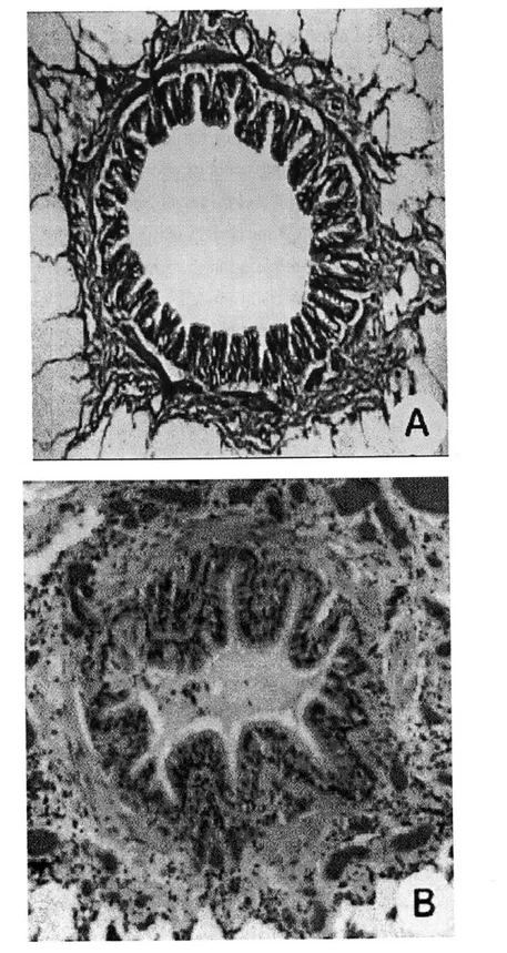
1.3 Histological cross-sections of a constricted normal (A) and asthmatic 24 (B) airway. Courtesy of University of British Columbia Pulmonary
Research Laboratory.
1.4 Diagram outlining the inflammatory network in the airway wall. 29 Responses of the various cell types to inflammatory mediators and their
contribution to hyperresponsiveness are also shown.
2.1 Schematic of two-layer airway model. 36
2.2 Schematic of epithelial barrier model. 48
2.3 Comparison of a single fold in a constricted airway and the finite 50 element model.
2.4 Tracings of a snap-frozen highly constricted guinea pig airway. 51 3.1 Finite element mesh of the two-layer normal airway model. 60 3.2 Finite element mesh of the two-layer asthmatic airway model. 60
3.3 Tube laws for normal and asthmatic airways. 62
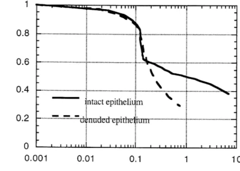
3.4 Tube law for asthmatic airway with intact epithelium and with denuded 65 epithelium.
3.5 Pressure-Area relationships for Generation 15 Airways calculated in 66 Hrousis.
3.6 Plot of normalized airway resistance (R*) vs. normalized smooth muscle 66 pressure (P*) for the representative normal airway, asthmatic airway,
and asthmatic airway denuded of epithelium.
3.7 Color band plots of normalized circumferential stress in a highly 69 constricted normal and asthmatic airway.
3.8 Color band plots of normalized radial stress in a highly constricted 71 normal and asthmatic airway.
3.9 Average normalized circumferential stress on epithelium within the fold 73 vs. normalized smooth muscle stress.
3.10 Ratio of compressive stress on epithelium to smooth muscle stress as a 73 function of normalized smooth muscle stress.
3.11 Band plots of pressure within the normal (A) and asthmatic (B) airways 83 at maximum constriction.
4.1 Hypothesis that mechanical stresses placed on airway epithelial cells 95 during smooth muscle constriction cause the epithelium to signal for
airway wall remodeling.
5.1 Schematic of air-liquid interface culture of rat tracheal epithelial cells on 99 Transwell-COLs.
5.2 Schematic of pressure apparatus and schematic of one Transwell with 101 transmembrane pressure application.
6.1 Hematoxylin-periodic acid Schiff's stained tissue cross section of rat 109 tracheal epithelial cells.
6.2 Transmission electron micrograph of RTE cells. 111
6.3 Representative Northern autoradiographs of our three candidate genes. 113 6.4 Expression of Egr-1 mRNA after 1 hour of 0, 2, 5, 10, or 20 cmH20 114
transmembrane pressure.
6.5 Transmembrane pressure magnitude response of candidate genes. 115 6.6 Fluorescent immunohistochemistry for Egr-1 protein. 117 7.1 Comparison of Egr-1 mRNA expression in cells exposed to 10 cmH2O 130
transmembrane or hydrostatic pressure.
7.2 Schematic of strained culture well under transmembrane pressure. 132 7.3 Comparison of Egr-1 mRNA expression in cells exposed to 10 cmH2O 134
transmembrane pressure with or without strain of the culture membrane.
7.4 Morphology of cell layers exposed to 10 cmH20 pressure and control. 137
7.5 Transmission electron micrograph of RTE cells subjected to 10 cmH20 141 transmembrane pressure for 6 hours.
7.6 Scanning electron micrograph of Transwell-COL membrane with 143 0.4 gm diameter pore size. Schematic of nucleopore membrane.
7.7 Culture substrate effects on Egr-1 mRNA expression. 145 A. 1.1 Comparison of Hookean and neohookean formulations in ADINA 157 A. 1.2 Pressure-Area relationships for Generation 15 Airways calculated in 158
Hrousis.
List of Tables
1.1 Changes in airway wall area of asthmatic airways (average percent 18 change from control cases). Adapted from ref. 54.
3.1 Subepithelial collagen layer thicknesses from various morphometric 54 studies.
3.2 Airway wall layer dimensions calculated from James et al. (53). 56 3.3 Outer layer thickness ratios calculated from Wiggs models. 57
3.4 Summary of model dimensions used. 59
7.1 Average membrane strains due to transmembrane pressure calculated 132 from maximum membrane deflection.
7.2 Average cell layer thicknesses of cultures exposed to 0, 10, or 20 cmH20 139
for 1 or 6 hours.
A.l.1 Model dimensions of Generation 15 airways in Hrousis model 155 calculations.
A. 1.2 Results of linearized buckling analyses of ABAQUS and ADINA 156 calculations.
Chapter 1
Introduction
1.1 Asthma: General Characteristics
Asthma affects 3-5% of the population, making it the most common respiratory disease (124). Currently, 15 million Americans suffer from asthma, twice as many as 15 years ago. Symptoms usually appear in early childhood and may persist over many years with varying severity. About 5000 Americans die each year from acute asthma attacks (29).
The major symptoms of asthma are cough, shortness of breath, wheezing and chest tightness. Asthma attacks occur spontaneously or are induced by a trigger stimulus such as inhaled allergen, inhaled irritants such as smoke and cold air, respiratory tract infection, and exercise (124). Symptoms are relieved by inhaled bronchodilators, which relax the constricting smooth muscle surrounding the airway, and by corticosteroids, which reduce the frequency and severity of symptoms (54).
Symptoms of asthma are preceded by a reduction in size of the airway lumen due to constriction of the surrounding smooth muscle. Characterization of airway caliber is determined by physiological measurements such as the forced expired volume in 1 s (FEV1), peak expiratory flow (PEF), and airway resistance (Raw) (54). During an asthma
attack, the reduction in ability to breathe is most acutely felt during exhalation and FEV1 and PEF both decrease due to increased Raw. Because asthmatics have a much greater propensity for increased airways resistance and smooth muscle activation, the airways are deemed hyperresponsive. Hyperresponsiveness includes both increased sensitivity of the airways (e.g., same response at a lower dose of agonist) and increased reactivity of the airways (e.g., greater response at the same dose of agonist). It is unclear what causes asthmatics to be hyperresponsive, but it is believed to be either biochemically mediated via increased sensitivity to an agonist, or bioniechanically mediated via changes in the structure of the airway wall. It is the biomechanically induced changes that are explored in this thesis.
1.2 Airway Structure
The airways of the lung form a branching structure, beginning with the trachea
(generation 0) and dividing into (usually) two smaller daughter branches at each bifurcation. Generations 0 - 15 are called the conducting airways, and generations 16-23
are termed the acinar airways. It is in the acinar airways where alveoli are present and gas is exchanged with the circulation (125). The basic structure of the tissue in the conducting airways is the same in all the generations, except that the larger airways (generations 0-6) contain cartilage in the wall. The other airways do not have cartilage and are termed "membranous" airways. In this thesis, only membranous airways will be considered and the term "airway" will refer to a "membranous airway".
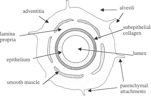
A cross section of a membranous bronchiole is depicted in Fig. 1.1. The nomenclature
used has been previously described (6) but is reiterated here for clarity. Surrounding the open space of the airway lumen is the epithelium. The epithelium rests on a thin basement membrane, which consists primarily of type IV collagen and laminin and is
maintained by the epithelial cells themselves. Beneath the basement membrane there
exists another fibrous layer consisting of collagens III and V, and fibronectin (98). This fibrous layer is maintained by fibroblast cells located within the airway wall (16). In this thesis, this layer is termed the subepithelial collagen layer (SCL), but in other studies it is also called (erroneously) the basement membrane, or the reticular lamina. Between the
basement membrane and the smooth muscle is the lamina propria, which consists of the
SCL and loose connective tissue (mostly proteoglycans), blood vessels, and connective
tissue cells. In this thesis, the SCL will be considered separately from the lamina propria, and the term lamina propria will refer only to the layer of loose connective tissue external to the SCL. External to the smooth muscle is the adventia, which is connected by sparse parenchymal attachments to the surrounding alveoli.
alveoli lamina collagen propria lumen epithelium smooth muscle parenchymal attachments
Figure 1.1: Sketch depicting key structures of a membranous bronchiole. Not to scale.
1.3 Asthma: A Disease Remodeling the Airways
1.3.1 Thickening of the airway wall
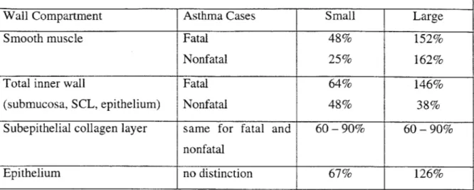
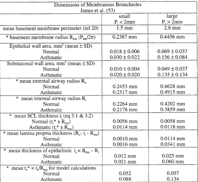
Several investigators have measured changes in the dimensions of asthmatic airways vs. normals, and the resounding conclusion is that all regions of the airway wall thicken in
asthma (20, 21, 49, 53, 54, 63, 126). A summary of these and other studies is given in ref. 54 and is presented here in Table 1.1. Table 1.1 contains data for regions of the airway wall internal to and including the smooth muscle layer. Airways were separated into size groups based on internal perimeter: <2 mm-small membranous bronchioles, 2
- 4 mm-large membranous bronchioles. The median values of the internal perimeters for these size groups (1.5 mm and 2.8 mm for small and large membranous bronchioles, respectively) were used to derive average values for airway dimensions. (54). Small membranous bronchioles are located approximately in generations 14 - 16 (terminal and transitional bronchioles), and large membranous bronchioles are in generations 10 - 14. (126, 128).
Cross-Sectional Area Changes in Cases of Asthma for Small and Large Membranous Bronchioles (Average Percent Increase from Control Cases)
Wall Compartment Asthma Cases Small Large
Smooth muscle Fatal 48% 152%
Nonfatal 25% 162%
Total inner wall Fatal 64% 146%
(submucosa, SCL, epithelium) Nonfatal 48% 38%
Subepithelial collagen layer same for fatal and 60 - 90% 60- 90% nonfatal
Epithelium no distinction 67% 126%
Table 1.1: Changes in airway wall area of asthmatic airways (average percent increase from control cases). Adapted from ref. 54.
1.3.2 Other structural changes in asthma
Microscopic examination of asthmatic airways reveals other aspects of airway remodeling. The epithelial cell layer appears "fragile" and may be denuded from the surface. Mucous glands are hypertrophied and there is an increased number of goblet cells. This enlargement of the mucus-secreting apparatus tends to occlude asthmatic airways with thick mucus plugs (124). Vascularity of the wall increases (72). Finally, the wall is infiltrated by various inflammatory cells such as eosinophils, lymphocytes, macrophages and mast cells. These cells release various growth factors, cytokines, and cytotoxic proteins that are known to induce cell damage and fibrosis (15). These structural changes, plus the overall increase in airway wall thickness, are all believed to contribute to airway hyperresponsiveness in asthma.
1.4 Hyperresponsiveness
It is not understood why asthmatics display hyperresponsiveness and normals do not. Airway responsiveness if measured by having the test subject inhale a prescribed dose of a constrictor agonist such as methacholine (MCh); following this inhalation, airway resistance (or FEV1) is measured. Asthmatics have steep dose-response curves that are
shifted to the left of normals (increased sensitivity) (55). It is generally accepted that normal healthy subjects eventually reach a plateau in their dose response curve, (i.e., a maximal response is reached and resistance does not increase with an increase in agonist dose). Asthmatic subjects, however, do not reach this plateau, and their airway resistance continues to increase sharply with increasing dose of MCh (132). There are several hypotheses explaining hyperresponsiveness, five of which are examined in the next sections.
1.4.1 Inflammation
Acute exposure to allergen causes an influx of inflammatory cells into the airway and release of the cells' mediators may lead to hyperresponsiveness. Mast cells may directly cause smooth muscle constriction by the release of histamine and synthesis of leukotrienes upon activation (19). Eosinophils release proteins that are toxic to epithelial cells and that breakdown the extracellular matrix. Breakdown of the protective epithelial cell barrier may cause hyperresponsiveness by directly exposing the smooth muscle to agonist (110). Despite the fact that inflammation tends to accompany asthma, the correlation between hyperresponsiveness and inflammation is not clear. While several groups have found that the presence of eosinophils, mast cells or activated T-cells have an inverse relationship with an asthmatic's FEV, several other groups have found no such relationship (43). These data demonstrate that hyperresponsiveness is not simply an inflammatory response, and that the mechanics of hyperresponsiveness must be considered.
1.4.2 Inner wall thickness: simple geometrical occlusion
Like most biological materials, the tissue of the airway wall has been shown to be relatively incompressible, at least during short periods of constriction, and the airway internal perimeter and wall area are relatively unchanged with different degrees of smooth muscle constriction (52). Thus, thickened asthmatic airways experience a greater degree of luminal obstruction with even normal amounts of smooth
normal (A) (thin inner region)
asthma (B)
(thick inner region) 0
Figure 1.2a: Hyperresponsiveness due to thickened inner region of an asthmatic airway. With the same degree of smooth muscle shortening, the airway resistance is much greater in asthma (Wiggs, James).
normal (A)
asthma (B)
Figure 1.2b: Hyperresponsiveness due to increased adventital thickness. Thicker adventitia reduces load on smooth muscle by the parenchyma, (depicted as unloaded springs in B), resulting in greater smooth muscle constriction upon activation.
normal (A) smooth muscle asthma (B) smooth muscle activation 'II
Figure 1.2c: Hyperresponsiveness due to increased smooth muscle mass. Thickened smooth muscle is able to generate more force, and cause greater airway obstruction (B)
(67)
normal (A)
asthma (B)
Figure 1.2d: Hyperresponsiveness due to increased subepithelial collagen layer thickness. A thickened SCL results in fewer folds upon buckling, creating a more compliant airway
muscle constriction (see Fig. 1.2a). Using morphometric measurements of normal and asthmatic airways, James et al. (53) determined that 55 - 62% muscle shortening is required to cause complete closure of normal membranous airways, but only 45 - 50%
muscle shortening is required to close asthmatic airways.
A geometrical model of the airway tree of both normal and asthmatic cases generated from morphometric data was used to generate theoretical dose-response curves (126). Using values of smooth muscle shortening of 20 - 40%, (which is the range observed during in vivo and in situ shortening), the normal airway model displayed the characteristic "plateau" in resistance with increasing "dose" of smooth muscle agonist. The asthmatic model, however, did not display a plateau in resistance within a physiologically reasonable range. The authors concluded that it is the thickening of the peripheral airways that has the most important effect on the relationship between smooth muscle shortening and airway narrowing. Indeed, asthmatic airways in generations 16 and distal experienced complete closure at smooth muscle shortening of 39% in the model, resulting in infinite resistance.
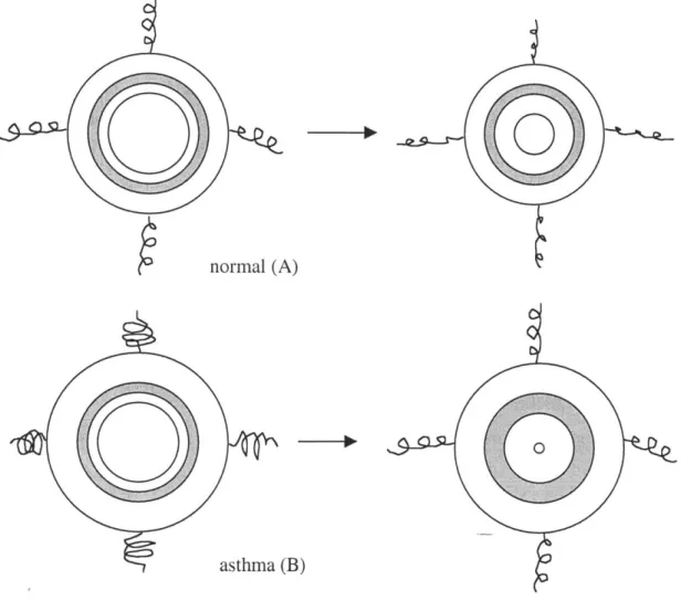
1.4.3 Adventitial thickening: uncoupling airway from parenchyma
A bronchiole is tethered to the lung parenchyma and is held open by lung recoil forces. Between these tethers and the smooth muscle of the airway lies the adventitia, the thickness of which is increased in asthma (63). This thickening can contribute to hyperresponsiveness by reducing the load on the smooth muscle, allowing it to shorten more easily in asthma. Thus, with smooth muscle shortening, the retarding force of the lung parenchyma on airway smooth muscle is "uncoupled" from the airway wall, and the smooth muscle is allowed to shorten further in asthma (Fig. 1.2b) (88, 63).
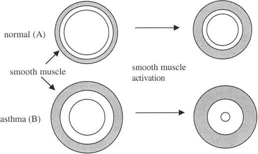
1.4.4 Smooth muscle
It has been observed that there is an increased amount of smooth muscle in the walls of asthmatic airways (20). Lambert and colleagues (67) developed a computational model of normal and asthmatic airways to determine the functional significance of increased smooth muscle mass. They incorporated several features in their model, including the geometrical model of Wiggs et al. (126), adventitial thickening (63), parenchymal
interdependence, and increased smooth muscle mass. They concluded that the increase in smooth muscle mass was the most important feature in airway remodeling. For a given maximal muscle stress, greater muscle thickness would allow for greater tension development, and thus more constriction of the lumen. (See Fig. 1.2c).
What all of the above models lack is the effect of tissue mechanics on smooth muscle shortening. There appears to be a maximum amount of force that smooth muscle can generate, and the stiffness of the airway wall and the recoil forces of the lung parenchyma resist the smooth muscle constriction. A constricted airway buckles into a many-lobed rosette pattern, not axisymmetrically as modeled in the simple geometrical models (see Fig. 1.3). Ignoring buckling in the geometrical models would grossly overestimate the stiffness of the airway to occlusion. Thus, the most realistic models of airway constriction include both the correct geometrical parameters and the mechanical behavior (i.e., buckling) of lung tissue. Much of previous work concerns the effects of inner wall mechanics on airway constriction, and so the effects of lung parenchyma are not
considered in this thesis.
While current models claim to incorporate airway wall mechanics, none of them realistically do so. In the model of airway constriction developed by Gunst and Stropp (41), the stiffness of the lung parenchyma surrounding the airway was included in the mechanics of constriction, but the inner airway wall was not modeled. In the more complete model of Lambert et al. (67) the inner airway mechanics were included along with models for smooth muscle force generation, lung recoil, and parenchymal interdependence. The inner airway was modeled, however, as a simple parallel elastic element whose stiffness was determined by Laplace's Law for a tube. Later, Lambert and others (68) developed a more realistic model of the inner airway by including wall buckling, but this model was also incomplete as it did not include the epithelium in the post-bucking behavior, and the lamina propria was modeled as a liquid. There is clearly a need for a more realistic model of inner airway wall mechanics in order to understand the physiology of asthmatic airway hyperresponsiveness.
Figure 1.3: Histological cross sections of a normal (A) and asthmatic (B) airway. Courtesy of University of British Columbia Pulmonary Research Laboratory.
1.4.5 Inner wall thickness: altered airway wall mechanics
It seems apparent from simple mechanics that a thickened inner airway wall would resist smooth muscle constriction more readily than a thinner wall. The airway does not constrict axisymmetrically, however, but at a certain pressure it buckles into a many-lobed shape, as shown in Fig. 1.3 (49, 65, 68, 127). After the airway buckles the muscle can constrict the airway much more easily, but the pattern of buckling greatly determines the ease of constriction. An airway with few folds buckles at a lower level of external force than an airway with many folds (65, 127). In Fig. 1.3a, the normal airway is buckled into a many-lobed pattern, which resists smooth muscle constriction and maintains luminal patency. The asthmatic airway in Fig. 1.3b has fewer folds which extend further into the lumen; these folds and the mucous plug have completely occluded this particular airway. There has been no systematic study to determine if asthmatic airways have fewer folds than normals, but it has been observed that the folds are deeper in an asthmatic (49).
There is debate over what determines the number of folds into which the airway wall will buckle. Macklem (73) has proposed that the parenchymal attachments surrounding the airway determine the buckling pattern, while Wagner and Mitzner (121) believe that the location of the blood vessels determines this pattern, as the vessels are arranged in a similar pattern as that of the folds. Two mechanical models of the airway wall, however, have determined that it is primarily the subepithelial collagen layer that determines the pattern of buckling (48, 68, 127). According to the model of Wiggs et al. (127), the structure and mechanical properties of all the wall components, particularly the elastic moduli of the various layers and their thicknesses, determine the number of folds into which the airway will buckle.
A schematic of a two-layer model of the airway wall is depicted in Fig. 1.2d, with a thin, stiff inner layer representing the SCL, and a thick, more compliant outer layer representing the submucosa. The airway with a thicker subepithelial collagen layer (model B) buckled with fewer folds than the airway with the thin SCL (model A). It is uncertain if the thicker subepithelial collagen layer makes the airway intrinsically stiffer
or less stiff, however. There are two models of the inner airway that attempt to resolve this question developed by Lambert et al. (68) and Hrousis (48).
In the two-layer model of Lambert et al. (65, 68), the thick outer layer representing the lamina propria (or submucosa) was modeled as being incapable of sustaining significant shear stress and so was modeled as a liquid. The basement membrane was modeled as a membrane that folded during constriction. They did not include the epithelial cell layer in the model. The membrane was constrained by muscle wall bounding (i.e., the membrane could not "poke" through the outer radius) and by a constant submucosal area during constriction. Smooth muscle constriction was modeled by placing a pressure on the outer radius. As the airway in the model narrowed, the membrane first buckled into two folds (n = 2, like a peanut shape), but quickly encountered the outer wall and was forced to develop more folds. This process continued until the appropriate value of n was determined by energy minimization. They compared their data with measurements of n, lumen area, and percent muscle shortening from sheep airways of varying wall dimensions.
The morphometric data of Lambert and colleagues (68) showed that the normalized lumen area decreased with the normalized submucosal area (i.e., a thinner airway was less occluded) and that the lumen area decreased with the number of folds (i.e., more folds were present in the more occluded airways.) This paradoxical behavior was explained using their two-layer model. Model airways with a thicker submucosa buckled with fewer folds, due to the more removed constraint of the outer wall of smooth muscle. The thickness to radius ratio of the membrane in the model was correlated with the submucosal thickness, which resulted in a thicker membrane (i.e., thicker basement membrane) employed in the model. While the thicker membrane buckled with fewer folds than a normal membrane, the increased stiffness of the folding membrane due to its increased thickness outweighed the reduction in stiffness caused by the decrease in n. Therefore, Lambert and colleagues concluded from their model that wall remodeling, particularly the increased thickness of the SCL, served to resist bronchoconstriction and reduce hyperresponsiveness.
There were several shortcomings in the analyses of Lambert et al. (68). First of all, the morphometric data that they obtained were from unremodeled, unactivated sheep airways. Any differences in dimensions or folding patterns among airways were simply due to random distribution present within the lung, and were not due to any remodeling or direct activation of smooth muscle. Human airways, on the other hand, are clearly remodeled in asthma, and differences in folding or dimensions among normal and asthmatic airways might not be due to random distribution but instead may be directly linked to the remodeling process. In the model of Lambert et al., the submucosa was modeled as a liquid unable to sustain shear stress, which was an obvious simplification that would likely affect the folding pattern and the ease of airway collapse. Also, the model airways initially collapsed in a peanut shape and the folds gradually increased in number. In vivo, however, the two-lobed collapse was not observed in airways with smooth muscle constriction, and the number of folds did not change significantly with degree of smooth muscle activation (48). Because of these discrepancies, a more realistic model of the airway wall is necessary.
The model developed by Hrousis (48) is believed to be more realistic and better captures the wall buckling behavior. We describe this model in more detail in the next chapter, but we present it briefly here for comparison with the model of Lambert (68). The airway model of Hrousis consisted of two layers of different dimensions and material properties that represented the internal submucosa and the subepithelial collagen layer. There were no geometrical constraints, and the number of folds was determined by linearized buckling analysis. Using this model, Hrousis concluded that the thickening of the subepithelial collagen layer was primarily responsible for the alteration in buckling pattern, and that the folding pattern was more important in determining luminal occlusion. Because the subepithelial collagen layer is thicker in asthmatic airways, it would be expected that these airways would have fewer folds and greater occlusion according to the model of Hrousis. This conclusion was supported by the study of Boulet et al. (14), where they found that airway hyperresponsiveness was correlated with the degree of subepithelial fibrosis in asthmatics.
1.5 Causes of Remodeling
The previous section examined the consequences of airway remodeling. Increased amounts of adventitial tissue, smooth muscle, and inner wall tissue are believed to lead to hyperresponsiveness. It is unknown what causes remodeling in the first place, but the general assumption is that airway inflammation is responsible. In allergic asthma, it is believed that the release of mediators after allergen binding to IgE-coated mast cells results in the influx of inflammatory cells into the airway wall (124). The network of cytokines and growth factors in the inflamed airway is extremely complicated. A brief sketch of this network and how it may lead to wall remodeling and hyperresponsiveness is presented in Fig. 1.4. The cellular responses to inflammatory mediators are numerous, but they generally tend to create hyperresponsiveness. The inflammatory cells maintain the inflammation, and the act of infiltrating the epithelial cell layer is thought to damage the integrity of the layer (12). Loss of the epithelial layer may expose the smooth muscle to agonist, leading to abnormal constriction, and may reduce the presence of the epithelial cell relaxing factors that reduce smooth muscle activation (124). The epithelial cells may respond directly to the inflammation by proliferation of the mucus-secreting apparatus, and by directing tissue remodeling and fibrosis (12). Smooth muscle cells might proliferate and undergo hypertrophy in response to inflammation, which may increase the amount the muscle band is able to shorten and the force it can generate (67). Finally, the fibroblasts may respond to inflammation by proliferation and increased production of extracellular matrix, which would lead to thickening of the airway wall (71). For a more complete description of wall remodeling, the reader is referred to a recent book that reviews all aspects of asthmatic airway wall remodeling (109). This thesis ultimately will not examine all the aspects of remodeling, but will concentrate on the thickened subepithelial collagen layer.
trigger allergen antigen Inflammatory T Cells Eosinophils Mast Cells Macrophages Cells *
0
7 Growth Factors Chemokines CytokinesInterleukins, TGF-P, PDGF, Endothelin, Leukotrienes, GM-CSF, Fibronectin, FGF, IGF, Cationic protein, Histamine, TNF-ot, etc.
\
Smooth Muscle CellsInflammatory Cells
Epithelial Cell
H1
Fibroblasts
-Continued-Figure 1.4: Diagram outlining the inflammatory network in the airway wall. Responses of the various cell types to inflammatory mediators and their contribution to hyperresponsiveness are given on the next page.
Epithelial Cells I--,'
Response
release more mediators
--- maintain activation
recruit more cells
Result Leading to Hyperresponsiveness -> *exacerbate inflammation infiltration process damages epithelium Inflammatory Cells damage, leakiness, denudation
goblet cell hyperplasia Epithelial Cells
-- proliferation
hypertrophy
- smooth muscle activation P- . loss of smooth muscle
"relaxing factors"
e cell layer thickening e mucus hypersecretion e lumen obstruction
* increased force generation - increased constriction Smooth Muscle Cells
.1. proliferation - subepithelial fibrosis
matrix synthesis -* . thickening of lamina
propria Fibroblasts
A'1
1.5.1 Fibroblasts: source of the thickened SCL
Wall fibrosis, primarily thickening of the subepithelial collagen layer, may reduce hyperresponsiveness by stiffening the airway wall (68), or may exacerbate airway narrowing by reducing the number of folds (48). We chose to focus on SCL thickening because the SCL appears largely to determine the mechanics of airway buckling, and because the thickening of the SCL is a remodeling feature unique to asthma (21, 56).
The subepithelial collagen layer is composed primarily of types III and V collagen, unlike the epithelial basement membrane which is composed of type IV collagen. Because of this difference in composition, the epithelium does not directly produce the matrix material in the SCL (98). Brewster et al. (16) found a strong correlation between the thickness of the SCL and the numbers of myofibroblasts (fibroblasts with contractile properties) in the tissue of asthmatic patients. They concluded that it is the myofibroblasts that synthesize and secrete the extracellular matrix material in the SCL.
1.5.2 Production of extracellular matrix by fibroblasts
The cytokine and growth factor network in an inflamed airway is extremely complicated. T cells, eosinophils, mast cells, and macrophages are all known to express growth factors and cytokines that can stimulate fibroblast proliferation and collagen synthesis (Roche, 1991). Some of the more important growth factors and cytokines implicated in asthmatic fibrosis are interleukin-1 (JL-1), platelet derived growth factor (PDGF), fibroblast growth factor (FGF) and transforming growth factor-P (TGF-P) (7). Another potentially important source for fibrotic agents is the epithelium. Because the fibrosis in asthma occurs directly beneath the epithelial basement membrane, the interaction between epithelial cells and fibroblasts has recently been considered as the most important relationship in SCL remodeling in asthma (90). Epithelial cells produce PDGF, TGF-P, IL-1, and endothelin-1 (ET-1) (90), which cause fibroblast proliferation and/or extracellular matrix production. It is unknown what would cause the epithelial cells to release these factors, but researchers have speculated that mediators from inflammatory cells (7) are responsible.
1.5.3 Alternative hypothesis for airway remodeling
It is now well established that mechanical forces play an important role in tissue remodeling and cellular homeostasis. Vascular endothelial cells are very sensitive to their mechanical environment and respond readily to changes in shear stress and wall strain. Perturbations in their mechanical environment cause the cells to realign, proliferate, or release fibrinolytic activators and growth factors in order to maintain tissue homeostasis ( 28, 94). Cardiac myocytes sense mechanical stretch and convert the signal into growth signals, which can lead to hypertrophy of the heart muscle (101). Small mechanical strains are critical in the continuous remodeling of bone tissue, the absence of which lead to bone deterioration and osteoporosis (138). Stretch and mechanical pressure act on the mesangial cells of the kidney. Abnormal amounts of pressure cause mesangial cell proliferation in vitro (60) and stretch leads to excess matrix accumulation (139, 95). Excess matrix and mesangial cell proliferation are linked to glomerular hypertension and renal disease.
The cells of the lung are also in a mechanically active environment, but the effects of mechanical forces on lung tissue have not been extensively studied. Stretching of alveolar epithelial cells resulted in Ca' mobilization and surfactant secretion (131). Intercellular calcium signaling was also initiated by mechanical perturbation of intact tracheal epithelium (32) and intracellular inositol 1,4,5-triphosphate (IP3) concentrations increased with stretch of airway epithelial cells (31). Mechanical strain inhibited wound healing and prostanoid synthesis of airway epithelial cells in vitro (103, 104). Fluid shear stress and stretch altered growth factor release from pleural mesothelial cells (123). Finally, high lung inflation due to positive end expiratory pressure increased mRNA levels of extracellular matrix components and growth factors in lung parenchyma (10). These studies demonstrate that cells in the lung respond to mechanical forces.
It is obvious from histological cross sections that the cells in a constricted airway are experiencing perturbations in their mechanical environment. Epithelial cells located deep within the folds appear to be pushed up against each other, thus experiencing a compressive normal stress. Computational models of a buckled airway show regions of
high shear stress and shear stress gradients near the epithelium, and regions of high and low pressure through which wall liquid could flow (127). As in other regions of the body, the cells of the airway may respond to these mechanical forces by remodeling their environment to accommodate these stresses. We employed computational modeling of the airway to determine the type and magnitude of these stresses on the epithelium in a buckled airway, then subjected airway epithelial cell cultures to these stresses and evaluated their molecular responses.
1.6 Goals of Thesis
This thesis will continue the investigation of airway remodeling in asthma in two ways. 1. The computational model of Hrousis (48) will be developed further to include the
effects of an epithelial cell layer on wall buckling. This model will be used to determine the stresses on the epithelial cells in a constricted, buckled airway.
2. It is well known that cells respond to mechanical forces by modifying their environment. This thesis will explore the hypothesis that mechanical forces in a constricted airway could stimulate the epithelial cells to release factors that would lead to airway wall remodeling. This is viewed as a possible parallel mechanism for airway remodeling and does not necessarily preclude effects caused by inflammation. An in vitro model of the normal stresses produced by the smooth muscle constriction on epithelial cells is developed herein. Molecular biology techniques are employed to determine changes in gene expression and protein synthesis of factors known to stimulate remodeling.
Ultimately, this work will combine computational modeling and molecular cell biology in a unique way to further our understanding of hyperresponsiveness in asthma.
1.7 Thesis Organization
In chapter 2, we present in brief the two-layer model of the airway developed by Hrousis (48). We develop the model further to incorporate the effects of the epithelium in the post-buckling behavior of the airway. In chapter 3, we present the results from our model calculations. We show that the epithelial cell layer has a very important effect in
determining the mechanics of airway constriction. The epithelial model is also used to calculate the stresses within a highly constricted airway; these calculations are used to validate the magnitude of compressive stresses utilized in our in vitro epithelial cell model.
In chapter 4, the effects of mechanical forces on various cell types and mechanotransduction mechanisms are reviewed. The literature on the response of pulmonary cells to mechanical stresses is reviewed in detail. We present the three candidate genes selected for study in our experiments and review how these genes are induced in other cell systems with mechanical stress. The genes selected to study are known to be mechanically transduced in other cell systems, may be produced by airway epithelium, and are believed to play a role in asthmatic airway remodeling. In chapter 5, we outline the protocols used to culture airway epithelial cells, to apply mechanical stress to the cells, to examine their molecular and morphometric responses, and to examine the mechanism of mechanotransduction.
We present the results of our transmembrane pressure experiments on airway epithelial cell cultures in chapter 6. Changes in expression of our candidate genes in response to pressure, and the timecourse of their expression are determined. Cell viability, cell morphology, and protein synthesis are also evaluated. We discuss the results of our experiments in the context of airway wall remodeling, and how knowledge of the epithelial response to mechanical forces helps complete our understanding of the remodeling process. In chapter 7 we explore the mechanism behind mechanotransduction of our epithelial cultures by transmembrane stress. Finally, in chapter 8 we summarize the conclusions developed in the thesis, and present suggestions for future work.
Chapter 2
Development of Finite Element Model
Much of the work in developing a finite element model of the inner airway wall was done by my predecessor, Constantine Hrousis (48). Thus, details of model development and finite element theory will not be fully described in this thesis, and the reader is directed to ref 48 for this information. Briefly, Hrousis constructed a two-layer model of the airway wall; the lamina propria was modeled as a thick, compliant outer layer, and the subepithelial collagen layer was modeled as a thin, stiff inner layer. This model was based on the assumption that these two layers would capture most of the buckling behavior; the lamina propria, due to its thickness, and the SCL, due to its stiffness, would exert the strongest influence of airway mechanics. The epithelium was assumed to compose a small part of the airway wall and to be highly compliant; thus it was excluded from the model because it was unlikely to play a role in buckling. The smooth muscle band was modeled as a very thin, uniformly constricting band surrounding the inner airway wall. This smooth muscle boundary condition resulted in the buckling behavior consistent with observation. As the smooth muscle constricts, the region of the airway external to the smooth muscle is assumed to 'deform axisymmetrically and to be decoupled from the inner airway's deformation. Therefore, for simplicity the adventitia and parenchymal attachments were not included in the model, as they were likely to deform axisymmetrically and without large resistance to smooth muscle shortening.
2.1 Two-Layer Model Components
The simple two-layer structure of the model was based on the geometry of the completely relaxed airway internal to the smooth muscle (Fig. 2.1). The lamina propria in vivo is a thick layer of loose connective tissue internal to the smooth muscle; thus the lamina propria was modeled as an outer layer of thickness t0 and stiffness E. The subepithelial
collagen layer in vivo is a thin layer of densely packed, organized collagen matrix (96); thus the SCL was modeled as a thin layer of thickness ti and stiffness E,, where t < to and Ei > E0. The base radius R was defined from the center of the lumen to the outer edge of the SCL in a completely relaxed airway.
smooth muscle band
lamina propria
to
E
0Figure 2.1: Schematic of two-layer airway model. The lamina propria is modeled as a thick, compliant outer layer of thickness to and stiffness E.. The SCL is modeled as a thin, stiff inner layer of thickness t1 and stiffness Ei. The base radius of the airway is the
distance from the lumen center to the outer edge of the SCL. The smooth muscle is modeled by a boundary condition of uniformly constricting hoop strain.
Only the lamina propria and the subepithelial collagen layer were included in the model of the airway wall. The epithelium was not included because it is likely to be much less stiff than the SCL, or even the lamina propria, and therefore would not provide significant resistance to collapse or affect the deformation behavior of the buckling. However, the epithelium would be expected to provide significant resistance to airway constriction once the airway has buckled and the folds are well developed. Therefore, a model of a folded epithelial cell layer was incorporated into the two-layer model after the buckling behavior was determined. This model will be described in a later section.
The smooth muscle action was incorporated into a boundary condition at the outer radius of the wall, as described later. The smooth muscle and any wall components external to the smooth muscle were assumed to deform axisymmetrically and therefore did not need to be modeled with continuum elements. This means that the smooth muscle layer, once activated, decouples the inner airway's deformation from the outer airway's deformation. Therefore, a fraction of the total load that the smooth muscle exerts would go towards axisymmetrically deforming the parenchyma and adventitia, and the rest would be used to buckle and collapse the inner airway. According to the computational model of Lambert and Pard (66), resistance to smooth muscle constriction provided by the adventitia or parenchymal attachments would be comparable to that of the inner airway wall at low lung recoil pressures; at high lung recoil pressure, the resistance due to the airway is negligible. For simplicity, we did not include the outer airway and parenchyma in our model, but it must be noted that it might play a role in maintaining airway patency, especially at low lung recoil pressures.
2.2 Mechanical Modeling Assumptions
2.2.1 Two-dimensional plane strain
The branching structure of the airways and the arrangement of the smooth muscle are highly three-dimensional. However, at locations between branch points, the smooth muscle and the wall geometry appear to have little variation along the axis. The mucosal folds in a constricted airway tend to run along the length of the airway without any variation in number along the airway length (133). Because there appears to be little
variation in the geometry along the length of the airways far enough away from the bifurcation, we modeled the airway as two-dimensional. In the case of two-dimensional deformation, an assumption must be made about the third (axial) direction. If the assumption is "plane stress," then all non-zero stresses lie in the plane and there is no force, only deformation, in the axial direction. If the assumption is "plane strain," then all non-zero strains lie in the plane and there is no strain, only stress, in the axial direction. Of these choices, the assumption of plane strain appears most reasonable because the airways are not likely to shorten axially during constriction. The smooth muscle wraps around the airway at an angle approximately 130 to the plane of a cross-section (70), which is a shallow angle and would likely have little effect on airway length during constriction (8).
2.2.2 Homogeneous isotropic layers
Both layers were assumed to be a homogeneous, isotropic continuum, which differ only in their thickness and assigned elastic modulus.
The assumptions of homogeneity and isotropy are likely to be reasonable for the lamina propria, as it is composed primarily of loose connective tissue and gel-like proteoglycans. In tissue sections of the lamina propria taken perpendicular to the airway axis, the fiber structure appears to be composed of random collagen coils. Parallel to the airway axis, however, the are straight fibers of elastin, which may result in some anisotropy of the lamina propria (22). There exist some discontinuities in composition with blood vessels and individual bundles of collagen and elastin. However, these anisotropies and inhomogeneities appear relatively small in size and would likely result in a roughly constant value of E throughout the layer.
The subepithelial collagen layer is significantly more ordered than the lamina propria, with collagen fibrils aligned approximately at a 450 angle to the airway axis (70). This orientation suggests that the collagen layer has an equal tendency to be deformed longitudinally or circumferentially, thus behaving relatively isotropically. Measurements of the tensile stiffness of the inner ovine tracheal wall showed a marked anisotropy, however, with a longitudinal stiffness approximately three times greater than the
circumferential stiffness (22). There have been no studies of the SCL properties in compression. For simplicity and for a lack of knowledge about the material properties, we assumed isotropic behavior. The size of the collagen bundles is small compared to the layer thickness, creating a layer that we could assume to be homogeneous. There is no clear boundary between the two layers in vivo, but the collagen content changes abruptly over a relatively short distance beneath the epithelium. The location of this jump in collagen content is taken to be the interface between the two discrete layers of
the model.
2.2.3 Incompressible Hookean and neohookean materials
Determining the correct constitutive law for biological materials is extremely difficult. The microstructure of the tissue is more complicated than the classic engineering materials, and the tissue is generally hydrated, with the material properties depending on the degree of hydration and the porosity. The stress-strain behavior of biological tissues is also difficult to determine, and is likely to behave in a non-linear fashion with large deformations. However, incorporating complicated material behavior into the finite element model is not likely to bring greater insight into airway wall mechanics, so the simplest linear material models are employed.
The simplest material constitutive law is that of an incompressible, linear-elastic (Hookean) material. The material is characterized by two constants: the Young's modulus (E), which represents the material stiffness, and Poisson's ratio (v), which represents the material compressibility. Other material constants that might be used to describe a Hookean material are the shear modulus (G), the bulk modulus (K), or the Lam6 constant (X). Only two constants are necessary to describe the material (here E and v are used) but conversion between constants is entirely equivalent.
An incompressible material has a Poisson's ratio (v) of 1/2 and an infinite bulk compression modulus K. Computationally, v = 0.5 is difficult to implement, and values of v > 0.45 are used to model "nearly incompressible" materials. Because biological tissues are composed primarily of water, they are assumed to be incompressible. The constituents of tissue (e.g., collagen, elastin, proteoglycans, water, etc.) when taken
separately, do appear to be incompressible. Tissues are bi-phasic in nature, however, with a porous matrix of connective tissue filled by cells, water, proteoglycans, and other proteins. Therefore, tissue could be compressed if sufficient time were allowed to elapse for the water to be squeezed out of the interstitial spaces. In the case of the airway wall, an assumption of incompressibility means that the time interval over which the smooth muscle constricts is sufficiently short that the water in the wall does not have time to significantly redistribute. Good estimates of this time for fluid movement and compressibility would require knowledge of the permeability of the tissue, which is unknown at this time.
Hooke's Law
Hooke's law describes the relationship between stress (the force per unit area in the current configuration) and strain for isotropic linear-elastic materials. The stress and strain tensors are symmetric, which means the off-diagonal shear stresses and strains are equal. For simplicity these tensors may be expressed in column matrix form.
stress tensor: " "2= T21 (2.1) L'12 T22 T12J dul
El
dx1 Es n 12 u (2.2) strain tensor: - E 22 f 2 _E12 E221 x2±rzl
+
ii2
and u(x) = the displacement as a function of reference position = y(x) - x. The strain tensor presented here is the small strain approximation of the Cauchy-Green strain tensor and is used in linear elastic theory. For two dimensional plane strain, the stress is related to the strain by the following tensor equation: